Embed Size (px)
Citation preview
MLAB 1227: COAGULATIONKERI BROPHY-MARTINEZ
Overview of Hemostasis:Part Two
FUNCTION OF PLATELETS
Surveillance of blood vessel continuityChecks endothelial lining for gaps and
breaksFill-in small gaps caused by separation of
endothelial cells Formation of primary hemostatic plug Surface for coagulation factors to
make secondary hemostatic plug Aid in healing injured tissue
FORMATION OF PRIMARY HEMOSTATIC PLUG
Once the platelets “normal” environment is changed, they become activated or adhesive
Stages of plug formationAdhesionSecretionAggregationStabilization
STAGE 1: PLATELET ADHESION Platelets attach to non-platelet surfaces, such as
collagen fibers in the subendothelium Platelets move from the blood vessels and into the
tissues. Exposure to surfaces in the tissues causes them to
bind to collagen with the presence of von Willebrand factor ( vWF) through the Glycoprotein Ib/IX receptor
Results in a bridge formation, which triggers a shape change in the platelets
Reversible No energy required
PLATELET ACTIVATION
Platelets undergo a shape change from disc to spiny sphere with projections
Activation required for 1O hemostatic plug formation
Activation continues until Ca ++ threshold met Platelets remain localized in injured area
http://tiny.cc/abmhmw
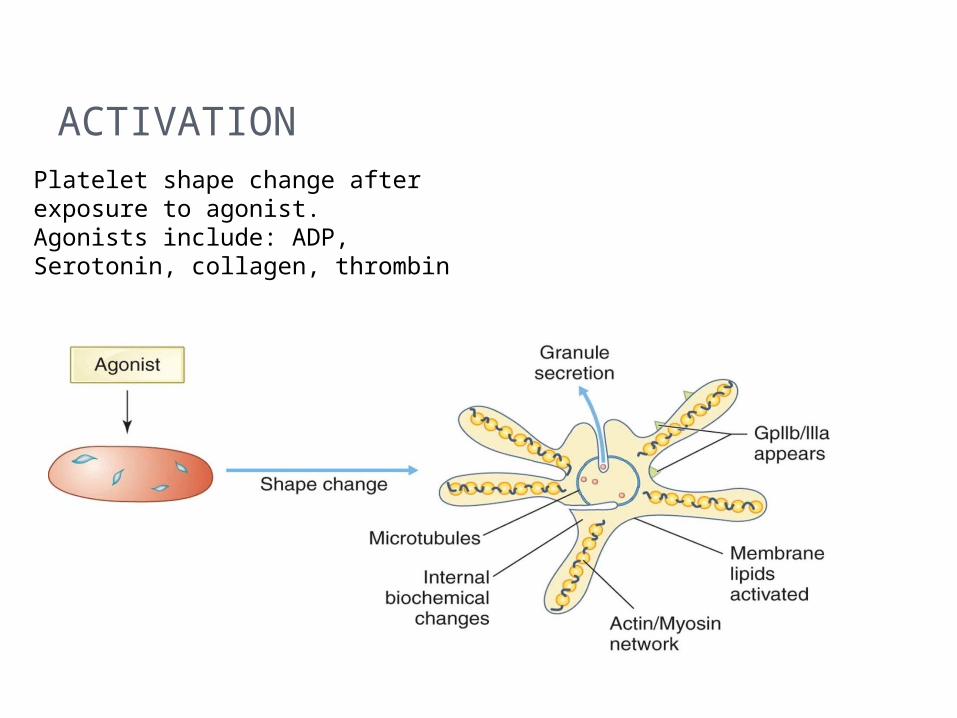
ACTIVATIONPlatelet shape change after exposure to agonist.Agonists include: ADP, Serotonin, collagen, thrombin
STAGE 2: PLATELET SECRETION/ PLATELET RELEASE REACTION Secretion
Requires ATP Provides a positive feedback by releasing more
agonists to stimulate more plt receptors Release of Granule contents
Causing vasoconstriction Release of ADP(agonist)= increased Ca+, plt
release, increase in fibrinogen receptors Trigger a secondary aggregation which is
irreversible
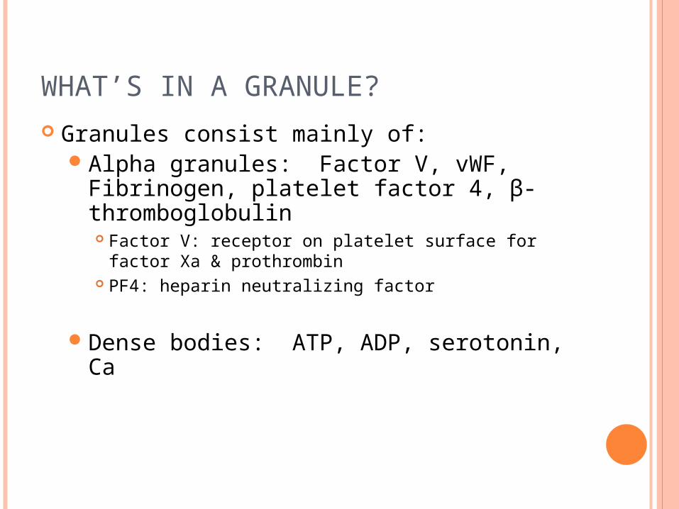
WHAT’S IN A GRANULE? Granules consist mainly of:
Alpha granules: Factor V, vWF, Fibrinogen, platelet factor 4, β-thromboglobulin Factor V: receptor on platelet surface for factor Xa &
prothrombin PF4: heparin neutralizing factor
Dense bodies: ATP, ADP, serotonin, Ca
SIDE NOTE
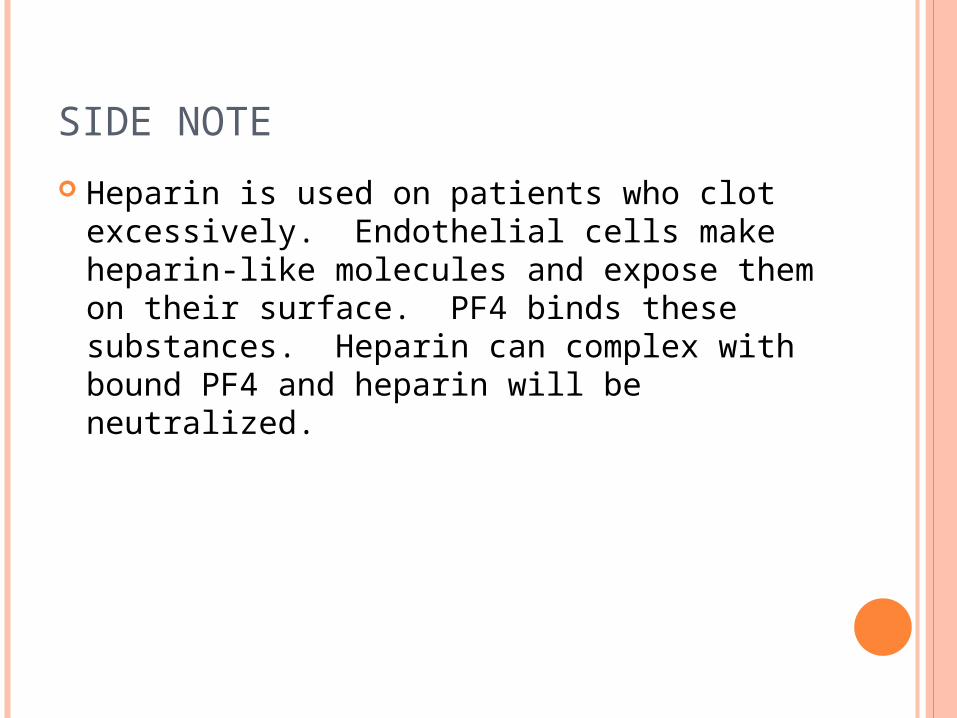
Heparin is used on patients who clot excessively. Endothelial cells make heparin-like molecules and expose them on their surface. PF4 binds these substances. Heparin can complex with bound PF4 and heparin will be neutralized.
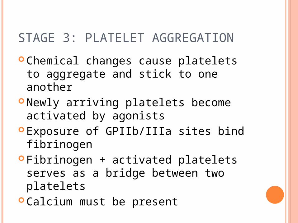
STAGE 3: PLATELET AGGREGATION
Chemical changes cause platelets to aggregate and stick to one another
Newly arriving platelets become activated by agonists
Exposure of GPIIb/IIIa sites bind fibrinogen
Fibrinogen + activated platelets serves as a bridge between two platelets
Calcium must be present
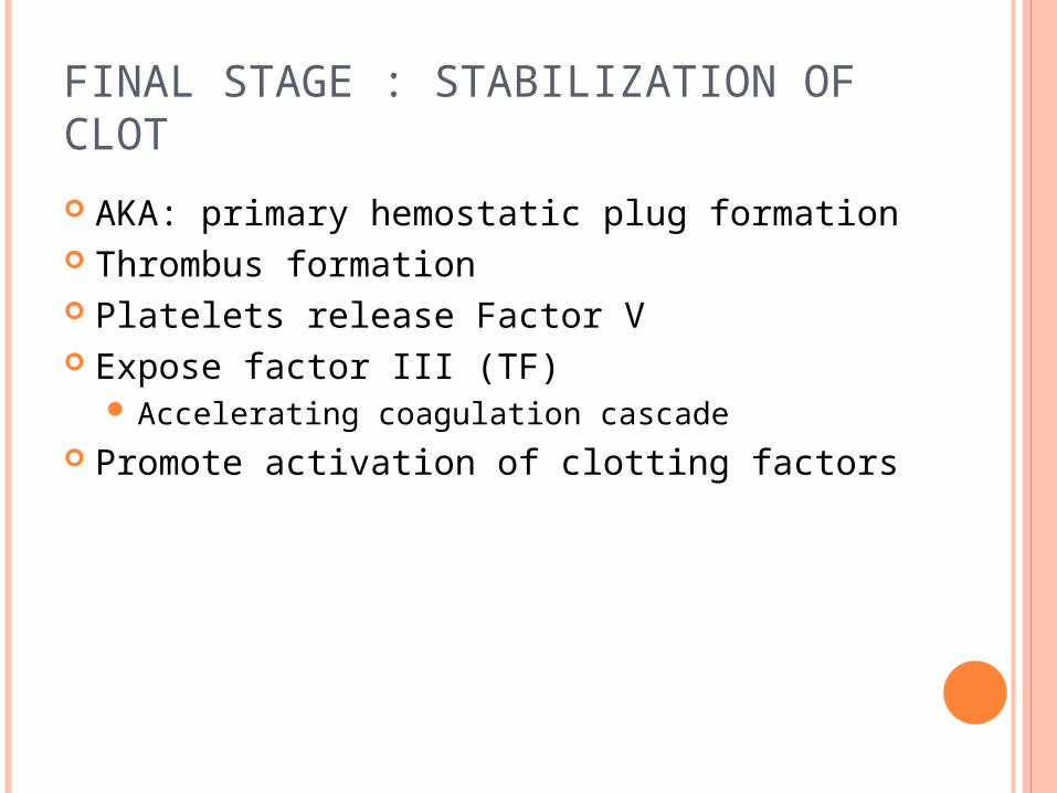
FINAL STAGE : STABILIZATION OF CLOT
AKA: primary hemostatic plug formation Thrombus formation Platelets release Factor V Expose factor III (TF)
Accelerating coagulation cascade Promote activation of clotting factors
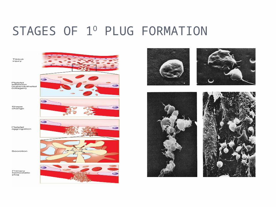
STAGES OF 1O PLUG FORMATION
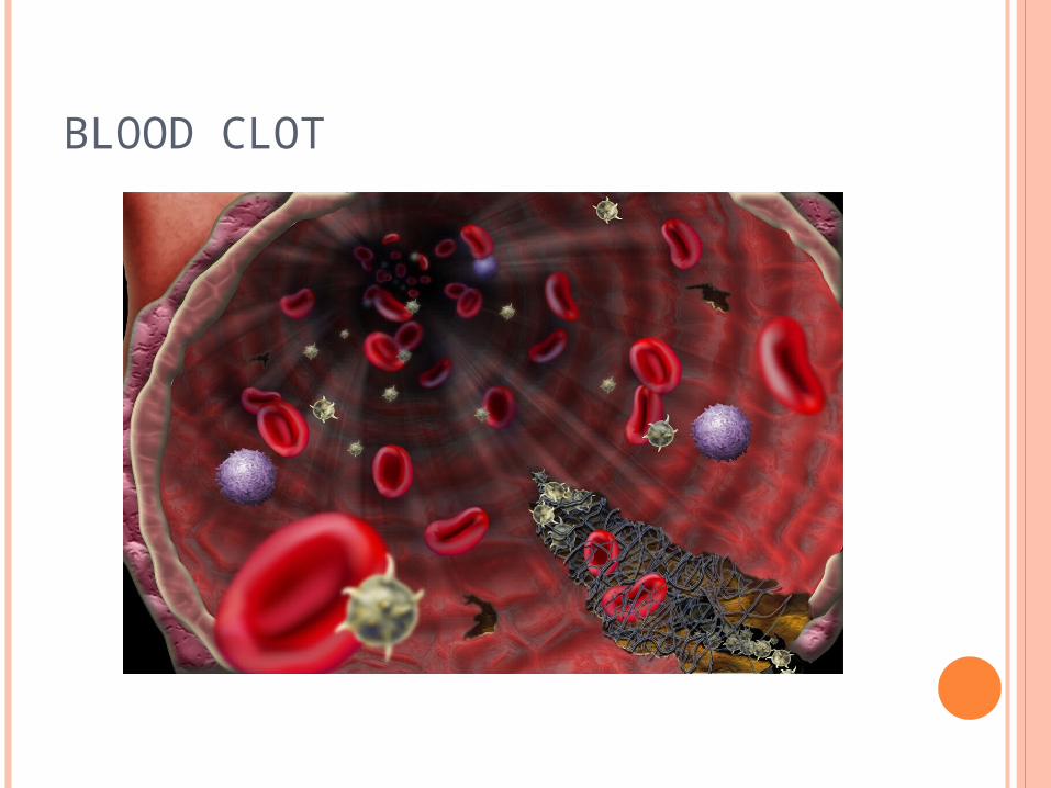
BLOOD CLOT
COAGULATION SYSTEM
Composed of 14 coagulation factors (serine proteases) which are interdependent (Factors I through XIII – there is no Factor VI – and PK and HMWK)◦ Inactive form of each is an enzyme precursor which is usually designated by a
Roman numeral but also given a name – Ex. Factor I fibrinogen. Numbers correspond to order of discovery NOT order in cascade.
◦ Active forms are usually designated by the letter “a” after the Roman numeral and may also have a different name – Ex. Ia Fibrin
◦ Cofactors are needed for many reactions in the cascade – Ex. Calcium, platelet factor 3 (PF3)
◦ Each molecule must be present in sufficient quantity as well as functioning normally
Final product is fibrin mesh or clot which completely stops bleeding◦ Secondary hemostasis
Slow contraction or retraction and lysis of the clot occurs
FIBRINOLYTIC SYSTEM
Plasminogen is converted to plasmin Plasmin enzymatically attacks the fibrin
molecule producing fibrin degradation products (FDPs, sometimes called FSPs) that are cleared from the circulation by macrophages
Fibrin is a product formed during hemostasis, tissue repair or inflammation
Fibrin plays a temporary role Once injury heals, the fibrin clot is lysed
COAGULATION INHIBITION SYSTEM
Provides balance and control of clotting mechanisms
Natural inhibitors and anticoagulants circulate in the plasma to:Prevent clotting when it’s not neededLimit or localize the clotting that is needed
Examples: Protein C and S, antithrombin III
REFERENCES
McKenzie, Shirlyn B., and J. Lynne. Williams. "Chapter 29." Clinical Laboratory Hematology. Boston: Pearson, 2010. Print.
Platelet Research Laboratories. Platelet Function. Retrieved from http://www.platelet-research.org/1/function_hemo.htm.