Embed Size (px)
Citation preview

Mitral valve disease – Mitral Regurgitation
Mitral valve fails to close completely, causing blood to flow back into the left
atrium during ventricular systole of multiple aetiology
www.anaesthesia.co.in
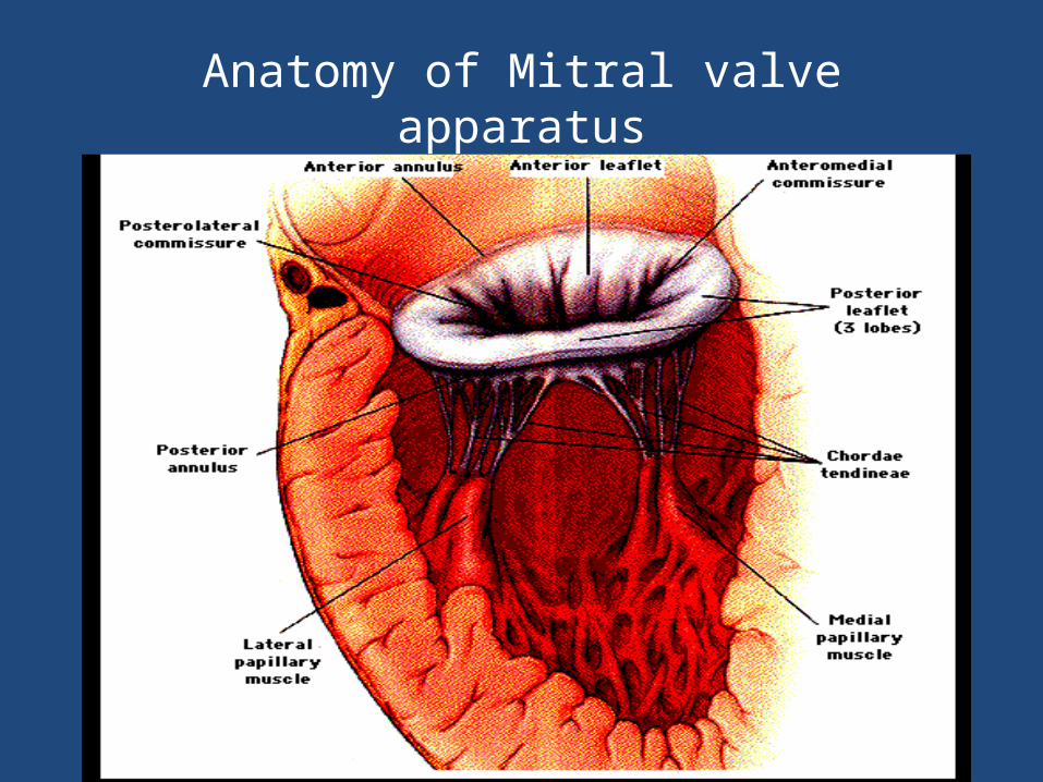
Anatomy of Mitral valve apparatus

• 45 year old women diagnosed c/o chronic mitral regurgitation posted for abdominal hysterectomy
• Natural history• Pathophysiology• Preoperative assessment• Perioperative management
Clinical presentation
• Depends on EtiologySeverity
of regurgitation Left ventricular functionPulmonary hypertensionAtrial fibrillationMixed valvular lesionCoronary artery disease & Hypertension
Etiopathology
• Valve leaflet Rheumatic Carditis SequlaeMyxomatous DegenerationCongenital Cleft Leaflet Marfan Syndrome Infective Endocarditis HOCM
Mitral valve prolapse (MVP)

Etiopathology
• Chordae Tendinae Rheumatic
Ischemic• Papillary Muscles
Ischemic• Annulus
Calcification - elderly patients Dilatation - functional MR

Pathophysiology
Regurgitation of LV SV• R
educed CO & LA dilatation
LA dilatation• A
trial fibrillation, Clot, Reactive PHT& Right heart failure
LV volume overload • L
VH (concentric) & LV dilatation
LV failure • P
ulmonary edema, systemic hypotension
Assessment of severity of MR
• Symptoms severity• Presence of PHT• Physical signs of CHF• ECHO
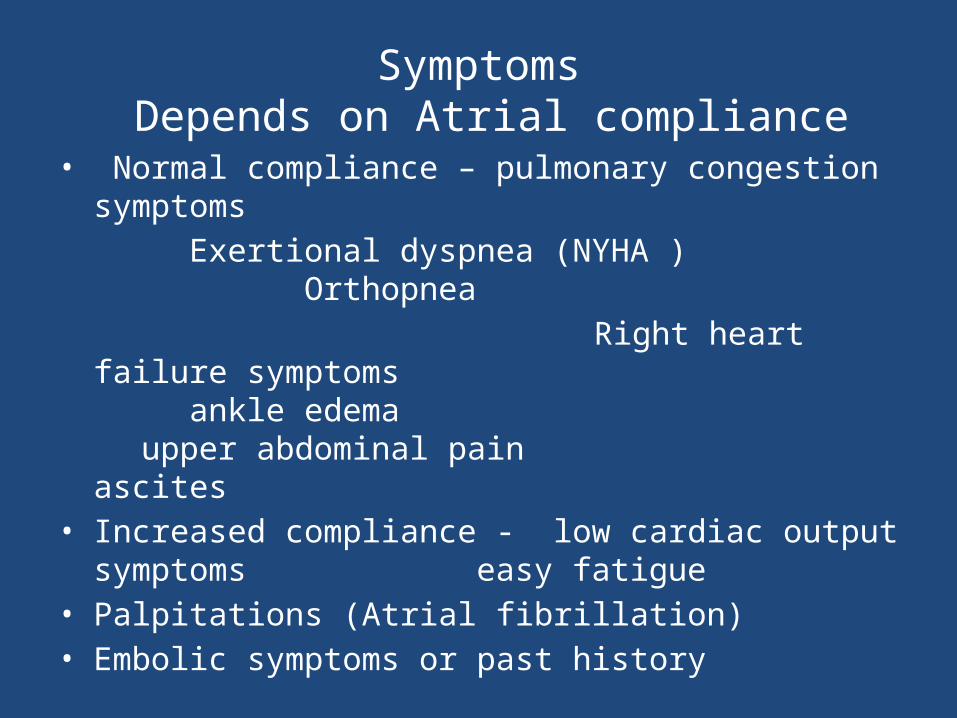
Symptoms Depends on Atrial compliance
• Normal compliance – pulmonary congestion symptomsExertional dyspnea (NYHA )
Orthopnea Right heart failure symptoms
ankle edema upper abdominal pain
ascites • Increased compliance - low cardiac output symptoms
easy fatigue • Palpitations (Atrial fibrillation)• Embolic symptoms or past history
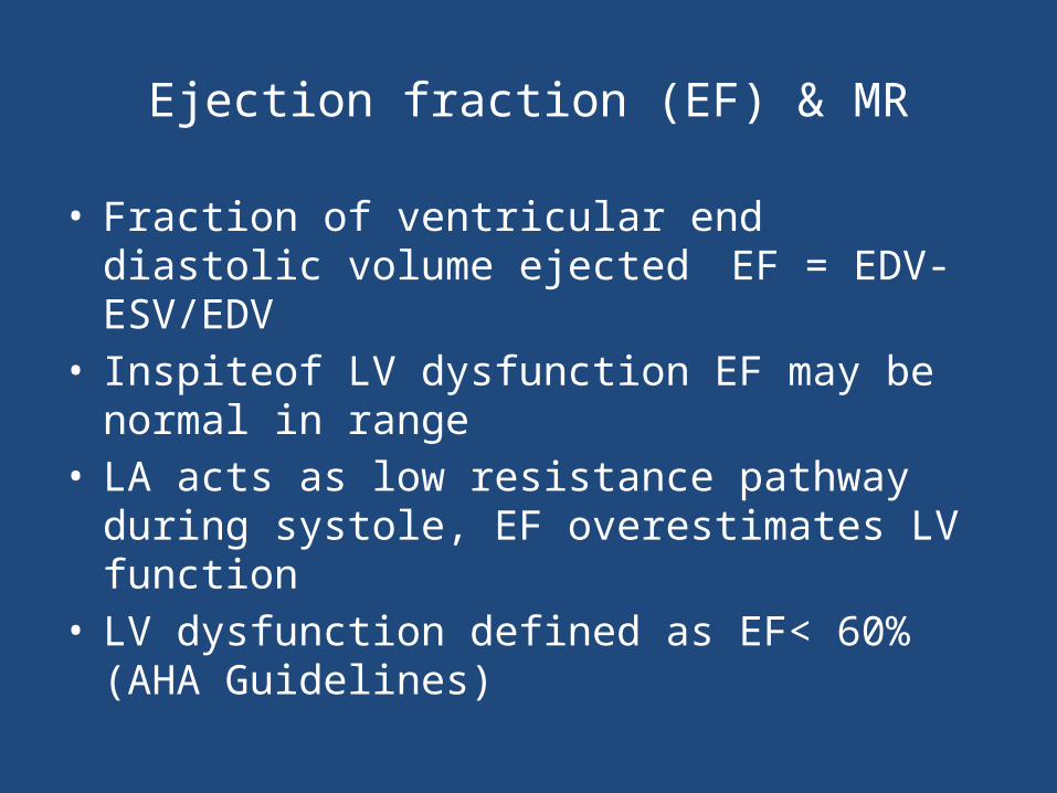
Ejection fraction (EF) & MR
• Fraction of ventricular end diastolic volume ejected EF = EDV- ESV/EDV
• Inspiteof LV dysfunction EF may be normal in range• LA acts as low resistance pathway during systole, EF
overestimates LV function• LV dysfunction defined as EF< 60% (AHA Guidelines)

Echocardiographic classification of MR

Echocardiographic assessment of regurgitant lesions by color – flow Doppler
Grade Description Criterion
0 None No regurgitation into receiving chmber
1+ Mild Regurgitant flow limited to area near valve
2+ Mild to moderate Regurgitant flow occupies up to 1/3 of LA
3+ Moderate to severe Regurgitant flow occupies up to 2/3 of LA
4+ Severe Regurgitant flow occupies most of receiving LA & flow reversal in pulmonary veins
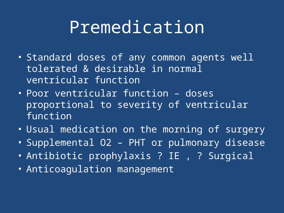
Premedication
• Standard doses of any common agents well tolerated & desirable in normal ventricular function
• Poor ventricular function – doses proportional to severity of ventricular function
• Usual medication on the morning of surgery• Supplemental O2 – PHT or pulmonary disease• Antibiotic prophylaxis ? IE , ? Surgical • Anticoagulation management

Monitoring
• Depends on LV function & procedure• Full hemodymaic monitoring – if plan for afterload
reduction with vasodilators• Color- flow Doppler TEE
quantify severity guides therapeutic interventions in severe lesions
• Pulmonary artery pressure monitoring

Anesthetic management objectives
• Tailored to severity of regurgitation jet, LV function• Avoid factors exacerbating the regurgitation
Bradycardia - ↑↑ LV end diastolic volumemitral annular dilatation
Acute rise in SVR Excess volume expansion – dilates LVMyocardial depression
• Prevent & promptly treat AF
Hemodynamic goalsMaintain forward systolic flow
Full, Fast & Vasodilated
Preload
• Augment LV preload prior to induction• Remember MR is dynamic• Excess ventricular distension →annular dilatation →
worsens MR

Heart rate & Rhythm
• Bradycardia detrimental ↑duration of systole→ prolongs regurgitation ↑ diastolic filling→ LV distension
• Sinus rhythm preferred LV filling less depends on atrial kick as compared to MS

Contractility
• EF underestimates LV systolic function • Avoid myocardial depression • How to manage hypotension?
Manipulate volume & heart rate• Persistent hypotension → inotropic support
DobutamineLow Dose EpinephrineMilrinone

Afterload
• Low SVR maximizes forward cardiac outputAdequate anesthetic depthSystemic vasodilatorsInodilatorsIABP – acute MR
• Alpha 1 agonists worsens MRincreases SVR & reflex bradycardia
• Temporary use of Ephedrine bolus preferable
Pulmonary hypertension
• PAP & PVR elevated in acute and chronic MR• Secondary RV dysfunction• Avoid factors increasing PVR
Hypoxia Hypercapnia Acidosis
High tidal volume High PEEP

Factors affecting Mitral valve repair
• Acute vs chronic MR• Patient’s symptom severity• Left ventricular function• Severity of mitral regurgitation• Feasibility of successessful repair• Combined valvular lesions• Complications of chronic MR
Choice of agents
• Preserved ventricular function – most anesthetic techniques tolerated well
• Spinal, epidural anesthesia tolerated (avoid bradycardia)
• Ventricular dysfunctionsensitive to volatile agentsopioid based anesthetic (avoid bradycardia)pancuronium with opioids useful
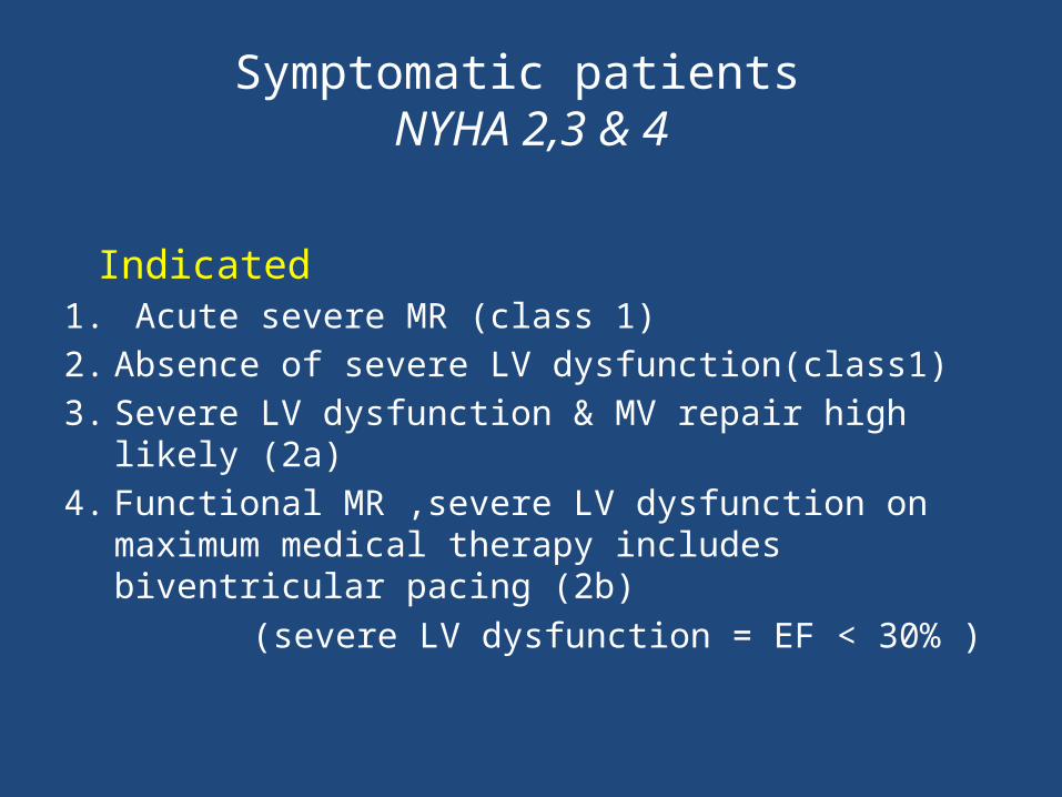
Symptomatic patients NYHA 2,3 & 4
Indicated 1. Acute severe MR (class 1)2. Absence of severe LV dysfunction(class1)3. Severe LV dysfunction & MV repair high likely (2a)4. Functional MR ,severe LV dysfunction on maximum
medical therapy includes biventricular pacing (2b) (severe LV dysfunction = EF < 30% )

Indications for Asymptomatic patients
• Chronic severe MR and mild to moderate LV dysfunction (class1)
• Chronic sever MR with normal LV function High likely successful repair (2a)New onset atrial fibrillation (2a)Pulmonary hypertension (2a)
• Not indicated (class 3)Normal LV function & doubt about feasibility
of repairMild or moderate MR
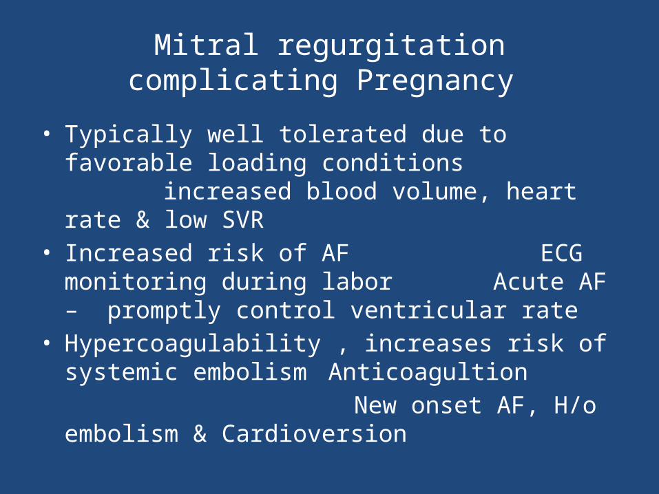
Mitral regurgitation complicating Pregnancy
• Typically well tolerated due to favorable loading conditionsincreased blood volume, heart rate & low SVR
• Increased risk of AF ECG monitoring during laborAcute AF – promptly control ventricular rate
• Hypercoagulability , increases risk of systemic embolismAnticoagultion
New onset AF, H/o embolism & Cardioversion
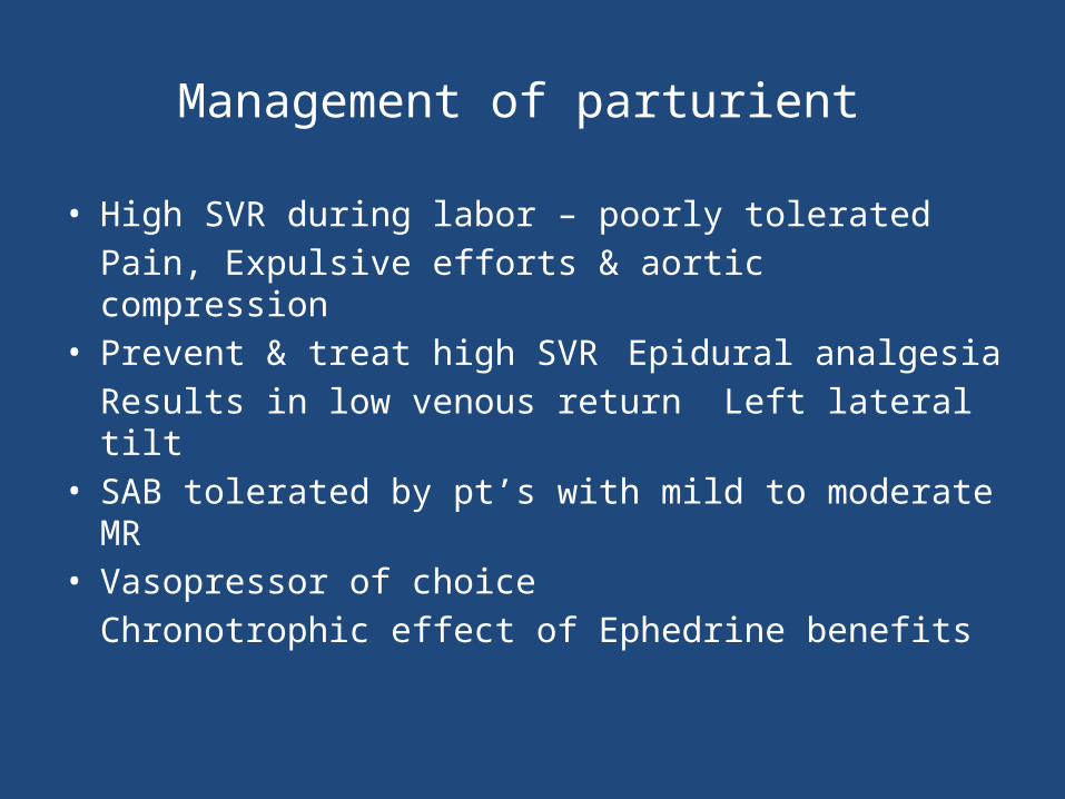
Management of parturient
• High SVR during labor – poorly toleratedPain, Expulsive efforts & aortic compression
• Prevent & treat high SVR Epidural analgesia
Results in low venous return Left lateral tilt
• SAB tolerated by pt’s with mild to moderate MR• Vasopressor of choice
Chronotrophic effect of Ephedrine benefits

Thank you
www.anaesthesia.co.in