Embed Size (px)
Citation preview
Mitochondrial-Produced
Reactive Oxygen Species
Matthew Zimmerman, PhD
Assistant Professor
Cellular & Integrative Physiology
University of Nebraska Medical Center
Summer 2012 BIOC 998-590 UNL
Karolinska Institutet
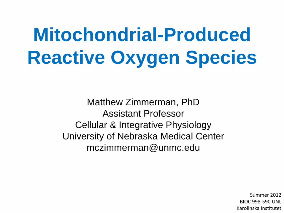
Lecture Outline
1. Complex I and Complex III – Primary
sources of ROS in mitochondria
2. Other sources of mitochondrial-
produced reactive oxygen species
(ROS)
NADPH oxidase (Nox4)??
3. Mitochondrial-localized antioxidants
4. Methods to measure mitochondrial-
produced ROS
5. Diseases associated with
mitochondrial-produced ROS
Amyotrophic lateral sclerosis
(ALS; aka Lou Gehrig’s
disease) Murphy MP. (2009) Biochem J. 417:1-13)
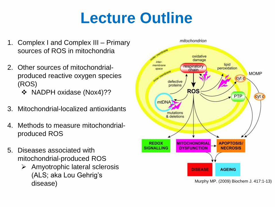
Sources of Reactive Oxygen Species
• Mitochondria
• NADPH oxidase
• Xanthine oxidase
• Lipoxygenase
• Nitric oxide synthases
Turrens JF. (2003) J Physiol. 552.2:335-344
NADPH oxidase
Mitochondrial-Produced ROS
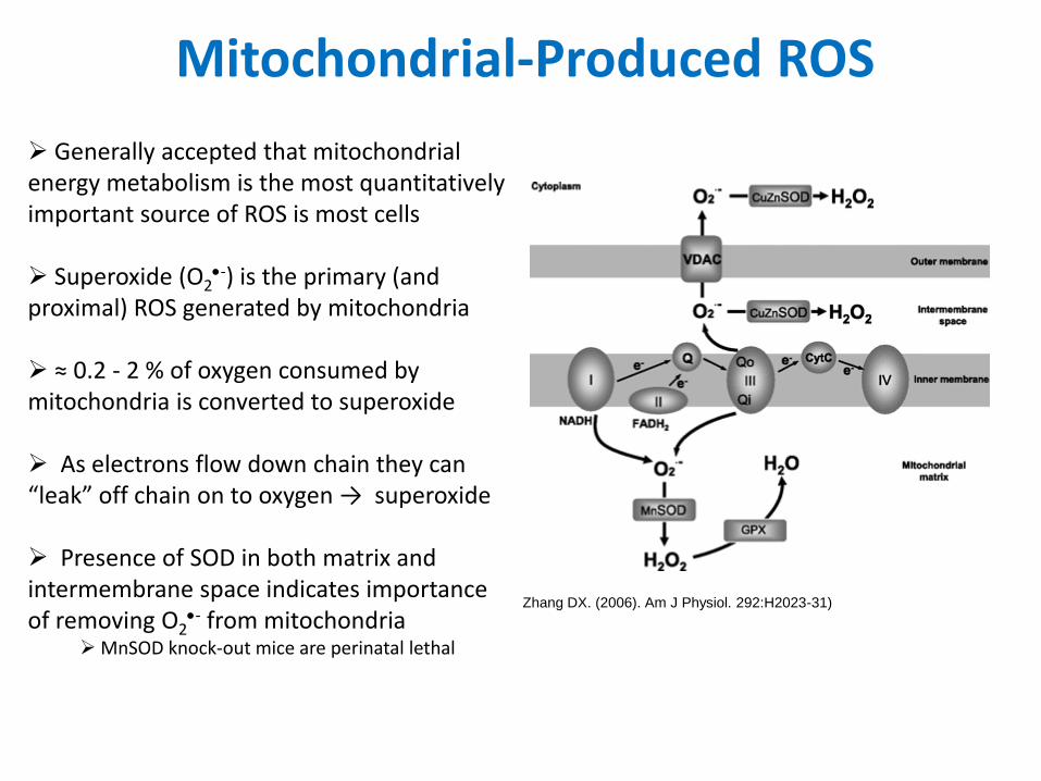
Generally accepted that mitochondrial energy metabolism is the most quantitatively important source of ROS is most cells
Superoxide (O2
-) is the primary (and proximal) ROS generated by mitochondria
≈ 0.2 - 2 % of oxygen consumed by mitochondria is converted to superoxide
As electrons flow down chain they can “leak” off chain on to oxygen → superoxide
Presence of SOD in both matrix and intermembrane space indicates importance of removing O2
- from mitochondria MnSOD knock-out mice are perinatal lethal
Zhang DX. (2006). Am J Physiol. 292:H2023-31)
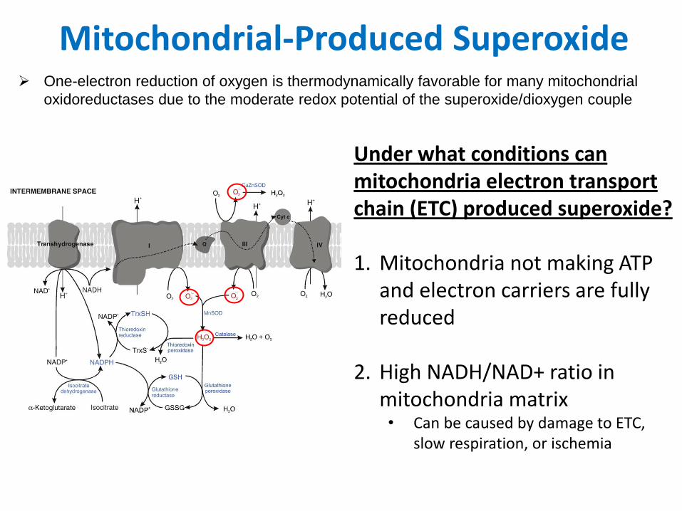
Mitochondrial-Produced Superoxide One-electron reduction of oxygen is thermodynamically favorable for many mitochondrial
oxidoreductases due to the moderate redox potential of the superoxide/dioxygen couple
Under what conditions can mitochondria electron transport chain (ETC) produced superoxide? 1. Mitochondria not making ATP
and electron carriers are fully reduced
2. High NADH/NAD+ ratio in mitochondria matrix • Can be caused by damage to ETC,
slow respiration, or ischemia
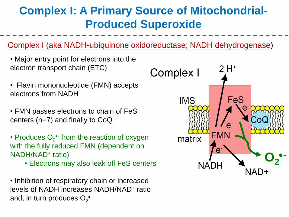
Complex I: A Primary Source of Mitochondrial-
Produced Superoxide
Complex I (aka NADH-ubiquinone oxidoreductase; NADH dehydrogenase)
• Major entry point for electrons into the
electron transport chain (ETC)
• Flavin mononucleotide (FMN) accepts
electrons from NADH
• FMN passes electrons to chain of FeS
centers (n=7) and finally to CoQ
• Produces O2- from the reaction of oxygen
with the fully reduced FMN (dependent on
NADH/NAD+ ratio)
• Electrons may also leak off FeS centers
• Inhibition of respiratory chain or increased
levels of NADH increases NADH/NAD+ ratio
and, in turn produces O2-
O2-
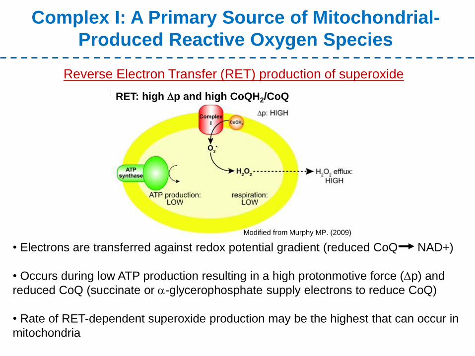
Complex I: A Primary Source of Mitochondrial-
Produced Reactive Oxygen Species
Reverse Electron Transfer (RET) production of superoxide
• Electrons are transferred against redox potential gradient (reduced CoQ NAD+)
• Occurs during low ATP production resulting in a high protonmotive force (Dp) and
reduced CoQ (succinate or a-glycerophosphate supply electrons to reduce CoQ)
• Rate of RET-dependent superoxide production may be the highest that can occur in
mitochondria
RET: high Dp and high CoQH2/CoQ
Modified from Murphy MP. (2009)
Complex I: A Primary Source of Mitochondrial-
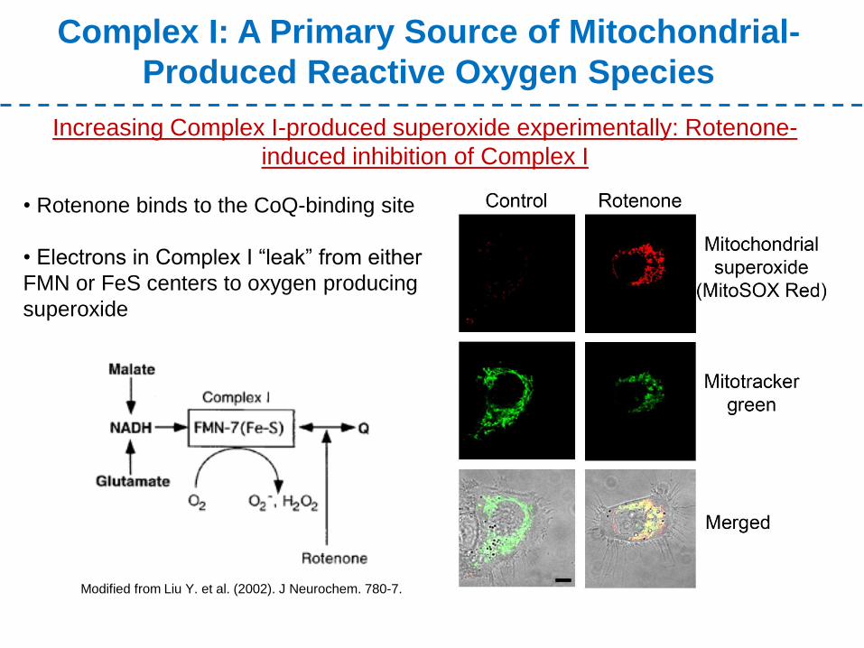
Produced Reactive Oxygen Species
Increasing Complex I-produced superoxide experimentally: Rotenone-
induced inhibition of Complex I
• Rotenone binds to the CoQ-binding site
• Electrons in Complex I “leak” from either
FMN or FeS centers to oxygen producing
superoxide
Modified from Liu Y. et al. (2002). J Neurochem. 780-7.
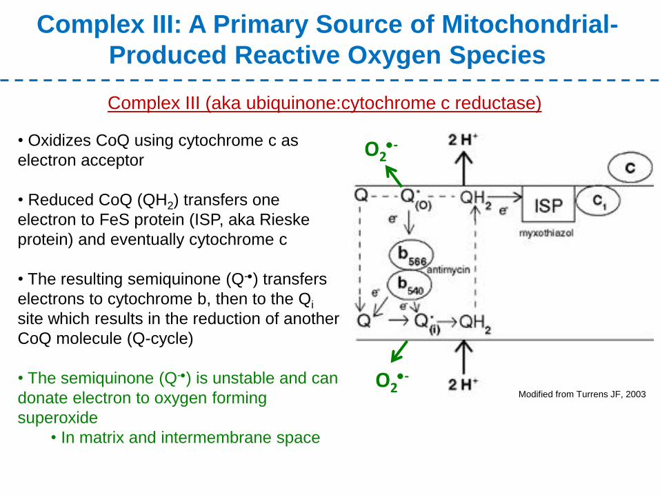
Complex III: A Primary Source of Mitochondrial-
Produced Reactive Oxygen Species
• Oxidizes CoQ using cytochrome c as
electron acceptor
• Reduced CoQ (QH2) transfers one
electron to FeS protein (ISP, aka Rieske
protein) and eventually cytochrome c
• The resulting semiquinone (Q-) transfers
electrons to cytochrome b, then to the Qi
site which results in the reduction of another
CoQ molecule (Q-cycle)
• The semiquinone (Q-) is unstable and can
donate electron to oxygen forming
superoxide
• In matrix and intermembrane space
Complex III (aka ubiquinone:cytochrome c reductase)
Modified from Turrens JF, 2003
O2-
O2-
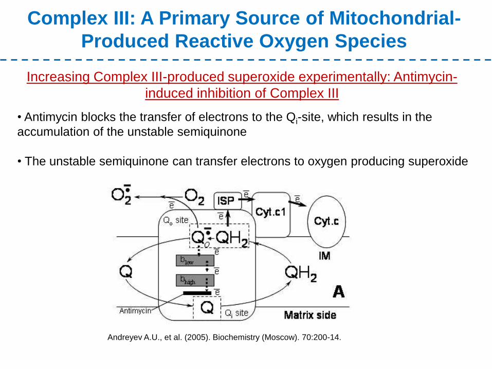
Complex III: A Primary Source of Mitochondrial-
Produced Reactive Oxygen Species
Increasing Complex III-produced superoxide experimentally: Antimycin-
induced inhibition of Complex III
• Antimycin blocks the transfer of electrons to the Qi-site, which results in the
accumulation of the unstable semiquinone
• The unstable semiquinone can transfer electrons to oxygen producing superoxide
Andreyev A.U., et al. (2005). Biochemistry (Moscow). 70:200-14.
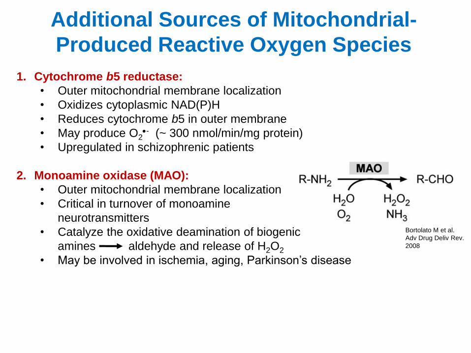
Additional Sources of Mitochondrial-
Produced Reactive Oxygen Species
1. Cytochrome b5 reductase:
• Outer mitochondrial membrane localization
• Oxidizes cytoplasmic NAD(P)H
• Reduces cytochrome b5 in outer membrane
• May produce O2- (~ 300 nmol/min/mg protein)
• Upregulated in schizophrenic patients
2. Monoamine oxidase (MAO):
• Outer mitochondrial membrane localization
• Critical in turnover of monoamine
neurotransmitters
• Catalyze the oxidative deamination of biogenic
amines aldehyde and release of H2O2
• May be involved in ischemia, aging, Parkinson’s disease
Bortolato M et al.
Adv Drug Deliv Rev.
2008
3. Dihyroorotate dehydrogenase (DHOH):
• Located at the outer surface of inner membrane
• In the process of pyrimidine nucleotide synthesis, DHOH converts
dihyroorotate to orotate
• Electron receptor is coenzyme Q and in absence of coenzyme Q produces
H2O2 (in vitro)
• Role in producing ROS in vivo remains unclear and controversial
4. Dehydrogenase of a-glycerophosphate:
• Located at the outer surface of inner membrane
• Uses coenzyme Q as electron receptor and catalyzes oxidation of
glycerol-3-phosphate to dihydroxyacetone
• Studies in mice and drosophila suggest it produces H2O2
5. Aconitase:
• Localized in matrix
• Catalyzes conversion of citrate to isocitrate (tricarboxylic acid (TCA) cycle)
• Inactivated by O2- and, in turn, produces OH most likely via Fe2+ release
Additional Sources of Mitochondrial-
Produced Reactive Oxygen Species
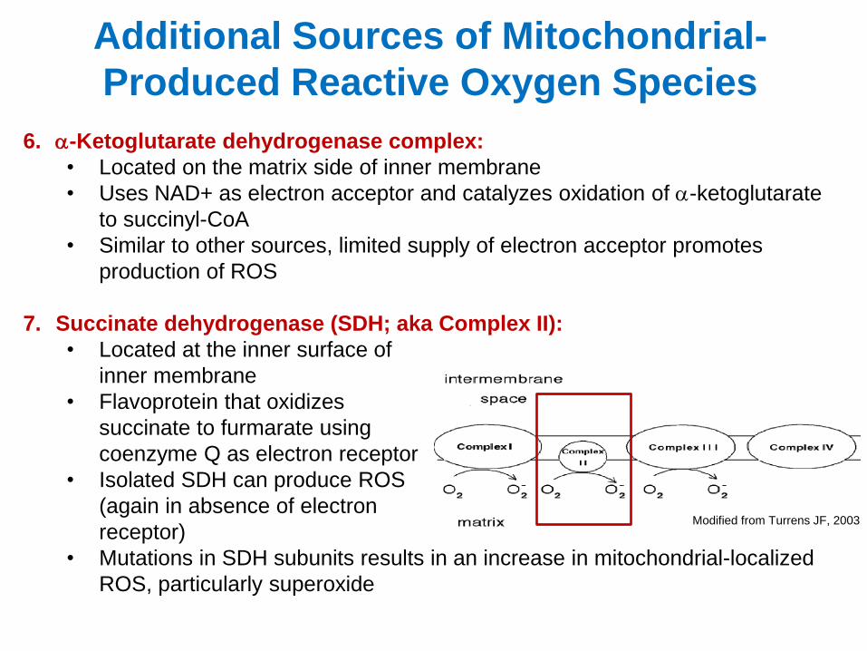
6. a-Ketoglutarate dehydrogenase complex:
• Located on the matrix side of inner membrane
• Uses NAD+ as electron acceptor and catalyzes oxidation of a-ketoglutarate
to succinyl-CoA
• Similar to other sources, limited supply of electron acceptor promotes
production of ROS
7. Succinate dehydrogenase (SDH; aka Complex II):
• Located at the inner surface of
inner membrane
• Flavoprotein that oxidizes
succinate to furmarate using
coenzyme Q as electron receptor
• Isolated SDH can produce ROS
(again in absence of electron
receptor)
• Mutations in SDH subunits results in an increase in mitochondrial-localized
ROS, particularly superoxide
Modified from Turrens JF, 2003
Additional Sources of Mitochondrial-
Produced Reactive Oxygen Species
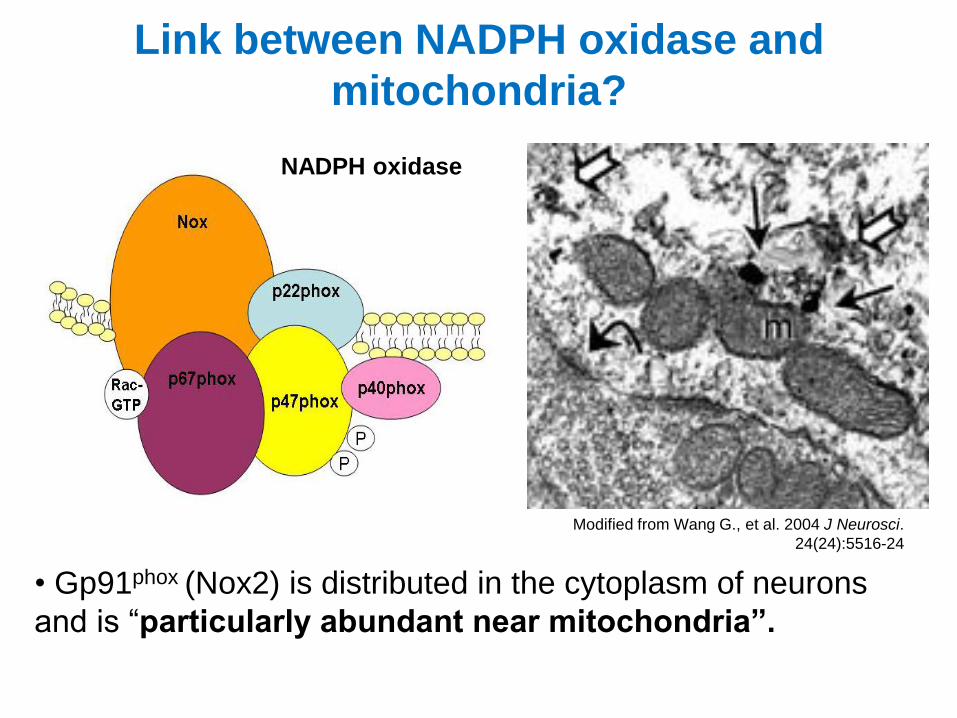
Link between NADPH oxidase and
mitochondria?
• Gp91phox (Nox2) is distributed in the cytoplasm of neurons
and is “particularly abundant near mitochondria”.
Modified from Wang G., et al. 2004 J Neurosci.
24(24):5516-24
NADPH oxidase
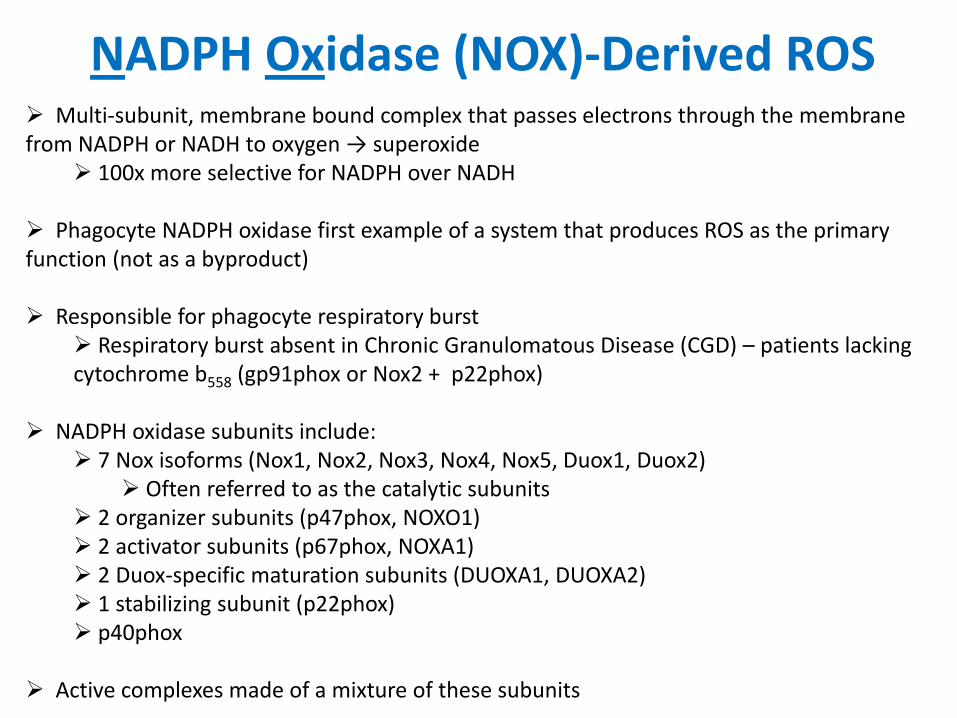
NADPH Oxidase (NOX)-Derived ROS Multi-subunit, membrane bound complex that passes electrons through the membrane from NADPH or NADH to oxygen → superoxide
100x more selective for NADPH over NADH
Phagocyte NADPH oxidase first example of a system that produces ROS as the primary function (not as a byproduct)
Responsible for phagocyte respiratory burst
Respiratory burst absent in Chronic Granulomatous Disease (CGD) – patients lacking cytochrome b558 (gp91phox or Nox2 + p22phox)
NADPH oxidase subunits include:
7 Nox isoforms (Nox1, Nox2, Nox3, Nox4, Nox5, Duox1, Duox2) Often referred to as the catalytic subunits
2 organizer subunits (p47phox, NOXO1) 2 activator subunits (p67phox, NOXA1) 2 Duox-specific maturation subunits (DUOXA1, DUOXA2) 1 stabilizing subunit (p22phox) p40phox
Active complexes made of a mixture of these subunits
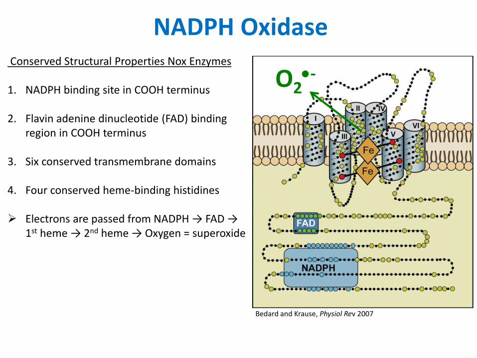
NADPH Oxidase Conserved Structural Properties Nox Enzymes 1. NADPH binding site in COOH terminus
2. Flavin adenine dinucleotide (FAD) binding
region in COOH terminus
3. Six conserved transmembrane domains
4. Four conserved heme-binding histidines
Electrons are passed from NADPH → FAD → 1st heme → 2nd heme → Oxygen = superoxide
Bedard and Krause, Physiol Rev 2007
O2-
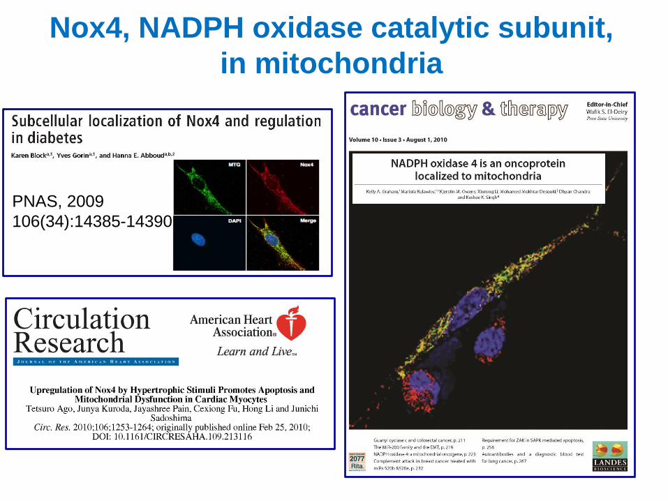
Nox4, NADPH oxidase catalytic subunit,
in mitochondria
PNAS, 2009
106(34):14385-14390
0
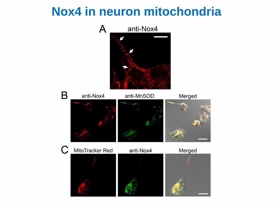
Nox4 in neuron mitochondria
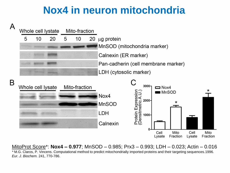
MitoProt Score*: Nox4 – 0.977; MnSOD – 0.985; Prx3 – 0.993; LDH – 0.023; Actin – 0.016 * M.G. Claros, P. Vincens. Computational method to predict mitochondrially imported proteins and their targeting sequences.1996.
Eur. J. Biochem. 241, 770-786.
Nox4 in neuron mitochondria
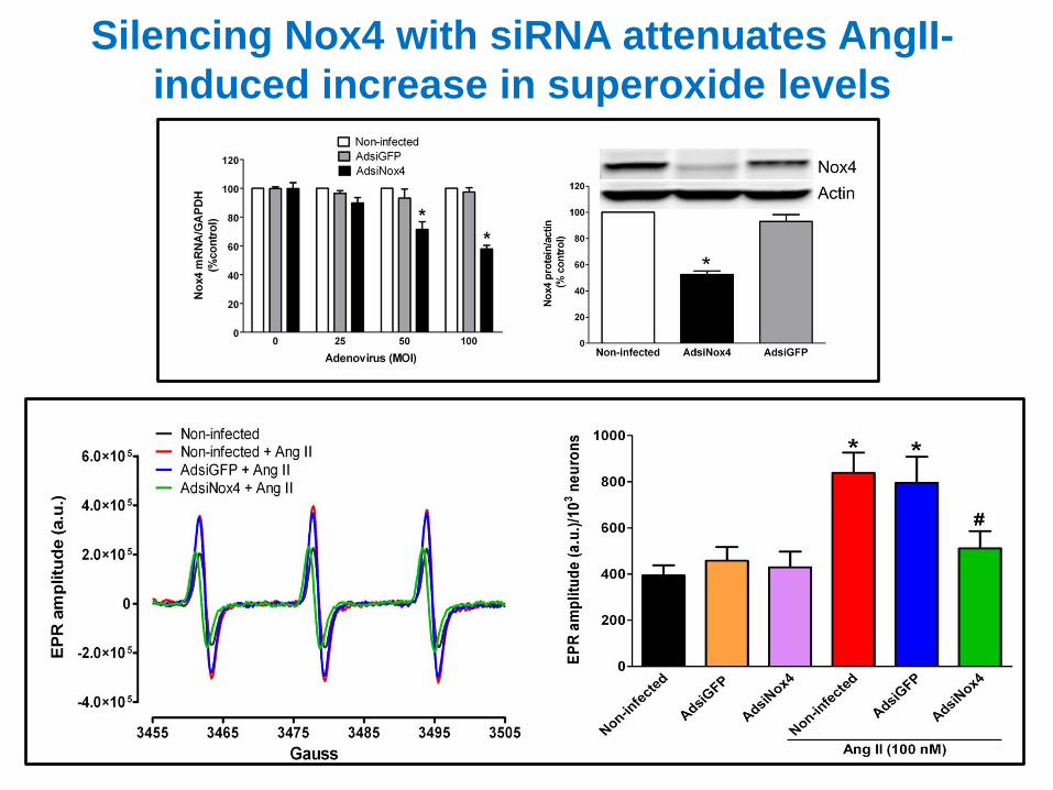
Silencing Nox4 with siRNA attenuates AngII-
induced increase in superoxide levels
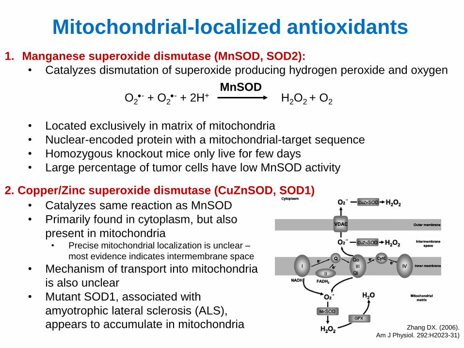
Mitochondrial-localized antioxidants
1. Manganese superoxide dismutase (MnSOD, SOD2):
• Catalyzes dismutation of superoxide producing hydrogen peroxide and oxygen
• Located exclusively in matrix of mitochondria
• Nuclear-encoded protein with a mitochondrial-target sequence
• Homozygous knockout mice only live for few days
• Large percentage of tumor cells have low MnSOD activity
O2- + O2
- + 2H+ H2O2 + O2
MnSOD
2. Copper/Zinc superoxide dismutase (CuZnSOD, SOD1)
• Catalyzes same reaction as MnSOD
• Primarily found in cytoplasm, but also
present in mitochondria • Precise mitochondrial localization is unclear –
most evidence indicates intermembrane space
• Mechanism of transport into mitochondria
is also unclear
• Mutant SOD1, associated with
amyotrophic lateral sclerosis (ALS),
appears to accumulate in mitochondria Zhang DX. (2006).
Am J Physiol. 292:H2023-31)
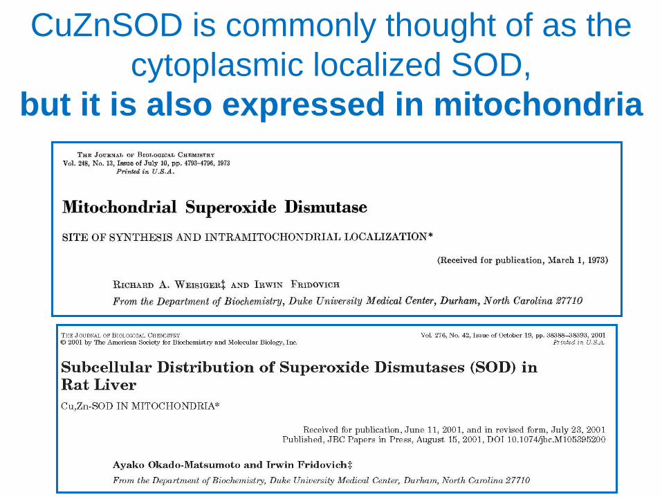
CuZnSOD is commonly thought of as the
cytoplasmic localized SOD,
but it is also expressed in mitochondria
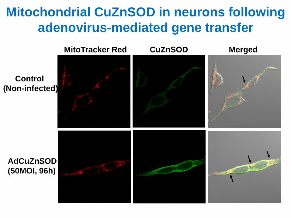
Mitochondrial CuZnSOD in neurons following
adenovirus-mediated gene transfer
Control
(Non-infected)
AdCuZnSOD
(50MOI, 96h)
MitoTracker Red CuZnSOD Merged
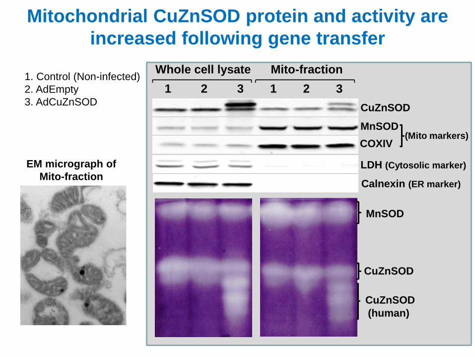
Mitochondrial CuZnSOD protein and activity are
increased following gene transfer
EM micrograph of
Mito-fraction
1 2 3 1 2 3 1. Control (Non-infected)
2. AdEmpty
3. AdCuZnSOD
Whole cell lysate Mito-fraction
MnSOD
LDH (Cytosolic marker)
COXIV
CuZnSOD
MnSOD
CuZnSOD
(human)
CuZnSOD
Calnexin (ER marker)
(Mito markers)
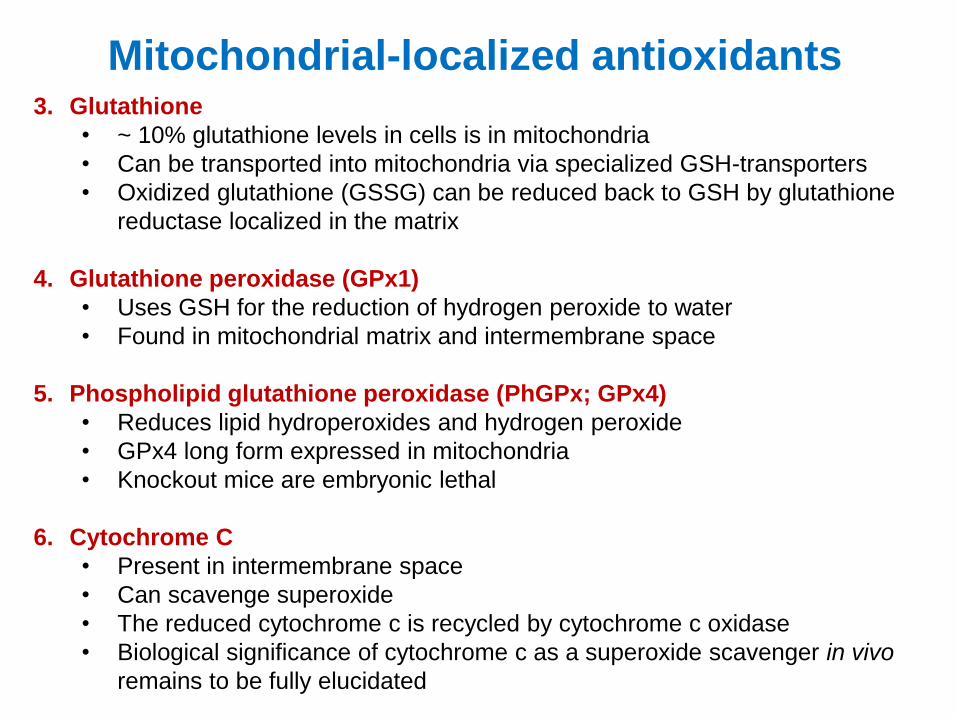
Mitochondrial-localized antioxidants 3. Glutathione
• ~ 10% glutathione levels in cells is in mitochondria
• Can be transported into mitochondria via specialized GSH-transporters
• Oxidized glutathione (GSSG) can be reduced back to GSH by glutathione
reductase localized in the matrix
4. Glutathione peroxidase (GPx1)
• Uses GSH for the reduction of hydrogen peroxide to water
• Found in mitochondrial matrix and intermembrane space
5. Phospholipid glutathione peroxidase (PhGPx; GPx4)
• Reduces lipid hydroperoxides and hydrogen peroxide
• GPx4 long form expressed in mitochondria
• Knockout mice are embryonic lethal
6. Cytochrome C
• Present in intermembrane space
• Can scavenge superoxide
• The reduced cytochrome c is recycled by cytochrome c oxidase
• Biological significance of cytochrome c as a superoxide scavenger in vivo
remains to be fully elucidated
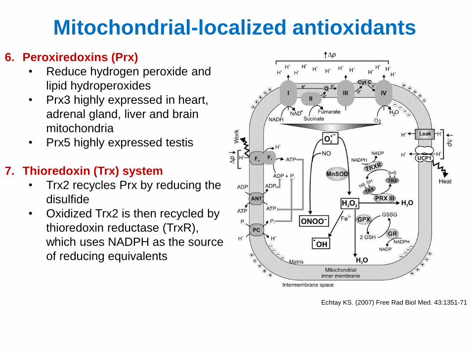
Mitochondrial-localized antioxidants
6. Peroxiredoxins (Prx)
• Reduce hydrogen peroxide and
lipid hydroperoxides
• Prx3 highly expressed in heart,
adrenal gland, liver and brain
mitochondria
• Prx5 highly expressed testis
7. Thioredoxin (Trx) system
• Trx2 recycles Prx by reducing the
disulfide
• Oxidized Trx2 is then recycled by
thioredoxin reductase (TrxR),
which uses NADPH as the source
of reducing equivalents
Echtay KS. (2007) Free Rad Biol Med. 43:1351-71
Smith R.A.J., et al. (2008) Ann NY
Acad Sci. 1147:105-111
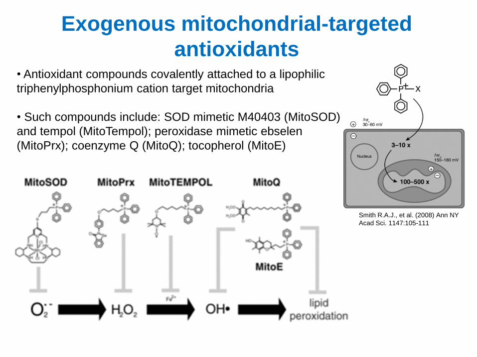
Exogenous mitochondrial-targeted
antioxidants • Antioxidant compounds covalently attached to a lipophilic
triphenylphosphonium cation target mitochondria
• Such compounds include: SOD mimetic M40403 (MitoSOD)
and tempol (MitoTempol); peroxidase mimetic ebselen
(MitoPrx); coenzyme Q (MitoQ); tocopherol (MitoE)
Methods for measuring mitochondrial-produced ROS
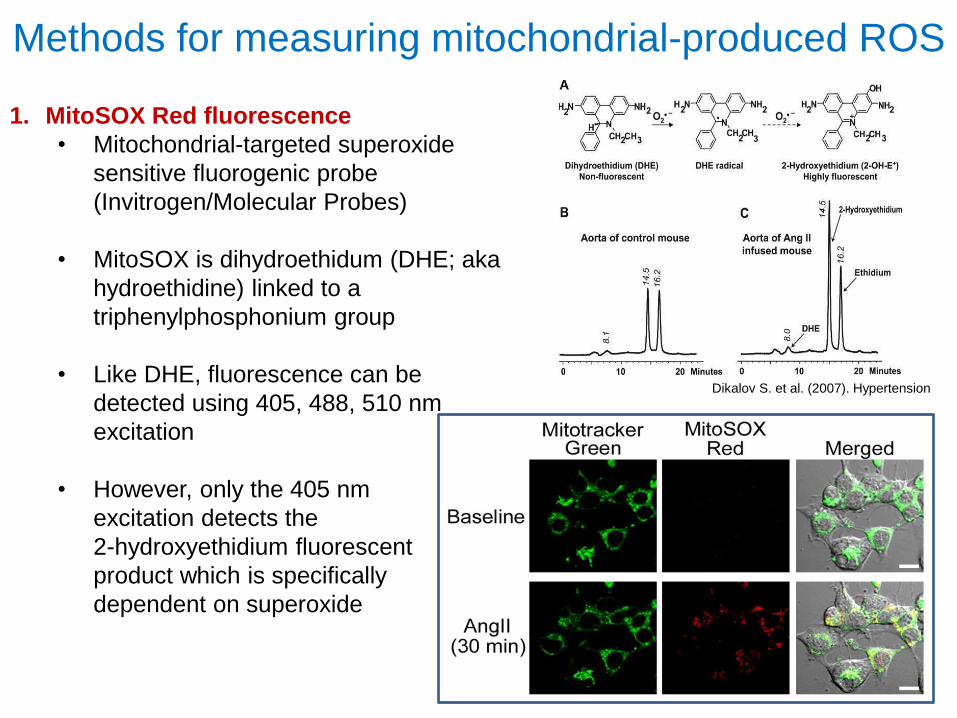
1. MitoSOX Red fluorescence
• Mitochondrial-targeted superoxide
sensitive fluorogenic probe
(Invitrogen/Molecular Probes)
• MitoSOX is dihydroethidum (DHE; aka
hydroethidine) linked to a
triphenylphosphonium group
• Like DHE, fluorescence can be
detected using 405, 488, 510 nm
excitation
• However, only the 405 nm
excitation detects the
2-hydroxyethidium fluorescent
product which is specifically
dependent on superoxide
Dikalov S. et al. (2007). Hypertension
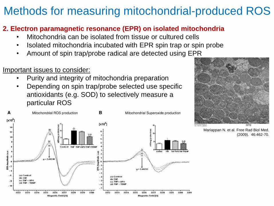
Methods for measuring mitochondrial-produced ROS
2. Electron paramagnetic resonance (EPR) on isolated mitochondria
• Mitochondria can be isolated from tissue or cultured cells
• Isolated mitochondria incubated with EPR spin trap or spin probe
• Amount of spin trap/probe radical are detected using EPR
Important issues to consider:
• Purity and integrity of mitochondria preparation
• Depending on spin trap/probe selected use specific
antioxidants (e.g. SOD) to selectively measure a
particular ROS
Mariappan N. et al. Free Rad Biol Med.
(2009). 46:462-70.
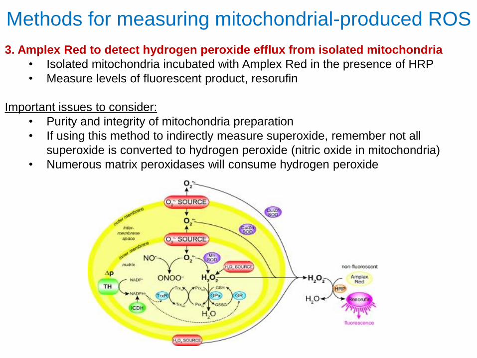
Methods for measuring mitochondrial-produced ROS
3. Amplex Red to detect hydrogen peroxide efflux from isolated mitochondria
• Isolated mitochondria incubated with Amplex Red in the presence of HRP
• Measure levels of fluorescent product, resorufin
Important issues to consider:
• Purity and integrity of mitochondria preparation
• If using this method to indirectly measure superoxide, remember not all
superoxide is converted to hydrogen peroxide (nitric oxide in mitochondria)
• Numerous matrix peroxidases will consume hydrogen peroxide
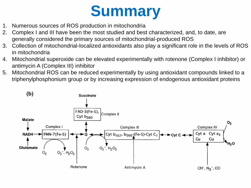
Summary 1. Numerous sources of ROS production in mitochondria
2. Complex I and III have been the most studied and best characterized, and, to date, are
generally considered the primary sources of mitochondrial-produced ROS
3. Collection of mitochondrial-localized antioxidants also play a significant role in the levels of ROS
in mitochondria
4. Mitochondrial superoxide can be elevated experimentally with rotenone (Complex I inhibitor) or
antimycin A (Complex III) inhibitor
5. Mitochondrial ROS can be reduced experimentally by using antioxidant compounds linked to a
triphenylphosphonium group or by increasing expression of endogenous antioxidant proteins

• Fatal neurodegenerative disease that specifically targets motor neurons in the spinal cord, brain stem, and cortex
• Most common adult motor neuron disease; 5,600 cases diagnosed each year in U.S.
• Disease onset usually begins with weakness in arms and legs and quickly progresses to total paralysis
• Patients generally die of respiratory failure 2-5 years after the first symptoms appear
• ALS often referred to as Lou Gehrig’s disease
Amyotrophic Lateral Sclerosis (ALS)
From alsa.org
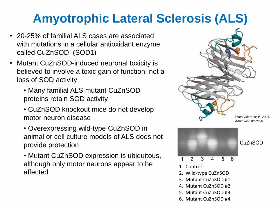
• 20-25% of familial ALS cases are associated
with mutations in a cellular antioxidant enzyme
called CuZnSOD (SOD1)
• Mutant CuZnSOD-induced neuronal toxicity is
believed to involve a toxic gain of function; not a
loss of SOD activity
• Many familial ALS mutant CuZnSOD
proteins retain SOD activity
• CuZnSOD knockout mice do not develop
motor neuron disease
• Overexpressing wild-type CuZnSOD in
animal or cell culture models of ALS does not
provide protection
• Mutant CuZnSOD expression is ubiquitous,
although only motor neurons appear to be
affected
From Valentine JS, 2005. Annu. Rev. Biochem
1. Control 2. Wild-type CuZnSOD 3. Mutant CuZnSOD #1 4. Mutant CuZnSOD #2 5. Mutant CuZnSOD #3 6. Mutant CuZnSOD #4
Amyotrophic Lateral Sclerosis (ALS)
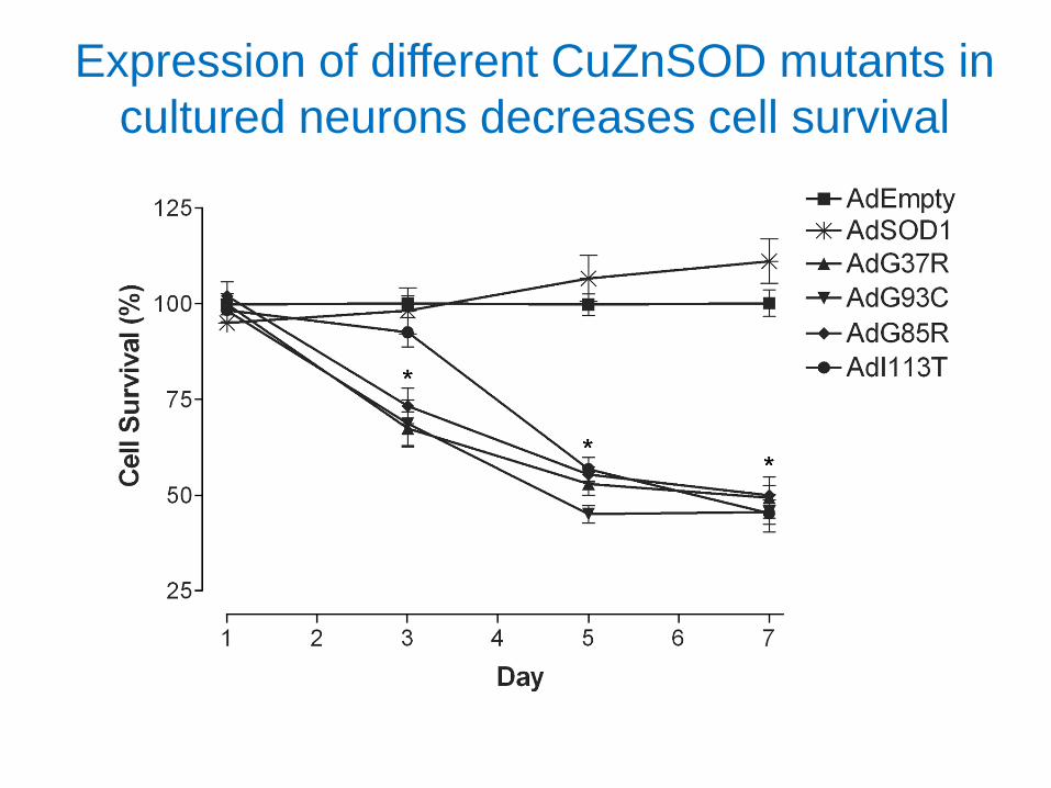
Expression of different CuZnSOD mutants in
cultured neurons decreases cell survival
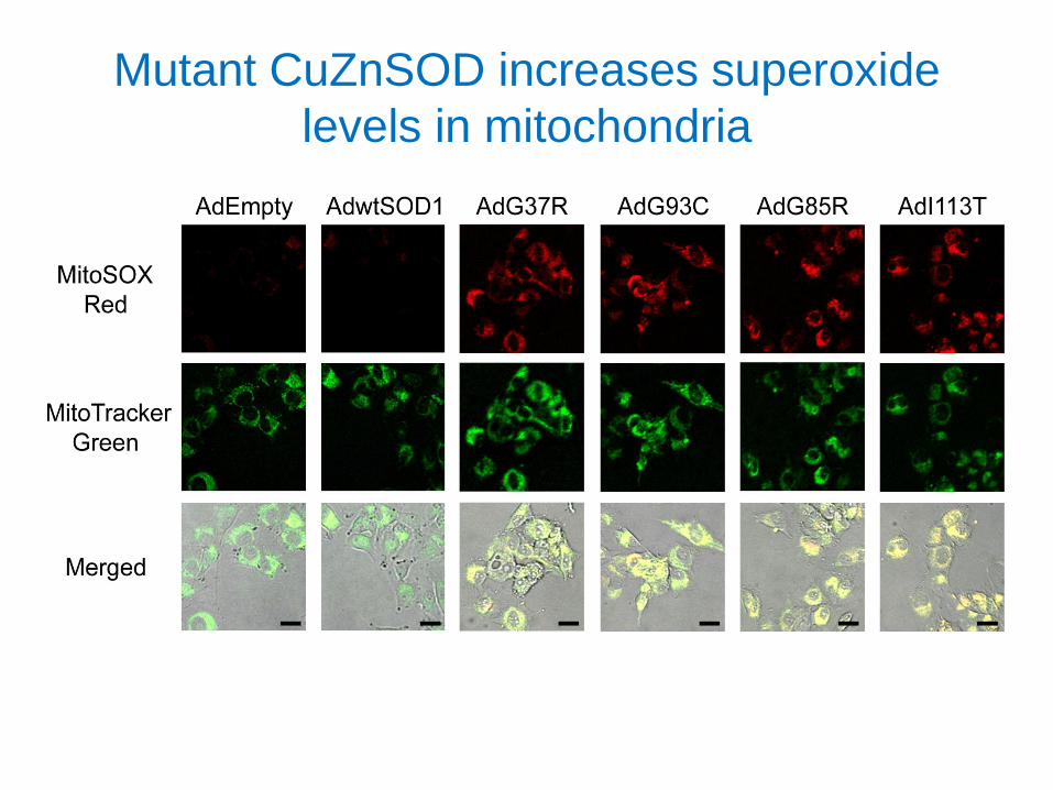
Mutant CuZnSOD increases superoxide
levels in mitochondria
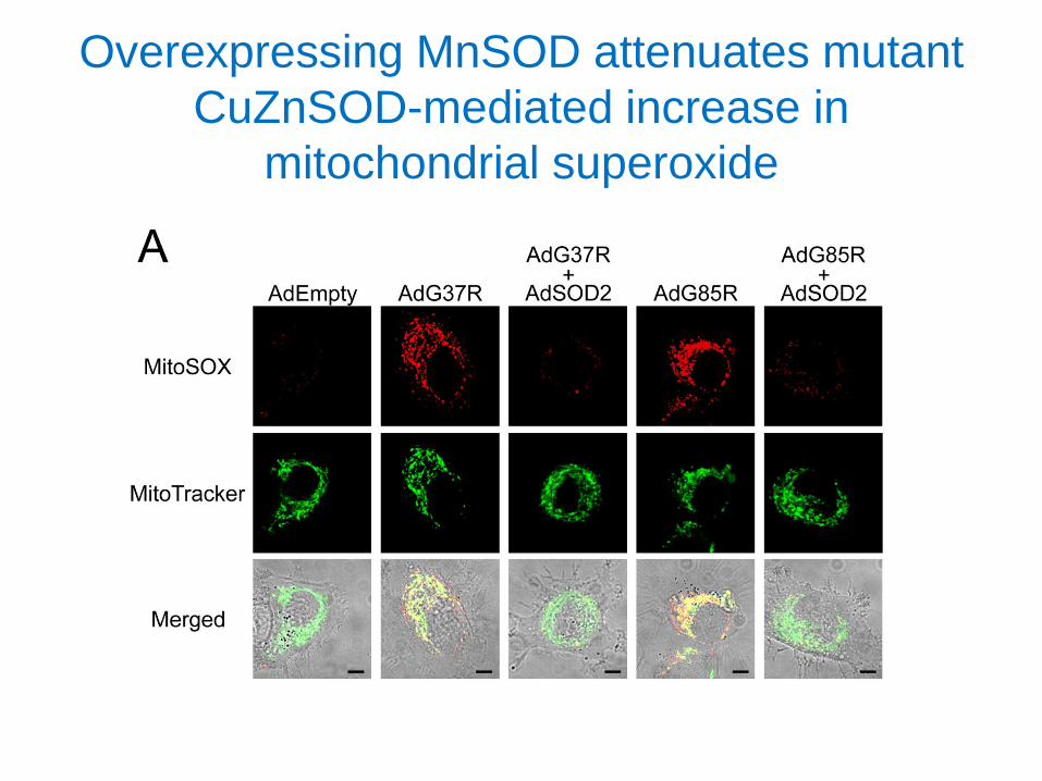
Overexpressing MnSOD attenuates mutant
CuZnSOD-mediated increase in
mitochondrial superoxide
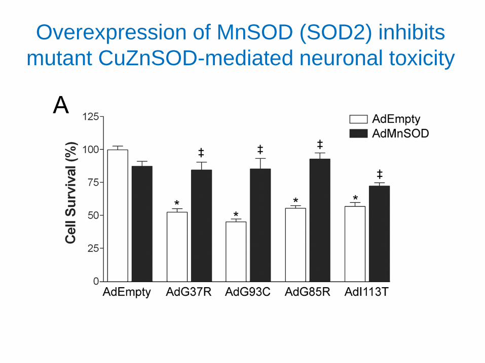
Overexpression of MnSOD (SOD2) inhibits
mutant CuZnSOD-mediated neuronal toxicity
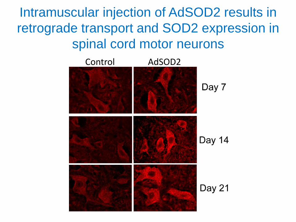
Intramuscular injection of AdSOD2 results in
retrograde transport and SOD2 expression in
spinal cord motor neurons
Control AdSOD2
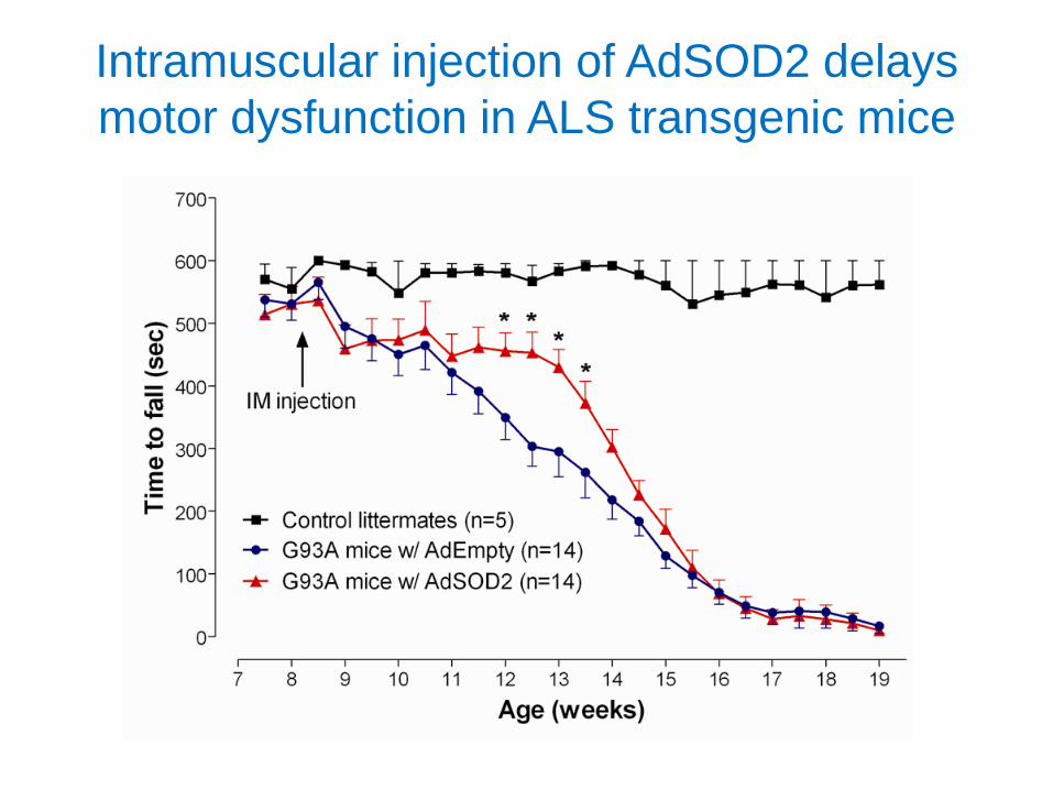
Intramuscular injection of AdSOD2 delays
motor dysfunction in ALS transgenic mice