Embed Size (px)
Citation preview
Int. J. Mol. Sci. 2013, 14, 6306-6344; doi:10.3390/ijms14036306
International Journal of
Molecular Sciences ISSN 1422-0067
www.mdpi.com/journal/ijms
Review
Mitochondria and Reactive Oxygen Species: Physiology and Pathophysiology
Subhashini Bolisetty 1 and Edgar A. Jaimes 1,2,*
1 Nephrology Division, University of Alabama at Birmingham, Birmingham, AL 35294, USA;
E-Mail: [email protected] 2 Veterans Affairs Medical Center, Birmingham, AL 35233, USA
* Author to whom correspondence should be addressed; E-Mail: [email protected];
Tel.: +1-205-934-0544, Fax: +1-205-976-6288.
Received: 10 January 2013; in revised form: 8 March 2013 / Accepted: 11 March 2013 /
Published: 19 March 2013
Abstract: The air that we breathe contains nearly 21% oxygen, most of which is utilized
by mitochondria during respiration. While we cannot live without it, it was perceived as a
bane to aerobic organisms due to the generation of reactive oxygen and nitrogen
metabolites by mitochondria and other cellular compartments. However, this dogma was
challenged when these species were demonstrated to modulate cellular responses through
altering signaling pathways. In fact, since this discovery of a dichotomous role of reactive
species in immune function and signal transduction, research in this field grew at an
exponential pace and the pursuit for mechanisms involved began. Due to a significant
number of review articles present on the reactive species mediated cell death, we have
focused on emerging novel pathways such as autophagy, signaling and maintenance of the
mitochondrial network. Despite its role in several processes, increased reactive species
generation has been associated with the origin and pathogenesis of a plethora of diseases.
While it is tempting to speculate that anti-oxidant therapy would protect against these
disorders, growing evidence suggests that this may not be true. This further supports our
belief that these reactive species play a fundamental role in maintenance of cellular and
tissue homeostasis.
Keywords: mitochondria; reactive oxygen species; nitric oxide; hydrogen peroxide;
mitochondria fission; mitochondria fusion; autophagy; mitochondria network; cell signaling
OPEN ACCESS
Int. J. Mol. Sci. 2013, 14 6307
1. Introduction
Mitochondria were first identified over a century ago and were initially termed as “bioblasts” by
Richard Altmann who described them as “elementary organisms” living inside cells [1]. In fact this
theory of endosymbiosis that mitochondria are the direct descendants of a bacterial endosymbiont is
still one of the most widely accepted theories of mitochondrial evolution [2]. The term mitochondria,
was later coined by Carl Benda and literally means “mitos-thread” and “chondrion-granule” [1]. The
role of mitochondria in the cell was initially presumed to be only to generate energy in the form of
adenosine triphosphate (ATP) and is still referred to as the “powerhouse of the cell”. However,
research in the past few decades has provided compelling evidence to suggest that mitochondria are
actively involved in a multitude of cellular activities including, signaling, proliferation and death. In
fact, while most eukaryotic cells contain mitochondria, the size, number and location of mitochondria
in a cell vary significantly based on the cellular needs. For instance, in neuronal cells, mitochondria
accumulate predominantly at high energy demanding sites such as presynaptic terminals, nodes of
Ranvier and active growth cones and branches [3]. Given the role of mitochondria in a variety of
cellular processes, it is not surprising that damage to the mitochondria has been implicated in the
pathogenesis of end-organ injury in a variety of diseases [4–23].
How does a single organelle control the fate of the cell? This question has captivated scientists and
studies over the past few decades have revealed fascinating mechanisms. An extensively studied and
established mechanism relies on the generation of free radical species that determine the outcome of
the cellular processes involved. This review will mainly focus on the role of these reactive
oxygen/nitrogen species in the modulation of cellular activities. We will summarize some of the major
pathways and molecular mechanisms that are regulated by reactive oxygen species (ROS) and reactive
nitrogen species (RNS) and given the wealth of knowledge that exists for these species, this review
will highlight recent developments in ROS/RNS mediated signaling pathways and their role in the
regulation of cellular processes, such as autophagy, mitochondria fusion and fission.
2. Mitochondria Structure and Function
Mitochondria are unique organelles as its structure provides compartmentalization of metabolism
(Figure 1A). They are very complex organelles that contain two phospholipid bilayers, by virtue of
which they can be categorized into 4 different segments: the outer membrane, inter-membrane space,
inner membrane and matrix [24–26].
The outer membrane of the organelle is identical to the plasma membrane in its content (equal ratio
of protein to phospholipid content by weight). It contains porins that allow molecules that are less than
5 KDa to freely diffuse through. However, larger proteins require the presence of a mitochondria
targeted sequence that will enable binding to specific transporters (translocase of the outer
membrane—TOM and inner membrane—TIM) on the membrane for entry into the organelle [26–29].
The outer membrane therefore mainly serves as a permeability barrier to the cytosolic components.
Until recently, it was presumed that the inter membrane space had no specific function and was
identical to the cytosol in its contents. However, emerging studies have suggested an important role for
this space in maintaining mitochondrial homeostasis, including protein sorting and lipid homeostasis
Int. J. Mol. Sci. 2013, 14 6308
(reviewed in [30]). The inner membrane of the mitochondria is perhaps the single most extensively
studied cell membrane component due to its relative importance in oxidative phosphorylation. This
membrane comprises of the highest number of proteins per phospholipid moiety in a cell. These
proteins are integral to the electron transport chain, ATP synthesis and transport [31,32]. The inner
membrane is also distinct from other membranes by the presence of cristae (invaginations of the
membrane), which allow for compartmentalization and increases the surface area. The inner membrane
is also less permeable to ions and molecules and helps in compartmentalization through separation of
the mitochondrial matrix from the cytosolic environment, thereby acting as an electrical insulator and
chemical barrier [32]. This helps in maintenance of the electron gradient across the membrane, which
enables generation of ATP. The mitochondrial matrix of mammalian cells contains the mitochondrial
DNA (16.5 kilobase genome) that encodes for nearly 13 proteins, some of which are involved in
oxidative phosphorylation. The remaining proteins required for the normal function of the
mitochondria are encoded by the nuclear genome and imported into the mitochondria [33]. The matrix
also contains a majority of the enzymes required for the citric acid cycle, which oxidizes acetyl
coenzyme A and in the process generates energy in the form of nicotinamide adenine dinucleotide
(NADH) and flavin adenine dinucleotide (FADH2). These molecules then serve as substrates for
oxidative phosphorylation by the proteins in the inner membrane to generate cellular energy in the
form of ATP.
3. Reactive Oxygen and Nitrogen Species
Halliwell and colleagues described free radicals as “any species capable of independent existence
that contains one or more unpaired electrons” [34]. The term ROS simply refers to a variety of reactive
molecules that are derived from oxygen and can be free radicals (superoxide (O2•−) or hydroxyl radical
(OH•)) or non-radicals (hydrogen peroxide (H2O2)). Similarly, they can be further classified as ions
(O2•−) or non-ions (H2O2). On the other hand, RNS refers to reactive species derived from nitrogen and
can be broadly classified as ions (peroxynitrite (ONOO−)) or non-ions (Nitric Oxide (NO•)). These
reactive species are formed at low levels during the execution of physiological functions of the cell. In
fact, the electron transport chain is responsible for most of the superoxide that is generated through
partial reduction of oxygen (Figure 1A) and is reviewed in detail in the following sections.
Furthermore, they are formed at different rates in a cell and differ in their activity. In terms of activity,
hydroxyl radical is the most reactive species known and is by in large responsible for the cytotoxic
effects of ROS. In contrast, reactive species such as nitric oxide and hydrogen peroxide are less
reactive and have shown to play an important role in several cellular activities. This is discussed in
detail in the following sections.
Other enzymatic systems responsible for the generation of these reactive species include but are not
limited to respiratory burst enzymes (such as NADPH oxidases –Nox1-5), amino acid oxidases,
cytochrome P450 enzymes, cycloxygenases, lipoxygenases, xanthine oxidase [35–41]. Addressing
each of these systems is beyond the scope of this review and hence we will focus specifically on
mitochondria associated reactive species.
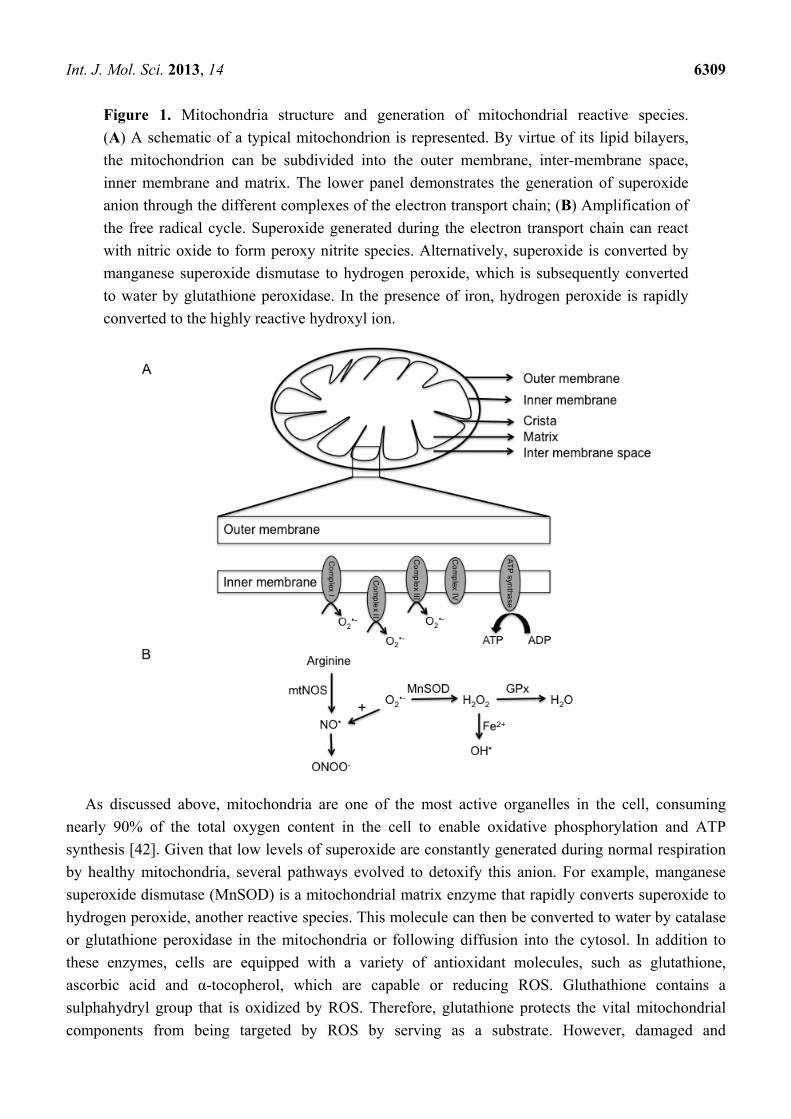
Int. J. Mol. Sci. 2013, 14 6309
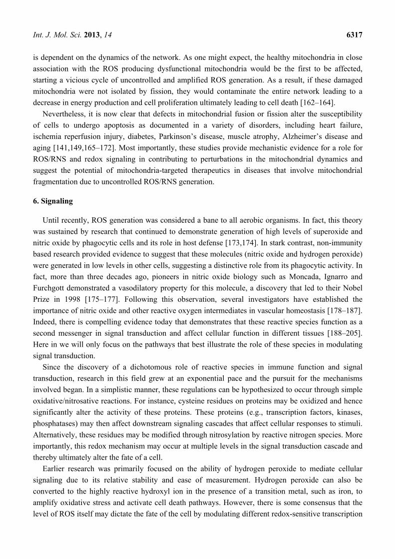
Figure 1. Mitochondria structure and generation of mitochondrial reactive species.
(A) A schematic of a typical mitochondrion is represented. By virtue of its lipid bilayers,
the mitochondrion can be subdivided into the outer membrane, inter-membrane space,
inner membrane and matrix. The lower panel demonstrates the generation of superoxide
anion through the different complexes of the electron transport chain; (B) Amplification of
the free radical cycle. Superoxide generated during the electron transport chain can react
with nitric oxide to form peroxy nitrite species. Alternatively, superoxide is converted by
manganese superoxide dismutase to hydrogen peroxide, which is subsequently converted
to water by glutathione peroxidase. In the presence of iron, hydrogen peroxide is rapidly
converted to the highly reactive hydroxyl ion.
As discussed above, mitochondria are one of the most active organelles in the cell, consuming
nearly 90% of the total oxygen content in the cell to enable oxidative phosphorylation and ATP
synthesis [42]. Given that low levels of superoxide are constantly generated during normal respiration
by healthy mitochondria, several pathways evolved to detoxify this anion. For example, manganese
superoxide dismutase (MnSOD) is a mitochondrial matrix enzyme that rapidly converts superoxide to
hydrogen peroxide, another reactive species. This molecule can then be converted to water by catalase
or glutathione peroxidase in the mitochondria or following diffusion into the cytosol. In addition to
these enzymes, cells are equipped with a variety of antioxidant molecules, such as glutathione,
ascorbic acid and α-tocopherol, which are capable or reducing ROS. Gluthathione contains a
sulphahydryl group that is oxidized by ROS. Therefore, glutathione protects the vital mitochondrial
components from being targeted by ROS by serving as a substrate. However, damaged and
Int. J. Mol. Sci. 2013, 14 6310
dysregulated mitochondria generate excessive amounts of superoxide which can damage several
mitochondrial components, including proteins, lipids and DNA. These reactions then lead to a vicious
cycle of further generation of reactive species and ultimately cell death. While hydrogen peroxide is a
reactive molecule, in the presence of transition metals, such as iron, it can be converted to hydroxyl ion
via the Fenton reaction. This iron is thought to be released through destabilization of ferritin and other
iron containing proteins by superoxide anion [43–46]. Alternatively, superoxide may react with nitric
oxide to generate peroxynitrite species. The details of these reactions are presented in Figure 1B.
Over the past few decades, research on these reactive species has soared in both physiology and
pathology. With the advent of novel technology, researchers have now been able to shed light on the
source of reactive species generation in the different sub-compartments of the mitochondria which has
been extensively reviewed by Lenoz [47]. To summarize, it was demonstrated that complex I, also
referred to as NADH CoQ reductase, catalyzes the transfer of electrons from NADH to coenzyme Q,
which is accompanied by translocation of protons from the matrix to the intermembrane space.
There is now evidence to suggest that complex I is involved in ROS production, specifically
superoxide [48–50]. Similarly, succinate dehydrogenase, complex II enzyme is responsible for the
reduction of CoQ and has also shown to be involved in generating low levels of superoxide
anion [48,51]. Complex III (ubiquinol cytochrome c reductase), on the other hand has shown to be
responsible for the superoxide generation in the intermembrane space. Superoxide generation by this
complex is significantly enhanced when the electron transfer is reduced, either due to inhibition in
respiration (actinamycin A) or an increase in membrane potential [52,53]. Interestingly, the
contribution of each of these enzymes to ROS production is different in different tissues and during
disease conditions. For instance, while complex III has been implicated as the major source of
superoxide in the heart, complex I seem to be of prime importance in the brain [54–57]. Additionally,
enzymes such as Glycerol-3-phosphate dehydrogenase, Monoamine oxidase, Dihydrolipoamide
dehydrogenase and Electron-transferring-flavoprotein dehydrogenase have also been implicated in
ROS production [58–63].
As evident from the reactions described above, most of the superoxide generated is either in the
matrix or on the inner membrane of the mitochondria that faces the matrix. While most of the reactive
species generated within the mitochondria is superoxide anion, MnSOD rapidly converts it to
hydrogen peroxide. Although hydrogen peroxide is more stable than superoxide, it can freely diffuse
out of the mitochondria into the cytosol, thereby reducing the harmful effects of these reactive species
to the mitochondria. It has also been suggested that in the presence of excessive superoxide, MnSOD is
oxidized and this further compromises the antioxidant capacity of the mitochondria and enhances
oxidative stress [64]. Alternatively, superoxide may be carried to the cytoplasm by voltage-dependent
anion channels [65].
Nitric oxide (NO•) is another reactive species that is generated by the mitochondria and research on
this molecule has gained momentum over the past few decades. Nitric oxide is generated during the
breakdown of arginine to citrulline by a family of NADPH-dependent enzymes called nitric oxide
synthases (NOS). The importance of this enzyme in physiology is underscored by the fact there are
several isoforms of NOS, including an endothelial constitutive isoform (NOS3), an inducible isoform
that is expressed in several cell types (NOS2) in response to pro-inflammatory stimuli and produces
large amounts of NO• and a neuronal isoform (NOS1). Recently, a mitochondrial isoform of NOS
Int. J. Mol. Sci. 2013, 14 6311
referred to as mtNOS has been described [66]. This isoform was found to be responsive to changes in
calcium concentration in the matrix and to play an important role in modulating mitochondrial
respiration. Following this initial study, several groups have identified the presence of mtNOS in the
mitochondria of cells from different tissues including liver, brain and kidney [67–72]. However, and in
spite of these studies there is still debate on the existence of this isoform and indeed some studies
suggest that the presence of NO• in the mitochondria may reflect pathways independent of NOS
activity that may include nitrite reductase activity and the electron transport chain [65,72–75]. Once
generated, NO• can inhibit respiration by binding to heme groups in the proteins of the electron
transport chain, including cytochrome c oxidase [76–81]. Irrespective of the source of NO•, its
presence in the mitochondria can alter the activity of a number of processes including respiration,
mitochondrial biogenesis and oxidative stress through increased production of reactive oxygen and
nitrogen species and thereby impact cell physiology [82–94].
As described above, mitochondria are a major source of reactive oxygen and nitrogen species.
Interestingly, these species in turn regulate the mitochondrial activity through several mechanisms,
including mitochondrial biogenesis, mtDNA damage, lipid peroxidation and mitochondrial membrane
permeability transition (reviewed in [83,86,89–98]). The physiological and pathological role of these
reactive species in cellular activities will be discussed below.
4. Autophagy
Autophagy (meaning self-eating) is an evolutionarily conserved catabolic process that involves an
intracellular degradation system in which cytoplasmic components, such as organelles, protein
aggregates and other macromolecules are directed to the lysosome through a physiologically regulated
process to maintain cell homeostasis. While three different types of autophagy (micro-, macro- and
chaperone-mediated autophagy) have been described, the most studied process in mammalian cells is
macro-autophagy. As the name suggests, it involves degradation of large moieties such as organelles
and protein aggregates through a tightly regulated process. This process begins with the isolation and
sequestration of the cytoplasmic components by a double-layered lipid membrane that forms an
autophagosome. This vesicle then fuses with a lysosome to form an autolysosomes where the
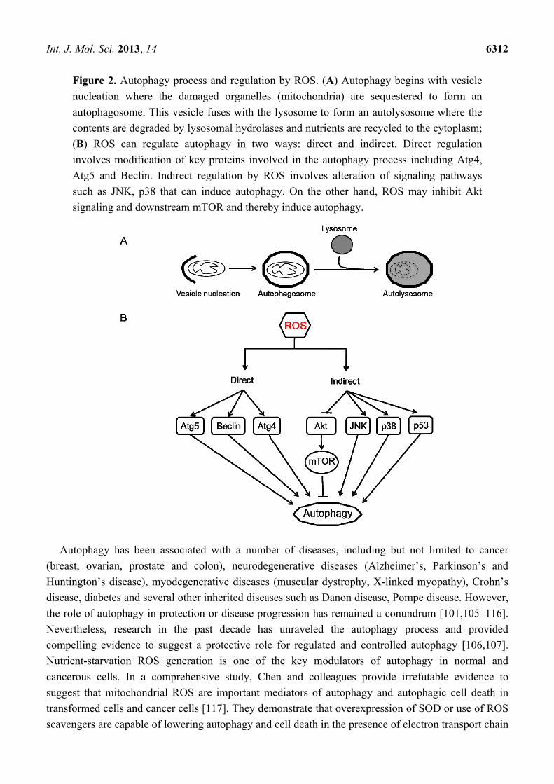
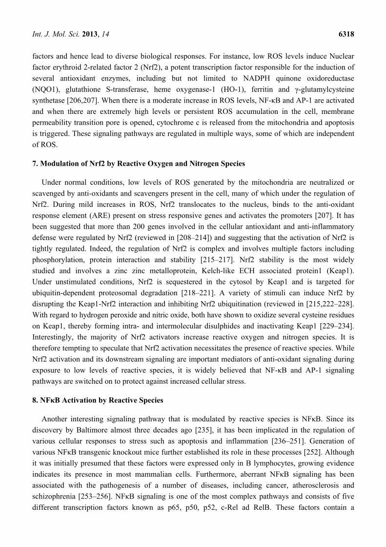
sequestered cellular components are degraded by the lysosomal enzymes (Figure 2A) [99–102].
Although this process may appear to be self-destructive, it is an extremely efficient recycling strategy
that is vital to maintain cell homeostasis and generates amino acids, fatty acids and energy (ATP)
that are used for macromolecular synthesis. There have been at least 35 Atg (Autophagy) related
genes identified in yeast whereas their mammalian orthologs have not been completely
characterized [103,104].
Int. J. Mol. Sci. 2013, 14 6312
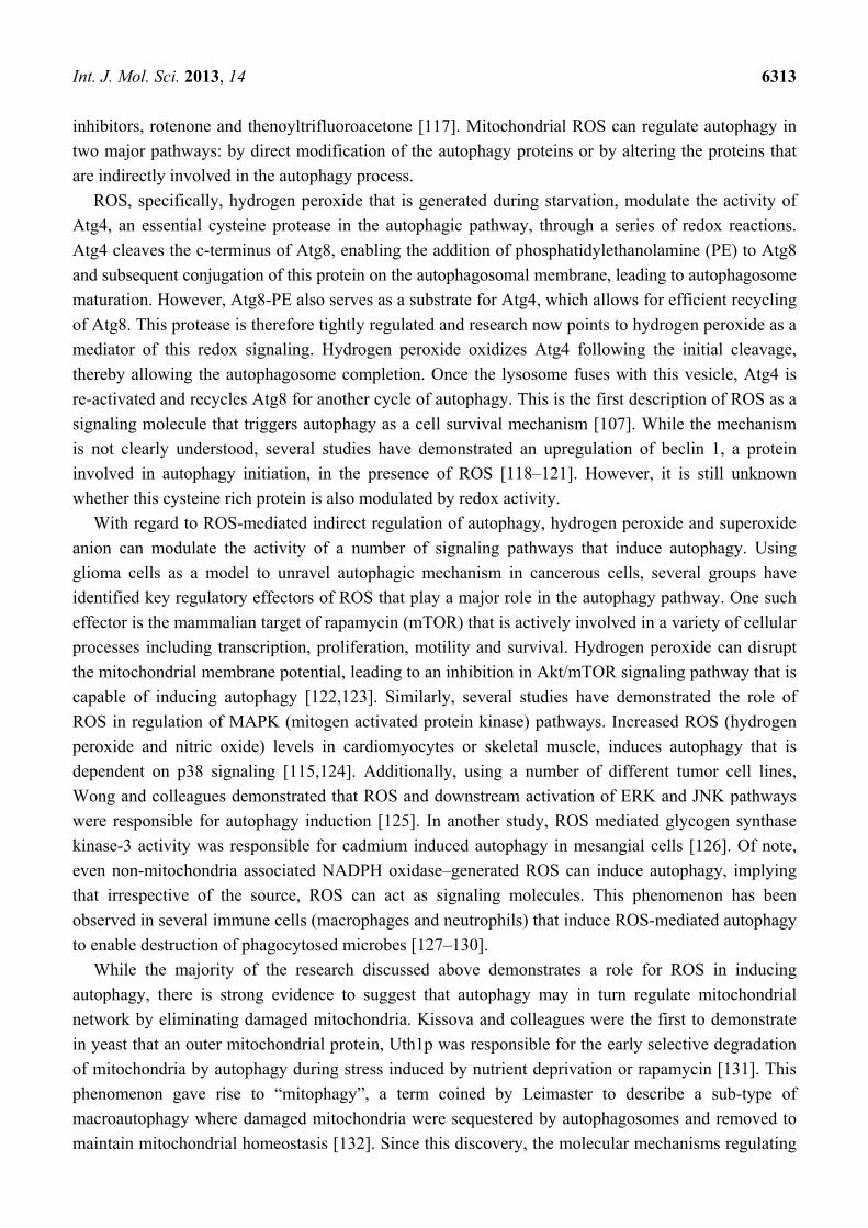
Figure 2. Autophagy process and regulation by ROS. (A) Autophagy begins with vesicle
nucleation where the damaged organelles (mitochondria) are sequestered to form an
autophagosome. This vesicle fuses with the lysosome to form an autolysosome where the
contents are degraded by lysosomal hydrolases and nutrients are recycled to the cytoplasm;
(B) ROS can regulate autophagy in two ways: direct and indirect. Direct regulation
involves modification of key proteins involved in the autophagy process including Atg4,
Atg5 and Beclin. Indirect regulation by ROS involves alteration of signaling pathways
such as JNK, p38 that can induce autophagy. On the other hand, ROS may inhibit Akt
signaling and downstream mTOR and thereby induce autophagy.
Autophagy has been associated with a number of diseases, including but not limited to cancer
(breast, ovarian, prostate and colon), neurodegenerative diseases (Alzheimer’s, Parkinson’s and
Huntington’s disease), myodegenerative diseases (muscular dystrophy, X-linked myopathy), Crohn’s
disease, diabetes and several other inherited diseases such as Danon disease, Pompe disease. However,
the role of autophagy in protection or disease progression has remained a conundrum [101,105–116].
Nevertheless, research in the past decade has unraveled the autophagy process and provided
compelling evidence to suggest a protective role for regulated and controlled autophagy [106,107].
Nutrient-starvation ROS generation is one of the key modulators of autophagy in normal and
cancerous cells. In a comprehensive study, Chen and colleagues provide irrefutable evidence to
suggest that mitochondrial ROS are important mediators of autophagy and autophagic cell death in
transformed cells and cancer cells [117]. They demonstrate that overexpression of SOD or use of ROS
scavengers are capable of lowering autophagy and cell death in the presence of electron transport chain
Int. J. Mol. Sci. 2013, 14 6313
inhibitors, rotenone and thenoyltrifluoroacetone [117]. Mitochondrial ROS can regulate autophagy in
two major pathways: by direct modification of the autophagy proteins or by altering the proteins that
are indirectly involved in the autophagy process.
ROS, specifically, hydrogen peroxide that is generated during starvation, modulate the activity of
Atg4, an essential cysteine protease in the autophagic pathway, through a series of redox reactions.
Atg4 cleaves the c-terminus of Atg8, enabling the addition of phosphatidylethanolamine (PE) to Atg8
and subsequent conjugation of this protein on the autophagosomal membrane, leading to autophagosome
maturation. However, Atg8-PE also serves as a substrate for Atg4, which allows for efficient recycling
of Atg8. This protease is therefore tightly regulated and research now points to hydrogen peroxide as a
mediator of this redox signaling. Hydrogen peroxide oxidizes Atg4 following the initial cleavage,
thereby allowing the autophagosome completion. Once the lysosome fuses with this vesicle, Atg4 is
re-activated and recycles Atg8 for another cycle of autophagy. This is the first description of ROS as a
signaling molecule that triggers autophagy as a cell survival mechanism [107]. While the mechanism
is not clearly understood, several studies have demonstrated an upregulation of beclin 1, a protein
involved in autophagy initiation, in the presence of ROS [118–121]. However, it is still unknown
whether this cysteine rich protein is also modulated by redox activity.
With regard to ROS-mediated indirect regulation of autophagy, hydrogen peroxide and superoxide
anion can modulate the activity of a number of signaling pathways that induce autophagy. Using
glioma cells as a model to unravel autophagic mechanism in cancerous cells, several groups have
identified key regulatory effectors of ROS that play a major role in the autophagy pathway. One such
effector is the mammalian target of rapamycin (mTOR) that is actively involved in a variety of cellular
processes including transcription, proliferation, motility and survival. Hydrogen peroxide can disrupt
the mitochondrial membrane potential, leading to an inhibition in Akt/mTOR signaling pathway that is
capable of inducing autophagy [122,123]. Similarly, several studies have demonstrated the role of
ROS in regulation of MAPK (mitogen activated protein kinase) pathways. Increased ROS (hydrogen
peroxide and nitric oxide) levels in cardiomyocytes or skeletal muscle, induces autophagy that is
dependent on p38 signaling [115,124]. Additionally, using a number of different tumor cell lines,
Wong and colleagues demonstrated that ROS and downstream activation of ERK and JNK pathways
were responsible for autophagy induction [125]. In another study, ROS mediated glycogen synthase
kinase-3 activity was responsible for cadmium induced autophagy in mesangial cells [126]. Of note,
even non-mitochondria associated NADPH oxidase–generated ROS can induce autophagy, implying
that irrespective of the source, ROS can act as signaling molecules. This phenomenon has been
observed in several immune cells (macrophages and neutrophils) that induce ROS-mediated autophagy
to enable destruction of phagocytosed microbes [127–130].
While the majority of the research discussed above demonstrates a role for ROS in inducing
autophagy, there is strong evidence to suggest that autophagy may in turn regulate mitochondrial
network by eliminating damaged mitochondria. Kissova and colleagues were the first to demonstrate
in yeast that an outer mitochondrial protein, Uth1p was responsible for the early selective degradation
of mitochondria by autophagy during stress induced by nutrient deprivation or rapamycin [131]. This
phenomenon gave rise to “mitophagy”, a term coined by Leimaster to describe a sub-type of
macroautophagy where damaged mitochondria were sequestered by autophagosomes and removed to
maintain mitochondrial homeostasis [132]. Since this discovery, the molecular mechanisms regulating
Int. J. Mol. Sci. 2013, 14 6314
mitophagy in yeast have been identified. However, in the mammalian system, research has been
mainly focused on parkin/pink1 (PTEN-induced putative kinase protein 1) mechanism. PINK1 is a
mitochondrial kinase that may serve as a guardian to the mitochondria through its ability to identify
depolarized mitochondria. Under normal conditions, PINK1 undergoes voltage-dependent lysis and is
removed from the mitochondria. However, in damaged mitochondria that have low membrane
potential, PINK1 accumulates on the mitochondrial surface and recruits parkin [133,134]. Parkin
ubiquitinates mitochondrial proteins, including voltage-dependent anion channel 1 (VDAC1), and
recruits autophagic machinery to the damaged mitochondria for removal [133,135–138]. Recently,
Egan and colleagues demonstrated that mammalian ortholog of Atg1, ULK-1 is required for mitophagy.
In the absence of this protein, there was a decrease in starvation-induced mitophagy and an associated
increase in the number of aberrant mitochondria in mouse embryonic fibroblasts and hepatocytes,
suggesting that ULK-1 is a key regulator of mitochondrial homeostasis [139].
In summary, these studies suggest that autophagy is a complex process that is regulated by ROS in
multiple ways (Figure 2B). Autophagy is a double-edged sword, with both cytoprotective and
cytotoxic capabilities. Whether it inhibits cell death by removing damaged organelles (mitochondria)
or induces cell death depends on many factors, including ROS. Mild ROS induces autophagy (or
mitophagy) as a survival mechanism to eliminate damaged mitochondria that are responsible for
uncontrolled ROS generation. On the contrary, high levels of ROS or sustained exposure to ROS can
alter signaling pathways that converge to induce autophagic or apoptotic cell death [116,140].
5. Mitochondria Network
Mitochondria are complex organelles that exist as a tubular network in cells. Two opposing
pathways that must exist in equilibrium sustain this dynamic filamentous network: mitochondria fusion
and fission. While the molecular mechanism and signaling involved in these two processes is only now
being understood, it is well established that dysregulation of these pathways can lead to cellular
dysfunction. Mitochondria fusion, mediated by mitofusin-1 and mitofusin-2 are responsible for the
lengthening and tethering of adjacent mitochondria to form a network. On the contrary, mitochondria
fission involves division of mitochondria and is mediated by fission-1 and dynamin-related protein 1
(Drp-1). Under healthy conditions, the mitochondrial tubular network is established through increased
fusion. However, during oxidative stress, mitochondria fission prevails and the filamentous network is
broken down to fragmented mitochondrial puncta [141,142].
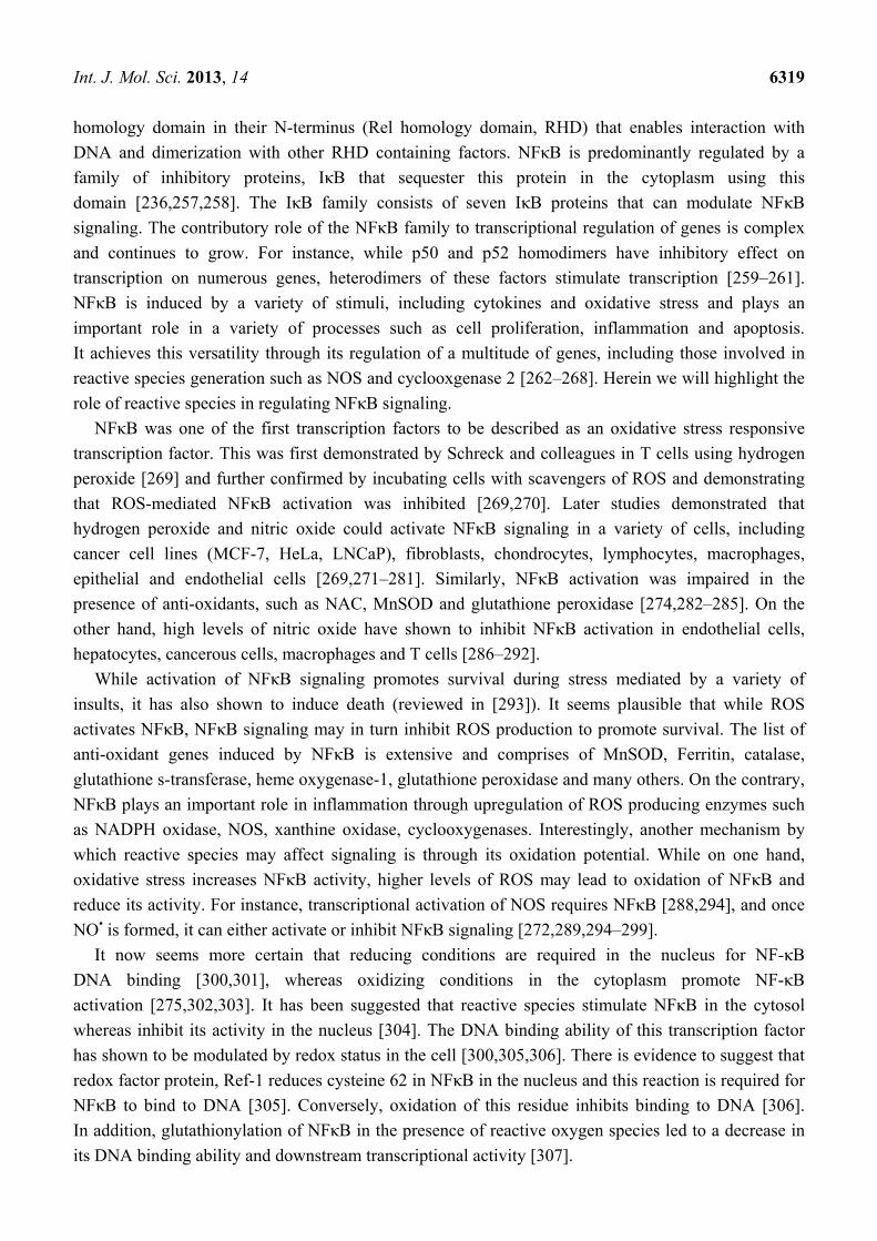
Hydrogen peroxide, a major contributor to oxidative damage in cells, was shown to induce
mitochondria fission in a variety of cells, including fibroblasts. This fragmentation has been shown to
be dose- and time-dependent and reversible, suggesting that mitochondria dynamics may be involved
in signaling during cellular stress [143–145].
In a recent study, Makino and colleagues demonstrated that superoxide anion was able to induce
mitochondrial fragmentation in coronary endothelial cells isolated from a diabetic mouse.
Supplementation with TEMPOL, a superoxide scavenger was able to inhibit mitochondrial
fragmentation, demonstrating a causal role for superoxide in this process [146]. This was also
corroborated by another study that treated HUVEC cells with hydrogen peroxide and demonstrate
mitochondria fragmentation, which was suppressed in the presence of an anti-oxidant, N-acetylcysteine
Int. J. Mol. Sci. 2013, 14 6315
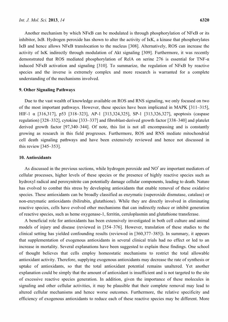
(Figure 3) [145]. In another study using neuronal cells, nitric oxide had a profound fission effect that
was rescued by antioxidants (Figure 4) [147,148]. Furthermore, mitochondrial fission in neurons
occurs prior to the onset of neuronal loss in an animal model of stroke [147]. This was further
demonstrated in the kidney following ischemia reperfusion and cisplatin nephrotoxicity [149,150].
Conversely, Yu et al. demonstrated that mitochondria fission plays an important role in ROS
overproduction and demonstrate that inhibition of fission reduces ROS generation by the
mitochondria [151]. While all these studies implied a role for ROS/RNS in mitochondria fission, a
recent study by Giedt and colleagues demonstrated that during simulated ischemia reperfusion in
endothelial cells, DRP-1 activation (phosphorylation and oligomerization) was enhanced. This was
inhibited by N-acetylcysteine or L-NG-Nitroarginine Methyl Ester (L-NAME, a NOS inhibitor),
suggesting that oxidative/nitrosative stress drives Drp1 activation and translocation to mitochondria,
which could be the underlying mechanism that induces mitochondrial fission in these studies [152,153].
These studies suggest that different species of ROS can initiate or act as a messenger in the signaling
process leading to mitochondrial fragmentation and when persistent, it can lead to chronic
mitochondria fission and apoptosis.
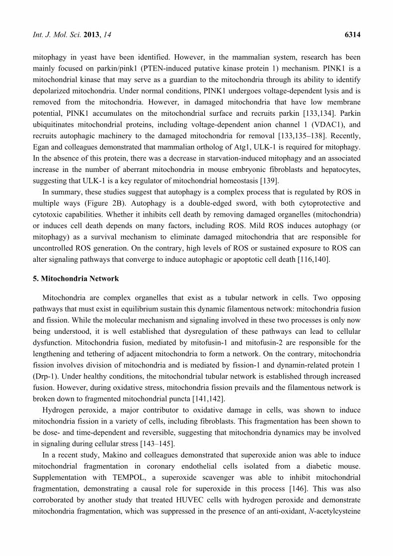
Figure 3. Changes in mitochondrial structure following hydrogen peroxide treatment.
(Top panels) Transmission electron micrographs of mitochondria in untreated (A) and
hydrogen peroxide treated cells (B,C,D). Electron dense granules (arrows) and fragmented
mitochondria were observed. Bar = 1 μm. (Bottom panels) Mitochondria were stained with
Mitotracker Red and existed as tubular (A), intermediate (B) (tubular with swollen regions)
and fragmented (C). Bar = 10 μm.
Int. J. Mol. Sci. 2013, 14 6316
Figure 3. Cont.
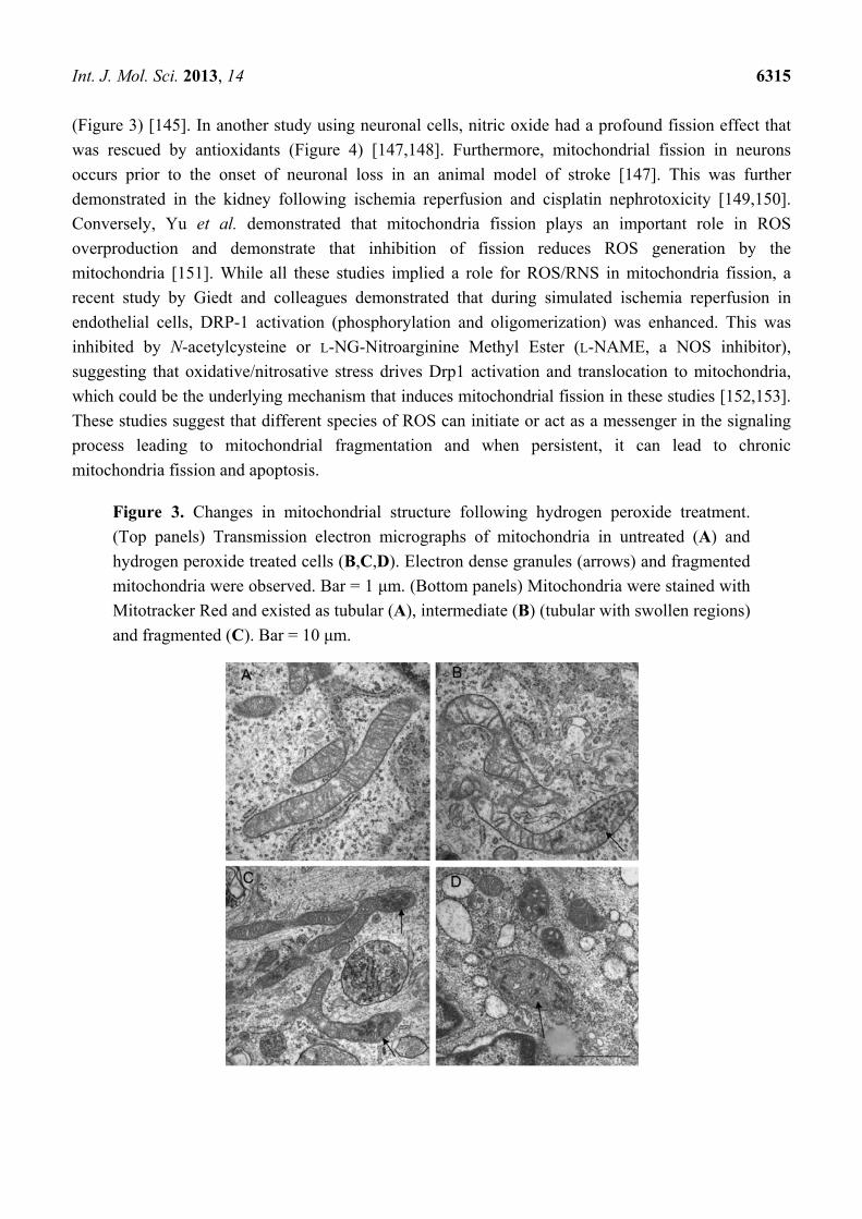
Figure 4. NO• triggers mitochondrial fission. (A) 3D time-lapse microscopy of
mitochondria undergoing fission in a dendritic arbor of a neuron. Neurons were transfected
with Mito-DsRed2, pretreated with the pan-caspase inhibitor zVAD-fmk methyl ester
(100 M), and exposed to SNOC (200 M). Images were 3D iso-surface rendered. Frames
depict representative time points of the movie demonstrating mitochondrial fragmentation
within 3 h of NO• exposure (upper panels; scale bar, 15 µm) and closeup views (lower
panels; scale bar = 3 µm).
While a majority of these studies point to the role of ROS/RNS to activating fission, there is also
evidence suggesting that inhibition of fission may be detrimental to the mitochondria network and
cellular homeostasis. Several studies have demonstrated that inhibition of fission led to a decrease in
cytochrome c release and delayed apoptosis [154–158]. Similarly, proteins involved in mitochondria
fusion have shown to play an anti-apoptotic role [157,159]. However, recent evidence in a number of
models suggests that inhibition of fission can lead to mitochondrial dysfunction, increase in
ROS levels accompanied by loss of mtDNA and concomitant reduction in energy production
and autophagy [160,161].
It is now evident from a variety of studies that ROS affects mitochondrial dynamics and leads to
fragmentation of mitochondria tubules. It is also apparent that the mitochondria network plays an
important role in mitochondrial function, including respiration, ATP production, apoptosis and
functional complementation of mitochondrial DNA mutations and/or damaged proteins. However, it
has been suggested that transient or low levels of ROS may in fact benefit the network by selectively
removing the dysfunctional mitochondria through autophagy. This idea was proposed using a simulation
model that suggested that the vulnerability of the mitochondria network to the harmful effects of ROS
Int. J. Mol. Sci. 2013, 14 6317
is dependent on the dynamics of the network. As one might expect, the healthy mitochondria in close
association with the ROS producing dysfunctional mitochondria would be the first to be affected,
starting a vicious cycle of uncontrolled and amplified ROS generation. As a result, if these damaged
mitochondria were not isolated by fission, they would contaminate the entire network leading to a
decrease in energy production and cell proliferation ultimately leading to cell death [162–164].
Nevertheless, it is now clear that defects in mitochondrial fusion or fission alter the susceptibility
of cells to undergo apoptosis as documented in a variety of disorders, including heart failure,
ischemia reperfusion injury, diabetes, Parkinson’s disease, muscle atrophy, Alzheimer’s disease and
aging [141,149,165–172]. Most importantly, these studies provide mechanistic evidence for a role for
ROS/RNS and redox signaling in contributing to perturbations in the mitochondrial dynamics and
suggest the potential of mitochondria-targeted therapeutics in diseases that involve mitochondrial
fragmentation due to uncontrolled ROS/RNS generation.
6. Signaling
Until recently, ROS generation was considered a bane to all aerobic organisms. In fact, this theory
was sustained by research that continued to demonstrate generation of high levels of superoxide and
nitric oxide by phagocytic cells and its role in host defense [173,174]. In stark contrast, non-immunity
based research provided evidence to suggest that these molecules (nitric oxide and hydrogen peroxide)
were generated in low levels in other cells, suggesting a distinctive role from its phagocytic activity. In
fact, more than three decades ago, pioneers in nitric oxide biology such as Moncada, Ignarro and
Furchgott demonstrated a vasodilatory property for this molecule, a discovery that led to their Nobel
Prize in 1998 [175–177]. Following this observation, several investigators have established the
importance of nitric oxide and other reactive oxygen intermediates in vascular homeostasis [178–187].
Indeed, there is compelling evidence today that demonstrates that these reactive species function as a
second messenger in signal transduction and affect cellular function in different tissues [188–205].
Here in we will only focus on the pathways that best illustrate the role of these species in modulating
signal transduction.
Since the discovery of a dichotomous role of reactive species in immune function and signal
transduction, research in this field grew at an exponential pace and the pursuit for the mechanisms
involved began. In a simplistic manner, these regulations can be hypothesized to occur through simple
oxidative/nitrosative reactions. For instance, cysteine residues on proteins may be oxidized and hence
significantly alter the activity of these proteins. These proteins (e.g., transcription factors, kinases,
phosphatases) may then affect downstream signaling cascades that affect cellular responses to stimuli.
Alternatively, these residues may be modified through nitrosylation by reactive nitrogen species. More
importantly, this redox mechanism may occur at multiple levels in the signal transduction cascade and
thereby ultimately alter the fate of a cell.
Earlier research was primarily focused on the ability of hydrogen peroxide to mediate cellular
signaling due to its relative stability and ease of measurement. Hydrogen peroxide can also be
converted to the highly reactive hydroxyl ion in the presence of a transition metal, such as iron, to
amplify oxidative stress and activate cell death pathways. However, there is some consensus that the
level of ROS itself may dictate the fate of the cell by modulating different redox-sensitive transcription
Int. J. Mol. Sci. 2013, 14 6318
factors and hence lead to diverse biological responses. For instance, low ROS levels induce Nuclear
factor erythroid 2-related factor 2 (Nrf2), a potent transcription factor responsible for the induction of
several antioxidant enzymes, including but not limited to NADPH quinone oxidoreductase
(NQO1), glutathione S-transferase, heme oxygenase-1 (HO-1), ferritin and γ-glutamylcysteine
synthetase [206,207]. When there is a moderate increase in ROS levels, NF-κB and AP-1 are activated
and when there are extremely high levels or persistent ROS accumulation in the cell, membrane
permeability transition pore is opened, cytochrome c is released from the mitochondria and apoptosis
is triggered. These signaling pathways are regulated in multiple ways, some of which are independent
of ROS.
7. Modulation of Nrf2 by Reactive Oxygen and Nitrogen Species
Under normal conditions, low levels of ROS generated by the mitochondria are neutralized or
scavenged by anti-oxidants and scavengers present in the cell, many of which under the regulation of
Nrf2. During mild increases in ROS, Nrf2 translocates to the nucleus, binds to the anti-oxidant
response element (ARE) present on stress responsive genes and activates the promoters [207]. It has
been suggested that more than 200 genes involved in the cellular antioxidant and anti-inflammatory
defense were regulated by Nrf2 (reviewed in [208–214]) and suggesting that the activation of Nrf2 is
tightly regulated. Indeed, the regulation of Nrf2 is complex and involves multiple factors including
phosphorylation, protein interaction and stability [215–217]. Nrf2 stability is the most widely
studied and involves a zinc zinc metalloprotein, Kelch-like ECH associated protein1 (Keap1).
Under unstimulated conditions, Nrf2 is sequestered in the cytosol by Keap1 and is targeted for
ubiquitin-dependent proteosomal degradation [218–221]. A variety of stimuli can induce Nrf2 by
disrupting the Keap1-Nrf2 interaction and inhibiting Nrf2 ubiquitination (reviewed in [215,222–228].
With regard to hydrogen peroxide and nitric oxide, both have shown to oxidize several cysteine residues
on Keap1, thereby forming intra- and intermolecular disulphides and inactivating Keap1 [229–234].
Interestingly, the majority of Nrf2 activators increase reactive oxygen and nitrogen species. It is
therefore tempting to speculate that Nrf2 activation necessitates the presence of reactive species. While
Nrf2 activation and its downstream signaling are important mediators of anti-oxidant signaling during
exposure to low levels of reactive species, it is widely believed that NF-κB and AP-1 signaling
pathways are switched on to protect against increased cellular stress.
8. NFκB Activation by Reactive Species
Another interesting signaling pathway that is modulated by reactive species is NFκB. Since its
discovery by Baltimore almost three decades ago [235], it has been implicated in the regulation of
various cellular responses to stress such as apoptosis and inflammation [236–251]. Generation of
various NFκB transgenic knockout mice further established its role in these processes [252]. Although
it was initially presumed that these factors were expressed only in B lymphocytes, growing evidence
indicates its presence in most mammalian cells. Furthermore, aberrant NFκB signaling has been
associated with the pathogenesis of a number of diseases, including cancer, atherosclerosis and
schizophrenia [253–256]. NFκB signaling is one of the most complex pathways and consists of five
different transcription factors known as p65, p50, p52, c-Rel ad RelB. These factors contain a
Int. J. Mol. Sci. 2013, 14 6319
homology domain in their N-terminus (Rel homology domain, RHD) that enables interaction with
DNA and dimerization with other RHD containing factors. NFκB is predominantly regulated by a
family of inhibitory proteins, IκB that sequester this protein in the cytoplasm using this
domain [236,257,258]. The IκB family consists of seven IκB proteins that can modulate NFκB
signaling. The contributory role of the NFκB family to transcriptional regulation of genes is complex
and continues to grow. For instance, while p50 and p52 homodimers have inhibitory effect on
transcription on numerous genes, heterodimers of these factors stimulate transcription [259–261].
NFκB is induced by a variety of stimuli, including cytokines and oxidative stress and plays an
important role in a variety of processes such as cell proliferation, inflammation and apoptosis.
It achieves this versatility through its regulation of a multitude of genes, including those involved in
reactive species generation such as NOS and cyclooxgenase 2 [262–268]. Herein we will highlight the
role of reactive species in regulating NFκB signaling.
NFκB was one of the first transcription factors to be described as an oxidative stress responsive
transcription factor. This was first demonstrated by Schreck and colleagues in T cells using hydrogen
peroxide [269] and further confirmed by incubating cells with scavengers of ROS and demonstrating
that ROS-mediated NFκB activation was inhibited [269,270]. Later studies demonstrated that
hydrogen peroxide and nitric oxide could activate NFκB signaling in a variety of cells, including
cancer cell lines (MCF-7, HeLa, LNCaP), fibroblasts, chondrocytes, lymphocytes, macrophages,
epithelial and endothelial cells [269,271–281]. Similarly, NFκB activation was impaired in the
presence of anti-oxidants, such as NAC, MnSOD and glutathione peroxidase [274,282–285]. On the
other hand, high levels of nitric oxide have shown to inhibit NFκB activation in endothelial cells,
hepatocytes, cancerous cells, macrophages and T cells [286–292].
While activation of NFκB signaling promotes survival during stress mediated by a variety of
insults, it has also shown to induce death (reviewed in [293]). It seems plausible that while ROS
activates NFκB, NFκB signaling may in turn inhibit ROS production to promote survival. The list of
anti-oxidant genes induced by NFκB is extensive and comprises of MnSOD, Ferritin, catalase,
glutathione s-transferase, heme oxygenase-1, glutathione peroxidase and many others. On the contrary,
NFκB plays an important role in inflammation through upregulation of ROS producing enzymes such
as NADPH oxidase, NOS, xanthine oxidase, cyclooxygenases. Interestingly, another mechanism by
which reactive species may affect signaling is through its oxidation potential. While on one hand,
oxidative stress increases NFκB activity, higher levels of ROS may lead to oxidation of NFκB and
reduce its activity. For instance, transcriptional activation of NOS requires NFκB [288,294], and once
NO• is formed, it can either activate or inhibit NFκB signaling [272,289,294–299].
It now seems more certain that reducing conditions are required in the nucleus for NF-κB
DNA binding [300,301], whereas oxidizing conditions in the cytoplasm promote NF-κB
activation [275,302,303]. It has been suggested that reactive species stimulate NFκB in the cytosol
whereas inhibit its activity in the nucleus [304]. The DNA binding ability of this transcription factor
has shown to be modulated by redox status in the cell [300,305,306]. There is evidence to suggest that
redox factor protein, Ref-1 reduces cysteine 62 in NFκB in the nucleus and this reaction is required for
NFκB to bind to DNA [305]. Conversely, oxidation of this residue inhibits binding to DNA [306].
In addition, glutathionylation of NFκB in the presence of reactive oxygen species led to a decrease in
its DNA binding ability and downstream transcriptional activity [307].
Int. J. Mol. Sci. 2013, 14 6320
Another mechanism by which NFκB can be modulated is through phosphorylation of NFκB or its
inhibitor, IκB. Hydrogen peroxide has shown to alter the activity of IκK, a kinase that phosphorylates
IκB and hence allows NFκB translocation to the nucleus [308]. Alternatively, ROS can increase the
activity of IκK indirectly through modulation of Akt signaling [309]. Furthermore, it was recently
demonstrated that ROS mediated phosphorylation of RelA on serine 276 is essential for TNF-α
induced NFκB activation and signaling [310]. To summarize, the regulation of NFκB by reactive
species and the inverse is extremely complex and more research is warranted for a complete
understanding of the mechanisms involved.
9. Other Signaling Pathways
Due to the vast wealth of knowledge available on ROS and RNS signaling, we only focused on two
of the most important pathways. However, these species have been implicated in MAPK [311–315],
HIF-1 α [316,317], p53 [318–323], AP-1 [313,324,325], SP-1 [313,326,327], apoptosis (caspase
regulation) [328–332], cytokine [333–337] and fibroblast-derived growth factor [338–340] and platelet
derived growth factor [97,340–344]. Of note, this list is not all encompassing and is constantly
growing as research in this field progresses. Furthermore, ROS and RNS mediate mitochondrial
cell death signaling pathways and have been extensively reviewed and hence not discussed in
this review [345–353].
10. Antioxidants
As discussed in the previous sections, while hydrogen peroxide and NO• are important mediators of
cellular processes, higher levels of these species or the presence of highly reactive species such as
hydroxyl radical and peroxynitrite can potentially damage cellular components, leading to death. Nature
has evolved to combat this stress by developing antioxidants that enable removal of these oxidative
species. These antioxidants can be broadly classified as enzymatic (superoxide dismutase, catalase) or
non-enzymatic antioxidants (bilirubin, glutathione). While they are directly involved in eliminating
reactive species, cells have evolved other mechanisms that can indirectly reduce or inhibit generation
of reactive species, such as heme oxygenase-1, ferritin, ceruloplasmin and glutathione transferase.
A beneficial role for antioxidants has been extensively investigated in both cell culture and animal
models of injury and disease (reviewed in [354–376]. However, translation of these studies to the
clinical setting has yielded confounding results (reviewed in [360,377–385]). In summary, it appears
that supplementation of exogenous antioxidants in several clinical trials had no effect or led to an
increase in mortality. Several explanations have been suggested to explain these findings. One school
of thought believes that cells employ homeostatic mechanisms to restrict the total allowable
antioxidant activity. Therefore, supplying exogenous antioxidants may decrease the rate of synthesis or
uptake of antioxidants, so that the total antioxidant potential remains unaltered. Yet another
explanation could be simply that the amount of antioxidant is insufficient and is not targeted to the site
of excessive reactive species generation. In addition, given the importance of these molecules in
signaling and other cellular activities, it may be plausible that their complete removal may lead to
altered cellular mechanisms and hence worse outcomes. Furthermore, the relative specificity and
efficiency of exogenous antioxidants to reduce each of these reactive species may be different. More
Int. J. Mol. Sci. 2013, 14 6321
importantly, the oxidants responsible for injury must be evaluated. For instance, in a model of
cisplatin-mediated renal epithelial injury, overexpression of MnSOD was protective whereas, catalase
overexpression was ineffective [386]. This supports the notion that injury may be mediated through
different oxidative and nitrosative species and future therapies must be targeted based on the
detrimental species generated. Therefore, despite the advances made in deciphering the molecular
mechanisms that are regulated by oxidants and antioxidants, translation of these pathways to benefit
mankind is still in its infancy and more studies are warranted.
11. Concluding Remarks
The field of free radicals has evolved over the past few decades and has significantly contributed to
understanding normal physiology and pathophysiology. Research has provided irrefutable evidence
that reactive oxygen and nitrogen species are important mediators of cellular response to stress and
they function through several mechanisms including, modulation of autophagy, mitochondrial
network, signaling and apoptosis. However, high levels of certain reactive species can contribute to
cell injury and progression of diseases. These studies granted opportunities for implementing the use
of antioxidants in clinical trials, only some of which provided promising results. Therefore, there is an
urgent need to comprehensively assess the amounts of different species generated during injury and
their relative role in the pathogenesis of disease. Targeting the detrimental reactive species through
antioxidant therapy would perhaps yield better outcomes in the clinical trials. In this review, we
provided a brief discussion of some of the major pathways that are regulated by ROS and RNS. This
review has focused on the complexities and dual roles of these species in cellular activities that affect
health and disease. This review will not only provide an understanding of the intricate underlying
mechanisms of the reactive species, but will also enable opportunities for the design and development
of effective novel therapeutic strategies.
Acknowledgments
This work was supported by an AHA grant 11POST7600074 (S.B.) a Veterans Affairs Program
Project Award 1IP1BX001595 (E.A.J) a Merit Review Award of the Veterans Affairs Administration
1I01BX001073 (E.A.J) and an NIH Grant R01ES014948 (E.A.J).
Conflict of Interest
The authors declare no conflict of interest.
References
1. Ernster, L.; Schatz, G. Mitochondria: A historical review. J. Cell Biol. 1981, 91, 227s–255s.
2. Gray, M.W.; Burger, G.; Lang, B.F. Mitochondrial evolution. Science 1999, 283, 1476–1481.
3. Hollenbeck, P.J.; Saxton, W.M. The axonal transport of mitochondria. J. Cell Sci. 2005, 118,
5411–5419.
4. Lenaz, G. The mitochondrial production of reactive oxygen species: Mechanisms and
implications in human pathology. IUBMB Life 2001, 52, 159–164.
Int. J. Mol. Sci. 2013, 14 6322
5. Swerdlow, R.H. Brain aging, Alzheimer’s disease, and mitochondria. Biochim. Biophys. Acta
2011, 1812, 1630–1639.
6. Griffiths, E.J. Mitochondria and heart disease. Adv. Exp. Med. Biol. 2012, 942, 249–267.
7. Birch-Machin, M.A. Mitochondria and skin disease. Clin. Exp. Dermatol. 2000, 25, 141–146.
8. Frohman, M.A. Mitochondria as integrators of signal transduction and energy production in
cardiac physiology and disease. J. Mol. Med. (Berl.) 2010, 88, 967–970.
9. Grattagliano, I.; Russmann, S.; Diogo, C.; Bonfrate, L.; Oliveira, P.J.; Wang, D.Q.; Portincasa, P.
Mitochondria in chronic liver disease. Curr. Drug Targets 2011, 12, 879–893.
10. Duchen, M.R. Mitochondria in health and disease: Perspectives on a new mitochondrial biology.
Mol. Aspects Med. 2004, 25, 365–451.
11. Smith, R.A.; Adlam, V.J.; Blaikie, F.H.; Manas, A.R.; Porteous, C.M.; James, A.M.; Ross, M.F.;
Logan, A.; Cocheme, H.M.; Trnka, J.; et al. Mitochondria-targeted antioxidants in the treatment
of disease. Ann. N. Y. Acad. Sci. 2008, 1147, 105–111.
12. Gobe, G.; Crane, D. Mitochondria, reactive oxygen species and cadmium toxicity in the kidney.
Toxicol. Lett. 2010, 198, 49–55.
13. Cloonan, S.M.; Choi, A.M. Mitochondria: Commanders of innate immunity and disease?
Curr. Opin. Immunol. 2012, 24, 32–40.
14. Nunnari, J.; Suomalainen, A. Mitochondria: In sickness and in health. Cell 2012, 148,
1145–1159.
15. Schapira, A.H. Mitochondrial diseases. Lancet 2012, 379, 1825–1834.
16. Armstrong, J.S. Mitochondrial medicine: Pharmacological targeting of mitochondria in disease.
Br. J. Pharmacol. 2007, 151, 1154–1165.
17. Minocherhomji, S.; Tollefsbol, T.O.; Singh, K.K. Mitochondrial regulation of epigenetics and its
role in human diseases. Epigenetics 2012, 7, 326–334.
18. Rocha, M.; Apostolova, N.; Hernandez-Mijares, A.; Herance, R.; Victor, V.M. Oxidative stress
and endothelial dysfunction in cardiovascular disease: Mitochondria-targeted therapeutics.
Curr. Med. Chem. 2010, 17, 3827–3841.
19. Diogo, C.V.; Grattagliano, I.; Oliveira, P.J.; Bonfrate, L.; Portincasa, P. Re-wiring the circuit:
Mitochondria as a pharmacological target in liver disease. Curr. Med. Chem. 2011, 18,
5448–5465.
20. Johannsen, D.L.; Ravussin, E. The role of mitochondria in health and disease. Curr. Opin.
Pharmacol. 2009, 9, 780–786.
21. Duchen, M.R. Roles of mitochondria in health and disease. Diabetes 2004, 53, S96–S102.
22. Duchen, M.R.; Szabadkai, G. Roles of mitochondria in human disease. Essays Biochem. 2010,
47, 115–137.
23. Hedskog, L.; Zhang, S.; Ankarcrona, M. Strategic role for mitochondria in Alzheimer’s disease
and cancer. Antioxid. Redox Signal. 2012, 16, 1476–1491.
24. Watson, K.; Haslam, J.M.; Linnane, A.W. Biogenesis of mitochondria. 13. The isolation of
mitochondrial structures from anaerobically grown Saccharomyces cerevisiae. J. Cell Biol. 1970,
46, 88–96.
25. Green, D.E.; Oda, T. On the unit of mitochondrial structure and function. J. Biochem. 1961, 49,
742–757.
Int. J. Mol. Sci. 2013, 14 6323
26. Neupert, W. Protein import into mitochondria. Annu. Rev. Biochem. 1997, 66, 863–917.
27. Pfanner, N.; Craig, E.A.; Honlinger, A. Mitochondrial preprotein translocase. Annu. Rev. Cell
Dev. Biol. 1997, 13, 25–51.
28. Dekker, P.J.; Ryan, M.T.; Brix, J.; Muller, H.; Honlinger, A.; Pfanner, N. Preprotein translocase
of the outer mitochondrial membrane: Molecular dissection and assembly of the general import
pore complex. Mol. Cell. Biol. 1998, 18, 6515–6524.
29. Yamamoto, H.; Itoh, N.; Kawano, S.; Yatsukawa, Y.; Momose, T.; Makio, T.; Matsunaga, M.;
Yokota, M.; Esaki, M.; Shodai, T.; et al. Dual role of the receptor Tom20 in specificity and
efficiency of protein import into mitochondria. Proc. Natl. Acad. Sci. USA 2011, 108, 91–96.
30. Herrmann, J.M.; Riemer, J. The intermembrane space of mitochondria. Antioxid. Redox Signal.
2010, 13, 1341–1358.
31. Davies, K.M.; Strauss, M.; Daum, B.; Kief, J.H.; Osiewacz, H.D.; Rycovska, A.; Zickermann, V.;
Kuhlbrandt, W. Macromolecular organization of ATP synthase and complex I in whole
mitochondria. Proc. Natl. Acad. Sci. USA 2011, 108, 14121–14126.
32. O’Rourke, B. Mitochondrial ion channels. Annu. Rev. Physiol. 2007, 69, 19–49.
33. Asin-Cayuela, J.; Gustafsson, C.M. Mitochondrial transcription and its regulation in mammalian
cells. Trends Biochem. Sci. 2007, 32, 111–117.
34. Halliwell, B.; Gutteridge, J.M. Oxygen toxicity, oxygen radicals, transition metals and disease.
Biochem. J. 1984, 219, 1–14.
35. Cline, M.J.; Lehrer, R.I. D-amino acid oxidase in leukocytes: A possible D-amino-acid-linked
antimicrobial system. Proc. Natl. Acad. Sci. USA 1969, 62, 756–763.
36. Ji, L.L. Antioxidant signaling in skeletal muscle: A brief review. Exp. Gerontol. 2007, 42,
582–593.
37. Kim, C.; Kim, J.Y.; Kim, J.H. Cytosolic phospholipase A2, lipoxygenase metabolites, and
reactive oxygen species. BMB Rep. 2008, 41, 555–559.
38. Nishino, T. The conversion of xanthine dehydrogenase to xanthine oxidase and the role of the
enzyme in reperfusion injury. J. Biochem. 1994, 116, 1–6.
39. Nishino, T.; Okamoto, K.; Eger, B.T.; Pai, E.F. Mammalian xanthine oxidoreductase—Mechanism
of transition from xanthine dehydrogenase to xanthine oxidase. FEBS J. 2008, 275, 3278–3289.
40. Sumimoto, H.; Miyano, K.; Takeya, R. Molecular composition and regulation of the Nox family
NAD(P)H oxidases. Biochem. Biophys. Res. Commun. 2005, 338, 677–686.
41. Zangar, R.C.; Davydov, D.R.; Verma, S. Mechanisms that regulate production of reactive
oxygen species by cytochrome P450. Toxicol. Appl. Pharmacol. 2004, 199, 316–331.
42. Brown, G.C. Control of respiration and ATP synthesis in mammalian mitochondria and cells.
Biochem. J. 1992, 284, 1–13.
43. Paul, T. Effect of a prolonged superoxide flux on transferrin and ferritin. Arch. Biochem.
Biophys. 2000, 382, 253–261.
44. Biemond, P.; Swaak, A.J.; van Eijk, H.G.; Koster, J.F. Superoxide dependent iron release from
ferritin in inflammatory diseases. Free Radic. Biol. Med. 1988, 4, 185–198.
45. Liochev, S.L. The role of iron-sulfur clusters in in vivo hydroxyl radical production. Free Radic.
Res. 1996, 25, 369–384.
Int. J. Mol. Sci. 2013, 14 6324
46. Flint, D.H.; Tuminello, J.F.; Emptage, M.H. The inactivation of Fe–S cluster containing
hydro-lyases by superoxide. J. Biol. Chem. 1993, 268, 22369–22376.
47. Lenaz, G. Mitochondria and reactive oxygen species. Which role in physiology and pathology?
Adv. Exp. Med. Biol. 2012, 942, 93–136.
48. McLennan, H.R.; Esposti, M.D. The contribution of mitochondrial respiratory complexes to the
production of reactive oxygen species. J. Bioenerg. Biomembr. 2000, 32, 153–162.
49. Fato, R.; Bergamini, C.; Leoni, S.; Strocchi, P.; Lenaz, G. Generation of reactive oxygen species
by mitochondrial complex I: Implications in neurodegeneration. Neurochem. Res. 2008, 33,
2487–2501.
50. Murphy, M.P. How mitochondria produce reactive oxygen species. Biochem. J. 2009, 417, 1–13.
51. Yankovskaya, V.; Horsefield, R.; Tornroth, S.; Luna-Chavez, C.; Miyoshi, H.; Leger, C.;
Byrne, B.; Cecchini, G.; Iwata, S. Architecture of succinate dehydrogenase and reactive oxygen
species generation. Science 2003, 299, 700–704.
52. Casteilla, L.; Rigoulet, M.; Penicaud, L. Mitochondrial ROS metabolism: Modulation by
uncoupling proteins. IUBMB Life 2001, 52, 181–188.
53. Starkov, A.A.; Fiskum, G. Myxothiazol induces H2O2 production from mitochondrial respiratory
chain. Biochem. Biophys. Res. Commun. 2001, 281, 645–650.
54. Barja, G. Mitochondrial oxygen radical generation and leak: Sites of production in states 4 and 3,
organ specificity, and relation to aging and longevity. J. Bioenerg. Biomembr. 1999, 31, 347–366.
55. Kushnareva, Y.; Murphy, A.N.; Andreyev, A. Complex I-mediated reactive oxygen species
generation: Modulation by cytochrome c and NAD(P)+ oxidation-reduction state. Biochem. J.
2002, 368, 545–553.
56. Barja, G.; Herrero, A. Localization at complex I and mechanism of the higher free radical
production of brain nonsynaptic mitochondria in the short-lived rat than in the longevous pigeon.
J. Bioenerg. Biomembr. 1998, 30, 235–243.
57. Turrens, J.F.; Boveris, A. Generation of superoxide anion by the NADH dehydrogenase of
bovine heart mitochondria. Biochem. J. 1980, 191, 421–427.
58. Miwa, S.; St-Pierre, J.; Partridge, L.; Brand, M.D. Superoxide and hydrogen peroxide production
by Drosophila mitochondria. Free Radic. Biol. Med. 2003, 35, 938–948.
59. Drahota, Z.; Chowdhury, S.K.; Floryk, D.; Mracek, T.; Wilhelm, J.; Rauchova, H.; Lenaz, G.;
Houstek, J. Glycerophosphate-dependent hydrogen peroxide production by brown adipose tissue
mitochondria and its activation by ferricyanide. J. Bioenerg. Biomembr. 2002, 34, 105–113.
60. Seifert, E.L.; Estey, C.; Xuan, J.Y.; Harper, M.E. Electron transport chain-dependent
and -independent mechanisms of mitochondrial H2O2 emission during long-chain fatty acid
oxidation. J. Biol. Chem. 2010, 285, 5748–5758.
61. Maurel, A.; Hernandez, C.; Kunduzova, O.; Bompart, G.; Cambon, C.; Parini, A.; Frances, B.
Age-dependent increase in hydrogen peroxide production by cardiac monoamine oxidase A in
rats. Am. J. Physiol. Heart Circ. Physiol. 2003, 284, H1460–H1467.
62. Starkov, A.A.; Fiskum, G.; Chinopoulos, C.; Lorenzo, B.J.; Browne, S.E.; Patel, M.S.;
Beal, M.F. Mitochondrial alpha-ketoglutarate dehydrogenase complex generates reactive oxygen
species. J. Neurosci. 2004, 24, 7779–7788.
Int. J. Mol. Sci. 2013, 14 6325
63. Tretter, L.; Adam-Vizi, V. Generation of reactive oxygen species in the reaction catalyzed by
alpha-ketoglutarate dehydrogenase. J. Neurosci. 2004, 24, 7771–7778.
64. MacMillan-Crow, L.A.; Crow, J.P.; Thompson, J.A. Peroxynitrite-mediated inactivation of
manganese superoxide dismutase involves nitration and oxidation of critical tyrosine residues.
Biochemistry 1998, 37, 1613–1622.
65. Lacza, Z.; Snipes, J.A.; Zhang, J.; Horvath, E.M.; Figueroa, J.P.; Szabo, C.; Busija, D.W.
Mitochondrial nitric oxide synthase is not eNOS, nNOS or iNOS. Free Radic. Biol. Med. 2003,
35, 1217–1228.
66. Ghafourifar, P.; Richter, C. Nitric oxide synthase activity in mitochondria. FEBS Lett. 1997, 418,
291–296.
67. Giulivi, C.; Poderoso, J.J.; Boveris, A. Production of nitric oxide by mitochondria. J. Biol. Chem.
1998, 273, 11038–11043.
68. Alvarez, S.; Valdez, L.B.; Zaobornyj, T.; Boveris, A. Oxygen dependence of mitochondrial nitric
oxide synthase activity. Biochem. Biophys. Res. Commun. 2003, 305, 771–775.
69. Lacza, Z.; Puskar, M.; Figueroa, J.P.; Zhang, J.; Rajapakse, N.; Busija, D.W. Mitochondrial
nitric oxide synthase is constitutively active and is functionally upregulated in hypoxia.
Free Radic. Biol. Med. 2001, 31, 1609–1615.
70. Bates, T.E.; Loesch, A.; Burnstock, G.; Clark, J.B. Immunocytochemical evidence for a
mitochondrially located nitric oxide synthase in brain and liver. Biochem. Biophys. Res.
Commun. 1995, 213, 896–900.
71. Haynes, V.; Elfering, S.; Traaseth, N.; Giulivi, C. Mitochondrial nitric-oxide synthase: Enzyme
expression, characterization, and regulation. J. Bioenerg. Biomembr. 2004, 36, 341–346.
72. Giulivi, C. Mitochondria as generators and targets of nitric oxide. Novartis Found. Symp. 2007,
287, 92–100; discussion 100–104.
73. Lacza, Z.; Kozlov, A.V.; Pankotai, E.; Csordas, A.; Wolf, G.; Redl, H.; Kollai, M.; Szabo, C.;
Busija, D.W.; Horn, T.F. Mitochondria produce reactive nitrogen species via an
arginine-independent pathway. Free Radic. Res. 2006, 40, 369–378.
74. Kozlov, A.V.; Staniek, K.; Nohl, H. Nitrite reductase activity is a novel function of mammalian
mitochondria. FEBS Lett. 1999, 454, 127–130.
75. Lacza, Z.; Pankotai, E.; Csordas, A.; Gero, D.; Kiss, L.; Horvath, E.M.; Kollai, M.; Busija, D.W.;
Szabo, C. Mitochondrial NO and reactive nitrogen species production: Does mtNOS exist?
Nitric. Oxide 2006, 14, 162–168.
76. Mason, M.G.; Nicholls, P.; Wilson, M.T.; Cooper, C.E. Nitric oxide inhibition of respiration
involves both competitive (heme) and noncompetitive (copper) binding to cytochrome c oxidase.
Proc. Natl. Acad. Sci. USA 2006, 103, 708–713.
77. Cooper, C.E.; Giulivi, C. Nitric oxide regulation of mitochondrial oxygen consumption II:
Molecular mechanism and tissue physiology. Am. J. Physiol. Cell Physiol. 2007, 292,
C1993–C2003.
78. Gladwin, M.T.; Shiva, S. The ligand binding battle at cytochrome c oxidase: How NO regulates
oxygen gradients in tissue. Circ. Res. 2009, 104, 1136–1138.
79. Brunori, M.; Forte, E.; Arese, M.; Mastronicola, D.; Giuffre, A.; Sarti, P. Nitric oxide and the
respiratory enzyme. Biochim. Biophys. Acta 2006, 1757, 1144–1154.
Int. J. Mol. Sci. 2013, 14 6326
80. Sarti, P.; Forte, E.; Giuffre, A.; Mastronicola, D.; Magnifico, M.C.; Arese, M. The chemical
interplay between nitric oxide and mitochondrial cytochrome c oxidase: Reactions, effectors and
pathophysiology. Int. J. Cell Biol. 2012, 2012, 571067.
81. Poderoso, J.J.; Carreras, M.C.; Lisdero, C.; Riobo, N.; Schopfer, F.; Boveris, A. Nitric oxide
inhibits electron transfer and increases superoxide radical production in rat heart mitochondria
and submitochondrial particles. Arch. Biochem. Biophys. 1996, 328, 85–92.
82. Brown, G.C.; Borutaite, V. Inhibition of mitochondrial respiratory complex I by nitric oxide,
peroxynitrite and S-nitrosothiols. Biochim. Biophys. Acta 2004, 1658, 44–49.
83. Nisoli, E.; Falcone, S.; Tonello, C.; Cozzi, V.; Palomba, L.; Fiorani, M.; Pisconti, A.; Brunelli, S.;
Cardile, A.; Francolini, M.; et al. Mitochondrial biogenesis by NO yields functionally active
mitochondria in mammals. Proc. Natl. Acad. Sci. USA 2004, 101, 16507–16512.
84. Shen, W.; Hintze, T.H.; Wolin, M.S. Nitric oxide. An important signaling mechanism between
vascular endothelium and parenchymal cells in the regulation of oxygen consumption.
Circulation 1995, 92, 3505–3512.
85. Brown, G.C. Nitric oxide and mitochondrial respiration. Biochim. Biophys. Acta 1999, 1411,
351–369.
86. Nisoli, E.; Clementi, E.; Paolucci, C.; Cozzi, V.; Tonello, C.; Sciorati, C.; Bracale, R.;
Valerio, A.; Francolini, M.; Moncada, S.; et al. Mitochondrial biogenesis in mammals: The role
of endogenous nitric oxide. Science 2003, 299, 896–899.
87. Xie, Y.W.; Kaminski, P.M.; Wolin, M.S. Inhibition of rat cardiac muscle contraction and
mitochondrial respiration by endogenous peroxynitrite formation during posthypoxic
reoxygenation. Circ. Res. 1998, 82, 891–897.
88. Radi, R.; Rodriguez, M.; Castro, L.; Telleri, R. Inhibition of mitochondrial electron transport by
peroxynitrite. Arch. Biochem. Biophys. 1994, 308, 89–95.
89. Piantadosi, C.A.; Suliman, H.B. Redox regulation of mitochondrial biogenesis. Free Radic. Biol.
Med. 2012, 53, 2043–2053.
90. Bourens, M.; Fontanesi, F.; Soto, I.C.; Liu, J.; Barrientos, A. Redox and reactive oxygen species
regulation of mitochondrial cytochrome c oxidase biogenesis. Antioxid. Redox Signal. 2012,
doi:10.1089/ars.2012.4847.
91. Nisoli, E.; Tonello, C.; Cardile, A.; Cozzi, V.; Bracale, R.; Tedesco, L.; Falcone, S.; Valerio, A.;
Cantoni, O.; Clementi, E.; et al. Calorie restriction promotes mitochondrial biogenesis by
inducing the expression of eNOS. Science 2005, 310, 314–317.
92. Nisoli, E.; Clementi, E.; Moncada, S.; Carruba, M.O. Mitochondrial biogenesis as a cellular
signaling framework. Biochem. Pharmacol. 2004, 67, 1–15.
93. Lee, H.C.; Wei, Y.H. Mitochondrial biogenesis and mitochondrial DNA maintenance of
mammalian cells under oxidative stress. Int. J. Biochem. Cell Biol. 2005, 37, 822–834.
94. Kong, X.; Wang, R.; Xue, Y.; Liu, X.; Zhang, H.; Chen, Y.; Fang, F.; Chang, Y. Sirtuin 3, a new
target of PGC-1alpha, plays an important role in the suppression of ROS and mitochondrial
biogenesis. PLoS One 2010, 5, e11707.
95. Piantadosi, C.A.; Carraway, M.S.; Haden, D.W.; Suliman, H.B. Protecting the permeability pore
and mitochondrial biogenesis. Novartis Found. Symp. 2007, 280, 266–276; discussion 276–280.
Int. J. Mol. Sci. 2013, 14 6327
96. Handy, D.E.; Loscalzo, J. Redox regulation of mitochondrial function. Antioxid. Redox Signal.
2012, 16, 1323–1367.
97. Le Bras, M.; Clement, M.V.; Pervaiz, S.; Brenner, C. Reactive oxygen species and the
mitochondrial signaling pathway of cell death. Histol. Histopathol. 2005, 20, 205–219.
98. Ma, Z.A. The role of peroxidation of mitochondrial membrane phospholipids in pancreatic
beta-cell failure. Curr. Diabetes Rev. 2012, 8, 69–75.
99. Mizushima, N.; Levine, B.; Cuervo, A.M.; Klionsky, D.J. Autophagy fights disease through
cellular self-digestion. Nature 2008, 451, 1069–1075.
100. Mizushima, N. Autophagy: Process and function. Genes Dev. 2007, 21, 2861–2873.
101. Kiffin, R.; Bandyopadhyay, U.; Cuervo, A.M. Oxidative stress and autophagy. Antioxid. Redox
Signal. 2006, 8, 152–162.
102. Mizushima, N.; Klionsky, D.J. Protein turnover via autophagy: Implications for metabolism.
Annu. Rev. Nutr. 2007, 27, 19–40.
103. Nakatogawa, H.; Suzuki, K.; Kamada, Y.; Ohsumi, Y. Dynamics and diversity in autophagy
mechanisms: Lessons from yeast. Nat. Rev. Mol. Cell Biol. 2009, 10, 458–467.
104. Klionsky, D.J.; Cregg, J.M.; Dunn, W.A., Jr.; Emr, S.D.; Sakai, Y.; Sandoval, I.V.; Sibirny, A.;
Subramani, S.; Thumm, M.; Veenhuis, M.; et al. A unified nomenclature for yeast
autophagy-related genes. Dev. Cell 2003, 5, 539–545.
105. Levine, B.; Mizushima, N.; Virgin, H.W. Autophagy in immunity and inflammation. Nature
2011, 469, 323–335.
106. Azad, M.B.; Chen, Y.; Gibson, S.B. Regulation of autophagy by reactive oxygen species (ROS):
Implications for cancer progression and treatment. Antioxid. Redox Signal. 2009, 11, 777–790.
107. Scherz-Shouval, R.; Shvets, E.; Fass, E.; Shorer, H.; Gil, L.; Elazar, Z. Reactive oxygen species
are essential for autophagy and specifically regulate the activity of Atg4. EMBO J. 2007, 26,
1749–1760.
108. Mathew, R.; White, E. Autophagy, stress, and cancer metabolism: What doesn’t kill you makes
you stronger. Cold Spring Harb. Symp. Quant. Biol. 2011, 76, 389–396.
109. Li, Z.Y.; Yang, Y.; Ming, M.; Liu, B. Mitochondrial ROS generation for regulation of
autophagic pathways in cancer. Biochem. Biophys. Res. Commun. 2011, 414, 5–8.
110. Ishdorj, G.; Li, L.; Gibson, S.B. Regulation of autophagy in hematological malignancies: Role of
reactive oxygen species. Leuk Lymphoma 2012, 53, 26–33.
111. Yu, L.; Alva, A.; Su, H.; Dutt, P.; Freundt, E.; Welsh, S.; Baehrecke, E.H.; Lenardo, M.J.
Regulation of an ATG7-beclin 1 program of autophagic cell death by caspase-8. Science 2004,
304, 1500–1502.
112. Reef, S.; Zalckvar, E.; Shifman, O.; Bialik, S.; Sabanay, H.; Oren, M.; Kimchi, A. A short
mitochondrial form of p19ARF induces autophagy and caspase-independent cell death. Mol. Cell
2006, 22, 463–475.
113. Cuervo, A.M. Autophagy and aging—When “all you can eat” is yourself. Sci. Aging Knowl.
Environ. 2003, 2003, 25.
114. Codogno, P.; Meijer, A.J. Autophagy and signaling: Their role in cell survival and cell death.
Cell Death Differ. 2005, 12, 1509–1518.
Int. J. Mol. Sci. 2013, 14 6328
115. Yuan, H.; Perry, C.N.; Huang, C.; Iwai-Kanai, E.; Carreira, R.S.; Glembotski, C.C.;
Gottlieb, R.A. LPS-induced autophagy is mediated by oxidative signaling in cardiomyocytes and
is associated with cytoprotection. Am. J. Physiol. Heart Circ. Physiol. 2009, 296, H470–H479.
116. Chen, Y.; McMillan-Ward, E.; Kong, J.; Israels, S.J.; Gibson, S.B. Oxidative stress induces
autophagic cell death independent of apoptosis in transformed and cancer cells. Cell Death
Differ. 2008, 15, 171–182.
117. Chen, Y.; McMillan-Ward, E.; Kong, J.; Israels, S.J.; Gibson, S.B. Mitochondrial
electron-transport-chain inhibitors of complexes I and II induce autophagic cell death mediated
by reactive oxygen species. J. Cell Sci. 2007, 120, 4155–4166.
118. Chen, Y.; Azad, M.B.; Gibson, S.B. Superoxide is the major reactive oxygen species regulating
autophagy. Cell Death Differ. 2009, 16, 1040–1052.
119. Yang, J.; Wu, L.J.; Tashino, S.; Onodera, S.; Ikejima, T. Reactive oxygen species and nitric
oxide regulate mitochondria-dependent apoptosis and autophagy in evodiamine-treated human
cervix carcinoma HeLa cells. Free Radic. Res. 2008, 42, 492–504.
120. Bolisetty, S.; Traylor, A.M.; Kim, J.; Joseph, R.; Ricart, K.; Landar, A.; Agarwal, A. Heme
oxygenase-1 inhibits renal tubular macroautophagy in acute kidney injury. J. Am. Soc. Nephrol.
2010, 21, 1702–1712.
121. Morse, D.; Lin, L.; Choi, A.M.; Ryter, S.W. Heme oxygenase-1, a critical arbitrator of cell death
pathways in lung injury and disease. Free Radic. Biol. Med. 2009, 47, 1–12.
122. Byun, Y.J.; Kim, S.K.; Kim, Y.M.; Chae, G.T.; Jeong, S.W.; Lee, S.B. Hydrogen peroxide
induces autophagic cell death in C6 glioma cells via BNIP3-mediated suppression of the mTOR
pathway. Neurosci. Lett. 2009, 461, 131–135.
123. Zhang, H.; Kong, X.; Kang, J.; Su, J.; Li, Y.; Zhong, J.; Sun, L. Oxidative stress induces parallel
autophagy and mitochondria dysfunction in human glioma U251 cells. Toxicol. Sci. 2009, 110,
376–388.
124. McClung, J.M.; Judge, A.R.; Powers, S.K.; Yan, Z. p38 MAPK links oxidative stress to
autophagy-related gene expression in cachectic muscle wasting. Am. J. Physiol. Cell Physiol.
2010, 298, C542–549.
125. Wong, C.H.; Iskandar, K.B.; Yadav, S.K.; Hirpara, J.L.; Loh, T.; Pervaiz, S. Simultaneous
induction of non-canonical autophagy and apoptosis in cancer cells by ROS-dependent ERK and
JNK activation. PLoS One 2010, 5, e9996.
126. Wang, S.H.; Shih, Y.L.; Kuo, T.C.; Ko, W.C.; Shih, C.M. Cadmium toxicity toward autophagy
through ROS-activated GSK-3beta in mesangial cells. Toxicol. Sci. 2009, 108, 124–131.
127. Huang, J.; Canadien, V.; Lam, G.Y.; Steinberg, B.E.; Dinauer, M.C.; Magalhaes, M.A.;
Glogauer, M.; Grinstein, S.; Brumell, J.H. Activation of antibacterial autophagy by NADPH
oxidases. Proc. Natl. Acad. Sci. USA 2009, 106, 6226–6231.
128. Mitroulis, I.; Kourtzelis, I.; Kambas, K.; Rafail, S.; Chrysanthopoulou, A.; Speletas, M.; Ritis, K.
Regulation of the autophagic machinery in human neutrophils. Eur. J. Immunol. 2010, 40,
1461–1472.
129. Huang, J.; Brumell, J.H. NADPH oxidases contribute to autophagy regulation. Autophagy 2009,
5, 887–889.

Int. J. Mol. Sci. 2013, 14 6329
130. Sanjuan, M.A.; Dillon, C.P.; Tait, S.W.; Moshiach, S.; Dorsey, F.; Connell, S.; Komatsu, M.;
Tanaka, K.; Cleveland, J.L.; Withoff, S.; et al. Toll-like receptor signalling in macrophages links
the autophagy pathway to phagocytosis. Nature 2007, 450, 1253–1257.
131. Kissova, I.; Deffieu, M.; Manon, S.; Camougrand, N. Uth1p is involved in the autophagic
degradation of mitochondria. J. Biol. Chem. 2004, 279, 39068–39074.
132. Lemasters, J.J. Selective mitochondrial autophagy, or mitophagy, as a targeted defense against
oxidative stress, mitochondrial dysfunction, and aging. Rejuvenation Res. 2005, 8, 3–5.
133. Narendra, D.P.; Jin, S.M.; Tanaka, A.; Suen, D.F.; Gautier, C.A.; Shen, J.; Cookson, M.R.;
Youle, R.J. PINK1 is selectively stabilized on impaired mitochondria to activate Parkin.
PLoS Biol. 2010, 8, e1000298.
134. Jin, S.M.; Lazarou, M.; Wang, C.; Kane, L.A.; Narendra, D.P.; Youle, R.J. Mitochondrial
membrane potential regulates PINK1 import and proteolytic destabilization by PARL. J. Cell Biol.
2010, 191, 933–942.
135. Narendra, D.; Tanaka, A.; Suen, D.F.; Youle, R.J. Parkin is recruited selectively to impaired
mitochondria and promotes their autophagy. J. Cell Biol. 2008, 183, 795–803.
136. Vives-Bauza, C.; Zhou, C.; Huang, Y.; Cui, M.; de Vries, R.L.; Kim, J.; May, J.;
Tocilescu, M.A.; Liu, W.; Ko, H.S.; et al. PINK1-dependent recruitment of Parkin to
mitochondria in mitophagy. Proc. Natl. Acad. Sci. USA 2010, 107, 378–383.
137. Matsuda, N.; Sato, S.; Shiba, K.; Okatsu, K.; Saisho, K.; Gautier, C.A.; Sou, Y.S.; Saiki, S.;
Kawajiri, S.; Sato, F.; et al. PINK1 stabilized by mitochondrial depolarization recruits Parkin to
damaged mitochondria and activates latent Parkin for mitophagy. J. Cell Biol. 2010, 189,
211–221.
138. Geisler, S.; Holmstrom, K.M.; Skujat, D.; Fiesel, F.C.; Rothfuss, O.C.; Kahle, P.J.; Springer, W.
PINK1/Parkin-mediated mitophagy is dependent on VDAC1 and p62/SQSTM1. Nat. Cell Biol.
2010, 12, 119–131.
139. Egan, D.F.; Shackelford, D.B.; Mihaylova, M.M.; Gelino, S.; Kohnz, R.A.; Mair, W.;
Vasquez, D.S.; Joshi, A.; Gwinn, D.M.; Taylor, R.; et al. Phosphorylation of ULK1 (hATG1) by
AMP-activated protein kinase connects energy sensing to mitophagy. Science 2011, 331,
456–461.
140. Nishida, K.; Yamaguchi, O.; Otsu, K. Crosstalk between autophagy and apoptosis in heart
disease. Circ. Res. 2008, 103, 343–351.
141. Youle, R.J.;van der Bliek, A.M. Mitochondrial fission, fusion, and stress. Science 2012, 337,
1062–1065.
142. Benard, G.; Karbowski, M. Mitochondrial fusion and division: Regulation and role in cell
viability. Semin. Cell Dev. Biol. 2009, 20, 365–374.
143. Pletjushkina, O.Y.; Lyamzaev, K.G.; Popova, E.N.; Nepryakhina, O.K.; Ivanova, O.Y.;
Domnina, L.V.; Chernyak, B.V.; Skulachev, V.P. Effect of oxidative stress on dynamics of
mitochondrial reticulum. Biochim. Biophys. Acta 2006, 1757, 518–524.
144. Fan, X.; Hussien, R.; Brooks, G.A. H2O2-induced mitochondrial fragmentation in C2C12
myocytes. Free Radic. Biol. Med. 2010, 49, 1646–1654.
Int. J. Mol. Sci. 2013, 14 6330
145. Jendrach, M.; Mai, S.; Pohl, S.; Voth, M.; Bereiter-Hahn, J. Short- and long-term alterations of
mitochondrial morphology, dynamics and mtDNA after transient oxidative stress. Mitochondrion
2008, 8, 293–304.
146. Makino, A.; Scott, B.T.; Dillmann, W.H. Mitochondrial fragmentation and superoxide anion
production in coronary endothelial cells from a mouse model of type 1 diabetes. Diabetologia
2010, 53, 1783–1794.
147. Barsoum, M.J.; Yuan, H.; Gerencser, A.A.; Liot, G.; Kushnareva, Y.; Graber, S.; Kovacs, I.;
Lee, W.D.; Waggoner, J.; Cui, J.; et al. Nitric oxide-induced mitochondrial fission is regulated
by dynamin-related GTPases in neurons. EMBO J. 2006, 25, 3900–3911.
148. Liot, G.; Bossy, B.; Lubitz, S.; Kushnareva, Y.; Sejbuk, N.; Bossy-Wetzel, E. Complex II
inhibition by 3-NP causes mitochondrial fragmentation and neuronal cell death via an
NMDA- and ROS-dependent pathway. Cell Death Differ. 2009, 16, 899–909.
149. Brooks, C.; Wei, Q.; Cho, S.G.; Dong, Z. Regulation of mitochondrial dynamics in acute kidney
injury in cell culture and rodent models. J. Clin. Invest. 2009, 119, 1275–1285.
150. Funk, J.A.; Schnellmann, R.G. Persistent disruption of mitochondrial homeostasis after acute
kidney injury. Am. J. Physiol. Renal Physiol. 2012, 302, F853–F864.
151. Yu, T.; Sheu, S.S.; Robotham, J.L.; Yoon, Y. Mitochondrial fission mediates high
glucose-induced cell death through elevated production of reactive oxygen species.
Cardiovasc. Res. 2008, 79, 341–351.
152. Giedt, R.J.; Yang, C.; Zweier, J.L.; Matzavinos, A.; Alevriadou, B.R. Mitochondrial fission in
endothelial cells after simulated ischemia/reperfusion: Role of nitric oxide and reactive oxygen
species. Free Radic. Biol. Med. 2012, 52, 348–356.
153. Solesio, M.E.; Prime, T.A.; Logan, A.; Murphy, M.P.; Del Mar Arroyo-Jimenez, M.; Jordan, J.;
Galindo, M.F. The mitochondria-targeted anti-oxidant MitoQ reduces aspects of mitochondrial
fission in the 6-OHDA cell model of Parkinson’s disease. Biochim. Biophys. Acta 2012, 1832,
174–182.
154. Estaquier, J.; Arnoult, D. Inhibiting Drp1-mediated mitochondrial fission selectively prevents the
release of cytochrome c during apoptosis. Cell Death Differ. 2007, 14, 1086–1094.
155. Frank, S.; Gaume, B.; Bergmann-Leitner, E.S.; Leitner, W.W.; Robert, E.G.; Catez, F.;
Smith, C.L.; Youle, R.J. The role of dynamin-related protein 1, a mediator of mitochondrial
fission, in apoptosis. Dev. Cell 2001, 1, 515–525.
156. Lee, Y.J.; Jeong, S.Y.; Karbowski, M.; Smith, C.L.; Youle, R.J. Roles of the mammalian
mitochondrial fission and fusion mediators Fis1, Drp1, and Opa1 in apoptosis. Mol. Biol. Cell
2004, 15, 5001–5011.
157. Sugioka, R.; Shimizu, S.; Tsujimoto, Y. Fzo1, a protein involved in mitochondrial fusion,
inhibits apoptosis. J. Biol. Chem. 2004, 279, 52726–52734.
158. James, D.I.; Parone, P.A.; Mattenberger, Y.; Martinou, J.C. hFis1, a novel component of the
mammalian mitochondrial fission machinery. J. Biol. Chem. 2003, 278, 36373–36379.
159. Olichon, A.; Baricault, L.; Gas, N.; Guillou, E.; Valette, A.; Belenguer, P.; Lenaers, G. Loss of
OPA1 perturbates the mitochondrial inner membrane structure and integrity, leading to
cytochrome c release and apoptosis. J. Biol. Chem. 2003, 278, 7743–7746.
Int. J. Mol. Sci. 2013, 14 6331
160. Parone, P.A.; Da Cruz, S.; Tondera, D.; Mattenberger, Y.; James, D.I.; Maechler, P.; Barja, F.;
Martinou, J.C. Preventing mitochondrial fission impairs mitochondrial function and leads to loss
of mitochondrial DNA. PLoS One 2008, 3, e3257.
161. Benard, G.; Bellance, N.; James, D.; Parrone, P.; Fernandez, H.; Letellier, T.; Rossignol, R.
Mitochondrial bioenergetics and structural network organization. J. Cell Sci. 2007, 120, 838–848.
162. Frank, M.; Duvezin-Caubet, S.; Koob, S.; Occhipinti, A.; Jagasia, R.; Petcherski, A.;
Ruonala, M.O.; Priault, M.; Salin, B.; Reichert, A.S. Mitophagy is triggered by mild oxidative
stress in a mitochondrial fission dependent manner. Biochim. Biophys. Acta 2012, 1823,
2297–2310.
163. Park, J.; Lee, J.; Choi, C. Mitochondrial network determines intracellular ROS dynamics and
sensitivity to oxidative stress through switching inter-mitochondrial messengers. PLoS One 2011,
6, e23211.
164. Jou, M.J. Pathophysiological and pharmacological implications of mitochondria-targeted reactive
oxygen species generation in astrocytes. Adv. Drug Deliv. Rev. 2008, 60, 1512–1526.
165. Yoon, Y.; Galloway, C.A.; Jhun, B.S.; Yu, T. Mitochondrial dynamics in diabetes. Antioxid.
Redox Signal. 2011, 14, 439–457.
166. Duncan, J.G. Mitochondrial dysfunction in diabetic cardiomyopathy. Biochim. Biophys. Acta
2011, 1813, 1351–1359.
167. Ong, S.B.; Hausenloy, D.J. Mitochondrial morphology and cardiovascular disease. Cardiovasc.
Res. 2010, 88, 16–29.
168. Romanello, V.; Guadagnin, E.; Gomes, L.; Roder, I.; Sandri, C.; Petersen, Y.; Milan, G.;
Masiero, E.; del Piccolo, P.; Foretz, M.; et al. Mitochondrial fission and remodelling contributes
to muscle atrophy. EMBO J. 2010, 29, 1774–1785.
169. Ong, S.B.; Subrayan, S.; Lim, S.Y.; Yellon, D.M.; Davidson, S.M.; Hausenloy, D.J. Inhibiting
mitochondrial fission protects the heart against ischemia/reperfusion injury. Circulation 2010,
121, 2012–2022.
170. Chen, L.; Gong, Q.; Stice, J.P.; Knowlton, A.A. Mitochondrial OPA1, apoptosis, and heart
failure. Cardiovasc. Res. 2009, 84, 91–99.
171. Nakamura, K.; Nemani, V.M.; Azarbal, F.; Skibinski, G.; Levy, J.M.; Egami, K.;
Munishkina, L.; Zhang, J.; Gardner, B.; Wakabayashi, J.; et al. Direct membrane association
drives mitochondrial fission by the Parkinson disease-associated protein alpha-synuclein. J. Biol.
Chem. 2011, 286, 20710–20726.
172. Wang, X.; Su, B.; Lee, H.G.; Li, X.; Perry, G.; Smith, M.A.; Zhu, X. Impaired balance of
mitochondrial fission and fusion in Alzheimer’s disease. J. Neurosci. 2009, 29, 9090–9103.
173. Babior, B.M.; Kipnes, R.S.; Curnutte, J.T. Biological defense mechanisms. The production by
leukocytes of superoxide, a potential bactericidal agent. J. Clin. Invest. 1973, 52, 741–744.
174. Babior, B.M.; Peters, W.A. The O2-producing enzyme of human neutrophils. Further properties.
J. Biol. Chem. 1981, 256, 2321–2323.
175. Palmer, R.M.; Ferrige, A.G.; Moncada, S. Nitric oxide release accounts for the biological activity
of endothelium-derived relaxing factor. Nature 1987, 327, 524–526.
Int. J. Mol. Sci. 2013, 14 6332
176. Gruetter, C.A.; Barry, B.K.; McNamara, D.B.; Gruetter, D.Y.; Kadowitz, P.J.; Ignarro, L.
Relaxation of bovine coronary artery and activation of coronary arterial guanylate cyclase by
nitric oxide, nitroprusside and a carcinogenic nitrosoamine. J. Cyclic Nucleotide Res. 1979, 5,
211–224.
177. Furchgott, R.F.; Carvalho, M.H.; Khan, M.T.; Matsunaga, K. Evidence for endothelium-dependent
vasodilation of resistance vessels by acetylcholine. Blood Vessels 1987, 24, 145–149.
178. Leclercq, B.; Jaimes, E.A.; Raij, L. Nitric oxide synthase and hypertension. Curr. Opin. Nephrol.
Hypertens 2002, 11, 185–189.
179. Tabima, D.M.; Frizzell, S.; Gladwin, M.T. Reactive oxygen and nitrogen species in pulmonary
hypertension. Free Radic. Biol. Med. 2012, 52, 1970–1986.
180. Tousoulis, D.; Kampoli, A.M.; Tentolouris, C.; Papageorgiou, N.; Stefanadis, C. The role of
nitric oxide on endothelial function. Curr. Vasc. Pharmacol. 2012, 10, 4–18.
181. Jaimes, E.A.; Hua, P.; Tian, R.X.; Raij, L. Human glomerular endothelium: Interplay among
glucose, free fatty acids, angiotensin II, and oxidative stress. Am. J. Physiol. Renal Physiol. 2010,
298, F125–F132.
182. Radomski, M.W.; Moncada, S. Regulation of vascular homeostasis by nitric oxide. Thromb
Haemost 1993, 70, 36–41.
183. Frazziano, G.; Champion, H.C.; Pagano, P.J. NADPH oxidase-derived ROS and the regulation of
pulmonary vessel tone. Am. J. Physiol. Heart Circ. Physiol. 2012, 302, H2166–H2177.
184. Satoh, K.; Berk, B.C.; Shimokawa, H. Vascular-derived reactive oxygen species for homeostasis
and diseases. Nitric Oxide 2011, 25, 211–215.
185. Erusalimsky, J.D.; Moncada, S. Nitric oxide and mitochondrial signaling: From physiology to
pathophysiology. Arterioscler. Thromb. Vasc. Biol. 2007, 27, 2524–2531.
186. Napoli, C.; Ignarro, L.J. Nitric oxide and pathogenic mechanisms involved in the development of
vascular diseases. Arch. Pharm. Res. 2009, 32, 1103–1108.
187. Zhou, M.S.; Jaimes, E.A.; Raij, L. Vascular but not cardiac remodeling is associated with
superoxide production in angiotensin II hypertension. J. Hypertens 2005, 23, 1737–1743.
188. Rigoulet, M.; Yoboue, E.D.; Devin, A. Mitochondrial ROS generation and its regulation:
Mechanisms involved in H2O2 signaling. Antioxid. Redox Signal. 2011, 14, 459–468.
189. Groeger, G.; Quiney, C.; Cotter, T.G. Hydrogen peroxide as a cell-survival signaling molecule.
Antioxid. Redox Signal. 2009, 11, 2655–2671.
190. Miller, A.A.; Drummond, G.R.; Sobey, C.G. Reactive oxygen species in the cerebral circulation:
Are they all bad? Antioxid. Redox Signal. 2006, 8, 1113–1120.
191. Kamsler, A.; Segal, M. Hydrogen peroxide as a diffusible signal molecule in synaptic plasticity.
Mol. NeuroBiol. 2004, 29, 167–178.
192. Kochevar, I.E. Singlet oxygen signaling: From intimate to global. Sci. STKE 2004, 2004, pe7.
193. Turpaev, K.T. Reactive oxygen species and regulation of gene expression. Biochemistry (Mosc.)
2002, 67, 281–292.
194. Kim, P.K.; Zamora, R.; Petrosko, P.; Billiar, T.R. The regulatory role of nitric oxide in apoptosis.
Int. Immunopharmacol. 2001, 1, 1421–1441.
Int. J. Mol. Sci. 2013, 14 6333
195. Martin, L.D.; Krunkosky, T.M.; Voynow, J.A.; Adler, K.B. The role of reactive oxygen and
nitrogen species in airway epithelial gene expression. Environ. Health Perspect. 1998, 106,
1197–1203.
196. Ho, J.J.; Man, H.S.; Marsden, P.A. Nitric oxide signaling in hypoxia. J. Mol. Med. (Berl.) 2012,
90, 217–231.
197. Martinez-Ruiz, A.; Cadenas, S.; Lamas, S. Nitric oxide signaling: Classical, less classical, and
nonclassical mechanisms. Free Radic. Biol. Med. 2011, 51, 17–29.
198. Bryan, N.S.; Bian, K.; Murad, F. Discovery of the nitric oxide signaling pathway and targets for
drug development. Front. Biosci. 2009, 14, 1–18.
199. Afanas’ev, I.B. Signaling functions of free radicals superoxide & nitric oxide under
physiological & pathological conditions. Mol. Biotechnol. 2007, 37, 2–4.
200. Buetler, T.M.; Krauskopf, A.; Ruegg, U.T. Role of superoxide as a signaling molecule. News
Physiol. Sci. 2004, 19, 120–123.
201. Veal, E.; Day, A. Hydrogen peroxide as a signaling molecule. Antioxid. Redox Signal. 2011, 15,
147–151.
202. Stone, J.R.; Yang, S. Hydrogen peroxide: A signaling messenger. Antioxid. Redox Signal. 2006,
8, 243–270.
203. Rhee, S.G. Redox signaling: Hydrogen peroxide as intracellular messenger. Exp. Mol. Med.
1999, 31, 53–59.
204. Lander, H.M. An essential role for free radicals and derived species in signal transduction.
FASEB J. 1997, 11, 118–124.
205. Hensley, K.; Robinson, K.A.; Gabbita, S.P.; Salsman, S.; Floyd, R.A. Reactive oxygen species,
cell signaling, and cell injury. Free Radic. Biol. Med. 2000, 28, 1456–1462.
206. Chen, X.L.; Kunsch, C. Induction of cytoprotective genes through Nrf2/antioxidant response
element pathway: A new therapeutic approach for the treatment of inflammatory diseases.
Curr. Pharm. Des. 2004, 10, 879–891.
207. Itoh, K.; Chiba, T.; Takahashi, S.; Ishii, T.; Igarashi, K.; Katoh, Y.; Oyake, T.; Hayashi, N.;
Satoh, K.; Hatayama, I.; et al. An Nrf2/small Maf heterodimer mediates the induction of phase II
detoxifying enzyme genes through antioxidant response elements. Biochem. Biophys. Res.
Commun. 1997, 236, 313–322.
208. Thimmulappa, R.K.; Mai, K.H.; Srisuma, S.; Kensler, T.W.; Yamamoto, M.; Biswal, S.
Identification of Nrf2-regulated genes induced by the chemopreventive agent sulforaphane by
oligonucleotide microarray. Cancer Res. 2002, 62, 5196–5203.
209. Chorley, B.N.; Campbell, M.R.; Wang, X.; Karaca, M.; Sambandan, D.; Bangura, F.; Xue, P.;
Pi, J.; Kleeberger, S.R.; Bell, D.A. Identification of novel NRF2-regulated genes by ChIP-Seq:
Influence on retinoid X receptor alpha. Nucleic Acids Res. 2012, 40, 7416–7429.
210. Taylor, R.C.; Acquaah-Mensah, G.; Singhal, M.; Malhotra, D.; Biswal, S. Network inference
algorithms elucidate Nrf2 regulation of mouse lung oxidative stress. PLoS Comput. Biol. 2008,
4, e1000166.
211. Numazawa, S.; Yoshida, T. Nrf2-dependent gene expressions: A molecular toxicological aspect.
J. Toxicol. Sci. 2004, 29, 81–89.
Int. J. Mol. Sci. 2013, 14 6334
212. Reddy, N.M.; Kleeberger, S.R.; Yamamoto, M.; Kensler, T.W.; Scollick, C.; Biswal, S.;
Reddy, S.P. Genetic dissection of the Nrf2-dependent redox signaling-regulated transcriptional
programs of cell proliferation and cytoprotection. Physiol. Genomics 2007, 32, 74–81.
213. Cheng, X.; Siow, R.C.; Mann, G.E. Impaired redox signaling and antioxidant gene expression in
endothelial cells in diabetes: A role for mitochondria and the nuclear factor-E2-related factor
2-Kelch-like ECH-associated protein 1 defense pathway. Antioxid. Redox Signal. 2011, 14,
469–487.
214. Van Muiswinkel, F.L.; Kuiperij, H.B. The Nrf2-ARE Signalling pathway: Promising drug target
to combat oxidative stress in neurodegenerative disorders. Curr. Drug Targets CNS Neurol.
Disord. 2005, 4, 267–281.
215. Kaspar, J.W.; Niture, S.K.; Jaiswal, A.K. Nrf2:INrf2 (Keap1) signaling in oxidative stress.
Free Radic. Biol. Med. 2009, 47, 1304–1309.
216. Apopa, P.L.; He, X.; Ma, Q. Phosphorylation of Nrf2 in the transcription activation domain by
casein kinase 2 (CK2) is critical for the nuclear translocation and transcription activation
function of Nrf2 in IMR-32 neuroblastoma cells. J. Biochem. Mol. Toxicol. 2008, 22, 63–76.
217. Numazawa, S.; Ishikawa, M.; Yoshida, A.; Tanaka, S.; Yoshida, T. Atypical protein kinase C
mediates activation of NF-E2-related factor 2 in response to oxidative stress. Am. J. Physiol. Cell
Physiol. 2003, 285, C334–C342.
218. Itoh, K.; Wakabayashi, N.; Katoh, Y.; Ishii, T.; Igarashi, K.; Engel, J.D.; Yamamoto, M. Keap1
represses nuclear activation of antioxidant responsive elements by Nrf2 through binding to the
amino-terminal Neh2 domain. Genes Dev. 1999, 13, 76–86.
219. Dinkova-Kostova, A.T.; Holtzclaw, W.D.; Kensler, T.W. The role of Keap1 in cellular protective
responses. Chem. Res. Toxicol. 2005, 18, 1779–1791.
220. Kobayashi, A.; Kang, M.I.; Okawa, H.; Ohtsuji, M.; Zenke, Y.; Chiba, T.; Igarashi, K.;
Yamamoto, M. Oxidative stress sensor Keap1 functions as an adaptor for Cul3-based E3 ligase
to regulate proteasomal degradation of Nrf2. Mol. Cell. Biol. 2004, 24, 7130–7139.
221. Zhang, D.D.; Lo, S.C.; Cross, J.V.; Templeton, D.J.; Hannink, M. Keap1 is a redox-regulated
substrate adaptor protein for a Cul3-dependent ubiquitin ligase complex. Mol. Cell. Biol. 2004,
24, 10941–10953.
222. Zhang, D.D. Mechanistic studies of the Nrf2-Keap1 signaling pathway. Drug Metab Rev. 2006,
38, 769–789.
223. Tong, K.I.; Kobayashi, A.; Katsuoka, F.; Yamamoto, M. Two-site substrate recognition model
for the Keap1-Nrf2 system: A hinge and latch mechanism. Biol. Chem. 2006, 387, 1311–1320.
224. Kensler, T.W.; Wakabayashi, N.; Biswal, S. Cell survival responses to environmental stresses via
the Keap1-Nrf2-ARE pathway. Annu. Rev. Pharmacol. Toxicol. 2007, 47, 89–116.
225. Villeneuve, N.F.; Lau, A.; Zhang, D.D. Regulation of the Nrf2-Keap1 antioxidant response by
the ubiquitin proteasome system: An insight into cullin-ring ubiquitin ligases. Antioxid. Redox
Signal. 2010, 13, 1699–1712.
226. Itoh, K.; Mimura, J.; Yamamoto, M. Discovery of the negative regulator of Nrf2, Keap1:
A historical overview. Antioxid. Redox Signal. 2010, 13, 1665–1678.
227. Baird, L.; Dinkova-Kostova, A.T. The cytoprotective role of the Keap1-Nrf2 pathway.
Arch. Toxicol. 2011, 85, 241–272.
Int. J. Mol. Sci. 2013, 14 6335
228. Tkachev, V.O.; Menshchikova, E.B.; Zenkov, N.K. Mechanism of the Nrf2/Keap1/ARE
signaling system. Biochemistry (Mosc.) 2011, 76, 407–422.
229. Dhakshinamoorthy, S.; Porter, A.G. Nitric oxide-induced transcriptional up-regulation of
protective genes by Nrf2 via the antioxidant response element counteracts apoptosis of
neuroblastoma cells. J. Biol. Chem. 2004, 279, 20096–20107.
230. Wilson, L.A.; Gemin, A.; Espiritu, R.; Singh, G. ets-1 is transcriptionally up-regulated by H2O2
via an antioxidant response element. FASEB J. 2005, 19, 2085–2087.
231. Ashino, T.; Yamanaka, R.; Yamamoto, M.; Shimokawa, H.; Sekikawa, K.; Iwakura, Y.;
Shioda, S.; Numazawa, S.; Yoshida, T. Negative feedback regulation of
lipopolysaccharide-induced inducible nitric oxide synthase gene expression by heme
oxygenase-1 induction in macrophages. Mol. Immunol. 2008, 45, 2106–2115.
232. Buckley, B.J.; Marshall, Z.M.; Whorton, A.R. Nitric oxide stimulates Nrf2 nuclear translocation
in vascular endothelium. Biochem. Biophys. Res. Commun. 2003, 307, 973–979.
233. Fourquet, S.; Guerois, R.; Biard, D.; Toledano, M.B. Activation of NRF2 by nitrosative agents
and H2O2 involves KEAP1 disulfide formation. J. Biol. Chem. 2010, 285, 8463–8471.
234. Motohashi, H.; Yamamoto, M. Nrf2-Keap1 defines a physiologically important stress response
mechanism. Trends Mol. Med. 2004, 10, 549–557.
235. Sen, R.; Baltimore, D. Multiple nuclear factors interact with the immunoglobulin enhancer
sequences. Cell 1986, 46, 705–716.
236. Ghosh, S.; May, M.J.; Kopp, E.B. NF-kappa B and Rel proteins: Evolutionarily conserved
mediators of immune responses. Annu. Rev. Immunol. 1998, 16, 225–260.
237. Farrow, S.N. Death receptors, NF-kappa B activation and apoptosis: The potential for therapeutic
intervention. Biochem. Soc. Trans. 1999, 27, 812–814.
238. Van Antwerp, D.J.; Martin, S.J.; Verma, I.M.; Green, D.R. Inhibition of TNF-induced apoptosis
by NF-kappa B. Trends Cell Biol. 1998, 8, 107–111.
239. Sonenshein, G.E. Rel/NF-kappa B transcription factors and the control of apoptosis. Semin.
Cancer Biol. 1997, 8, 113–119.
240. Francis, D.A.; Sen, R.; Rothstein, T.L. Receptor-specific regulation of NF-kappa B, c-Myc and
Fas-mediated apoptosis in primary B cells. Curr. Top. MicroBiol. Immunol. 1997, 224, 83–90.
241. Wang, H.; Cho, C.H. Effect of NF-kappaB signaling on apoptosis in chronic
inflammation-associated carcinogenesis. Curr. Cancer Drug Targets 2010, 10, 593–599.
242. Piotrowska, A.; Izykowska, I.; Podhorska-Okolow, M.; Zabel, M.; Dziegiel, P. The structure of
NF-kappaB family proteins and their role in apoptosis. Postepy Hig. Med. Dosw. (Online) 2008,
62, 64–74.
243. Sen, R. Control of B lymphocyte apoptosis by the transcription factor NF-kappaB. Immunity
2006, 25, 871–883.
244. Kucharczak, J.; Simmons, M.J.; Fan, Y.; Gelinas, C. To be, or not to be: NF-kappaB is the
answer—Role of Rel/NF-kappaB in the regulation of apoptosis. Oncogene 2003, 22, 8961–8982.
245. Barkett, M.; Gilmore, T.D. Control of apoptosis by Rel/NF-kappaB transcription factors.
Oncogene 1999, 18, 6910–6924.
246. DiDonato, J.A.; Mercurio, F.; Karin, M. NF-kappaB and the link between inflammation and
cancer. Immunol. Rev. 2012, 246, 379–400.
Int. J. Mol. Sci. 2013, 14 6336
247. Ben-Neriah, Y.; Karin, M. Inflammation meets cancer, with NF-kappaB as the matchmaker.
Nat. Immunol. 2011, 12, 715–723.
248. Baker, R.G.; Hayden, M.S.; Ghosh, S. NF-kappaB, inflammation, and metabolic disease.
Cell Metab. 2011, 13, 11–22.
249. Sanz, A.B.; Sanchez-Nino, M.D.; Ramos, A.M.; Moreno, J.A.; Santamaria, B.; Ruiz-Ortega, M.;
Egido, J.; Ortiz, A. NF-kappaB in renal inflammation. J. Am. Soc. Nephrol. 2010, 21, 1254–1262.
250. Lawrence, T. The nuclear factor NF-kappaB pathway in inflammation. Cold Spring Harb.
Perspect. Biol. 2009, 1, a001651.
251. Lawrence, T.; Fong, C. The resolution of inflammation: Anti-inflammatory roles for NF-kappaB.
Int. J. Biochem. Cell Biol. 2010, 42, 519–523.
252. Gerondakis, S.; Grumont, R.; Gugasyan, R.; Wong, L.; Isomura, I.; Ho, W.; Banerjee, A.
Unravelling the complexities of the NF-kappaB signalling pathway using mouse knockout and
transgenic models. Oncogene 2006, 25, 6781–6799.
253. Monaco, C.; Andreakos, E.; Kiriakidis, S.; Mauri, C.; Bicknell, C.; Foxwell, B.; Cheshire, N.;
Paleolog, E.; Feldmann, M. Canonical pathway of nuclear factor kappa B activation selectively
regulates proinflammatory and prothrombotic responses in human atherosclerosis. Proc. Natl.
Acad. Sci. USA 2004, 101, 5634–5639.
254. Song, X.Q.; Lv, L.X.; Li, W.Q.; Hao, Y.H.; Zhao, J.P. The interaction of nuclear factor-kappa B
and cytokines is associated with schizophrenia. Biol. Psychiatry 2009, 65, 481–488.
255. Yamamoto, Y.; Gaynor, R.B. Therapeutic potential of inhibition of the NF-kappaB pathway in
the treatment of inflammation and cancer. J. Clin. Invest. 2001, 107, 135–142.
256. Yamamoto, Y.; Gaynor, R.B. Role of the NF-kappaB pathway in the pathogenesis of human
disease states. Curr. Mol. Med. 2001, 1, 287–296.
257. Hoffmann, A.; Natoli, G.; Ghosh, G. Transcriptional regulation via the NF-kappaB signaling
module. Oncogene 2006, 25, 6706–6716.
258. Yamamoto, Y.; Gaynor, R.B. IkappaB kinases: Key regulators of the NF-kappaB pathway.
Trends Biochem. Sci. 2004, 29, 72–79.
259. Hansen, S.K.; Guerrini, L.; Blasi, F. Differential DNA sequence specificity and regulation of
HIV-1 enhancer activity by cRel-RelA transcription factor. J. Biol. Chem. 1994, 269,
22230–22237.
260. Brown, A.M.; Linhoff, M.W.; Stein, B.; Wright, K.L.; Baldwin, A.S., Jr.; Basta, P.V.; Ting, J.P.
Function of NF-kappa B/Rel binding sites in the major histocompatibility complex class II
invariant chain promoter is dependent on cell-specific binding of different NF-kappa B/Rel
subunits. Mol. Cell Biol. 1994, 14, 2926–2935.
261. Plaksin, D.; Baeuerle, P.A.; Eisenbach, L. KBF1 (p50 NF-kappa B homodimer) acts as a
repressor of H-2Kb gene expression in metastatic tumor cells. J. Exp. Med. 1993, 177, 1651–1662.
262. Kone, B.C.; Schwobel, J.; Turner, P.; Mohaupt, M.G.; Cangro, C.B. Role of NF-kappa B in the
regulation of inducible nitric oxide synthase in an MTAL cell line. Am. J. Physiol. 1995, 269,
F718–F729.
263. Pahan, K.; Sheikh, F.G.; Liu, X.; Hilger, S.; McKinney, M.; Petro, T.M. Induction of nitric-oxide
synthase and activation of NF-kappaB by interleukin-12 p40 in microglial cells. J. Biol. Chem.
2001, 276, 7899–7905.
Int. J. Mol. Sci. 2013, 14 6337
264. Fouad, D.; Siendones, E.; Costan, G.; Muntane, J. Role of NF-kappaB activation and nitric oxide
expression during PGE protection against d-galactosamine-induced cell death in cultured rat
hepatocytes. Liver Int. 2004, 24, 227–236.
265. Griscavage, J.M.; Wilk, S.; Ignarro, L.J. Inhibitors of the proteasome pathway interfere with
induction of nitric oxide synthase in macrophages by blocking activation of transcription factor
NF-kappa B. Proc. Natl. Acad. Sci. USA 1996, 93, 3308–3312.
266. Lee, K.M.; Kang, B.S.; Lee, H.L.; Son, S.J.; Hwang, S.H.; Kim, D.S.; Park, J.S.; Cho, H.J.
Spinal NF-kB activation induces COX-2 upregulation and contributes to inflammatory pain
hypersensitivity. Eur. J. NeuroSci. 2004, 19, 3375–3381.
267. Schmedtje, J.F., Jr.; Ji, Y.S.; Liu, W.L.; DuBois, R.N.; Runge, M.S. Hypoxia induces
cyclooxygenase-2 via the NF-kappaB p65 transcription factor in human vascular endothelial
cells. J. Biol. Chem. 1997, 272, 601–608.
268. Jobin, C.; Morteau, O.; Han, D.S.; Balfour Sartor, R. Specific NF-kappaB blockade selectively
inhibits tumour necrosis factor-alpha-induced COX-2 but not constitutive COX-1 gene expression
in HT-29 cells. Immunology 1998, 95, 537–543.
269. Schreck, R.; Rieber, P.; Baeuerle, P.A. Reactive oxygen intermediates as apparently widely used
messengers in the activation of the NF-kappa B transcription factor and HIV-1. EMBO J. 1991,
10, 2247–2258.
270. Schreck, R.; Meier, B.; Mannel, D.N.; Droge, W.; Baeuerle, P.A. Dithiocarbamates as potent
inhibitors of nuclear factor kappa B activation in intact cells. J. Exp. Med. 1992, 175, 1181–1194.
271. Schreck, R.; Grassmann, R.; Fleckenstein, B.; Baeuerle, P.A. Antioxidants selectively suppress
activation of NF-kappa B by human T-cell leukemia virus type I Tax protein. J. Virol 1992, 66,
6288–6293.
272. Lander, H.M.; Sehajpal, P.; Levine, D.M.; Novogrodsky, A. Activation of human peripheral
blood mononuclear cells by nitric oxide-generating compounds. J. Immunol. 1993, 150,
1509–1516.
273. Shibanuma, M.; Kuroki, T.; Nose, K. Inhibition by N-acetyl-L-cysteine of interleukin-6 mRNA
induction and activation of NF kappa B by tumor necrosis factor alpha in a mouse fibroblastic
cell line, Balb/3T3. FEBS Lett. 1994, 353, 62–66.
274. Manna, S.K.; Zhang, H.J.; Yan, T.; Oberley, L.W.; Aggarwal, B.B. Overexpression of
manganese superoxide dismutase suppresses tumor necrosis factor-induced apoptosis and
activation of nuclear transcription factor-kappaB and activated protein-1. J. Biol. Chem. 1998,
273, 13245–13254.
275. Meyer, M.; Schreck, R.; Baeuerle, P.A. H2O2 and antioxidants have opposite effects on
activation of NF-kappa B and AP-1 in intact cells: AP-1 as secondary antioxidant-responsive
factor. EMBO J. 1993, 12, 2005–2015.
276. Kim, S.J.; Hwang, S.G.; Shin, D.Y.; Kang, S.S.; Chun, J.S. p38 kinase regulates nitric
oxide-induced apoptosis of articular chondrocytes by accumulating p53 via NFkappa
B-dependent transcription and stabilization by serine 15 phosphorylation. J. Biol. Chem. 2002,
277, 33501–33508.
Int. J. Mol. Sci. 2013, 14 6338
277. Lee, S.; Chung, J.; Ha, I.S.; Yi, K.; Lee, J.E.; Kang, H.G.; Choi, I.; Oh, K.H.; Kim, J.Y.;
Surh, C.D.; et al. Hydrogen peroxide increases human leukocyte adhesion to porcine aortic
endothelial cells via NFkappaB-dependent up-regulation of VCAM-1. Int. Immunol. 2007, 19,
1349–1359.
278. Zhang, J.; Johnston, G.; Stebler, B.; Keller, E.T. Hydrogen peroxide activates NFkappaB and the
interleukin-6 promoter through NFkappaB-inducing kinase. Antioxid. Redox Signal. 2001, 3,
493–504.
279. Waldow, T.; Witt, W.; Weber, E.; Matschke, K. Nitric oxide donor-induced persistent inhibition
of cell adhesion protein expression and NFkappaB activation in endothelial cells. Nitric Oxide
2006, 15, 103–113.
280. Nam, H.Y.; Choi, B.H.; Lee, J.Y.; Lee, S.G.; Kim, Y.H.; Lee, K.H.; Yoon, H.K.; Song, J.S.;
Kim, H.J.; Lim, Y. The role of nitric oxide in the particulate matter (PM2.5)-induced NFkappaB
activation in lung epithelial cells. Toxicol. Lett. 2004, 148, 95–102.
281. Tsai, S.H.; Lin-Shiau, S.Y.; Lin, J.K. Suppression of nitric oxide synthase and the
down-regulation of the activation of NFkappaB in macrophages by resveratrol. Br. J. Pharmacol.
1999, 126, 673–680.
282. Schreck, R.; Albermann, K.; Baeuerle, P.A. Nuclear factor kappa B: An oxidative
stress-responsive transcription factor of eukaryotic cells (a review). Free Radic. Res. Commun.
1992, 17, 221–237.
283. Packer, L.; Suzuki, Y.J. Vitamin E and alpha-lipoate: Role in antioxidant recycling and
activation of the NF-kappa B transcription factor. Mol. Aspects Med. 1993, 14, 229–239.
284. Suzuki, Y.J.; Packer, L. Inhibition of NF-kappa B DNA binding activity by alpha-tocopheryl
succinate. Biochem. Mol. Biol. Int. 1993, 31, 693–700.
285. Kretz-Remy, C.; Mehlen, P.; Mirault, M.E.; Arrigo, A.P. Inhibition of I kappa B-alpha
phosphorylation and degradation and subsequent NF-kappa B activation by glutathione
peroxidase overexpression. J. Cell Biol. 1996, 133, 1083–1093.
286. Aravindan, N.; Mohan, S.; Herman, T.S.; Natarajan, M. Nitric oxide-mediated inhibition of
NFkappaB regulates hyperthermia-induced apoptosis. J. Cell Biochem. 2009, 106, 999–1009.
287. Gilad, E.; Wong, H.R.; Zingarelli, B.; Virag, L.; O’Connor, M.; Salzman, A.L.; Szabo, C.
Melatonin inhibits expression of the inducible isoform of nitric oxide synthase in murine
macrophages: Role of inhibition of NFkappaB activation. FASEB J. 1998, 12, 685–693.
288. Matthews, J.R.; Botting, C.H.; Panico, M.; Morris, H.R.; Hay, R.T. Inhibition of NF-kappaB
DNA binding by nitric oxide. Nucleic Acids Res. 1996, 24, 2236–2242.
289. Peng, H.B.; Libby, P.; Liao, J.K. Induction and stabilization of I kappa B alpha by nitric oxide
mediates inhibition of NF-kappa B. J. Biol. Chem. 1995, 270, 14214–14219.
290. Taylor, B.S.; Kim, Y.M.; Wang, Q.; Shapiro, R.A.; Billiar, T.R.; Geller, D.A. Nitric oxide
down-regulates hepatocyte-inducible nitric oxide synthase gene expression. Arch. Surg. 1997,
132, 1177–1183.
291. Sekkai, D.; Aillet, F.; Israel, N.; Lepoivre, M. Inhibition of NF-kappaB and HIV-1 long terminal
repeat transcriptional activation by inducible nitric oxide synthase 2 activity. J. Biol. Chem.
1998, 273, 3895–3900.
Int. J. Mol. Sci. 2013, 14 6339
292. Ohkita, M.; Takaoka, M.; Shiota, Y.; Nojiri, R.; Matsumura, Y. Nitric oxide inhibits endothelin-1
production through the suppression of nuclear factor kappa B. Clin. Sci. (Lond.) 2002, 103,
68S–71S.
293. Perkins, N.D.; Gilmore, T.D. Good cop, bad cop: The different faces of NF-kappaB. Cell Death
Differ. 2006, 13, 759–772.
294. Xie, Q.W.; Kashiwabara, Y.; Nathan, C. Role of transcription factor NF-kappa B/Rel in
induction of nitric oxide synthase. J. Biol. Chem. 1994, 269, 4705–4708.
295. Muhl, H.; Pfeilschifter, J. Amplification of nitric oxide synthase expression by nitric oxide in
interleukin 1 beta-stimulated rat mesangial cells. J. Clin. Invest. 1995, 95, 1941–1946.
296. Zouki, C.; Jozsef, L.; Ouellet, S.; Paquette, Y.; Filep, J.G. Peroxynitrite mediates
cytokine-induced IL-8 gene expression and production by human leukocytes. J. Leukoc Biol.
2001, 69, 815–824.
297. Colasanti, M.; Persichini, T.; Menegazzi, M.; Mariotto, S.; Giordano, E.; Caldarera, C.M.;
Sogos, V.; Lauro, G.M.; Suzuki, H. Induction of nitric oxide synthase mRNA expression.
Suppression by exogenous nitric oxide. J. Biol. Chem. 1995, 270, 26731–26733.
298. Park, S.K.; Lin, H.L.; Murphy, S. Nitric oxide regulates nitric oxide synthase-2 gene expression
by inhibiting NF-kappaB binding to DNA. Biochem. J. 1997, 322, 609–613.
299. Chen, F.; Lu, Y.; Castranova, V.; Rojanasakul, Y.; Miyahara, K.; Shizuta, Y.; Vallyathan, V.;
Shi, X.; Demers, L.M. Nitric oxide inhibits HIV tat-induced NF-kappaB activation. Am. J.
Pathol. 1999, 155, 275–284.
300. Matthews, J.R.; Wakasugi, N.; Virelizier, J.L.; Yodoi, J.; Hay, R.T. Thioredoxin regulates the
DNA binding activity of NF-kappa B by reduction of a disulphide bond involving cysteine 62.
Nucleic Acids Res. 1992, 20, 3821–3830.
301. Mitomo, K.; Nakayama, K.; Fujimoto, K.; Sun, X.; Seki, S.; Yamamoto, K. Two different
cellular redox systems regulate the DNA-binding activity of the p50 subunit of NF-kappa B
in vitro. Gene 1994, 145, 197–203.
302. Satriano, J.; Schlondorff, D. Activation and attenuation of transcription factor NF-κB in mouse
glomerular mesangial cells in response to tumor necrosis factor-alpha, immunoglobulin G, and
adenosine 3':5'-cyclic monophosphate. Evidence for involvement of reactive oxygen species.
J. Clin. Invest. 1994, 94, 1629–1636.
303. Schreck, R.; Baeuerle, P.A. Assessing oxygen radicals as mediators in activation of inducible
eukaryotic transcription factor NF-kappa B. Methods Enzymol. 1994, 234, 151–163.
304. Kabe, Y.; Ando, K.; Hirao, S.; Yoshida, M.; Handa, H. Redox regulation of NF-kappaB
activation: Distinct redox regulation between the cytoplasm and the nucleus. Antioxid. Redox
Signal. 2005, 7, 395–403.
305. Nishi, T.; Shimizu, N.; Hiramoto, M.; Sato, I.; Yamaguchi, Y.; Hasegawa, M.; Aizawa, S.;
Tanaka, H.; Kataoka, K.; Watanabe, H.; et al. Spatial redox regulation of a critical cysteine
residue of NF-kappa B in vivo. J. Biol. Chem. 2002, 277, 44548–44556.
306. Toledano, M.B.; Leonard, W.J. Modulation of transcription factor NF-kappa B binding activity
by oxidation-reduction in vitro. Proc. Natl. Acad. Sci. USA 1991, 88, 4328–4332.
Int. J. Mol. Sci. 2013, 14 6340
307. Pineda-Molina, E.; Klatt, P.; Vazquez, J.; Marina, A.; Garcia de Lacoba, M.; Perez-Sala, D.;
Lamas, S. Glutathionylation of the p50 subunit of NF-kappaB: A mechanism for redox-induced
inhibition of DNA binding. Biochemistry 2001, 40, 14134–14142.
308. Kamata, H.; Manabe, T.; Oka, S.; Kamata, K.; Hirata, H. Hydrogen peroxide activates IkappaB
kinases through phosphorylation of serine residues in the activation loops. FEBS Lett. 2002, 519,
231–237.
309. Vandermoere, F.; El Yazidi-Belkoura, I.; Adriaenssens, E.; Lemoine, J.; Hondermarck, H.
The antiapoptotic effect of fibroblast growth factor-2 is mediated through nuclear factor-kappaB
activation induced via interaction between Akt and IkappaB kinase-beta in breast cancer cells.
Oncogene 2005, 24, 5482–5491.
310. Jamaluddin, M.; Wang, S.; Boldogh, I.; Tian, B.; Brasier, A.R. TNF-alpha-induced
NF-kappaB/RelA Ser(276) phosphorylation and enhanceosome formation is mediated by an
ROS-dependent PKAc pathway. Cell Signal. 2007, 19, 1419–1433.
311. Go, Y.M.; Gipp, J.J.; Mulcahy, R.T.; Jones, D.P. H2O2-dependent activation of GCLC-ARE4
reporter occurs by mitogen-activated protein kinase pathways without oxidation of cellular
glutathione or thioredoxin-1. J. Biol. Chem. 2004, 279, 5837–5845.
312. Kamata, H.; Manabe, T.; Kakuta, J.; Oka, S.; Hirata, H. Multiple redox regulation of the cellular
signaling system linked to AP-1 and NFkappaB: Effects of N-acetylcysteine and H2O2 on the
receptor tyrosine kinases, the MAP kinase cascade, and IkappaB kinases. Ann. N. Y. Acad. Sci.
2002, 973, 419–422.
313. Vayalil, P.K.; Iles, K.E.; Choi, J.; Yi, A.K.; Postlethwait, E.M.; Liu, R.M. Glutathione suppresses
TGF-beta-induced PAI-1 expression by inhibiting p38 and JNK MAPK and the binding of AP-1,
SP-1, and Smad to the PAI-1 promoter. Am. J. Physiol. Lung Cell Mol. Physiol. 2007, 293,
L1281–L1292.
314. Finocchietto, P.V.; Franco, M.C.; Holod, S.; Gonzalez, A.S.; Converso, D.P.;
Antico Arciuch, V.G.; Serra, M.P.; Poderoso, J.J.; Carreras, M.C. Mitochondrial nitric oxide
synthase: A masterpiece of metabolic adaptation, cell growth, transformation, and death.
Exp. Biol. Med. (Maywood) 2009, 234, 1020–1028.
315. Carreras, M.C.; Poderoso, J.J. Mitochondrial nitric oxide in the signaling of cell integrated
responses. Am. J. Physiol. Cell Physiol. 2007, 292, C1569–C1580.
316. Gorlach, A.; Diebold, I.; Schini-Kerth, V.B.; Berchner-Pfannschmidt, U.; Roth, U.;
Brandes, R.P.; Kietzmann, T.; Busse, R. Thrombin activates the hypoxia-inducible factor-1
signaling pathway in vascular smooth muscle cells: Role of the p22(phox)-containing NADPH
oxidase. Circ. Res. 2001, 89, 47–54.
317. Shatrov, V.A.; Sumbayev, V.V.; Zhou, J.; Brune, B. Oxidized low-density lipoprotein (oxLDL)
triggers hypoxia-inducible factor-1alpha (HIF-1alpha) accumulation via redox-dependent
mechanisms. Blood 2003, 101, 4847–4849.
318. Li, C.Q.; Robles, A.I.; Hanigan, C.L.; Hofseth, L.J.; Trudel, L.J.; Harris, C.C.; Wogan, G.N.
Apoptotic signaling pathways induced by nitric oxide in human lymphoblastoid cells expressing
wild-type or mutant p53. Cancer Res. 2004, 64, 3022–3029.
319. Rainwater, R.; Parks, D.; Anderson, M.E.; Tegtmeyer, P.; Mann, K. Role of cysteine residues in
regulation of p53 function. Mol. Cell Biol. 1995, 15, 3892–3903.
Int. J. Mol. Sci. 2013, 14 6341
320. Yang, F.; von Knethen, A.; Brune, B. Modulation of nitric oxide-evoked apoptosis by the
p53-downstream target p21(WAF1/CIP1). J. Leukoc. Biol. 2000, 68, 916–922.
321. Giaccia, A.J.; Kastan, M.B. The complexity of p53 modulation: Emerging patterns from
divergent signals. Genes Dev. 1998, 12, 2973–2983.
322. Li, J.M.; Fan, L.M.; George, V.T.; Brooks, G. Nox2 regulates endothelial cell cycle arrest and
apoptosis via p21cip1 and p53. Free Radic. Biol. Med. 2007, 43, 976–986.
323. Liu, B.; Chen, Y.; St Clair, D.K. ROS and p53: A versatile partnership. Free Radic. Biol. Med.
2008, 44, 1529–1535.
324. Sun, Y.; Oberley, L.W. Redox regulation of transcriptional activators. Free Radic. Biol. Med.
1996, 21, 335–348.
325. Roebuck, K.A. Oxidant stress regulation of IL-8 and ICAM-1 gene expression: Differential
activation and binding of the transcription factors AP-1 and NF-kappaB (Review). Int. J. Mol.
Med. 1999, 4, 223–230.
326. Wu, X.; Bishopric, N.H.; Discher, D.J.; Murphy, B.J.; Webster, K.A. Physical and functional
sensitivity of zinc finger transcription factors to redox change. Mol. Cell. Biol. 1996, 16,
1035–1046.
327. Akiba, S.; Chiba, M.; Mukaida, Y.; Sato, T. Involvement of reactive oxygen species and SP-1 in
fibronectin production by oxidized LDL. Biochem. Biophys. Res. Commun. 2003, 310, 491–497.
328. Hampton, M.B.; Orrenius, S. Dual regulation of caspase activity by hydrogen peroxide:
Implications for apoptosis. FEBS Lett. 1997, 414, 552–556.
329. Higuchi, M.; Honda, T.; Proske, R.J.; Yeh, E.T. Regulation of reactive oxygen species-induced
apoptosis and necrosis by caspase 3-like proteases. Oncogene 1998, 17, 2753–2760.
330. Moungjaroen, J.; Nimmannit, U.; Callery, P.S.; Wang, L.; Azad, N.; Lipipun, V.;
Chanvorachote, P.; Rojanasakul, Y. Reactive oxygen species mediate caspase activation and
apoptosis induced by lipoic acid in human lung epithelial cancer cells through Bcl-2
down-regulation. J. Pharmacol. Exp. Ther. 2006, 319, 1062–1069.
331. Fadeel, B.; Ahlin, A.; Henter, J.I.; Orrenius, S.; Hampton, M.B. Involvement of caspases in
neutrophil apoptosis: Regulation by reactive oxygen species. Blood 1998, 92, 4808–4818.
332. Izeradjene, K.; Douglas, L.; Tillman, D.M.; Delaney, A.B.; Houghton, J.A. Reactive oxygen
species regulate caspase activation in tumor necrosis factor-related apoptosis-inducing
ligand-resistant human colon carcinoma cell lines. Cancer Res. 2005, 65, 7436–7445.
333. Vlahopoulos, S.; Boldogh, I.; Casola, A.; Brasier, A.R. Nuclear factor-kappaB-dependent
induction of interleukin-8 gene expression by tumor necrosis factor alpha: Evidence for an
antioxidant sensitive activating pathway distinct from nuclear translocation. Blood 1999, 94,
1878–1889.
334. Kurdi, M.; Booz, G.W. Evidence that IL-6-type cytokine signaling in cardiomyocytes is inhibited
by oxidative stress: Parthenolide targets JAK1 activation by generating ROS. J. Cell Physiol.
2007, 212, 424–431.
335. Schulze-Osthoff, K.; Beyaert, R.; Vandevoorde, V.; Haegeman, G.; Fiers, W. Depletion of the
mitochondrial electron transport abrogates the cytotoxic and gene-inductive effects of TNF.
EMBO J. 1993, 12, 3095–3104.
Int. J. Mol. Sci. 2013, 14 6342
336. De Keulenaer, G.W.; Ushio-Fukai, M.; Yin, Q.; Chung, A.B.; Lyons, P.R.; Ishizaka, N.;
Rengarajan, K.; Taylor, W.R.; Alexander, R.W.; Griendling, K.K. Convergence of
redox-sensitive and mitogen-activated protein kinase signaling pathways in tumor necrosis
factor-alpha-mediated monocyte chemoattractant protein-1 induction in vascular smooth muscle
cells. Arterioscler. Thromb. Vasc. Biol. 2000, 20, 385–391.
337. Shimada, T.; Watanabe, N.; Hiraishi, H.; Terano, A. Redox regulation of interleukin-8
expression in MKN28 cells. Dig. Dis. Sci. 1999, 44, 266–273.
338. Lo, Y.Y.; Cruz, T.F. Involvement of reactive oxygen species in cytokine and growth factor
induction of c-fos expression in chondrocytes. J. Biol. Chem. 1995, 270, 11727–11730.
339. Maulik, N.; Das, D.K. Redox signaling in vascular angiogenesis. Free Radic. Biol. Med. 2002,
33, 1047–1060.
340. Eyries, M.; Collins, T.; Khachigian, L.M. Modulation of growth factor gene expression in
vascular cells by oxidative stress. Endothelium 2004, 11, 133–139.
341. Sundaresan, M.; Yu, Z.X.; Ferrans, V.J.; Irani, K.; Finkel, T. Requirement for generation of H2O2
for platelet-derived growth factor signal transduction. Science 1995, 270, 296–299.
342. Morgan, M.J.; Liu, Z.G. Reactive oxygen species in TNFalpha-induced signaling and cell death.
Mol. Cells 2010, 30, 1–12.
343. Circu, M.L.; Aw, T.Y. Reactive oxygen species, cellular redox systems, and apoptosis.
Free Radic. Biol. Med. 2010, 48, 749–762.
344. Fruehauf, J.P.; Meyskens, F.L., Jr. Reactive oxygen species: A breath of life or death?
Clin. Cancer Res. 2007, 13, 789–794.
345. Simon, H.U.; Haj-Yehia, A.; Levi-Schaffer, F. Role of reactive oxygen species (ROS) in
apoptosis induction. Apoptosis 2000, 5, 415–418.
346. Kizaki, M.; Xian, M.; Sagawa, M.; Ikeda, Y. Induction of apoptosis via the modulation of
reactive oxygen species (ROS) production in the treatment of myeloid leukemia. Curr. Pharm.
Biotechnol. 2006, 7, 323–329.
347. Leon, L.; Jeannin, J.F.; Bettaieb, A. Post-translational modifications induced by nitric oxide
(NO): Implication in cancer cells apoptosis. Nitric. Oxide 2008, 19, 77–83.
348. Tarr, J.M.; Eggleton, P.; Winyard, P.G. Nitric oxide and the regulation of apoptosis in tumour
cells. Curr. Pharm. Des. 2006, 12, 4445–4468.
349. Li, C.Q.; Wogan, G.N. Nitric oxide as a modulator of apoptosis. Cancer Lett. 2005, 226, 1–15.
350. Brune, B. The intimate relation between nitric oxide and superoxide in apoptosis and cell
survival. Antioxid. Redox Signal. 2005, 7, 497–507.
351. Vieira, H.; Kroemer, G. Mitochondria as targets of apoptosis regulation by nitric oxide.
IUBMB Life 2003, 55, 613–616.
352. Brune, B.; Zhou, J.; von Knethen, A. Nitric oxide, oxidative stress, and apoptosis. Kidney Int.
Suppl. 2003, S22–S24.
353. Park, J.B. Phagocytosis induces superoxide formation and apoptosis in macrophages. Exp. Mol.
Med. 2003, 35, 325–335.
354. Valko, M.; Leibfritz, D.; Moncol, J.; Cronin, M.T.; Mazur, M.; Telser, J. Free radicals and
antioxidants in normal physiological functions and human disease. Int. J. Biochem. Cell Biol.
2007, 39, 44–84.
Int. J. Mol. Sci. 2013, 14 6343
355. Ghosh, N.; Ghosh, R.; Mandal, S.C. Antioxidant protection: A promising therapeutic
intervention in neurodegenerative disease. Free Radic. Res. 2011, 45, 888–905.
356. Iborra, M.; Moret, I.; Rausell, F.; Bastida, G.; Aguas, M.; Cerrillo, E.; Nos, P.; Beltran, B.
Role of oxidative stress and antioxidant enzymes in Crohn’s disease. Biochem. Soc. Trans. 2011,
39, 1102–1106.
357. Dumont, M.; Lin, M.T.; Beal, M.F. Mitochondria and antioxidant targeted therapeutic strategies
for Alzheimer’s disease. J. Alzheimers Dis. 2010, 20, S633–S643.
358. De Vries, H.E.; Witte, M.; Hondius, D.; Rozemuller, A.J.; Drukarch, B.; Hoozemans, J.;
van Horssen, J. Nrf2-induced antioxidant protection: A promising target to counteract
ROS-mediated damage in neurodegenerative disease? Free Radic. Biol. Med. 2008, 45,
1375–1383.
359. De Araujo, D.P.; Lobato Rde, F.; Cavalcanti, J.R.; Sampaio, L.R.; Araujo, P.V.; Silva, M.C.;
Neves, K.R.; Fonteles, M.M.; Sousa, F.C.; Vasconcelos, S.M. The contributions of antioxidant
activity of lipoic acid in reducing neurogenerative progression of Parkinson’s disease: A review.
Int. J. NeuroSci. 2011, 121, 51–57.
360. Aliev, G.; Obrenovich, M.E.; Reddy, V.P.; Shenk, J.C.; Moreira, P.I.; Nunomura, A.; Zhu, X.;
Smith, M.A.; Perry, G. Antioxidant therapy in Alzheimer’s disease: Theory and practice.
Mini Rev. Med. Chem. 2008, 8, 1395–1406.
361. Zarjou, A.; Sanders, P.W.; Mehta, R.L.; Agarwal, A. Enabling innovative translational research
in acute kidney injury. Clin. Transl. Sci. 2012, 5, 93–101.
362. Aziz, M.H.; Kumar, R.; Ahmad, N. Cancer chemoprevention by resveratrol: In vitro and in vivo
studies and the underlying mechanisms (review). Int. J. Oncol. 2003, 23, 17–28.
363. Turan, B.; Vassort, G. Vitamin E in oxidant stress-related cardiovascular pathologies: Focus on
experimental studies. Curr. Pharm. Des. 2011, 17, 2155–2169.
364. Turan, B. Role of antioxidants in redox regulation of diabetic cardiovascular complications.
Curr. Pharm. Biotechnol. 2010, 11, 819–836.
365. Violi, F.; Cangemi, R. Antioxidants and cardiovascular disease. Curr. Opin. Investig Drugs
2005, 6, 895–900.
366. Maxwell, S.R.; Lip, G.Y. Free radicals and antioxidants in cardiovascular disease. Br. J. Clin.
Pharmacol. 1997, 44, 307–317.
367. Tylicki, L.; Rutkowski, B.; Horl, W.H. Antioxidants: A possible role in kidney protection.
Kidney Blood Press Res. 2003, 26, 303–314.
368. Day, B.J. Antioxidants as potential therapeutics for lung fibrosis. Antioxid. Redox Signal. 2008,
10, 355–370.
369. Kinnula, V.L.; Crapo, J.D. Superoxide dismutases in the lung and human lung diseases.
Am. J. Respir Crit. Care Med. 2003, 167, 1600–1619.
370. Russell, G.A. Antioxidants and neonatal lung disease. Eur. J. Pediatr. 1994, 153, S36–S41.
371. Kamat, C.D.; Gadal, S.; Mhatre, M.; Williamson, K.S.; Pye, Q.N.; Hensley, K. Antioxidants in
central nervous system diseases: Preclinical promise and translational challenges. J. Alzheimers
Dis. 2008, 15, 473–493.
372. Durante, W. Targeting heme oxygenase-1 in vascular disease. Curr. Drug Targets 2010, 11,
1504–1516.
Int. J. Mol. Sci. 2013, 14 6344
373. Subramanian, S.; Kalyanaraman, B.; Migrino, R.Q. Mitochondrially targeted antioxidants for the
treatment of cardiovascular diseases. Recent Pat. Cardiovasc. Drug Discov. 2010, 5, 54–65.
374. Sawyer, D.B. Oxidative stress in heart failure: What are we missing? Am. J. Med. Sci. 2011, 342,
120–124.
375. Wu, J.; Hecker, J.G.; Chiamvimonvat, N. Antioxidant enzyme gene transfer for ischemic
diseases. Adv. Drug Deliv. Rev. 2009, 61, 351–363.
376. Golden, T.R.; Patel, M. Catalytic antioxidants and neurodegeneration. Antioxid. Redox Signal.
2009, 11, 555–570.
377. Poljsak, B.; Milisav, I. The neglected significance of “antioxidative stress”. Oxid. Med. Cell
Longev. 2012, 2012, 480895.
378. Firuzi, O.; Miri, R.; Tavakkoli, M.; Saso, L. Antioxidant therapy: Current status and future
prospects. Curr. Med. Chem. 2011, 18, 3871–3888.
379. Mecocci, P.; Polidori, M.C. Antioxidant clinical trials in mild cognitive impairment and
Alzheimer’s disease. Biochim. Biophys. Acta 2012, 1822, 631–638.
380. Shamseer, L.; Adams, D.; Brown, N.; Johnson, J.A.; Vohra, S. Antioxidant micronutrients for
lung disease in cystic fibrosis. Cochrane Database Syst. Rev. 2010,
doi:10.1002/14651858.CD007020.pub2.
381. Nunez-Cordoba, J.M.; Martinez-Gonzalez, M.A. Antioxidant vitamins and cardiovascular
disease. Curr. Top. Med. Chem. 2011, 11, 1861–1869.
382. Bonnefont-Rousselot, D.; Collin, F. Melatonin: Action as antioxidant and potential applications
in human disease and aging. Toxicology 2010, 278, 55–67.
383. Lee, H.P.; Zhu, X.; Casadesus, G.; Castellani, R.J.; Nunomura, A.; Smith, M.A.; Lee, H.G.;
Perry, G. Antioxidant approaches for the treatment of Alzheimer’s disease. Expert Rev.
Neurother. 2010, 10, 1201–1208.
384. Farbstein, D.; Kozak-Blickstein, A.; Levy, A.P. Antioxidant vitamins and their use in preventing
cardiovascular disease. Molecules 2010, 15, 8098–8110.
385. Levonen, A.L.; Vahakangas, E.; Koponen, J.K.; Yla-Herttuala, S. Antioxidant gene therapy for
cardiovascular disease: Current status and future perspectives. Circulation 2008, 117, 2142–2150.
386. Davis, C.A.; Nick, H.S.; Agarwal, A. Manganese superoxide dismutase attenuates
Cisplatin-induced renal injury: Importance of superoxide. J. Am. Soc. Nephrol. 2001, 12,
2683–2690.
© 2013 by the authors; licensee MDPI, Basel, Switzerland. This article is an open access article
distributed under the terms and conditions of the Creative Commons Attribution license
(http://creativecommons.org/licenses/by/3.0/).