Embed Size (px)
Citation preview
Missed opportunity for
Diagnosing Bacterial
Meningitis
An Ongoing Hospital Based Struggle
By
Dr. Mohammed Saleh Abdallah
1
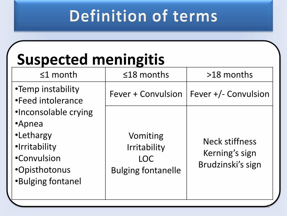
Suspected meningitis
≤1 month ≤18 months >18 months
•Temp instability •Feed intolerance •Inconsolable crying •Apnea •Lethargy •Irritability •Convulsion •Opisthotonus •Bulging fontanel
Fever + Convulsion Fever +/- Convulsion
Vomiting Irritability
LOC Bulging fontanelle
Neck stiffness Kerning’s sign
Brudzinski’s sign
2
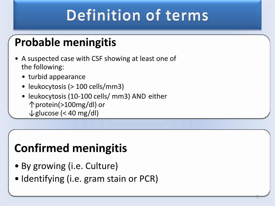
Probable meningitis • A suspected case with CSF showing at least one of
the following:
• turbid appearance
• leukocytosis (> 100 cells/mm3)
• leukocytosis (10-100 cells/ mm3) AND either ↑protein(>100mg/dl) or ↓glucose (< 40 mg/dl)
Confirmed meningitis
• By growing (i.e. Culture)
• Identifying (i.e. gram stain or PCR)
3
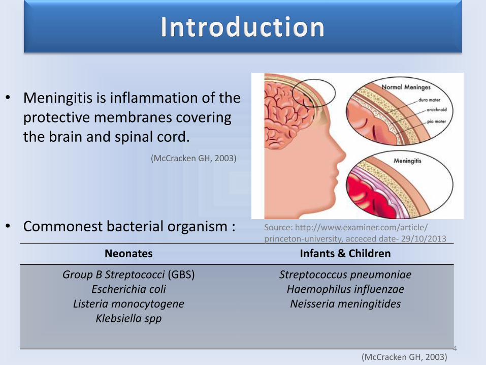
• Meningitis is inflammation of the protective membranes covering the brain and spinal cord. (McCracken GH, 2003)
• Commonest bacterial organism :
Neonates Infants & Children
Group B Streptococci (GBS) Escherichia coli
Listeria monocytogene Klebsiella spp
Streptococcus pneumoniae Haemophilus influenzae Neisseria meningitides
4 (McCracken GH, 2003)
Source: http://www.examiner.com/article/ princeton-university, acceced date- 29/10/2013
5
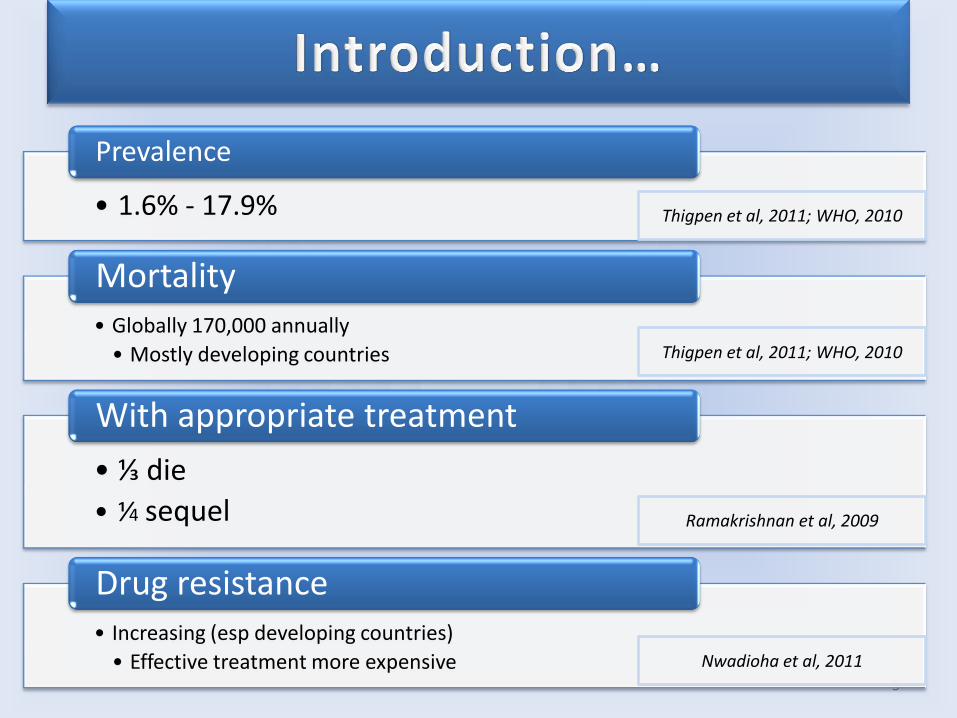
• 1.6% - 17.9%
Prevalence
• Globally 170,000 annually
• Mostly developing countries
Mortality
• ⅓ die
• ⅟4 sequel
With appropriate treatment
• Increasing (esp developing countries)
• Effective treatment more expensive
Drug resistance
Thigpen et al, 2011; WHO, 2010
Ramakrishnan et al, 2009
Nwadioha et al, 2011
Thigpen et al, 2011; WHO, 2010
6
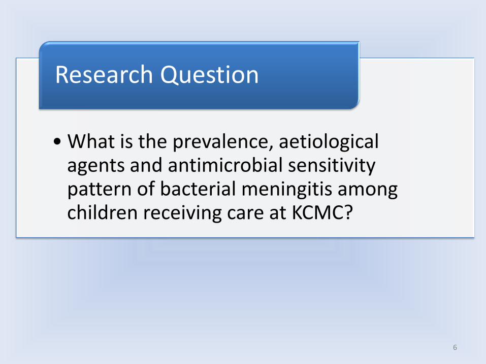
• What is the prevalence, aetiological agents and antimicrobial sensitivity pattern of bacterial meningitis among children receiving care at KCMC?
Research Question
7
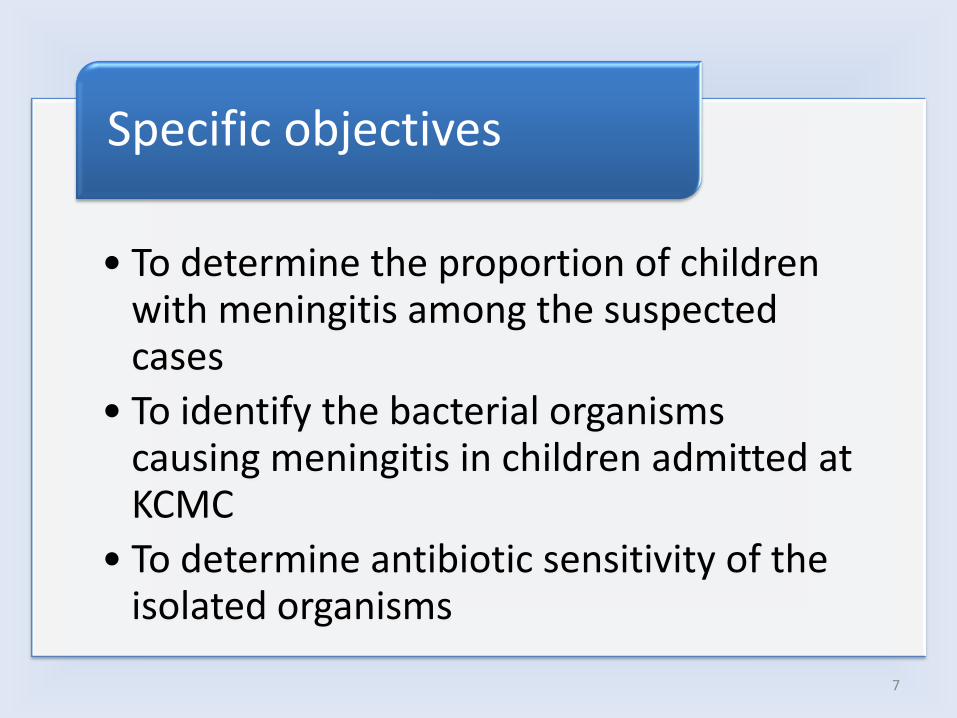
• To determine the proportion of children with meningitis among the suspected cases
• To identify the bacterial organisms causing meningitis in children admitted at KCMC
• To determine antibiotic sensitivity of the isolated organisms
Specific objectives
8
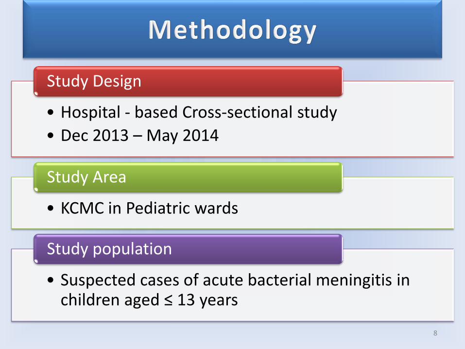
• Hospital - based Cross-sectional study
• Dec 2013 – May 2014
Study Design
• KCMC in Pediatric wards
Study Area
• Suspected cases of acute bacterial meningitis in children aged ≤ 13 years
Study population
9
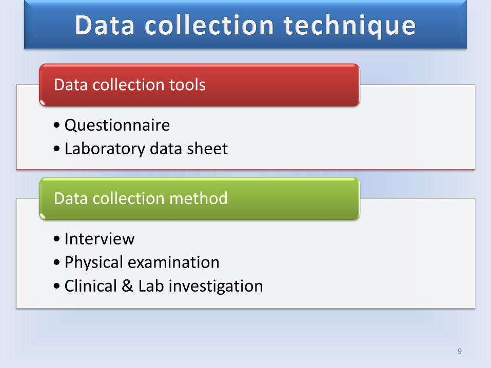
• Questionnaire
• Laboratory data sheet
Data collection tools
• Interview
• Physical examination
• Clinical & Lab investigation
Data collection method
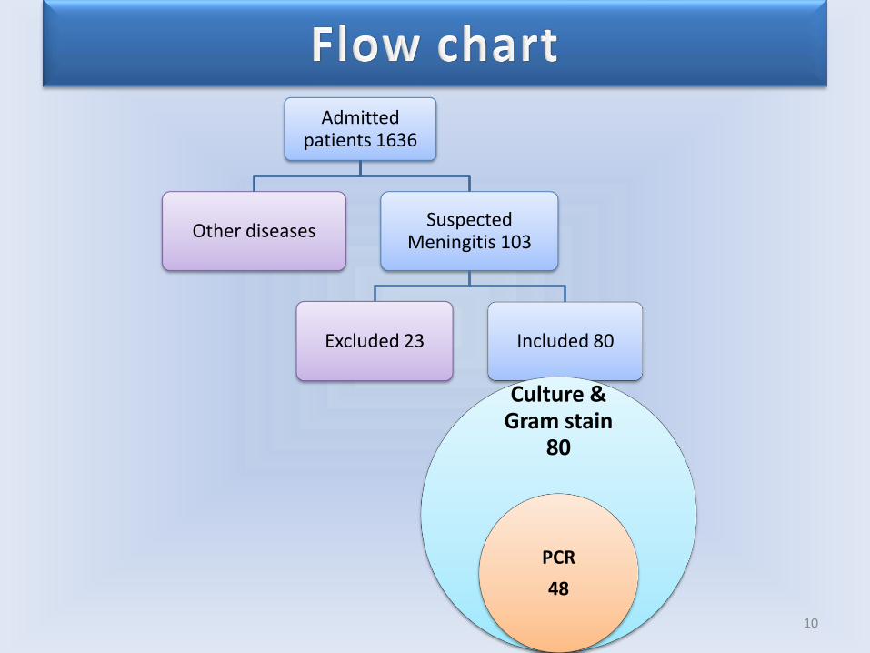
Admitted patients 1636
Other diseases Suspected
Meningitis 103
Excluded 23 Included 80
10
Culture & Gram stain
80
PCR
48
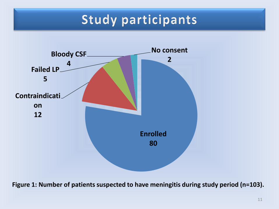
Enrolled 80
Contraindication 12
Failed LP 5
Bloody CSF 4
No consent 2
Figure 1: Number of patients suspected to have meningitis during study period (n=103).
11
12
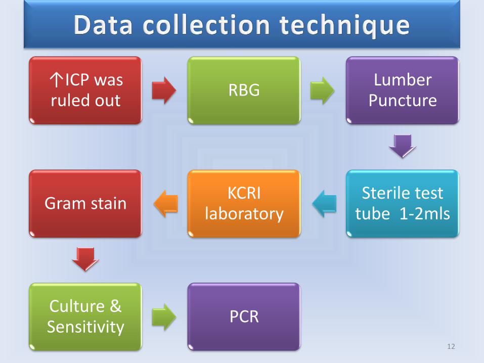
↑ICP was ruled out
RBG Lumber
Puncture
Sterile test tube 1-2mls
KCRI laboratory
Gram stain
Culture & Sensitivity
PCR

13
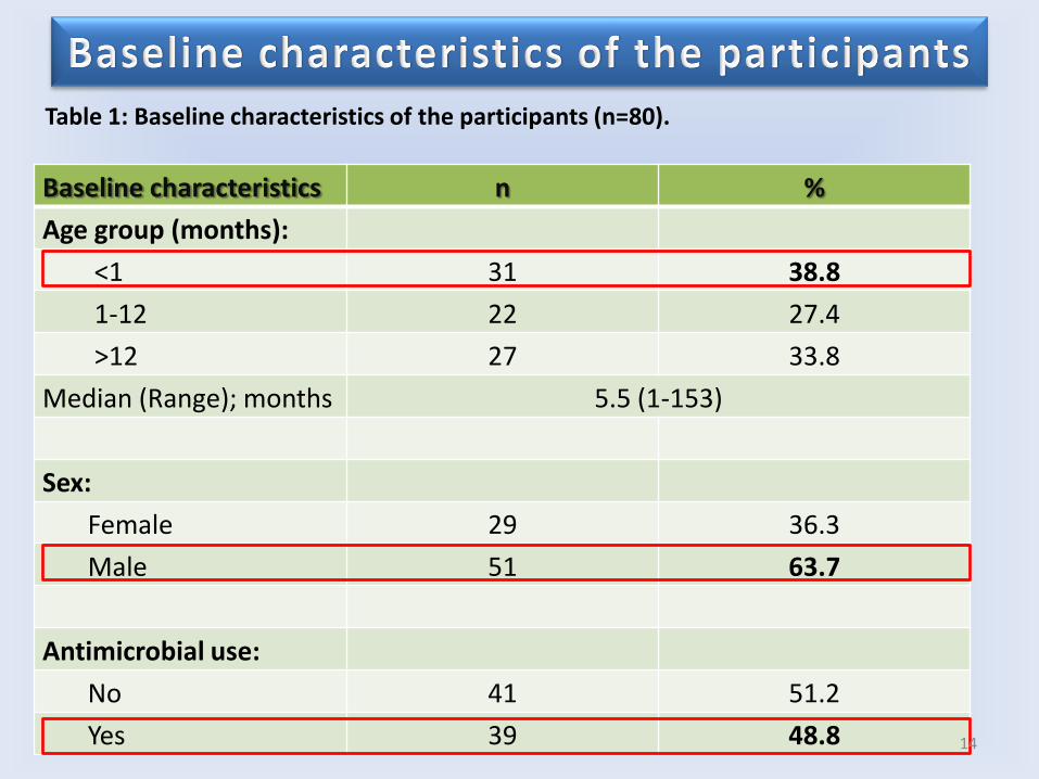
Baseline characteristics n %
Age group (months):
<1 31 38.8
1-12 22 27.4
>12 27 33.8
Median (Range); months 5.5 (1-153)
Sex:
Female 29 36.3
Male 51 63.7
Antimicrobial use:
No 41 51.2
Yes 39 48.8
Table 1: Baseline characteristics of the participants (n=80).
14
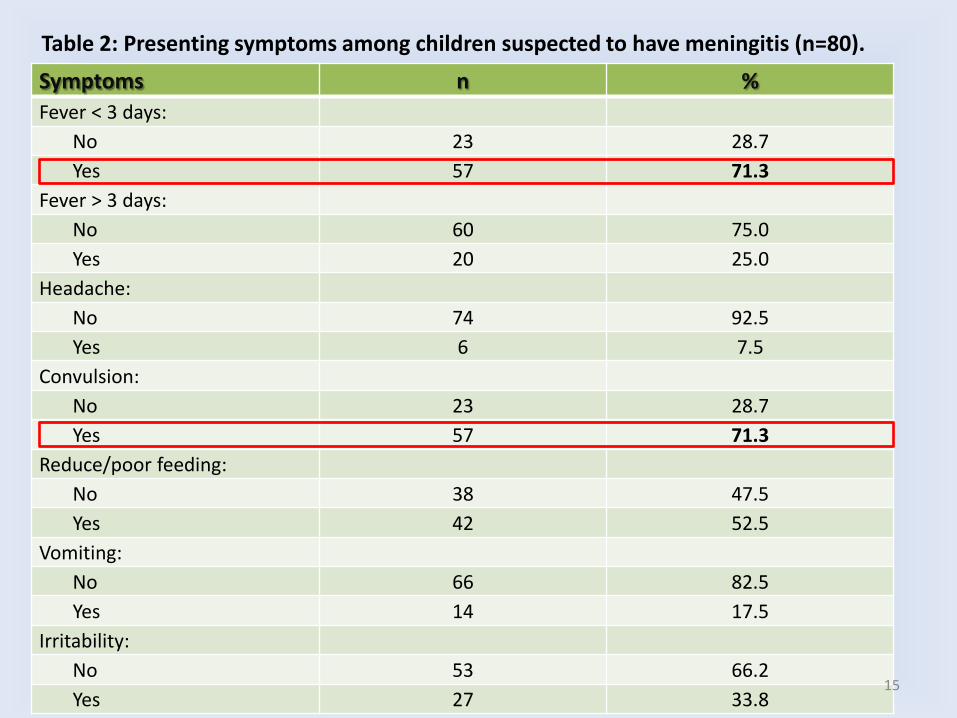
Symptoms n %
Fever < 3 days:
No 23 28.7
Yes 57 71.3
Fever > 3 days:
No 60 75.0
Yes 20 25.0
Headache:
No 74 92.5
Yes 6 7.5
Convulsion:
No 23 28.7
Yes 57 71.3
Reduce/poor feeding:
No 38 47.5
Yes 42 52.5
Vomiting:
No 66 82.5
Yes 14 17.5
Irritability:
No 53 66.2
Yes 27 33.8
Table 2: Presenting symptoms among children suspected to have meningitis (n=80).
15
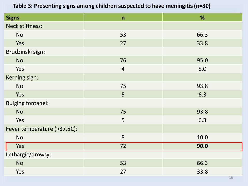
Signs n %
Neck stiffness:
No 53 66.3
Yes 27 33.8
Brudzinski sign:
No 76 95.0
Yes 4 5.0
Kerning sign:
No 75 93.8
Yes 5 6.3
Bulging fontanel:
No 75 93.8
Yes 5 6.3
Fever temperature (>37.5C):
No 8 10.0
Yes 72 90.0
Lethargic/drowsy:
No 53 66.3
Yes 27 33.8
Table 3: Presenting signs among children suspected to have meningitis (n=80)
16
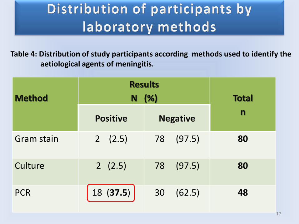
Method
Results
N (%)
Total
n Positive Negative
Gram stain 2 (2.5) 78 (97.5) 80
Culture 2 (2.5) 78 (97.5) 80
PCR 18 (37.5) 30 (62.5) 48
Table 4: Distribution of study participants according methods used to identify the aetiological agents of meningitis.
17
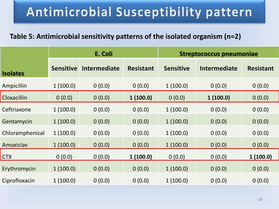
Isolates
E. Coli Streptococcus pneumoniae
Sensitive Intermediate Resistant Sensitive Intermediate Resistant
Ampicillin 1 (100.0) 0 (0.0) 0 (0.0) 1 (100.0) 0 (0.0) 0 (0.0)
Cloxacillin 0 (0.0) 0 (0.0) 1 (100.0) 0 (0.0) 1 (100.0) 0 (0.0)
Ceftriaxone 1 (100.0) 0 (0.0) 0 (0.0) 1 (100.0) 0 (0.0) 0 (0.0)
Gentamycin 1 (100.0) 0 (0.0) 0 (0.0) 1 (100.0) 0 (0.0) 0 (0.0)
Chloramphenical 1 (100.0) 0 (0.0) 0 (0.0) 1 (100.0) 0 (0.0) 0 (0.0)
Amoxiclav 1 (100.0) 0 (0.0) 0 (0.0) 1 (100.0) 0 (0.0) 0 (0.0)
CTX 0 (0.0) 0 (0.0) 1 (100.0) 0 (0.0) 0 (0.0) 1 (100.0)
Erythromycin 1 (100.0) 0 (0.0) 0 (0.0) 1 (100.0) 0 (0.0) 0 (0.0)
Ciprofloxacin 1 (100.0) 0 (0.0) 0 (0.0) 1 (100.0) 0 (0.0) 0 (0.0)
Table 5: Antimicrobial sensitivity patterns of the isolated organism (n=2)
18
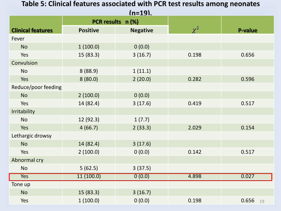
Table 5: Clinical features associated with PCR test results among neonates (n=19).
Clinical features
PCR results n (%)
P-value Positive Negative
Fever
No 1 (100.0) 0 (0.0)
Yes 15 (83.3) 3 (16.7) 0.198 0.656
Convulsion
No 8 (88.9) 1 (11.1)
Yes 8 (80.0) 2 (20.0) 0.282 0.596
Reduce/poor feeding
No 2 (100.0) 0 (0.0)
Yes 14 (82.4) 3 (17.6) 0.419 0.517
Irritability
No 12 (92.3) 1 (7.7)
Yes 4 (66.7) 2 (33.3) 2.029 0.154
Lethargic drowsy
No 14 (82.4) 3 (17.6)
Yes 2 (100.0) 0 (0.0) 0.142 0.517
Abnormal cry
No 5 (62.5) 3 (37.5)
Yes 11 (100.0) 0 (0.0) 4.898 0.027
Tone up
No 15 (83.3) 3 (16.7)
Yes 1 (100.0) 0 (0.0) 0.198 0.656 19
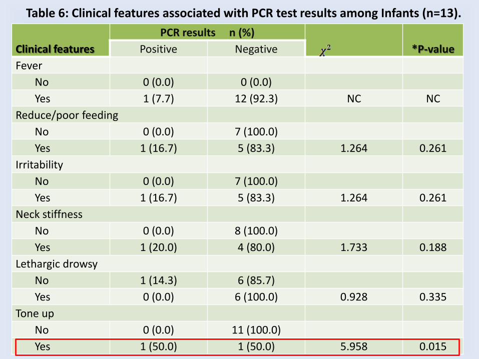
Table 6: Clinical features associated with PCR test results among Infants (n=13).
Clinical features
PCR results n (%)
*P-value Positive Negative
Fever
No 0 (0.0) 0 (0.0)
Yes 1 (7.7) 12 (92.3) NC NC
Reduce/poor feeding
No 0 (0.0) 7 (100.0)
Yes 1 (16.7) 5 (83.3) 1.264 0.261
Irritability
No 0 (0.0) 7 (100.0)
Yes 1 (16.7) 5 (83.3) 1.264 0.261
Neck stiffness
No 0 (0.0) 8 (100.0)
Yes 1 (20.0) 4 (80.0) 1.733 0.188
Lethargic drowsy
No 1 (14.3) 6 (85.7)
Yes 0 (0.0) 6 (100.0) 0.928 0.335
Tone up
No 0 (0.0) 11 (100.0)
Yes 1 (50.0) 1 (50.0) 5.958 0.015

21
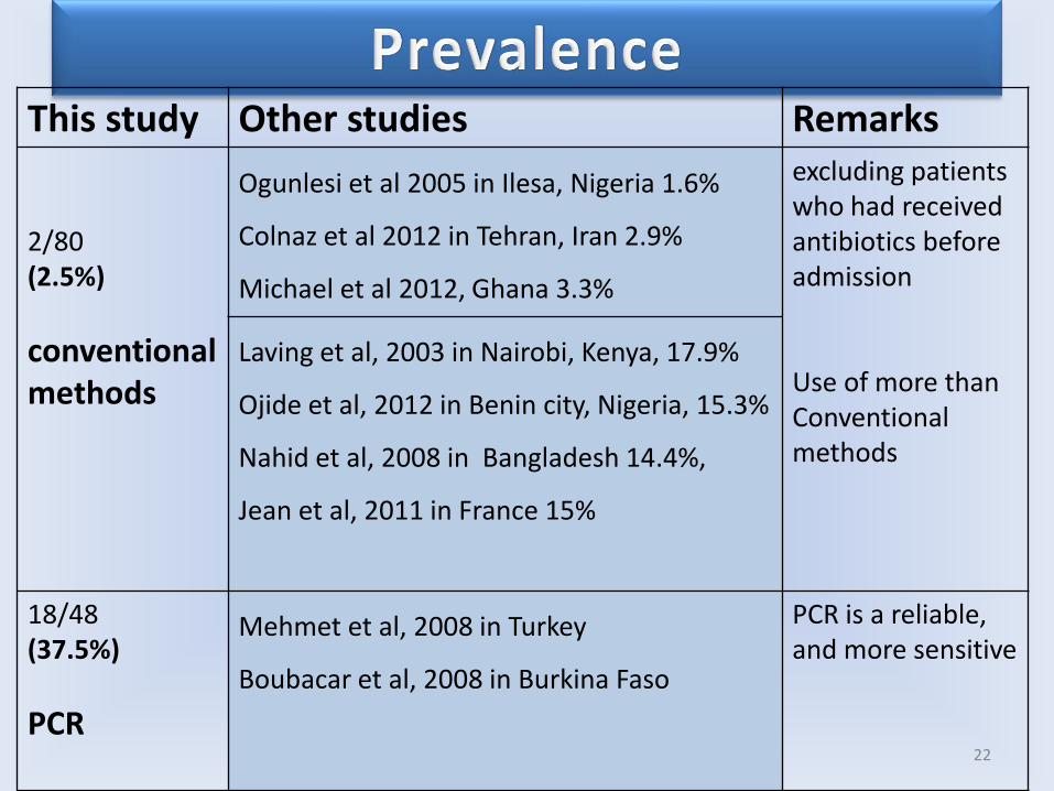
This study Other studies Remarks 2/80 (2.5%)
conventional methods
Ogunlesi et al 2005 in Ilesa, Nigeria 1.6%
Colnaz et al 2012 in Tehran, Iran 2.9%
Michael et al 2012, Ghana 3.3%
excluding patients who had received antibiotics before admission Use of more than Conventional methods
Laving et al, 2003 in Nairobi, Kenya, 17.9%
Ojide et al, 2012 in Benin city, Nigeria, 15.3%
Nahid et al, 2008 in Bangladesh 14.4%,
Jean et al, 2011 in France 15%
18/48 (37.5%)
PCR
Mehmet et al, 2008 in Turkey
Boubacar et al, 2008 in Burkina Faso
PCR is a reliable, and more sensitive
22
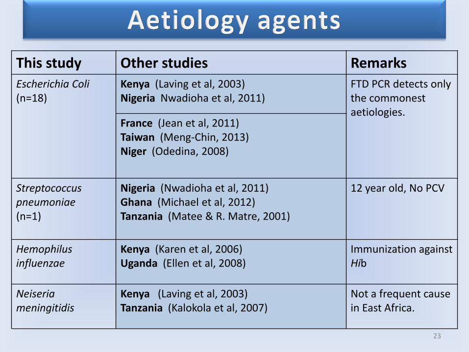
This study Other studies Remarks
Escherichia Coli (n=18)
Kenya (Laving et al, 2003) Nigeria Nwadioha et al, 2011)
FTD PCR detects only the commonest aetiologies. France (Jean et al, 2011)
Taiwan (Meng-Chin, 2013) Niger (Odedina, 2008)
Streptococcus pneumoniae (n=1)
Nigeria (Nwadioha et al, 2011) Ghana (Michael et al, 2012) Tanzania (Matee & R. Matre, 2001)
12 year old, No PCV
Hemophilus influenzae
Kenya (Karen et al, 2006) Uganda (Ellen et al, 2008)
Immunization against Hib
Neiseria meningitidis
Kenya (Laving et al, 2003) Tanzania (Kalokola et al, 2007)
Not a frequent cause in East Africa.
23
24
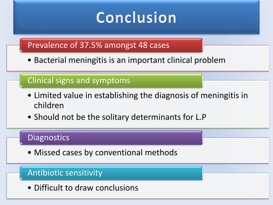
• Bacterial meningitis is an important clinical problem
Prevalence of 37.5% amongst 48 cases
• Limited value in establishing the diagnosis of meningitis in children
• Should not be the solitary determinants for L.P
Clinical signs and symptoms
• Missed cases by conventional methods
Diagnostics
• Difficult to draw conclusions
Antibiotic sensitivity
25
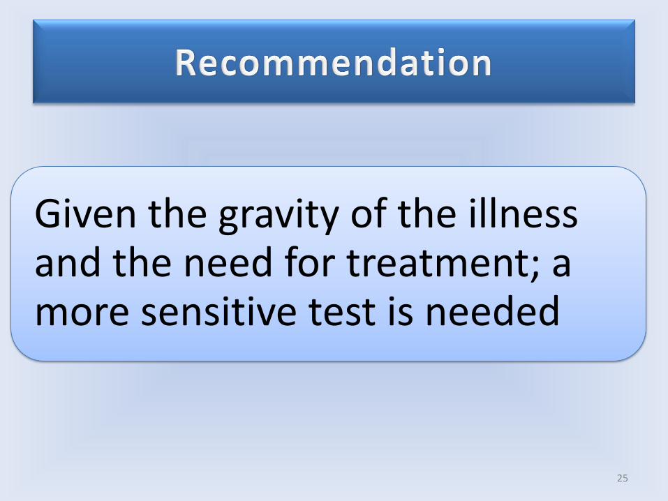
Given the gravity of the illness and the need for treatment; a more sensitive test is needed
26
Ministry of Health, Tanzania
Pediatric Department
KCRI-Biotech lab
Parents /guardians of participants

27
THANK YOU
• Ojide CK, Lofor PVO, Ozumba UC. Childhood Acute Bacterial Meningitis in Benin City, Nigeria. Nig Hosp Pract Vol. 10 (1-2), 2012, 9-17
• Shaban L, Siam R. Prevalence and antimicrobial resinstance pattern of bacterial meningitis in Egypt. Ann. Clin. Microbiol. Antimicrob. 2009; 8: 26-29.
• Kalokola FM, Mwakagile D, Mwamtemi H, Msangi V, Kabengula R, et al. An audit report of bacterial meninigitis among children admitted at Muhimbili National Hospital, Dar es salaam, Tanzania. Tanzania Med. J. 2007; 22: 5-8.
• Kwang Sik Kim. Acute bacterial meningitis in infants and children. Lancet Infect Dis 2010; 10:32-42
• Thigpen MC, Whitney CG, Messonnier NE, Zell ER, Lynfield R, Hadler JL, et al. Emerging Infections Programs Network. Bacterial meningitis in the United States, 1998-2007. N Engl J Med. 2011;364:2016-25.
• http://www.who.int/nuvi/meningitis/en/index.html last updated Feb, 2012 • Mado SM, Aikhionbare, HA Akpede GO. Pattern and antimicrobial sensitivity of pathogens in acute bacterial meningitis beyond neonatal period at Ahmadu
Bello University Teaching Hospital (ABUTH) Shika, Zaria. Niger J Paed 2013; 40 (1): 70 –74
• John VB, Alexander EP, Mary PES, Peter M, Anthony HB, Susan ER (2002). Haemophilus influenzae type b (Hib) Meningitis in the prevaccine era: a global review of incidence, age distributions, and casefatality rates, WHO/V&B/02.18,
• Muhamed-Kheir T, Ala-Eddine D (2010). Impact of changing epidemiology on vaccination strategies in Africa. Future Microbiol., 5(6): 837-839. • Nwadioha SI, FMCPath1, Onwuezube I, FMCPath2, Egesie J O, FMCPath3, Kashibu E, FMLS4 , Nwokedi EOP, FMCPath. BACTERIAL ISOLATES FROM
CEREBROSPINAL FLUID OF SUSPECTED ACUTE MENINGITIS IN NIGERIAN CHILDREN. emedpub – International Infectious Diseases : Vol 1:8 • Forbes BA, Sahm DF, Weisfeld AS. Streptococcus Meningitis and other infections of the central nervous system. In: Laboratory Manual of Bailey & Scotts
diagnostic microbiology. 12th Ed. Mosby Elsevier publication 2007, pp 907-916.
• Thaver D, Ali SA, Zaidi AK. Antimicrobial resistance among Neonatal pathogens in developing countries. Pediatric Infect Dis J. 2009;28 (1 suppl): s19-21 • Best J, Hughes S. Evidence behind the WHO Guidelines: hospital care for children–what are the useful clinical features of bacterial meningitis found in
infants and children? J Trop Pediatr 2008;54:83–86. • Weber MW, Carlin JB, Gatchalian S et al. Predictors of neonatal sepsis in developing countries. Pediatr Infect Dis J 2003;22:711–716. • • Brien, K. L, Wolfson L. J., Watt J. P., Henkle E., Deloria-Knoll M., McCall N., et al. 2009. Burden of disease caused by Streptococcus pneumoniae in children
younger than 5 years: global estimates. Lancet 374:893-902. • • Watt, J. P., Wolfson, L.J. O'Brien, K. L., Henkle, E. Deloria-Knoll, M., McCall, N., et al. 2009. Burden of disease caused by Haemophilus influenzae type b in
children younger than 5 years: global estimates. Lancet 374:903-911. • • Karanika M, Vasilopoulou VA, Katsioulis AT, Papastergiou P, Theodoridou MN, et al. (2009) Diagnostic Clinical and Laboratory Findings in Response to
Predetermining Bacterial Pathogen: Data from the Meningitis Registry. PLoS ONE 4(7): e6426. doi:10.1371/journal.pone.0006426 • • Thaver, D. & Zaidi, AK.( 2009). Burden of neonatal infections in developing countries: a review of evidence from community-based studies. Pediatr Infect
Dis J. Jan; 28(1 Suppl), pp. S3-9. 28