Embed Size (px)
Citation preview

AR
TICLE
v o l u m e 3 · n o . 1 1
© 2012 International Mycological Association
You are free to share - to copy, distribute and transmit the work, under the following conditions:Attribution: Youmustattributetheworkinthemannerspecifiedbytheauthororlicensor(butnotinanywaythatsuggeststhattheyendorseyouoryouruseofthework). Non-commercial: Youmaynotusethisworkforcommercialpurposes.No derivative works: Youmaynotalter,transform,orbuilduponthiswork.For any reuse or distribution, you must make clear to others the license terms of this work, which can be found at http://creativecommons.org/licenses/by-nc-nd/3.0/legalcode. Any of the above conditions can be waived if you get permission from the copyright holder. Nothing in this license impairs or restricts the author’s moral rights.
FOR
THC
OM
ING
ME
ETI
NG
S
(30)
INtroductIoN
Tibouchina granulosa (Melastomataceae), the Brazilianglorytree, is a fast growing tree native that occurs in the Atlantic ForestofBrazil.Itisacommonandimportantcomponentofthenativeflora,particularlyinsecondaryforests.Itisalsoahighly prized ornamental, which is widely used in gardensand parks because of its spectacular violet or pink blossoms that each year appear prior and duringEaster.The periodof lent (quaresma, in Portuguese) is an important periodin the catholic tradition, from which the Brazilian commonnameforthistree,“quaresmeira”isderived(Lorenzi2002).Usually there is a high demand for young T. granulosa plants in the garden nursery market in Brazil, particularly for thevariety that produces pink flowers. Nevertheless, diseasesare known to be a limiting factor in nursery production of T. granulosa. However, very little is known about the diseases affecting this tree. The record of Chrysoporthe cubensis (syn.:Cryphonectria cubensis) on T. granulosa inBrazilbySeixas et al.(2004)isthesolerecordofafungalpathogenonthishostintheBraziliandatabase(http://pragawall.cenargen.embrapa.br/aiqweb/michtml/fichahp.asp?id=1912)–andthere are only three records of fungi on this host in the USDA fungaldatabase(Farr&Rossman2010). Thisissomewhatsurprising for such a common plant in the neotropics, and possibly reflects the limitedexistingknowledgeaboutplantpathogenic fungi occurring on wild plants in this region.Here we clarify the identity of the fungus associated with a severe foliage blight (often evolving into a form of die-back) of quaresmeira.This is one of themostwidespread
and damaging diseases affecting T. granulosa inthefield,ingardens, and also in nurseries.
MAterIAl ANd Methods
Isolates Samples of young abnormal branches of Tibouchina granulosa bearing diseased leaves were collected at two localities in Brazil (states of Rio de Janeiro and MinasGerais),driedinaplantpressandbroughttothelaboratoryfor further examination. Representative specimens of thefungus were deposited in the herbarium at the Universidade Federal de Viçosa (VIC). Pure cultures were obtained bytransferofconidia,usingasterilefine-pointedneedle, fromlesionsontoplatescontainingVBA(vegetablebroth-agar)asdescribedinPereiraet al. (2003).Pureculturesaredepositedin the fungal culture collection at the Universidade Federal deViçosa and also at theCBS-KNAWFungal BiodiversityCentre (CBS) in Utrecht, TheNetherlands. RepresentativevoucherspecimensaredepositedinVICandatCBS.
DNA isolation, amplification and analysesGenomic DNA was isolated from fungal mycelium grownonMEA, using theUltraCleanTMMicrobial DNA IsolationKit (MoBio Laboratories, Inc., Solana Beach, CA, USA)according to the manufacturer’s protocols. The primersV9G (de Hoog & Gerrits van den Ende 1998) and LR5(Vilgalys&Hester1990)wereused toamplifypart of thenuclearrDNAoperonspanningthe3’endofthe18SrRNA
doi:10.5598/imafungus.2012.03.01.01 IMA FuNgus · voluMe 3 · No 1: 1–7
Pilidiella tibouchinae sp. nov. associated with foliage blight of Tibouchina granulosa (quaresmeira) in Brazil
BrunoE.C.Miranda1,RobertW.Barreto1,PedroW.Crous2,andJohannesZ.Groenewald2
1UniversidadeFederaldeViçosa,DepartamentodeFitopatologia,36570-000,Viçosa,MG,Brazil;correspondingauthore-mail:RobertW.Barreto,[email protected],P.O.Box85167,3508AD,Utrecht,TheNetherlands
Abstract: Tibouchina granulosa (Melastomataceae), Brazilian glorytree (Brazilian common name –quaresmeira),acommontreeoftheAtlanticForestofBrazil,iswidelyusedasanornamentalforitsvioletorpinkblossoms.Littleisknownaboutfungaldiseasesaffectingthisspecies,althoughtheserepresentaknownlimitationforitscultivationinnurseries.Amongthesethereisafoliageblightthatoccursincombinationwithdistortionofbranchapicesanddie-back.AconsistentassociationofaspeciesofPilidiella with the diseased tissueswasobserved.ThefunguswasisolatedinpurecultureandbasedonitsmorphologyandDNAphylogeny,we conclude that it represents a new species, for which the name Pilidiella tibouchinae isintroduced.
Article info:Submitted:9January2012;Accepted:29February2012;Published:5April2012.
Key words: ConiellaDiaporthalesITSLSUSordariomycetessystematics

i m a f u n G u S
Miranda et al.A
RTI
CLE
2
gene (SSU), the internal transcribed spacer 1, the 5.8SrRNAgene,theinternaltranscribedspacer2(ITS)andthefirst900basesatthe5’endofthe28SrRNAgene(LSU).Theprimers ITS4(Whiteet al.1990)andLSU1Fd(Crouset al. 2009a) were used as internal sequence primers toensuregoodqualitysequencesovertheentirelengthoftheamplicon. The PCR conditions, sequence alignment, andsubsequentphylogeneticanalysisfollowedthemethodsofCrouset al. (2006,2009b).Additionally, partial translationelongationfactor1-alpha(TEF)sequencesweredeterminedas described by Bensch et al. (2010). Sequences werecomparedwiththesequencesavailableinNCBI’sGenBanknucleotide (nr) database using a megablast search andalignmentswereconstructedbasedontheseresultsforITS.ForLSU,thenovelsequencewereaddedtoanalignmentmodified from Lamprecht et al. 2011 (TreeBASE studyS11805).Sequencesderived in this studywere lodgedatGenBank, thealignment inTreeBASE (www.treebase.org/treebase/index.html),andtaxonomicnoveltiesinMycoBank(www.MycoBank.org;Crouset al.2004).
MorphologySlides containing fungal structures were mounted in lactophenol or lactofuchsin, with 30 measurementsdetermined per structure.Sectionswere preparedwith thehelpofafreezingmicrotome(MicromHM520).Observationsof fungal structures and measurements, as well as preparation ofphotographs,wereperformedwithanOlympusBX51lightmicroscope fitted with an Olympus E330 camera. Colonycharactersandpigmentproductionwerenotedafter6dofgrowthon2%maltextractagar(MEA)andpotatocarrotagar(PCA)(Crouset al.2009c)platesincubatedat25ºC.Colonycolours (surface and reverse) were rated according to thecolourchartsofRayner(1970).
results
PhylogenyApproximately 1700 bases, spanning the ITS and LSUregions, were obtained from the sequenced culture.
Coniella fragariae
30 changes
Harknessia eucalypti AY720745
AY339313
AY339343
EU301037
AY339338
HQ264189
AY339320
AY339319
AY339317
AY339316
BECM2
BECM1
GU062317
AY339346
HQ166057
AY339342
AY339348
AY339344
DQ914688
EU301051
AY339324
AY339323
AY339333
AY339334
AY339332
100
100
98
85
90 100
94
51
65
72
75
Coniella australiensis
Pilidiella eucalyptorum
Pilidiella macrospora
Pilidiella crousii
Pilidiella tibouchinae
Pilidiella granati
Pilidiella sp.
Pilidiella quercicola
Schizoparme straminea
Pilidiella diplodiella
Pilidiella diplodiopsis
Fig. 1.Thefirstof24equallymostparsimonioustreesobtainedfromaheuristicsearchwith50randomtaxonadditionsoftheITSsequencealignment(Treelength=229,CI=0.834,RI=0.904,RC=0.754).Thescalebarshows30changes,andbootstrapsupportvaluesfrom1000replicatesareshownatthenodes.Accessionnumbersofex-typestrainsareshowninbold andthenovelspeciesinthisstudyinred.Branchespresent in thestrictconsensus treeare thickenedand the treewasrooted toasequenceofHarknessia eucalypti (GenBankaccessionno.AY720745).
Pilidiella tibouchinae sp. nov.A
RTIC
LE
v o l u m e 3 · n o . 1 3
The ITS region was used in the phylogenetic analysis todeterminespecies-rankrelationships(Fig.1)andtheLSUregion for the generic placement (Fig. 2). The manuallyadjusted ITS alignment contained 25 taxa including theoutgroupsequenceand,ofthe501charactersusedinthephylogenetic analysis, 86 were parsimony-informative, 49werevariableandparsimony-uninformative,and366were
constant. Twenty-four equally most parsimonious treeswere retained from theheuristic search, the first ofwhichisshowninFig.1.ThephylogenetictreeoftheITSregion(Fig.1)showsthattheobtainedsequencesclusterbetweenPilidiella crousii and Pilidiella granati. The manuallyadjusted LSU alignment contained 54 taxa including thetwooutgroupsequencesand,ofthe840charactersusedin
10 changes
Magnaporthe grisea AB026819Gaeumannomyces graminis var. avenae AF362556
Coniochaetidium savoryi AY346276Coniochaeta velutina EU999180
Calosphaeria pulchella AY761075Togninia novae-zealandiae AY761081
Phaeoacremonium sphinctrophorum DQ173151Asterosporium asterospermum AB553745Asterosporium asterospermum AB553741
Mazzantia napelli AF408368Diaporthe perjuncta AF408356Diaporthe pustulata AF408358Diaporthe padi AF408354
Diaporthe angelicae AY196781Phaeocytostroma ambiguum FR748102
Stenocarpella macrospora DQ377934Stenocarpella maydis DQ377936
Phaeocytostroma sacchari FR748105Diaporthe detrusa AF408349
Phaeocytostroma plurivorum FR748104Phaeocytostroma megalosporum FR748103
Valsa ceratosperma AF408386Valsella adhaerens AF408388
Leucostoma niveum AF408367Greeneria uvicola AF362570
Melanconiella spodiaea AF408370Endothiella gyrosa AF362555
Cryphonectria macrospora AF408340Cryphonectria nitschkei AF408341
Ophiovalsa betulae AF408375Phragmoporthe conformis AF408377
Gnomonia setacea AF362563Melanconis stilbostoma AF408374Melanconis marginalis AF408373Melanconis alni AF362566
Wuestneia molokaiensis AF408390Harknessia eucalypti AF408363Harknessia gibbosa EF110615
Pilidiella eucalyptorum EU754150Coniella australiensis AF408336Coniella fragariae AF362553BECM2 Pilidiella tibouchinae
BECM1 Pilidiella tibouchinaeSchizoparme botrytidis AF408383Pilidiella macrospora AY339292Coniella musaiaensis AF408337Schizoparme straminea AY339296Pilidiella granati AF408379
Pilidiella quercicola AY339293Pilidiella diplodiopsis AY339287Pilidiella diplodiella AY339286Pilidiella diplodiopsis AY339288Pilidiella sp. AY339295
Pilidiella castaneicola AF408378
Key to families:Ca: CalosphaeriaceaeCr: CryphonectriaceaeD: DiaporthaceaeG: GnomoniaceaeM: MelanconidiaceaeT: TogniniaceaeV: Valsaceae
100
100
97
88
100
98
100
60
70
6061
8078
73
100
93
63
89
58
96
100
57
62
82
59
100
53
79
98
100
57
92
88
T
D
M2
M1Cr
G
V
M3
Fam
ily
Ca
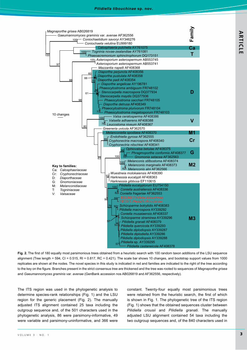
Fig. 2.Thefirstof180equallymostparsimonioustreesobtainedfromaheuristicsearchwith100randomtaxonadditionsoftheLSUsequencealignment(Treelength=594,CI=0.515,RI=0.817,RC=0.421).Thescalebarshows10changes,andbootstrapsupportvaluesfrom1000replicatesareshownatthenodes.Thenovelspeciesinthisstudyisindicatedinredandfamiliesareindicatedtotherightofthetreeaccordingtothekeyonthefigure.BranchespresentinthestrictconsensustreearethickenedandthetreewasrootedtosequencesofMagnaporthe grisea and Gaeumannomyces graminisvar.avenae(GenBankaccessionnosAB026819andAF362556,respectively).

i m a f u n G u S
Miranda et al.A
RTI
CLE
4
thephylogeneticanalysis,190wereparsimony-informative,54 were variable and parsimony-uninformative, and 596wereconstant.Fromthisheuristicsearch,180equallymostparsimonioustreeswereretained,thefirstofwhichisshowninFig.2.Phylogeneticanalysisof theLSUregion(Fig.1)confirmstheplacementofthenovelsequencesinPilidiella.ThepartialTEFsequencesdidnothaveanyhighidentitytothosesequencesavailableinGenBank(datanotshown).
TaxonomyA pycnidial coelomycete was regularly associated with diseased tissues on the samples collected at the two separate localities. Its morphology conformed to that ofspecies in the genus Pilidiella(NagRaj1993,vanNiekerket al. 2004),althoughitappearedtorepresentadistincttaxon.Accordingly, a new species name is introduced below to accommodate the fungus occurring on T. granulosa.
Pilidiella tibouchinae B.E.C.Miranda,R.W.Barreto&Crous,sp. nov.MycoBankMB563992(Fig.3)
Etymology:Namedafterthehostgenusonwhichitoccurs,Tibouchina.
Diagnosis: Similar to Pilidiella eucalyptorum but lacking conidial germ slits, and similar to P. petrakioidea but lacking mucoidappendagesonconidia.
Type: Brazil: Minas Gerais: Viçosa, campus of the Universidade Federal de Viçosa, on leaves of Tibouchina granulosa, 8 March 2010, B. C. Miranda (VIC 31443 –holotype;CBSH-20827–isotype;culturesex-holotypeCPC18511,CPC18512=CBS131595).
(GenBankaccessionnumbersforVIC31443andVIC31444:ITS=JQ281774,JQ281775;LSU=JQ281776,JQ281777;TEF=JQ281778,JQ281779)
Other specimen examined: Brazil: Minas Gerais: Viçosa, campus of the Universidade Federal de Viçosa, on leaves of T. granulosa, 17May2010,B. C. Miranda(VIC31444).
Fig. 3. Pilidiella tibouchinae.A–d.LeafspotsandcurlingonTibouchina granulosa.e.Colonyonoatmealagar.F, g. Vertical section through pycnidia.h, I.Conidiogenouscells.J, K.Conidia.Bars=10µm.

Pilidiella tibouchinae sp. nov.A
RTIC
LE
v o l u m e 3 · n o . 1 5
Description: Lesionsonlivingleavesandyoungstems,firstlyas adaxial straw-coloured necrotic spots, mostly appearing near the leaf veins, becoming yellowish to greyish with a dark brown to dark purple border, irregularly shaped, coalescing and leading to necrosis and distortion of large parts of the leaf lamina; loss of necrotic leaf parts usually creatingthe impression of insect damage. In conjunction to thesesymptoms, a shortening of branch internodes, leaf distortion, bud death, necrosis and die-back of young stems are also observed. Stunting and decline of severely affected plantsare observed even for adult plants.Conidiomata pycnidial, adaxial, subcuticular, solitary, globose to depressed globose, 42.5–75 × 75–112.5 μm, wall composed of dark greyishbrown textura angularis of 1–3 cell layers, 7–12 μm thick,dark brown; dehiscence ostiolate, central; conidiophores formed on a dense, basal, cushion-like aggregation of hyaline cells, mostly reduced to conidiogenous cells, subcylindrical, branched below, 8–15 × 3–4 μm, smooth,hyaline, 1–2-septate. Conidiogenous cells enteroblastic, phialidic with apical periclinal thickening, 5–10 × 2–3 μm,smooth, hyaline, with minute collarette, and covered in mucilage.Conidia mostly broadly ellipsoidal, often somewhat flattenedononeside,oblong,subreniform,ovoidtosubovoid,10–13×6–8μm (l:b= 1.7), apex rounded, subtruncate atbase, hilum sometimes slightly protuberant, aseptate, hyaline when immature, becoming smoky-brown at maturity, smooth, guttulate (usually with one large guttule but sometimesbiguttulateoreguttulate).
Culture characteristics: (MEA or PCA either under a 12 hlightregimeorinthedark):Coloniesfast-growing(upto86mmdiamafter6d);flat,occasionallyslightlyraisedcentrally;mostly composed of immersed mycelium, aerial mycelium mostly sparse (but very dense cottony to woolly aerialmyceliumonMEA in the dark); cottony towoolly to spiderweb-like white to grey olivaceous, sometimes with some small cinnamon areas centrally, occasionally becoming powdery towards the periphery, abundant olivaceous black fruit bodies crowdedinzoneringsonMEA/light.OnPCAgreyishblacktogreenishblackcentrally,withsaffronmargininreverse;blackfruitbodieslessabundant,andinmoredistinctringsonPCA/light.
dIscussIoN
The fungus on Tibouchina granulosa clearly belongs to the Coniella/Pilidiella-complex that has Schizoparme teleomorphs (Schizoparmaceae, Diaporthales; Rossmanet al. 2007). Fungi in Schizoparmaceae include several species associated with foliar diseases, sometimes occurring as secondary invaders of plant tissues infected byother organismsor injuredbyother causes (Ferreiraet al. 1997).ThereisnorecordofanyteleomorphicspeciesofSchizoparmaceae in association with members of the genus Tibouchina, and a single doubtful record of a Coniella on another member of the Melastomataceae, Miconia serrulata
(Farr & Rossman 2010). Several members of Myrtales, which according toBremeret al. (2003) includes up to 14families, are known hosts of Schizoparmaceae (Farr &Rossman 2010). For instance, several species are knownfrom Myrtaceae (Acca, Blepharocalyx, Eucalyptus, Eugenia, Heteropyxis, Myrcia, Syzygium), Lythraceae (Lythrum, Punica), and Combretaceae (Anogeissus, Anogeissus, Terminalia)(vanNiekerket al.2004,Farr&Rossman2010)
Sutton(1980)andNagRaj(1993)treatedPilidiella as a synonym of Coniella.However,basedonanalysesof largesubunit (LSU)nuclear ribosomalDNA (nrDNA) sequences,Castleburyet al. (2002) concluded thatPilidiella is distinct from Coniella.Pilidiella has two main morphological criteria separating it from Coniella: the presence of conidia that are hyaline when young becoming pale brown with age (consistentlybrowninConiella) (Castleburyet al.2002)andhaving a length to breadth ratio larger than 1.5 (equal toor smaller than1.5 forConiella) (vanNiekerket al. 2004).Pigmentationaloneisdifficulttointerpret(Table1),althoughP. tibouchinae has hyaline conidia that become smoky brown atmaturityanda l:b ratioof1.7.Additionally the resultsofthe phylogenetic analysis place P. tibouchinae in the Pilidiella clade, distinct from Coniella (Figs 1–2). Considering thecombination of morphological and molecular data, we prefer to place this fungus in the genus Pilidiella. Nevertheless,several species in the group still need to be re-examined, as isevidentfromFig.2,wherePilidiella eucalyptorum clusters in the Coniella clade, and C. musariensis clusters in the Pilidiellaclade.
Pycnidia in P. tibouchinae are small when compared to the species of Schizoparmaceae treated by Sutton (1980), Nag Raj (1993), and van Niekerk et al. (2004).Morphologically, conidia of P. tibouchinae show some similarity to that of P. eucalyptorum and P. petrakioidea. However, conidia of P. tibouchinae lack conidial germ slits (present in P. eucalyptorum) and mucoid appendages(presentinP. petrakioidea).Italsohasthinnerpycnidialwalls(7–12μm), than those inP. eucalyptorum (to25µmthick), and has hyaline to pale smoky-brown conidia, whereas those of P. eucalyptorumaremediumtodarkreddishbrown.Furthermore, P. tibouchinae also differs from P. petrakioidea in conidial morphology (narrowly ellipsoidal with acutelyrounded apices in P. petrakioidea) and a l:b ratio largerthan 1.9. Five species ofConiella have been described in association with members of the Myrtaceae: C. australiensis, C. castaneicola, C. costae, C. fragariae, and C. minima. Considering the close morphological similarity of Pilidiella and Coniella, the conidial morphology of these species is also provided here for comparison with that of P. tibouchinae (Table1).
Pilidiella tibouchinaeisthefirstspeciesofthegenus to be described on a host belonging to Melastomataceae, on which it appears to be associated with a rather serious foliar and diebackdisease.Furtherinvestigationsaimedatclarifyingthepathological status of the fungus on Tibouchina granulosa, and evaluating potential disease control measures are now urgentlyrequired,andwillbereportedelsewhere.

i m a f u n G u S
Miranda et al.A
RTI
CLE
6
AcKNowledgeMeNts
We thank the technical staff ofCBS,Arien van Iperen (cultures),MarjanVermaas(photographicplates),andMiekeStarink-Willemse(DNA isolation, amplification and sequencing) for their invaluableassistance.
reFereNces
AaHAvander (1983)AnewspeciesofConiella.Proceedings of the Koninklijke Nederlandse Akademie van Wetenschappen, C 86:121–125.
AhmadS (1967)Contributions to the fungi ofWest Pakistan–VI.Biologica 13:15–42.
Bensch K, Groenewald JZ, Dijksterhuis J, Starink-Willemse M,Andersen B, Summerell BA, ShinH-D, Dugan FM, SchroersH-J, Braun U, Crous PW (2010) Species and ecologicaldiversity within the Cladosporium cladosporioides complex (Davidiellaceae, Capnodiales).Studies in Mycology 67:1–94.
BremerB,BremerK,ChaseMW,RevealJL,SoltisDE,et al.(2003)Anupdateoftheangiospermphylogenygroupclassificationforthe orders and families of flowering plants:APG II.Botanical Journal of the Linnean Society 141:399–436.
CrousPW,GamsW,Stalpers JA,RobertV,StegehuisG (2004)MycoBank:anonlineinitiativetolaunchmycologyintothe21stcentury.Studies in Mycology 50:19–22.
Crous PW, Groenewald JZ, Risède J-M, Simoneau P, HydeKD (2006) Calonectria species and their Cylindrocladium anamorphs:specieswithclavatevesicles.Studies in Mycology 55:213–226.
CrousPW,GroenewaldJZ,SummerellBA,WingfieldBD,WingfieldMJ (2009b) Co-occurring species of Teratosphaeria on Eucalyptus.Persoonia 22:38–48.
Crous PW, Schoch CL, Hyde KD, WoodAR, Gueidan C, HoogGSDe,Groenewald JZ (2009a)Phylogenetic lineages in theCapnodiales.Studies in Mycology 64:17–47.
Crous PW, Verkley GJM, Groenewald JZ, Samson RA (eds)(2009c)Fungal Biodiversity.[CBSLaboratoryManualSeries 1.]CentraalbureauvoorSchimmelcultures,Utrecht.
CastleburyLA,RossmanAY,JaklitschWJ,VasilyevaLN(2002)Apreliminary overview of the Diaporthales based on large subunit nuclearribosomalDNAsequences.Mycologia 94:1017–1031.
DianeseJC,MedeirosRB,SantosLTP,SuttonBC(1993)Coniella costaesp.nov.on leavesofMyrcia tomentosa fromBraziliancerrado.Mycological Research 97:1234–1236.
FAO (1996) International Standard for Phytosanitary Measures. Rome: Secretariat of the International Plant ProtectionConvention.
Farr DF, Rossman AY (2010) Fungal databases. Beltsville, MD:Systematic Mycology and Microbiology Laboratory, ARS,USDA;http://nt.ars-grin.gov/fungaldatabasews/.
FerreiraFA,AlfenasAC,CoelhoL (1997)Portas-de-entradaparaConiella fragariaeem folhasdeeucalipto.Revista Árvore 21: 307–311.
FirdousiSA,SharmaCD,VyasKM(1994)AnewspeciesofConiella fromIndia.Acta Botanica Indica 22:134–135.ta
ble
1. Conidialm
orphologyofselected
Con
iella
and
Pili
diel
la s
peci
es re
cord
ed fr
om m
embe
rs o
f Myr
tale
s.
spec
ies
size
l:b ra
tesh
ape
App
enda
geg
erm
slit
ref
eren
ce
Con
iella
aus
tralie
nsis
(9–)10–11(–14)×(6–)7–8(–10)μm
1.4
Broadlyellipsoidal
+-
vanNiekerke
t al.(2004)
C. c
asta
naei
cola
13–29×2.5–3.5μm
7.3
Fuso
id to
falc
ate
+-
NagRaj(1993)
C. c
osta
e 19–28×7–7.5μm
3.2
Fuso
id to
elli
psoi
d-
-D
iane
se e
t al.(1993)
C. d
elic
ata
7–9×2.5–3μm
2.9
Elli
psoi
d-
-Sutton(1980)
C. f
raga
riae
(8–)9–10(–12.5)×
(5–)6–7(–8)μ
m1.5
Elli
psoi
d+
+vanNiekerke
t al.(2004)
in o
lder
con
idia
C. m
acro
spor
a (18.3–)25–29(–32.5)×
(13–)16–20(–21.5)µ
m1.5
Ovoid,ellipsoid,pyriform,globoid
+-
vanderA
a(1983)
C. m
inim
a
6.5–7.5×3.5–4.5μm
1.5
Glo
boid
to s
ubgl
oboi
d-
-Sutton(1969)
C. t
erm
inal
iae
2–8×2–3.5µm
2:01
Glo
bose
to s
ubgl
obos
e-
-Fi
rdou
si e
t al.(1994)
Pili
diel
la c
rous
ii(6–)7–12(–13.5)×
(2.5–)3–5μm
2.2
Narrowlyellipsoidtoellipsoid
--
Raj
eshk
umar
et a
l.(2011)
P. d
iplo
diel
la
(10–)12–15(–19)×
(4–)5–6μm
2.3
Narrowlyellipsoid
+ -
vanNiekerke
t al.(2004)
P.eu
caly
ptor
um
(9–)10–12(–14)×(6–)7–8μm
1.6
Broadlyellipsoidorlimoniform
unco
mm
on
+vanNiekerke
t al.(2004)
P. g
rana
ti 9–16×3–4.5μm
2.8
Elli
psoi
d+
-NagRaj(1993)
P. ja
mbo
lana
19–22×3.5–4µm
5.7
Elo
ngat
e-fu
soid
--
Ahm
ad(1967)
P. p
etra
kioi
dea
12–14.5×6.5–8μm
1.9
Narrowlyellipsoid
+-
NagRaj(1993)
P. ti
bouc
hina
e 10–13×6–8μm
1.7
Broadlyellipsoid
--
Thispublication

Pilidiella tibouchinae sp. nov.A
RTIC
LE
v o l u m e 3 · n o . 1 7
HoogGSde,GerritsvandenEndeAHG(1998)Moleculardiagnosticsof clinical strains of filamentous basidiomycetes.Mycoses 41: 183–189.
Lamprecht SC, Crous PW, Groenewald JZ, Tewoldemedhin YT,MarasasWFO (2011)Diaporthaceae associated with root and crownrotofmaize.IMA Fungus 2:13–24.
Lorenzi H (2002) Árvores brasileiras: manual de identificação e cultivo de plantas arbóreas do Brasil.Vol. 1. 4aedição.NovaOdessa,SP:InstitutoPlantarum.
Nag Raj TR (1993)Coelomycetous Anamorphs with Appendage-bearing Conidia.Waterloo:MycologuePublications.
NiekerkJMvan,GroenewaldJZ,VerkleyGJM,FouriePH,WingfieldMJ, Crous PW (2004) Systematic reappraisal of Coniella and Pilidiella, with specific reference to species occurring onEucalyptus and VitisinSouthAfrica.Mycological Research 108: 283–303.
RaynerRW(1970)A Mycological Colour Chart.Kew:CommonwealthMycologicalInstitute.
RossmanAY,FarrDF,CastleburyLA(2007)Areviewofthephylogenyand biology of the Diaporthales. Mycoscience 48:135–144.
Sutton BC (1969) Type studies of Coniella, Anthasthoopa, and Cyclodomella.Canadian Journal of Botany 47:603–608.
SuttonBC(1980)The Coelomycetes: fungi imperfecti with pycnidia, acervuli and stromata. Kew: Commonwealth MycologicalInstitute.
USDA/Fungal Database. Available at http://nt.ars-grin.gov/fungaldatabases/index.cfm.(accessed20March2010).
Vilgalys R, Hester M (1990) Rapid genetic identification andmappingofenzymaticallyamplifiedribosomalDNAfromseveralCryptococcusspecies.Journal of Bacteriology 172:4238–4246.
WhiteTJ,BrunsT, Lee J,Taylor J (1990)Amplification anddirectsequencing of fungal ribosomalRNAgenes for phylogenetics.In: Innis MA, Gelfand DH, Sninsky JJ, White TJ (eds), PCR Protocols: a guide to methods and applications:315–322.SanDiego:AcademicPress.