Embed Size (px)
Citation preview
RESEARCH Open Access
miR-155 induction in microglial cells suppressesJapanese encephalitis virus replication andnegatively modulates innate immune responsesSiddhika Pareek, Saugata Roy, Bharti Kumari, Pratistha Jain, Arup Banerjee* and Sudhanshu Vrati*
Abstract
Background: Microglial cells, which are resident macrophages of the central nervous system, play important rolesin immune responses and pathogenesis. Japanese encephalitis virus (JEV) is a neurotropic virus that infectsmicroglial cells in brain. Several microRNAs including miR-155 and miR-146a play an important role in defining themicroglia inflammatory profile. In this study, we have investigated the effect of miR-155 and miR-146a modulationon JEV infection as well as innate immune responses in human microglial cells.
Methods: In vitro studies were performed in JEV-infected human microglial CHME3 cells. miR-155 or miR-146a wereoverexpressed and total RNA and protein were extracted following JEV-infection. Expression of genes involved ininnate immune responses was studied by PCR array, quantitative real-time PCR (qPCR), western blot andFluorescence activated cell sorter (FACS). JEV replication was monitored by studying the viral RNA by qPCR, proteinby western blot, and titres by plaque assay.
Results: Overexpression of miR-155 in CHME3 cells resulted in significantly reduced JEV replication whereas miR-146aoverexpression had an insignificant effect. Additionally, interferon regulatory factor 8 (IRF8) and complement factor H(CFH) were induced during JEV infection; however, this induction was attenuated in miR-155 overexpressing cellsfollowing JEV infection. Further, JEV-induced NF-κB regulated downstream gene expression was attenuated. Interestingly,an increased level of CD45, a negative regulator of microglia activation and a reduced phosphorylated-SignalTransducers and Activators of Transcription (p-STAT1) expression was observed in miR-155 overexpressing cells uponJEV infection.
Conclusion: Induction of miR-155 in human microglial cells may negatively modulate JEV-induced innate immunegene expression and may have a beneficial role in limiting JEV replication in human microglial cells.
Keywords: NeurimmiRs, JEV, CD45, Microglia activation
BackgroundJapanese encephalitis (JE) is an acute central nervoussystem (CNS) inflammatory disease caused by infectionwith Japanese encephalitis virus (JEV); a small, envel-oped, plus-strand RNA virus belonging to the Flaviviri-dae family. It is the leading cause of viral encephalitis insouth-east Asia, India, and China where three billionpeople are at risk of contracting the disease, yet itspathogenesis remains poorly understood. While neurons
are believed to be the primary target of JEV in the brain,a recent report has suggested that microglial cells can bedirectly infected with JEV [1,2]. Microglial cells are theresident immune cells of the CNS and have a criticalrole in host defense against invading pathogens. However,substantial evidence suggests that infection of microgliaby JEV may actually lead to neuronal cell death throughuncontrolled production of pro-inflammatory cytokines.Therefore, downregulation of cytokine production canserve to dampen the inflammatory response and can con-tribute to better virus clearance and increased protectionagainst JEV [3]. Indeed, inhibition of chronic neuroinflam-mation, particularly due to microglial activation, has been
* Correspondence: [email protected]; [email protected] and Infectious Disease Research Center, Translational Health Scienceand Technology Institute, 496, Udyog Vihar Phase-III, Gurgaon 122016, India
JOURNAL OF NEUROINFLAMMATION
© 2014 Pareek et al.; licensee BioMed Central Ltd. This is an Open Access article distributed under the terms of the CreativeCommons Attribution License (http://creativecommons.org/licenses/by/4.0), which permits unrestricted use, distribution, andreproduction in any medium, provided the original work is properly credited. The Creative Commons Public DomainDedication waiver (http://creativecommons.org/publicdomain/zero/1.0/) applies to the data made available in this article,unless otherwise stated.
Pareek et al. Journal of Neuroinflammation 2014, 11:97http://www.jneuroinflammation.com/content/11/1/97
suggested to be a practical strategy in the treatment ofneurodegenerative diseases [4,5].Recently, a new class of regulatory RNAs, called
microRNAs (miRNAs) have emerged that modulateimmune response and play key regulatory roles in virus-host interactions. These miRNAs serve as universal reg-ulators of differentiation, activation, and polarization ofmammalian cells including microglia and macrophagesin normal and diseased CNS [6]. Thus, modulation ofcellular miRNA expression during viral infection may bean important determinant of disease outcome [7]. Re-cent reports suggested that a subset of miRNAs, termedas NeurimmiRs, co-exist in the brain and peripheral or-gans [8,9]. These miRNAs can affect both neuronal andimmune functions and thus constitute important thera-peutic targets for those diseases that affect both theimmune system and brain functions. Among them,miR-155 and miR-146a are multifunctional and widelyreported to modulate different stages of innate immuneresponse during inflammation and infection [10-12].Thus, miR-155 and miR-146a were shown to increase inthe microglial cells in response to stimulation with Lipo-polysaccharides (LPS) and Polyinosinic-polycytidylic acid(poly(I:C), respectively, and they seem to play a funda-mental role in the microglial inflammatory profile[13,14]. These miRNAs are also associated with inter-feron (IFN) signaling pathways [15,16]. Moreover, miR-155 and miR-146a not only modulate Toll-like receptors(TLRs)-mediated innate immune response, but also tar-get complement regulatory proteins and facilitate com-plement activation [17-19]. This phenomenon is veryimportant to eliminate the virus from infected cells. Fur-thermore, both miR-146a and miR-155 have been shownto play an important role in viral infection. For example,Wu et al. [20] reported an increased dengue virus 2(DENV2) replication in miR-146a overexpressing cells,whereas overexpression of miR-155 significantly sup-pressed human immunodeficiency virus (HIV) infectionin activated macrophages [21].Since JEV is a neurotropic virus it is likely that Neur-
immiRs play an important role in virus replication andimmunopathology. Using a global miRNA array we haveidentified differentially expressed NeurimmiRs in humanmicroglial cells during the course of JEV infection. Ofthese, we have focused our study on miR-155 andmiR-146a and have investigated their effect on JEV repli-cation and their role in the modulation of microglia-mediated innate immune response during JEV infection.For this purpose, in vitro studies were performed inJEV-infected human microglial CHME3 cells. Our re-sults indicate that miR-155 induction might have abeneficial role for the host by limiting JEV replicationthrough modulation of microglia-mediated innate im-mune responses.
Materials and methodsCells, antibodies, miRNA mimics, and inhibitorsHuman microglial cells (CHME3) were provided by theNational Brain Research Centre, Manesar, India. Porcinestable kidney (PS) cell line was procured from NationalCentre for Cell Science, Pune, India. CHME3 cells weregrown in Dulbecco’s Modified Eagle’s Medium (DMEM,Invitrogen, Carlsbad, CA, USA) and PS cells in Eagle’sMinimal Essential Medium (MEM, Invitrogen, Carlsbad,CA, USA) supplemented with 10% fetal bovine serum(FBS), 2 mM L-glutamine, and 100 μg/ml penicillin-streptomycin (Invitrogen, Carlsbad, CA, USA). Primaryantibodies against MyD88, Ikkε, IRF8, p-STAT1, STAT1and HRP-conjugated secondary antibodies were pur-chased from Cell Signaling Technology, Beverly, MA,USA. CD-45 antibodies were from BD Biosciences, Sanjose, CA, USA. Rabbit antibody against JEV NS1 proteinwas produced in-house. MicroRNA mimics and inhibi-tors were from Invitrogen, Carlsbad, CA, USA (AssayID: MC12601, mimic for hsa-miR-155-5p; MH12601, in-hibitor for hsa-miR-155-5p; MC10722, mimic for hsa-miR-146a-5p; MH10722, inhibitor for hsa-miR-146a-5p,mirVana® miRNA Mimic Control #1). NF-κB inhibitorammonium pyrrolidine dithiocarbamate, and PI3K in-hibitor LY294002 were from Sigma-Aldrich, Saint Louis,MO, USA.
Transfection of cells and virus infectionThe P20778 strain of JEV was propagated in PS cellsand titrated by plaque assay [22]. Microglial cells wereseeded in 6-well tissue culture plates at a density of0.5 × 106 cells/well and transfected 24 hours later usingLipofectamine 2000® reagent (Invitrogen, Carlsbad, CA,USA) according to the manufacturer’s protocol. For over-expression or inhibition studies, 25 pmol of a miRNAmimic or inhibitor, respectively, was transfected. Cellswere washed with 1 × PBS after 24 hours transfection andinfected with JEV at multiplicity of infection 5 (MOI = 5).Culture supernatant was collected for virus titration asplaque forming unit (PFU)/ml and cells lysate was usedfor protein and RNA studies at different times post-infection (pi).
RT2 Profiler PCR arrayThe Human NFκB Signaling Targets PCR array (#PAHS-0225Z) (SA Biosciences/Qiagen, Hilden, Germany) wasused to determine the profile of genes associated withthe human innate and adaptive immune responses. TotalRNA was extracted from JEV-infected and uninfected orspecific mimic- and inhibitor-transfected CHME3 cellsusing RNeasy mini kit (Qiagen, Hilden, Germany) withinclusion of a DNase I treatment step. cDNA was pre-pared from 1 μg total RNA using a RT2 PCR array firststrand kit (Qiagen, Hilden, Germany). Quantitative real-
Pareek et al. Journal of Neuroinflammation 2014, 11:97 Page 2 of 13http://www.jneuroinflammation.com/content/11/1/97
time PCR (qPCR) was performed with an ABI PRISM7500 (Applied Biosystems, Foster City, CA, USA) ac-cording to the manufacturer’s instructions.
Western blot analysisControl and treated microglial cells were washed with1 × PBS and lysed in cell lysis buffer (Sigma-Aldrich,Saint Louis, MO, USA) in the presence of protease in-hibitor cocktail (Roche Diagnostics, Basel, Switzerland).The concentration of the protein lysate was determinedusing the Bradford method. The protein sample (50 μg)was electrophoresed on 10% sodium dodecyl sulfate-polyacrylamide gel (SDS-PAGE) and transferred onto anitrocellulose membrane. The membrane was thenblocked using 5% non-fat dry milk in PBST (PBS con-taining 0.05% Tween-20) for one hour at room tem-perature on a shaker. After blocking, the membrane wasincubated with rabbit anti-human primary antibodyovernight at 4°C with gentle shaking. After three washesof 10 minutes each with 1 × PBST, blots were incubatedwith anti-rabbit horseradish peroxidase (HRP) conju-gated secondary antibody for one hour with gentle shak-ing at room temperature. After three washes of themembrane for 10 minutes each with PBST the westernblots were developed using chemiluminescence reagents(Santa Cruz Biotechnology, Dallas, Texas, USA). After-wards membranes were stripped using stripping buffer(10% SDS, Tris-Cl pH-6.8, 0.8% β-mercaptoethanol) andre-probed with Glyceraldehyde 3-phosphate dehydro-genase (GAPDH) antibodies. The protein levels werenormalized with the GAPDH levels.
Flow cytometry assayExpression of CD-45 isoforms on CHME3 cells was exam-ined by fluorescence activated cell sorter (FACS) analysis.Cells were fixed using 2% para-formaldehyde and stainedwith CD-45 antibody conjugated with Alexa-488 dye (BDBiosciences, San jose, CA, USA) as per the manufacturer’sinstruction. Samples were analyzed on FACS Calibur cellanalyser (BD Biosciences, San jose, CA, USA). Data wereanalyzed using FlowJo flow cytometry analysis software(Tree Star Inc, Ashland, OR, USA).
Quantitation of mRNA expressionCells were lysed in TRIzol reagent (Invitrogen, Carlsbad,CA, USA) for RNA isolation and the total RNA was iso-lated using RNeasy kit (Qiagen, Hilden, Germany). RNAconcentration was quantified using a NanoDrop 2000spectrophotometer (Thermo Fisher, Waltham, MA, USA).RNA (1 μg) was reverse transcribed using prime scriptfirst-strand cDNA Synthesis kit (Takara Bio Inc. Otsu,Shiga, Japan) in a 20 μl reaction according to the manufac-turer’s protocol. The expression of cellular genes was stud-ied by qPCR with the fluorescent DNA-binding dye SYBR
green (Power SYBR Green PCR master kit; Applied Bio-systems, Foster City, CA, USA) by the real-time fluores-cence detection method. JEV RNA was quantified asdescribed earlier [23]. Each quantitative PCR reaction wasperformed in triplicate and the mean threshold cycle (Ct)value for each sample was used for data analysis. TheRNA transcript levels were normalized to that of GAPDH.
Quantitation of miRNA expressionFor detection of mature miRNAs, 10 ng total RNA wasreverse transcribed in vitro to cDNA using the TaqManMicroRNA Reverse Transcription kit (Applied Biosystems,Foster City, CA, USA) according to the manufacturer’sinstructions. All miRNAs were assayed individually byTaqMan probe-directed real-time PCR (Reporter-FAM,Quencher-NFQ-MGB, Applied Biosystems, Foster City,CA, USA) using ABI 7500 Fast Real-Time PCR system(Applied Biosystems, Foster City, CA, USA). The follow-ing thermal cycling profile was used for the PCR analysis:95°C for 15 minutes, 40 cycles at 94°C for 15 seconds, and55°C for 30 seconds. Each qPCR reaction was performedin triplicate and the mean Ct value for each sample wasused for data analysis. Expression levels of miRNAs werenormalized to that of U6 snRNA.
Statistical analysisAll experiments were performed in triplicate. Gene expres-sion profiling data were analyzed statistically using one-way analysis of variance (ANOVA) following Bonferroni’smultiple comparison tests. Data were presented as themean ± SD; statistical significance of difference (P value)for two means was assessed using an unpaired Student’st-test using the GraphPad Prism 5 software (GraphPad,San Diego, CA, USA), and P < 0.05 was consideredsignificant.
ResultsJEV infection modulates expression of NeurimmiRs inmicroglial cellsIn order to understand how JEV infection modulates im-mune pathogenesis-related miRNAs, we carried out aglobal human miRNA array study to identify differen-tially expressed miRNAs in human microglial CHME3cells in response to JEV infection. Of the several miR-NAs modulated during the course of infection, we fo-cused here on a subset of seven miRNAs that havepreviously been defined as NeurimmiRs [8,9]. Followingthe bioinformatics and statistical analysis, we found thatsix of these NeurimmiRs were differentially expressed inCHME3 cells during JEV infection (Figure 1A). We ob-served that miR-125b and miR-132 remained down-regulated throughout the course of infection. WhilemiR-146a and miR-326 were up-regulated at an earlytime point (6 hours pi), these were down-regulated as
Pareek et al. Journal of Neuroinflammation 2014, 11:97 Page 3 of 13http://www.jneuroinflammation.com/content/11/1/97
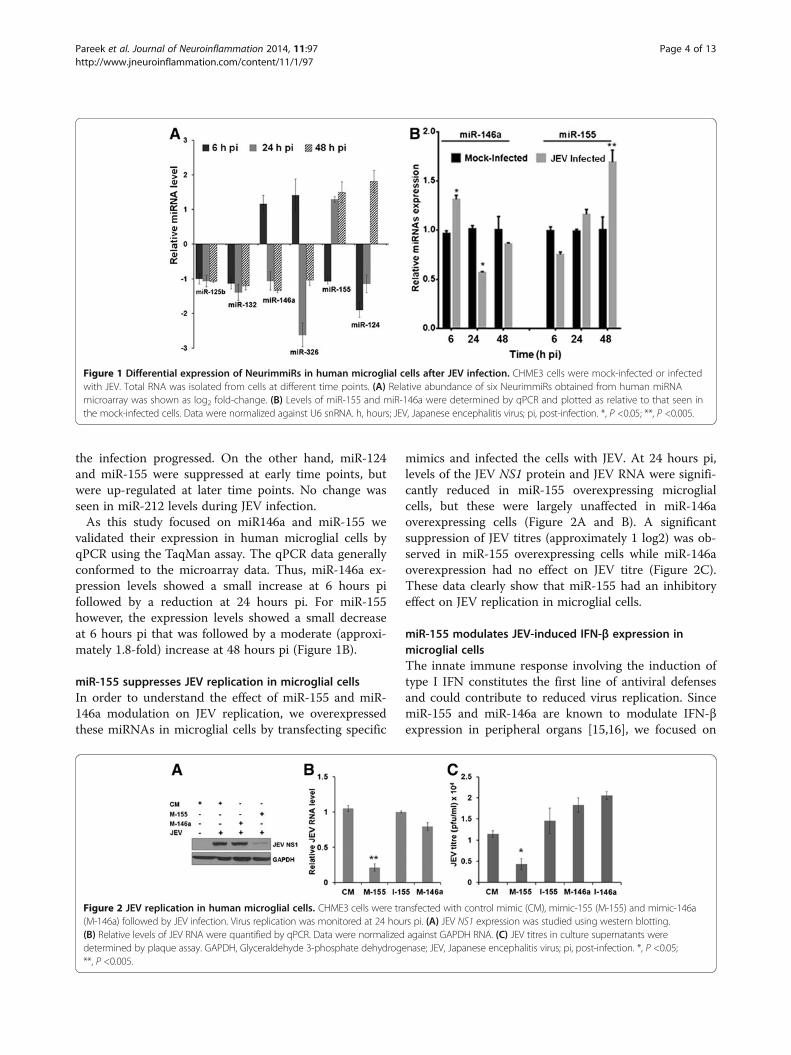
the infection progressed. On the other hand, miR-124and miR-155 were suppressed at early time points, butwere up-regulated at later time points. No change wasseen in miR-212 levels during JEV infection.As this study focused on miR146a and miR-155 we
validated their expression in human microglial cells byqPCR using the TaqMan assay. The qPCR data generallyconformed to the microarray data. Thus, miR-146a ex-pression levels showed a small increase at 6 hours pifollowed by a reduction at 24 hours pi. For miR-155however, the expression levels showed a small decreaseat 6 hours pi that was followed by a moderate (approxi-mately 1.8-fold) increase at 48 hours pi (Figure 1B).
miR-155 suppresses JEV replication in microglial cellsIn order to understand the effect of miR-155 and miR-146a modulation on JEV replication, we overexpressedthese miRNAs in microglial cells by transfecting specific
mimics and infected the cells with JEV. At 24 hours pi,levels of the JEV NS1 protein and JEV RNA were signifi-cantly reduced in miR-155 overexpressing microglialcells, but these were largely unaffected in miR-146aoverexpressing cells (Figure 2A and B). A significantsuppression of JEV titres (approximately 1 log2) was ob-served in miR-155 overexpressing cells while miR-146aoverexpression had no effect on JEV titre (Figure 2C).These data clearly show that miR-155 had an inhibitoryeffect on JEV replication in microglial cells.
miR-155 modulates JEV-induced IFN-β expression inmicroglial cellsThe innate immune response involving the induction oftype I IFN constitutes the first line of antiviral defensesand could contribute to reduced virus replication. SincemiR-155 and miR-146a are known to modulate IFN-βexpression in peripheral organs [15,16], we focused on
Figure 1 Differential expression of NeurimmiRs in human microglial cells after JEV infection. CHME3 cells were mock-infected or infectedwith JEV. Total RNA was isolated from cells at different time points. (A) Relative abundance of six NeurimmiRs obtained from human miRNAmicroarray was shown as log2 fold-change. (B) Levels of miR-155 and miR-146a were determined by qPCR and plotted as relative to that seen inthe mock-infected cells. Data were normalized against U6 snRNA. h, hours; JEV, Japanese encephalitis virus; pi, post-infection. *, P <0.05; **, P <0.005.
Figure 2 JEV replication in human microglial cells. CHME3 cells were transfected with control mimic (CM), mimic-155 (M-155) and mimic-146a(M-146a) followed by JEV infection. Virus replication was monitored at 24 hours pi. (A) JEV NS1 expression was studied using western blotting.(B) Relative levels of JEV RNA were quantified by qPCR. Data were normalized against GAPDH RNA. (C) JEV titres in culture supernatants weredetermined by plaque assay. GAPDH, Glyceraldehyde 3-phosphate dehydrogenase; JEV, Japanese encephalitis virus; pi, post-infection. *, P <0.05;**, P <0.005.
Pareek et al. Journal of Neuroinflammation 2014, 11:97 Page 4 of 13http://www.jneuroinflammation.com/content/11/1/97
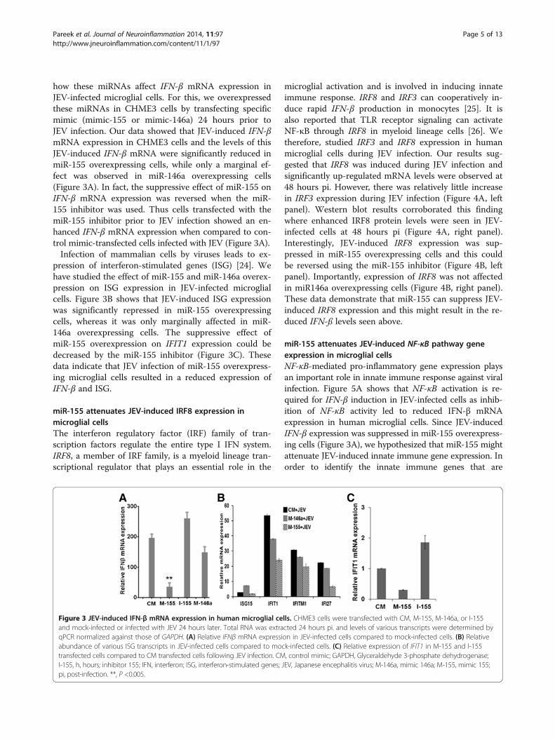
how these miRNAs affect IFN-β mRNA expression inJEV-infected microglial cells. For this, we overexpressedthese miRNAs in CHME3 cells by transfecting specificmimic (mimic-155 or mimic-146a) 24 hours prior toJEV infection. Our data showed that JEV-induced IFN-βmRNA expression in CHME3 cells and the levels of thisJEV-induced IFN-β mRNA were significantly reduced inmiR-155 overexpressing cells, while only a marginal ef-fect was observed in miR-146a overexpressing cells(Figure 3A). In fact, the suppressive effect of miR-155 onIFN-β mRNA expression was reversed when the miR-155 inhibitor was used. Thus cells transfected with themiR-155 inhibitor prior to JEV infection showed an en-hanced IFN-β mRNA expression when compared to con-trol mimic-transfected cells infected with JEV (Figure 3A).Infection of mammalian cells by viruses leads to ex-
pression of interferon-stimulated genes (ISG) [24]. Wehave studied the effect of miR-155 and miR-146a overex-pression on ISG expression in JEV-infected microglialcells. Figure 3B shows that JEV-induced ISG expressionwas significantly repressed in miR-155 overexpressingcells, whereas it was only marginally affected in miR-146a overexpressing cells. The suppressive effect ofmiR-155 overexpression on IFIT1 expression could bedecreased by the miR-155 inhibitor (Figure 3C). Thesedata indicate that JEV infection of miR-155 overexpress-ing microglial cells resulted in a reduced expression ofIFN-β and ISG.
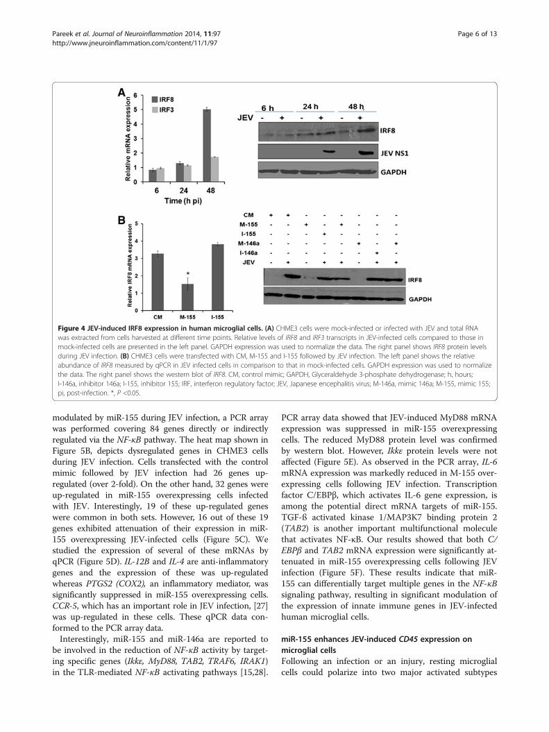
miR-155 attenuates JEV-induced IRF8 expression inmicroglial cellsThe interferon regulatory factor (IRF) family of tran-scription factors regulate the entire type I IFN system.IRF8, a member of IRF family, is a myeloid lineage tran-scriptional regulator that plays an essential role in the
microglial activation and is involved in inducing innateimmune response. IRF8 and IRF3 can cooperatively in-duce rapid IFN-β production in monocytes [25]. It isalso reported that TLR receptor signaling can activateNF-κB through IRF8 in myeloid lineage cells [26]. Wetherefore, studied IRF3 and IRF8 expression in humanmicroglial cells during JEV infection. Our results sug-gested that IRF8 was induced during JEV infection andsignificantly up-regulated mRNA levels were observed at48 hours pi. However, there was relatively little increasein IRF3 expression during JEV infection (Figure 4A, leftpanel). Western blot results corroborated this findingwhere enhanced IRF8 protein levels were seen in JEV-infected cells at 48 hours pi (Figure 4A, right panel).Interestingly, JEV-induced IRF8 expression was sup-pressed in miR-155 overexpressing cells and this couldbe reversed using the miR-155 inhibitor (Figure 4B, leftpanel). Importantly, expression of IRF8 was not affectedin miR146a overexpressing cells (Figure 4B, right panel).These data demonstrate that miR-155 can suppress JEV-induced IRF8 expression and this might result in the re-duced IFN-ß levels seen above.
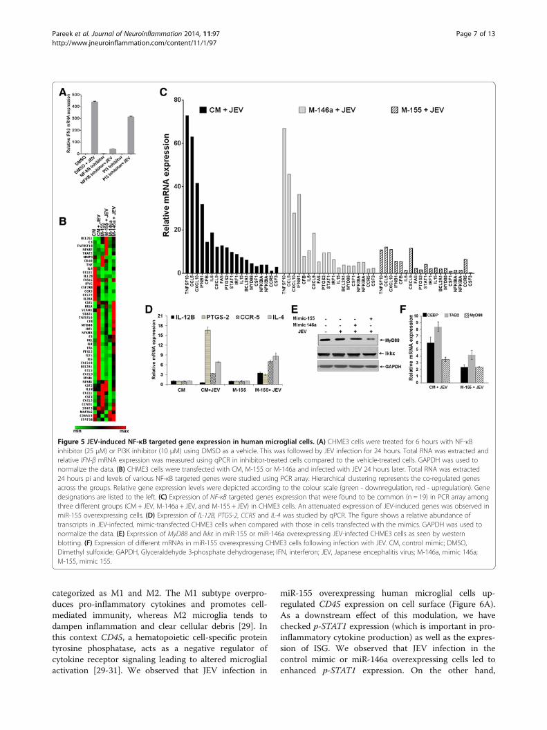
miR-155 attenuates JEV-induced NF-κB pathway geneexpression in microglial cellsNF-κB-mediated pro-inflammatory gene expression playsan important role in innate immune response against viralinfection. Figure 5A shows that NF-κB activation is re-quired for IFN-β induction in JEV-infected cells as inhib-ition of NF-κB activity led to reduced IFN-β mRNAexpression in human microglial cells. Since JEV-inducedIFN-β expression was suppressed in miR-155 overexpress-ing cells (Figure 3A), we hypothesized that miR-155 mightattenuate JEV-induced innate immune gene expression. Inorder to identify the innate immune genes that are
Figure 3 JEV-induced IFN-β mRNA expression in human microglial cells. CHME3 cells were transfected with CM, M-155, M-146a, or I-155and mock-infected or infected with JEV 24 hours later. Total RNA was extracted 24 hours pi. and levels of various transcripts were determined byqPCR normalized against those of GAPDH. (A) Relative IFNβ mRNA expression in JEV-infected cells compared to mock-infected cells. (B) Relativeabundance of various ISG transcripts in JEV-infected cells compared to mock-infected cells. (C) Relative expression of IFIT1 in M-155 and I-155transfected cells compared to CM transfected cells following JEV infection. CM, control mimic; GAPDH, Glyceraldehyde 3-phosphate dehydrogenase;I-155, h, hours; inhibitor 155; IFN, interferon; ISG, interferon-stimulated genes; JEV, Japanese encephalitis virus; M-146a, mimic 146a; M-155, mimic 155;pi, post-infection. **, P <0.005.
Pareek et al. Journal of Neuroinflammation 2014, 11:97 Page 5 of 13http://www.jneuroinflammation.com/content/11/1/97
modulated by miR-155 during JEV infection, a PCR arraywas performed covering 84 genes directly or indirectlyregulated via the NF-κB pathway. The heat map shown inFigure 5B, depicts dysregulated genes in CHME3 cellsduring JEV infection. Cells transfected with the controlmimic followed by JEV infection had 26 genes up-regulated (over 2-fold). On the other hand, 32 genes wereup-regulated in miR-155 overexpressing cells infectedwith JEV. Interestingly, 19 of these up-regulated geneswere common in both sets. However, 16 out of these 19genes exhibited attenuation of their expression in miR-155 overexpressing JEV-infected cells (Figure 5C). Westudied the expression of several of these mRNAs byqPCR (Figure 5D). IL-12B and IL-4 are anti-inflammatorygenes and the expression of these was up-regulatedwhereas PTGS2 (COX2), an inflammatory mediator, wassignificantly suppressed in miR-155 overexpressing cells.CCR-5, which has an important role in JEV infection, [27]was up-regulated in these cells. These qPCR data con-formed to the PCR array data.Interestingly, miR-155 and miR-146a are reported to
be involved in the reduction of NF-κB activity by target-ing specific genes (Ikkε, MyD88, TAB2, TRAF6, IRAK1)in the TLR-mediated NF-κB activating pathways [15,28].
PCR array data showed that JEV-induced MyD88 mRNAexpression was suppressed in miR-155 overexpressingcells. The reduced MyD88 protein level was confirmedby western blot. However, Ikkε protein levels were notaffected (Figure 5E). As observed in the PCR array, IL-6mRNA expression was markedly reduced in M-155 over-expressing cells following JEV infection. Transcriptionfactor C/EBPβ, which activates IL-6 gene expression, isamong the potential direct mRNA targets of miR-155.TGF-ß activated kinase 1/MAP3K7 binding protein 2(TAB2) is another important multifunctional moleculethat activates NF-κB. Our results showed that both C/EBPβ and TAB2 mRNA expression were significantly at-tenuated in miR-155 overexpressing cells following JEVinfection (Figure 5F). These results indicate that miR-155 can differentially target multiple genes in the NF-κBsignaling pathway, resulting in significant modulation ofthe expression of innate immune genes in JEV-infectedhuman microglial cells.
miR-155 enhances JEV-induced CD45 expression onmicroglial cellsFollowing an infection or an injury, resting microglialcells could polarize into two major activated subtypes
Figure 4 JEV-induced IRF8 expression in human microglial cells. (A) CHME3 cells were mock-infected or infected with JEV and total RNAwas extracted from cells harvested at different time points. Relative levels of IRF8 and IRF3 transcripts in JEV-infected cells compared to those inmock-infected cells are presented in the left panel. GAPDH expression was used to normalize the data. The right panel shows IRF8 protein levelsduring JEV infection. (B) CHME3 cells were transfected with CM, M-155 and I-155 followed by JEV infection. The left panel shows the relativeabundance of IRF8 measured by qPCR in JEV infected cells in comparison to that in mock-infected cells. GAPDH expression was used to normalizethe data. The right panel shows the western blot of IRF8. CM, control mimic; GAPDH, Glyceraldehyde 3-phosphate dehydrogenase; h, hours;I-146a, inhibitor 146a; I-155, inhibitor 155; IRF, interferon regulatory factor; JEV, Japanese encephalitis virus; M-146a, mimic 146a; M-155, mimic 155;pi, post-infection. *, P <0.05.
Pareek et al. Journal of Neuroinflammation 2014, 11:97 Page 6 of 13http://www.jneuroinflammation.com/content/11/1/97
categorized as M1 and M2. The M1 subtype overpro-duces pro-inflammatory cytokines and promotes cell-mediated immunity, whereas M2 microglia tends todampen inflammation and clear cellular debris [29]. Inthis context CD45, a hematopoietic cell-specific proteintyrosine phosphatase, acts as a negative regulator ofcytokine receptor signaling leading to altered microglialactivation [29-31]. We observed that JEV infection in
miR-155 overexpressing human microglial cells up-regulated CD45 expression on cell surface (Figure 6A).As a downstream effect of this modulation, we havechecked p-STAT1 expression (which is important in pro-inflammatory cytokine production) as well as the expres-sion of ISG. We observed that JEV infection in thecontrol mimic or miR-146a overexpressing cells led toenhanced p-STAT1 expression. On the other hand,
Figure 5 JEV-induced NF-κB targeted gene expression in human microglial cells. (A) CHME3 cells were treated for 6 hours with NF-κBinhibitor (25 μM) or PI3K inhibitor (10 μM) using DMSO as a vehicle. This was followed by JEV infection for 24 hours. Total RNA was extracted andrelative IFN-β mRNA expression was measured using qPCR in inhibitor-treated cells compared to the vehicle-treated cells. GAPDH was used tonormalize the data. (B) CHME3 cells were transfected with CM, M-155 or M-146a and infected with JEV 24 hours later. Total RNA was extracted24 hours pi and levels of various NF-κB targeted genes were studied using PCR array. Hierarchical clustering represents the co-regulated genesacross the groups. Relative gene expression levels were depicted according to the colour scale (green - downregulation, red - upregulation). Genedesignations are listed to the left. (C) Expression of NF-κB targeted genes expression that were found to be common (n = 19) in PCR array amongthree different groups (CM + JEV, M-146a + JEV, and M-155 + JEV) in CHME3 cells. An attenuated expression of JEV-induced genes was observed inmiR-155 overexpressing cells. (D) Expression of IL-12B, PTGS-2, CCR5 and IL-4 was studied by qPCR. The figure shows a relative abundance oftranscripts in JEV-infected, mimic-transfected CHME3 cells when compared with those in cells transfected with the mimics. GAPDH was used tonormalize the data. (E) Expression of MyD88 and Ikkε in miR-155 or miR-146a overexpressing JEV-infected CHME3 cells as seen by westernblotting. (F) Expression of different mRNAs in miR-155 overexpressing CHME3 cells following infection with JEV. CM, control mimic; DMSO,Dimethyl sulfoxide; GAPDH, Glyceraldehyde 3-phosphate dehydrogenase; IFN, interferon; JEV, Japanese encephalitis virus; M-146a, mimic 146a;M-155, mimic 155.
Pareek et al. Journal of Neuroinflammation 2014, 11:97 Page 7 of 13http://www.jneuroinflammation.com/content/11/1/97
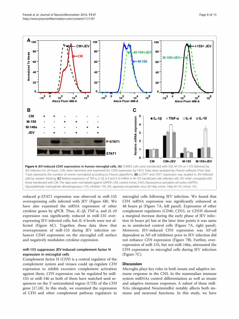
reduced p-STAT1 expression was observed in miR-155overexpressing cells infected with JEV (Figure 6B). Wehave also examined the mRNA expression of othercytokine genes by qPCR. Thus, IL-1β, TNF-α, and IL-10expression was significantly reduced in miR-155 over-expressing JEV-infected cells, but IL-4 levels were not af-fected (Figure 6C). Together, these data show thatoverexpression of miR-155 during JEV infection en-hances CD45 expression on the microglial cell surfaceand negatively modulates cytokine expression.
miR-155 suppresses JEV-induced complement factor Hexpression in microglial cellsComplement factor H (CFH) is a central regulator of thecomplement system and viruses could up-regulate CFHexpression to inhibit excessive complement activationagainst them. CFH expression can be regulated by miR-155 or miR-146 as both of them have matched seed se-quences on the 3’-untranslated region (UTR) of the CFHgene [17,18]. In this study, we examined the expressionof CFH and other complement pathway regulators in
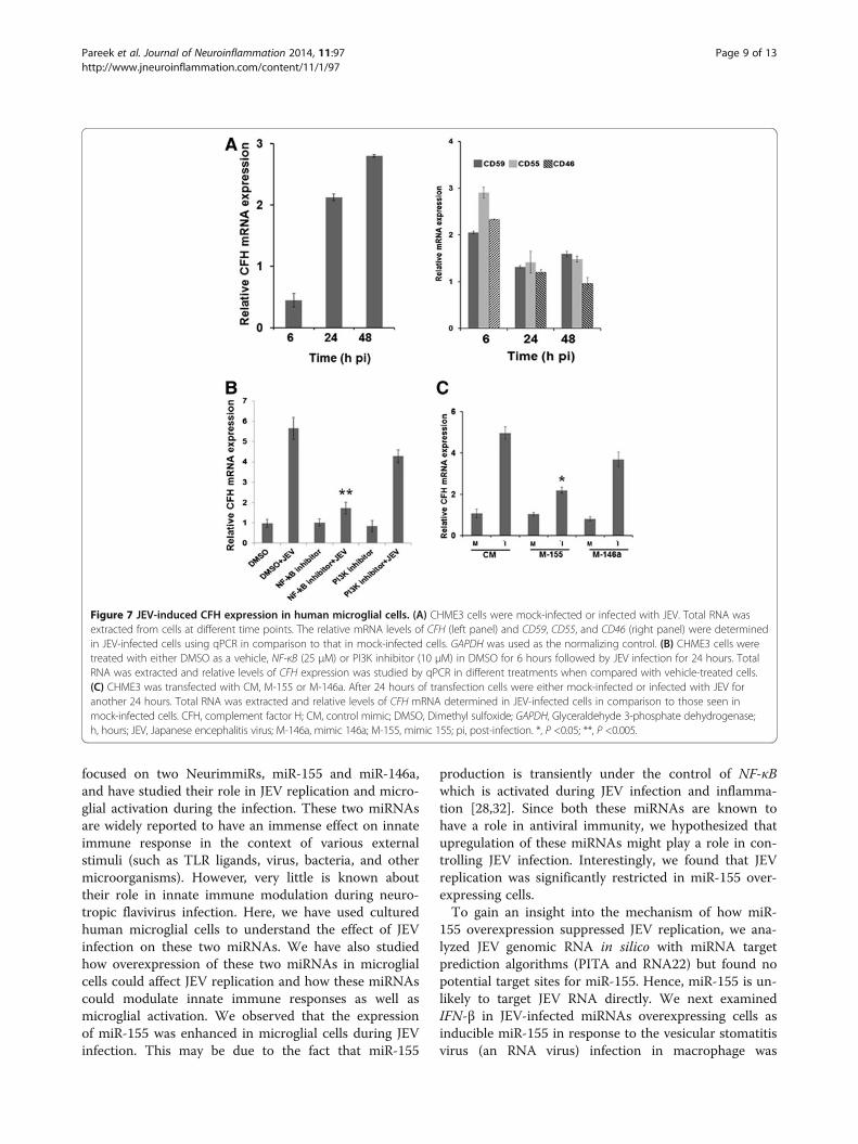
microglial cells following JEV infection. We found thatCFH mRNA expression was significantly enhanced at48 hours pi (Figure 7A, left panel). Expression of othercomplement regulators (CD46, CD55, or CD59) showeda marginal increase during the early phase of JEV infec-tion (6 hours pi) but at the later time points it was sameas in uninfected control cells (Figure 7A, right panel).Moreover, JEV-induced CFH expression was NF-κBdependent as NF-κB inhibition prior to JEV infection didnot enhance CFH expression (Figure 7B). Further, over-expression of miR-155, but not miR-146a, attenuated theCFH expression in microglial cells during JEV infection(Figure 7C).
DiscussionMicroglia plays key roles in both innate and adaptive im-mune response in the CNS. In the mammalian immunesystem miRNAs control differentiation as well as innateand adaptive immune responses. A subset of these miR-NAs (designated NeurimmiRs) notably affects both im-mune and neuronal functions. In this study, we have
Figure 6 JEV-induced CD45 expressions in human microglial cells. (A) CHME3 cells were transfected with CM, M-155 or I-155 followed byJEV infection for 24 hours. Cells were harvested and examined for CD45 expression by FACS. Data were analyzed by FlowJo software (Tree Star).Y-axis represents the number of events normalized according to FlowJo algorithms. (B) p-STAT1 and STAT1 expression was studied in JEV-infectedcells by western blotting. (C) Relative expression of TNF-α, IL-1β, IL-4 and IL10 mRNA in M-155 transfected cells infected with JEV when compared withthose transfected with CM. The data were normalized against GAPDH. CM, control mimic; FACS, fluorescence activated cell sorter; GAPDH,Glyceraldehyde 3-phosphate dehydrogenase; I-155, inhibitor 155; JEV, Japanese encephalitis virus; M-146a, mimic 146a; M-155, mimic 155.
Pareek et al. Journal of Neuroinflammation 2014, 11:97 Page 8 of 13http://www.jneuroinflammation.com/content/11/1/97
focused on two NeurimmiRs, miR-155 and miR-146a,and have studied their role in JEV replication and micro-glial activation during the infection. These two miRNAsare widely reported to have an immense effect on innateimmune response in the context of various externalstimuli (such as TLR ligands, virus, bacteria, and othermicroorganisms). However, very little is known abouttheir role in innate immune modulation during neuro-tropic flavivirus infection. Here, we have used culturedhuman microglial cells to understand the effect of JEVinfection on these two miRNAs. We have also studiedhow overexpression of these two miRNAs in microglialcells could affect JEV replication and how these miRNAscould modulate innate immune responses as well asmicroglial activation. We observed that the expressionof miR-155 was enhanced in microglial cells during JEVinfection. This may be due to the fact that miR-155
production is transiently under the control of NF-κBwhich is activated during JEV infection and inflamma-tion [28,32]. Since both these miRNAs are known tohave a role in antiviral immunity, we hypothesized thatupregulation of these miRNAs might play a role in con-trolling JEV infection. Interestingly, we found that JEVreplication was significantly restricted in miR-155 over-expressing cells.To gain an insight into the mechanism of how miR-
155 overexpression suppressed JEV replication, we ana-lyzed JEV genomic RNA in silico with miRNA targetprediction algorithms (PITA and RNA22) but found nopotential target sites for miR-155. Hence, miR-155 is un-likely to target JEV RNA directly. We next examinedIFN-β in JEV-infected miRNAs overexpressing cells asinducible miR-155 in response to the vesicular stomatitisvirus (an RNA virus) infection in macrophage was
Figure 7 JEV-induced CFH expression in human microglial cells. (A) CHME3 cells were mock-infected or infected with JEV. Total RNA wasextracted from cells at different time points. The relative mRNA levels of CFH (left panel) and CD59, CD55, and CD46 (right panel) were determinedin JEV-infected cells using qPCR in comparison to that in mock-infected cells. GAPDH was used as the normalizing control. (B) CHME3 cells weretreated with either DMSO as a vehicle, NF-κB (25 μM) or PI3K inhibitor (10 μM) in DMSO for 6 hours followed by JEV infection for 24 hours. TotalRNA was extracted and relative levels of CFH expression was studied by qPCR in different treatments when compared with vehicle-treated cells.(C) CHME3 was transfected with CM, M-155 or M-146a. After 24 hours of transfection cells were either mock-infected or infected with JEV foranother 24 hours. Total RNA was extracted and relative levels of CFH mRNA determined in JEV-infected cells in comparison to those seen inmock-infected cells. CFH, complement factor H; CM, control mimic; DMSO, Dimethyl sulfoxide; GAPDH, Glyceraldehyde 3-phosphate dehydrogenase;h, hours; JEV, Japanese encephalitis virus; M-146a, mimic 146a; M-155, mimic 155; pi, post-infection. *, P <0.05; **, P <0.005.
Pareek et al. Journal of Neuroinflammation 2014, 11:97 Page 9 of 13http://www.jneuroinflammation.com/content/11/1/97
shown to induce type I IFN signaling and inhibit viralreplication [16]. However, to our surprise we found sig-nificant reduction in JEV-induced IFN-β as well asdownstream ISG mRNA expression in cells where miR-155 was overexpressed. This suggests that miR-155 usesdifferent mechanisms to exert an antiviral effect againstJEV in microglial cells. In fact, Swaminathan et al. [21]have shown that miR-155 exerts an anti-HIV-1 effect bytargeting several HIV-1 dependency factors involved inpost-entry, pre-integration events, leading to severely di-minished HIV-1 infection. Thus, miR-155 can use differ-ent mechanisms for its antiviral effect depending uponthe virus and the environmental stimuli generated dur-ing the particular virus infection of a certain cell type.In order to understand reduced IFN-β expression in
JEV-infected miR-155 overexpressing cells, we checkedthe expression of IRFs. Among the several IRFs reported,we focused our study on IRF8 as it plays a major role inIFN signaling, response to infection, and maturation ofmyeloid lineages cells [33]. IRF8 is also involved in therapid induction of IFN-β in human monocytes [25].Moreover, IRF8 may activate a program of gene expres-sion that transforms microglia into a reactive phenotype[34]. In this study, we observed that JEV infection inmicroglia can induce IRF8 expression, but the same ex-pression was attenuated in miR-155 overexpressing cellsand may be a possible reason for reduced IFN-β produc-tion in miR-155 overexpressing cells. Interestingly, bio-informatics analysis predicted IRF8 as one of thepotential targets for miR-155. We cloned 3’-UTR ofIRF8 in the luciferase reporter system for miRNA targetvalidation but found no change in the luciferase readoutin miR-155 overexpressing cells (data not shown). Wehave shown that IRF8 is induced during JEV infection.Since JEV replication and titres are reduced in miR-155overexpressing cells, it could result in reduced IRF8induction.IRF8 is also involved in TLR-mediated NF-κB activa-
tion [26]. Since JEV-induced IRF8 expression in miR-155overexpressing cells was attenuated, we sought to under-stand its effect on NF-κB pathway. For this we examined84 genes by RT-PCR array whose expression and func-tion, either directly or indirectly, depend on NF-κBactivation. We found several genes, including IFN-β,MyD88, STAT1, PTGS2, and IL-12B, which are inducedby JEV and show attenuated expression in miR-155 over-expressing cells. Recently, CCR5 receptor expression in amouse model of JE was reported to play an importantrole in recovery as well as promote host survival againstJEV [27]. Interestingly, increased CCR5 expression wasalso observed in our cell culture based study. Thus,modulation of NF-κB mediated signaling pathway genesby JEV-induced miR-155 expression might play a role inreduced JEV replication in microglial cells.
Specific induction of active SHP2 phosphatase dephos-phorylates IRF8, which in turn becomes an active repres-sor and down-regulates TLR-mediated gene expression[35]. In hematopoietic cell lineage CD45 (a Src-homology2 domain (SH2)-containing protein tyrosine phosphatase)plays an important role in regulating cytokine receptormediated signal [36]. Analysis of microglia ex vivo revealedthat IRF8-deficient microglia had significantly increasedlevels of CD45 [37]. Thus attenuated IRF8 expression inmiR-155 overexpressing cells may result in enhancedCD45 expression on microglial cells.Following an infection, resting microglial cells can get
activated into M1 or M2 phenotypes. The M1 phenotypeis a pro-inflammatory state and induces neuropathology,whereas M2 is anti-inflammatory state that could have aneuroprotective role [36]. It is reported that CD45 cannegatively regulate CD40L-CD40-induced microglial M1activation; an effect leading to the promotion of the M2phenotype. Moreover, this CD45-mediated activationstate appears to decrease harmful cytokine production[36]. In this study, we observed an increased CD45 ex-pression in JEV-infected miR-155 overexpressing cellsand relatively reduced p-STAT1 expression. Further,mRNA expression for pro-inflammatory cytokines IL-1βand TNF-α was reduced in miR-155 overexpressing cells.CD45 has also been shown to down-regulate NF-κB, animportant mediator of pro-inflammatory cytokines [38].Thus, increased CD45 expression in JEV-induced miR-155 expressing cells may explain reduced expression ofNF-κB pathway genes in our study. Taken together, thesedata show that miR-155 induction can modulate theJEV-induced microglial activation to a state that may bebeneficial to the host. Further in vivo study is needed toclarify this issue.Complement activation is considered to be an important
component of innate immune response against invadingpathogens. CFH is one of the regulators which negativelyregulate the complement activation. Viruses can utilizeCFH to evade the innate immune response. The non-structural protein NS1 of the West Nile virus, a flavivirus,inhibits complement activation by binding to CFH [39].CFH can be induced by NF-κB activation and can be regu-lated through miRNAs [17]. Interestingly, both miR-155and miR-146a have the same target sequence in the 3’-UTR of CFH and this has been experimentally proven forboth the miRNAs [17]. We have shown that JEV infectioncan induce CFH expression in human and mouse micro-glial cells and this induction is attenuated in miR-155overexpressing cells. Therefore, it is possible that miR-155expression in JEV-infected microglial cells inhibits CFHexpression, which in turn may benefit the host by facilitat-ing complement activation against JEV.In a study published earlier this month, Thounaojam
et al. [40] showed an up-regulation of miR-155 in mouse
Pareek et al. Journal of Neuroinflammation 2014, 11:97 Page 10 of 13http://www.jneuroinflammation.com/content/11/1/97
microglial cells (BV-2) and in the mouse and humanbrain during JEV infection. They suggested that miR-155had a pro-inflammatory role as its inhibition decreasedTBK-1, IRF3/7, and NF-κB phosphorylation both in BV-2 cells as well as in the mouse brain. These results pointto a role for miR-155 that is opposite to what we haveobserved in our study. A major difference between thesetwo studies is that while results reported by Thounaojamet al. [40] are derived from mouse BV-2 cells and themouse brain where various different kinds of cells mayget infected with JEV, our results are derived entirelyfrom in vitro cultured human microglial cells CHME3.Additionally, the GP78 strain of JEV used by Thounaojamet al. [40] is a slow growing virus, both in cultured cells andthe mouse brain, with lesions in the virus-cell fusionprocess [22]. Another reason for these differences may berelated to the smaller increase (approximately 4-fold) ofmiR-155 during JEV infection in BV2 cells [40], comparedwith the super maximal concentration in CHME3 cells(approximately 100-fold) that occurred during miR-155overexpression in the present study. Several reports havesuggested that, depending on the expression level, miR-155can modulate cellular functions by targeting genes in differ-ent pathways. Ceppi et al. [41] showed that low levelmiR-155 expression enables the activation of p38 MAPKpathway, favoring IL-1β expression, which induces inflam-mation in an autocrine manner. However, the same path-way was inhibited when the miR-155 expression level wentup significantly, ultimately reflecting an altered immuneprofile. Similar observations were made by Xiao et al. [42]who reported a 3-fold induction of miR-155 and pro-inflammatory cytokine responses during Helicobacter pyl-ori infection, whereas the overexpression of miR-155negatively regulated the pro-inflammatory responses.Intracellular signaling pathways that are concomi-
tantly activated by the same stimulus often interactwith one another through a cross regulatory feed-back mechanism. The miR-155 is a multifunctionalmicroRNA and it can modulate inflammatory re-sponses in both a positive and negative way [40-48].Besides its positive role in NF-κB activation andsubsequent pro-inflammatory response, accumulatingevidence has demonstrated that it can constitute anegative feedback loop in the NF-κB signalingpathway by targeting multiple key proteins, whichultimately leads to repression of, or at least the limi-tation of NF-κB activation in response to viral ormicrobial stimuli [41,42,45-48]. Therefore, miR-155could modulate inflammation depending on variousfactors including its expression level, the cell type,and the environmental stimuli.Overall our study suggests that miR-155 modulation
can act on multiple levels to control JEV infection ofmicroglial cells and induce innate immune responses
that may be beneficial to the host. It can enhance CD45expression, reduce pro-inflammatory cytokines and CFHexpression by targeting several key genes, and suppressJEV replication in microglial cells. These data point tomiR-155 playing an important role in modulating JEV-induced microglial activation that may be beneficial inlimiting JEV infection in the host. Additional studies,both in vitro and in vivo, are needed to further under-stand the role of miR-155 during JEV infection inswitching microglial activation towards the neuroprotec-tive state.
AbbreviationsANOVA: Analysis Of Variance; CCR5: C-C Chemokine Receptor type 5;CEBPβ: CCAAT/Enhancer Binding Protein beta; CFH: Complement Factor H;CNS: Central Nervous System; COX-2: Cyclooxygenase-2; DENV 2: DengueVirus 2; DMEM: Dulbecco’s Modified Eagle’s Medium; DMSO: Di MethylSulfoxide; FACS: Fluorescence Activated Cell Sorter; FBS: Fetal Bovine Serum;GAPDH: Glyceraldehyde 3-Phosphate Dehydrogenase; HIV: HumanImmunodeficiency Virus; HRP: Horseradish Peroxidase; IRF: InterferonRegulatory Factor; ISG: Interferon Stimulated Genes; JE: Japanese Encephalitis;JEV: Japanese Encephalitis Virus; LPS: Lipopolysaccharide; MEM: MinimalEssential Medium; MOI: Multiplicity Of Infection; MyD88: MyeloidDifferentiation primary response gene (88); NF-kB: Nuclear Factor kappa lightchain enhancer of activated B cells; NFQ-MGB: Non fluorescent Quencher-Minor Groove Binder; PCR: Polymerase Chain Reaction; PFU: Plaque FormingUnit; PI3K: Phosphatidylinositol-4,5-bisphosphate 3-kinase; Poly (I:C):Polyinosinic-polycytidylic acid; PS: Porcine Stable kidney cell line; p-STAT1:Phosphorylated-Signal Transducers and Activators of Transcription; PTGS-2:Prostaglandin-endoperoxide Synthase 2; qPCR: Quantitative real-time PCR; SDS-PAGE: Sodium Dodecyl Sulfate-Polyacrylamide Gel; STAT1: Signal Transducersand Activators of Transcription; TBK1: TANK Binding Kinase 1; TLR: Toll LikeReceptor; TNF-α: Tumor Necrosis Factor-alpha.
Competing interestsThe authors declare that they have no competing interests.
Authors’ contributionsAB was responsible for experimental design, data analysis, and drafting themanuscript. SP, BK, and PJ performed the RNA extraction, PCR array, RT-PCR,miRNA assay, western blot, and FACS. SR and BK performed the viruspreparation, cell culture, and transfection and virus infection experiments. SVand AB conceived the idea, supervised the experiments, and participated inediting the manuscript. All authors have read and approved the finalmanuscript.
AcknowledgementsThis work was supported by the Department of Biotechnology (DBT),Government of India grants number BT/MB/01/VIDRC/08 and BT/PR6714/MED/29/617/2012. We are thankful to Dr Manpreet Kaur for her excellenttechnical advice. We are thankful to Dr Anirban Basu (National BrainResearch Centre, Manesar, India) for providing the human microglial cells(CHME3).
Received: 24 March 2014 Accepted: 13 May 2014Published: 29 May 2014
References1. Thongtan T, Cheepsunthorn P, Chaiworakul V, Rattanarungsan C, Wikan N,
Smith DR: Highly permissive infection of microglial cells by Japaneseencephalitis virus: a possible role as a viral reservoir. Microbes Infect 2010,12:37–45.
2. Chen CJ, Ou YC, Lin SY, Raung SL, Liao SL, Lai CY, Chen SY, Chen JH: Glialactivation involvement in neuronal death by Japanese encephalitis virusinfection. J Gen Virol 2010, 91:1028–1037.
3. Das S, Dutta K, Kumawat KL, Ghoshal A, Adhya D, Basu A: Abrogatedinflammatory response promotes neurogenesis in a murine model ofJapanese encephalitis. PLoS One 2011, 6:e17225.
Pareek et al. Journal of Neuroinflammation 2014, 11:97 Page 11 of 13http://www.jneuroinflammation.com/content/11/1/97
4. Kobayashi K, Imagama S, Ohgomori T, Hirano K, Uchimura K, Sakamoto K,Hirakawa A, Takeuchi H, Suzumura A, Ishiguro N, Kadomatsu K: Minocyclineselectively inhibits M1 polarization of microglia. Cell Death Dis 2013,4:e525.
5. Mishra MK, Basu A: Minocycline neuroprotects, reduces microglialactivation, inhibits caspase 3 induction, and viral replication followingJapanese encephalitis. J Neurochem 2008, 105:1582–1595.
6. Ponomarev ED, Veremeyko T, Weiner HL: MicroRNAs are universalregulators of differentiation, activation, and polarization of microglia andmacrophages in normal and diseased CNS. Glia 2013, 61:91–103.
7. Skalsky RL, Cullen BR: Viruses, microRNAs, and host interactions. Annu RevMicrobiol 2010, 64:123–141.
8. Soreq H, Wolf Y: NeurimmiRs: microRNAs in the neuroimmune interface.Trends Mol Med 2011, 17:548–555.
9. Wanet A, Tacheny A, Arnould T, Renard P: miR-212/132 expression andfunctions: within and beyond the neuronal compartment. Nucleic AcidsRes 2012, 40:4742–4753.
10. Lindsay MA: microRNAs and the immune response. Trends Immunol 2011,29:343–351.
11. Elton TS, Selemon H, Elton SM, Parinandi NL: Regulation of the MIR155 hostgene in physiological and pathological processes. Gene 2013, 532:1–12.
12. O’Connell RM, Rao DS, Chaudhuri AA, Baltimore D: Physiological andpathological roles for microRNAs in the immune system. Nat RevImmunol 2010, 10:111–122.
13. Cardoso AL, Guedes JR, Pereira de Almeida L, Pedroso de Lima MC:miR-155 modulates microglia-mediated immune response bydown-regulating SOCS-1 and promoting cytokine and nitric oxideproduction. Immunology 2012, 135:73–88.
14. Saba R, Gushue S, Huzarewich RLCH, Manguiat K, Medina S, Robertson C,Booth SA: MicroRNA 146a (miR-146a) is overexpressed during priondisease and modulates the innate immune response and the microglialactivation state. PLoS One 2012, 7:e30832.
15. Hou J, Wang P, Lin L, Liu X, Ma F, An H, Wang Z, Cao X: MicroRNA-146afeedback inhibits RIG-I-dependent Type I IFN production inmacrophages by targeting TRAF6, IRAK1, and IRAK2. J Immunol 2009,183:2150–2158.
16. Wang P, Hou J, Lin L, Wang C, Liu X, Li D, Ma F, Wang Z, Cao X: InduciblemicroRNA-155 feedback promotes type I IFN signaling in antiviral innateimmunity by targeting suppressor of cytokine signaling 1. J Immunol 2010,185:6226–6233.
17. Lukiw WJ, Surjyadipta B, Dua P, Alexandrov PN: Common micro RNAs(miRNAs) target complement factor H (CFH) regulation in Alzheimer’sdisease (AD) and in age-related macular degeneration (AMD).Int J Biochem Mol Biol 2012, 3:105–116.
18. Lukiw WJ, Alexandrov PN: Regulation of complement factor H (CFH) bymultiple miRNAs in Alzheimer’s disease (AD) brain. Mol Neurobiol 2012,46:11–19.
19. Li YY, Alexandrov PN, Pogue AI, Zhao Y, Bhattacharjee S, Lukiw WJ:miRNA-155 upregulation and complement factor H deficits in Down’sSyndrome. Nuroreport 2012, 23:168–173.
20. Wu S, He L, Li Y, Wang T, Feng L, Jiang L, Zhang P, Huang X: miR-146afacilitates replication of dengue virus by dampening interferon inductionby targeting TRAF6. J Infect 2013, 67:329–341.
21. Swaminathan G, Rossi F, Sierra LJ, Gupta A, Navas-Martín S, Martín-García J:A role for microRNA-155 modulation in the anti-HIV-1 effects of Toll-likereceptor 3 stimulation in macrophages. PLoS Pathog 2012, 8:e1002937.
22. Vrati S, Agarwal V, Malik P, Wani SA, Saini M: Molecular characterization ofan Indian isolate of Japanese encephalitis virus that shows an extendedlag phase during growth. J Gen Virol 1999, 80:1665–1671.
23. Kalia M, Khasa R, Sharma M, Nain M, Vrati S: Japanese encephalitis virusinfects neuronal cells through a clathrin-independent endocyticmechanism. J Virol 2013, 87:148–157.
24. El-Ekiaby N, Hamdi N, Negm M, Ahmed R, Zekri AR, Esmat G, Abdelaziz AI:Repressed induction of interferon-related microRNAs miR-146a andmiR-155 in peripheral blood mononuclear cells infected with HCVgenotype 4. FEBS Open Bio 2012, 2:179–186.
25. Li P, Wong JJ, Sum C, Sin WX, Ng KQ, Koh MB, Chin KC: IRF8 and IRF3cooperatively regulate rapid interferon-β induction in human bloodmonocytes. Blood 2011, 117:2847–2854.
26. Tsujimura H, Tamura T, Kong HJ, Nishiyama A, Ishii KJ, Klinman DM, Ozato K:Toll-like receptor 9 signaling activates NF-kappaB through IFN regulatory
factor-8/IFN consensus sequence binding protein in dendritic cells.J Immunol 2004, 172:6820–6827.
27. Larena M, Regner M, Lobigs M: The chemokine receptor CCR5, atherapeutic target for HIV/AIDS antagonists. Is critical for recovery in amouse model of Japanese encephalitis. PLoS One 2012, 7:e44834.
28. Boldin MP, Baltimore D: MicroRNAs, new effectors and regulators of NF-κB.Immunol Rev 2012, 246:205–220.
29. Town T, Nikolic V, Tan J: The microglial “activation” continuum: frominnate to adaptive responses. J Neuroinflammation 2005, 2:24.
30. Irie-Sasaki J, Sasaki T, Matsumoto W, Opavsky A, Cheng M, Welstead G,Griffiths E, Krawczyk C, Richardson CD, Aitken K, Iscove N, Koretzky G,Johnson P, Liu P, Rothstein DM, Penninger JM: CD45 is a JAK phosphataseand negatively regulates cytokine receptor signaling. Nature 2001,409:349–354.
31. Hermiston ML, Xu Z, Weiss A: CD45: a critical regulator of signalingthresholds in immune cells. Annu Rev Immunol 2003, 21:107–137.
32. Cremer TJ, Fatehchand K, Shah P, Gillette D, Patel H, Marsh RL, Besecker BY,Rajaram MV, Cormet-Boyaka E, Kanneganti TD, Schlesinger LS, Butchar JP,Tridandapani S: MiR-155 induction by microbes/microbial ligands requiresNF-κB-dependent de novo protein synthesis. Front Cell Infect Microbiol 2012,2:73.
33. Berghout J, Langlais D, Radovanovic I, Tam M, MacMicking JD, Stevenson MM,Gros P: Irf8-regulated genomic responses drive pathological inflammationduring cerebral malaria. PLoS Pathog 2013, 9:e1003491.
34. Horiuchi M, Wakayama K, Itoh A, Kawai K, Pleasure D, Ozato K, Itoh T:Interferon regulatory factor 8/interferon consensus sequence bindingprotein is a critical transcription factor for the physiological phenotypeof microglia. J Neuroinflammation 2012, 9:227.
35. Fragale A, Stellacci E, Ilari R, Remoli AL, Lanciotti A, Perrotti E, Shytaj I, Orsatti R,Lawrence HR, Lawrence NJ, Wu J, Rehli M, Ozato K, Battistini A: Critical role ofIRF-8 in negative regulation of TLR3 expression by Src homology 2domain-containing protein tyrosine phosphatase-2 activity in humanmyeloid dendritic cells. J Immunol 2011, 186:1951–1962.
36. Salemi J, Obregon DF, Cobb A, Reed S, Sadic E, Jin J, Fernandez F, Tan J,Giunta B: Flipping the switches: CD40 and CD45 modulation of microglialactivation states in HIV associated dementia (HAD). Mol Neurodegener 2011,6:3.
37. Masuda T, Tsuda M, Yoshinaga R, Tozaki-Saitoh H, Ozato K, Tamura T, Inoue K:IRF8 is a critical transcription factor for transforming microglia into areactive phenotype. Cell Rep 2012, 1:334–340.
38. Baur A, Garber S, Peterlin BM: Effects of CD45 on NF-kappa B. Implicationsfor replication of HIV-1. J Immunol 1994, 152:976–983.
39. Chung KM, Liszewski MK, Nybakken G, Davis AE, Townsend RR, Fremont DH,Atkinson JP, Diamond MS: West Nile virus nonstructural protein NS1inhibits complement activation by binding the regulatory protein factorH. Proc Natl Acad Sci USA 2006, 103:19111–19116.
40. Thounaojam MC, Kundu K, Kaushik DK, Swaroop S, Mahadevan A, Shankar SK,Basu A: MicroRNA-155 regulates Japanese encephalitis virus inducedinflammatory response by targeting src homology 2-containing inositolphosphatase-1 (SHIP1). J Virol 2014, 88:4798–4810.
41. Ceppi M, Pereira PM, Dunand-Sauthier I, Barras E, Reith W, Santos MA, Pierre P:MicroRNA-155 modulates the interleukin-1 signaling pathway in activatedhuman monocyte-derived dendritic cells. Proc Natl Acad Sci USA 2009,106:2735–2740.
42. Xiao B, Liu Z, Li BS, Tang B, Li W, Guo G, Shi Y, Wang F, Wu Y, Tong WD, Guo H,Mao XH, Zou QM: Induction of microRNA-155 during Helicobacter pyloriinfection and its negative regulatory role in the inflammatory response.J Infect Dis 2009, 200:916–925.
43. Xu C, Ren G, Cao G, Chen Q, Shou P, Zheng C, Du L, Han X, Jiang M, Yang Q,Lin L, Wang G, Yu P, Zhang X, Cao W, Brewer G, Wang Y, Shi Y:miR-155 regulates immune modulatory properties of mesenchymalstem cells by targeting TAK1-binding protein 2. J Biol Chem 2013,288:11074–11079.
44. Sullivan RP, Fogel LA, Leong JW, Schneider SE, Wong R, Romee R, Thai TH,Sexl V, Matkovich SJ, Dorn GW 2nd, French AR, Fehniger TA: MicroRNA-155tunes both the threshold and extent of NK cell activation via targetingof multiple signaling pathways. J Immunol 2013, 12:5904–5913.
45. Zhou H, Huang X, Cui H, Luo X, Tang Y, Chen S, Wu L, Shen N: miR-155and its star-form partner miR-155* cooperatively regulate type Iinterferon production by human plasmacytoid dendritic cells.Blood 2010, 116:5885–5894.
Pareek et al. Journal of Neuroinflammation 2014, 11:97 Page 12 of 13http://www.jneuroinflammation.com/content/11/1/97
46. Lu F, Weidmer A, Liu CG, Volinia S, Croce CM, Lieberman PM: Epstein-Barrvirus-induced miR-155 attenuates NF-kappaB signaling and stabilizeslatent virus persistence. J Virol 2008, 82:10436–10443.
47. Tang B, Xiao B, Liu Z, Li N, Zhu ED, Li BS, Xie QH, Zhuang Y, Zou QM,Mao XH: Identification of MyD88 as a novel target of miR-155, involvedin negative regulation of Helicobacter pylori-induced inflammation.FEBS Lett 2010, 584:1481–1486.
48. Ma X, Becker Buscaglia LE, Barker JR, Li Y: MicroRNAs in NF-kappaB signaling.J Mol Cell Biol 2011, 3:159–166.
doi:10.1186/1742-2094-11-97Cite this article as: Pareek et al.: miR-155 induction in microglial cellssuppresses Japanese encephalitis virus replication and negativelymodulates innate immune responses. Journal of Neuroinflammation2014 11:97.
Submit your next manuscript to BioMed Centraland take full advantage of:
• Convenient online submission
• Thorough peer review
• No space constraints or color figure charges
• Immediate publication on acceptance
• Inclusion in PubMed, CAS, Scopus and Google Scholar
• Research which is freely available for redistribution
Submit your manuscript at www.biomedcentral.com/submit
Pareek et al. Journal of Neuroinflammation 2014, 11:97 Page 13 of 13http://www.jneuroinflammation.com/content/11/1/97