Embed Size (px)
Citation preview
Miosis Control and Other Surgical Pearls
Visit http://tinyurl.com/miosiscme for online testing and instant CME certificate.
Complicated CataractCase Discussions in
Jointly provided by New York Eye and Ear Infirmary of Mount Sinai and MedEdicus LLC
This continuing medical education activity is supported through an unrestricted educational grant from Omeros Corporation.
Distributed with
CME Monograph
Proceedings From a CME Symposium During AAO 2015
Original Release: April 1, 2016 • Last Review: March 1, 2016 • Expiration: April 30, 2017
FacultyEdward J. Holland, MD (Chair)
Eric D. Donnenfeld, MDBonnie An Henderson, MD
Terry Kim, MD

2
Learning Method and Medium This educational activity consists of a supplementand ten (10) study questions. The participant should,in order, read the learning objectives contained atthe beginning of this supplement, read thesupplement, answer all questions in the post test,and complete the Activity Evaluation/Credit Requestform. To receive credit for this activity, please followthe instructions provided on the post test andActivity Evaluation/Credit Request form. Thiseducational activity should take a maximum of 1.5 hours to complete.
Content Source This continuing medical education (CME) activitycaptures content from a CME symposium held onNovember 16, 2015, in Las Vegas, Nevada.
Activity Description Preventing intraoperative miosis and controllingpostoperative inflammation and pain are importantfor the success of cataract surgery and patientsatisfaction. A variety of modalities can be used toachieve these goals, and the options have expandedwith recent product introductions. The purpose of thisactivity is to update ophthalmologists on the availableapproaches and to present techniques for managing afew additional challenges of cataract surgery.
Target Audience This activity is intended for ophthalmologists.
Learning Objectives Upon completion of this activity, participants will bebetter able to: • Identify optimal strategies for control of
postoperative pain and inflammation for patientsundergoing intraocular lens replacement
• Evaluate the benefits and risks ofpharmacological agents for managing miosis
• Appraise the safety and efficacy of mechanicaldevices for managing miosis
• Incorporate evidence-based approaches formydriasis maintenance in complicated cataract cases
Accreditation Statement This activity has been planned and implemented inaccordance with the accreditation requirements andpolicies of the Accreditation Council for ContinuingMedical Education (ACCME) through the jointprovidership of New York Eye and Ear Infirmary ofMount Sinai and MedEdicus LLC. The New York Eyeand Ear Infirmary of Mount Sinai is accredited bythe ACCME to provide continuing medical educationfor physicians.
In July 2013, the Accreditation Council forContinuing Medical Education (ACCME)awarded New York Eye and Ear Infirmaryof Mount Sinai “Accreditation withCommendation,” for six years as aprovider of continuing medical educationfor physicians, the highest accreditationstatus awarded by the ACCME.
AMA Credit Designation Statement The New York Eye and Ear Infirmary of Mount Sinaidesignates this enduring material for a maximum of1.5 AMA PRA Category 1 Credits™. Physiciansshould claim only the credit commensurate with theextent of their participation in the activity.
Grantor Statement This continuing medical education activity issupported through an unrestricted educational grantfrom Omeros Corporation.
Disclosure Policy Statement It is the policy of New York Eye and Ear Infirmary ofMount Sinai that the faculty and anyone in aposition to control activity content disclose any realor apparent conflicts of interest relating to the topicsof this educational activity, and also disclosediscussions of unlabeled/unapproved uses of drugs
or devices during their presentation(s). New York Eye and Ear Infirmary of Mount Sinai hasestablished policies in place that will identify andresolve all conflicts of interest prior to thiseducational activity. Full disclosure of faculty/planners and their commercial relationships, if any, follows.
DisclosuresEric D. Donnenfeld, MD, had a financial agreementor affiliation during the past year with the followingcommercial interests in the form of Consultant/Advisory Board: Abbott Laboratories Inc; AcuFocus,Inc; Alcon; Allergan; AqueSys, Inc; Bausch & LombIncorporated; Beaver-Visitec International; CarlZeiss Meditec, Inc; ELENZA, Inc; GlaukosCorporation; Icon Bioscience; Kala Pharmaceuticals;Katena Products, Inc; LacriScience, LLC; MatiTherapeutics, Inc; Merck & Co, Inc; MimetogenPharmaceuticals; NovaBay Pharmaceuticals, Inc;Novaliq GmbH Germany; OcuHub LLC; OmerosCorporation; Pfizer Inc; PRN PharmaceuticalResearch Network, LLC; ReSearch PharmaceuticalServices, Inc; Shire; Strathspey Crown; TearLabCorporation; TLC Laser Eye Centers; TrueVision;and Versant Venture Management, LLC; OwnershipInterest: AcuFocus, Inc; AqueSys, Inc; ELENZA, Inc;Glaukos Corporation; LacriScience, LLC; MatiTherapeutics, Inc; Mimetogen Pharmaceuticals;NovaBay Pharmaceuticals, Inc; OcuHub LLC;ReSearch Pharmaceutical Services, Inc; SARcodeBioscience, Inc; Strathspey Crown; TearLabCorporation; TrueVision; and Versant VentureManagement LLC.
Bonnie An Henderson, MD, had a financialagreement or affiliation during the past year withthe following commercial interests in the form ofConsultant/Advisory Board: Abbott Laboratories Inc;Alcon; Bausch & Lomb Incorporated; ClarVistaMedical, Inc; Regeneron Pharmaceuticals, Inc; andStealth BioTherapeutics Inc.
Edward J. Holland, MD, had a financial agreement or affiliation during the past year with the followingcommercial interests in the form of Consultant/Advisory Board: Alcon; Bausch & Lomb Incorporated;Kala Pharmaceuticals; Mati Therapeutics, Inc; PRNPharmaceutical Research Network, LLC; ReSearchPharmaceutical Services, Inc; Senju PharmaceuticalCo, Ltd; TearLab Corporation; and TearScience;Contracted Research: Alcon; Mati Therapeutics Inc;PRN Pharmaceutical Research Network, LLC; andSenju Pharmaceutical Co, Ltd; Honoraria frompromotional, advertising or non-CME servicesreceived directly from commercial interests or theirAgents (eg, Speakers Bureaus): Alcon; Bausch &Lomb Incorporated; Omeros Corporation; SenjuPharmaceutical Co, Ltd; and TearScience; Other/TravelSupport: Alcon; and Bausch & Lomb Incorporated.
Terry Kim, MD, had a financial agreement oraffiliation during the past year with the followingcommercial interests in the form of Consultant/Advisory Board: Actavis/Allergan; Acuity AdvisorsLLC; Alcon; Bausch & Lomb Incorporated; CoDaTherapeutics, Inc; Foresight Biotherapeutics, Inc;Kala Pharmaceuticals; NovaBay Pharmaceuticals,Inc; Ocular Systems; Ocular Therapeutix, Inc;Oculeve Inc; Omeros Corporation; PowerVision, Inc;Presbyopia Therapies, LLC; Shire; StealthBioTherapeutics Inc; TearLab Corporation;TearScience; and Valeant; Honoraria frompromotional, advertising or non-CME servicesreceived directly from commercial interests or theirAgents (eg, Speakers Bureaus): Alcon; Bausch &Lomb Incorporated; Omeros Corporation; andValeant; Ownership Interest: Ocular Therapeutix, Inc;and Omeros Corporation.
New York Eye and Ear Infirmary of Mount Sinai Peer Review Disclosure Priti Batta, MD, has no relevant commercialrelationships to disclose.
FacultyEdward J. Holland, MD (Chair)Professor of Ophthalmology The University of CincinnatiDirector, Cornea ServicesCincinnati Eye Institute Cincinnati, Ohio
Eric D. Donnenfeld, MD Clinical Professor of OphthalmologyNew York University
Langone Medical CenterNew York, New York Founding PartnerOphthalmic Consultants of Long IslandRockville Centre, New York
Bonnie An Henderson, MDClinical Professor of OphthalmologyTufts University School of MedicineOphthalmic Consultants of BostonBoston, Massachusetts
Terry Kim, MDProfessor of OphthalmologyDuke University School of MedicineChief, Cornea and External
Disease DivisionDirector, Refractive Surgery ServiceDuke University Eye CenterDurham, North Carolina
CME Reviewer for New York Eye and Ear Infirmaryof Mount Sinai
Priti Batta, MDAssistant Professor of OphthalmologyIcahn School of Medicine at Mount SinaiDirector, Medical Student EducationAssistant Director, Comprehensive
Ophthalmology ServiceNew York Eye and Ear Infirmary
of Mount SinaiNew York, New York
This CME activity is copyrighted to MedEdicus LLC©2016. All rights reserved.
3
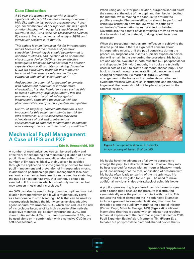
Despite the advances occurring in cataract surgery over the past 4 decades, pupilsize remains a critical determinant of surgical success. A small pupil limitsintraocular visualization and the area of the operative field (Figure 1). As a result, asmall pupil increases the difficulty of performing multiple steps in cataract surgeryand the risk of complications. Capsulorhexis, phacoemulsification, corticalcleanup, intraocular lens (IOL) implantation, and, in the case of a toric IOL, IOLpositioning, are all more challenging when the pupil is small, and cornealendothelial trauma, posterior capsule rupture, lens fragment retention, incompletecortex removal, and vitreous loss are more likely to occur.1-3 Operating through asmall pupil also increases the chance of inadvertent iris touch during surgery,which will cause patient discomfort and pupil constriction, risks iris damage, andleads to increased postoperative inflammation and pain.
Significant pain after cataract surgery, whether it is associated with operatingthrough a small pupil or not, can be alarming for patients and may be morecommon than surgeons realize. According to the findings of a systematic reviewof data from 21 published articles, up to 35% of patients reported moderate-to-severe pain in the early postoperative period after cataract surgery.4
The adverse outcomes associated with operating through a small pupil and thepotential negative impact of surgery-related pain and inflammation on patientperceptions of the overall surgical experience and postoperative recovery need tobe considered against the fact that today’s cataract surgery population has highexpectations for a comfortable procedure and an uneventful postoperative coursewith rapid vision rehabilitation.
Sponsored Supplement
Editorial Support Disclosures Cheryl Guttman Krader; Cynthia Tornallyay, RD,MBA, CHCP; Diane McArdle, PhD; Kimberly Corbin,CHCP; Barbara Aubel; and Michelle Ong have norelevant commercial relationships to disclose.
Disclosure Attestation The contributing physicians listed above haveattested to the following: 1) that the relationships/affiliations noted will not
bias or otherwise influence their involvement inthis activity;
2) that practice recommendations given relevant tothe companies with whom they have relationships/affiliations will be supported by the best availableevidence or, absent evidence, will be consistentwith generally accepted medical practice; and
3) that all reasonable clinical alternatives will bediscussed when making practice recommendations.
Off-Label Discussion This educational activity might contain discussion ofevidence-based and/or investigational uses of agentsthat are not indicated by the FDA. For additionalinformation about approved uses, including approvedindications, contraindications, and warnings, pleaserefer to the official prescribing information for eachproduct for discussion of approved indications,contraindications, and warnings.
For Digital Editions System Requirements:If you are viewing this activity online, please ensurethe computer you are using meets the followingrequirements:• Operating System: Windows or Macintosh • Media Viewing Requirements: Flash Player orAdobe Reader
• Supported Browsers: Microsoft Internet Explorer,Firefox, Google Chrome, Safari, and Opera
• A good Internet connection
New York Eye and Ear Infirmary of MountSinai Privacy & Confidentiality Policieshttp://www.nyee.edu/health-professionals/cme/enduring-activities
CME Provider Contact Information For questions about this activity, call 212-979-4383.
To Obtain AMA PRA Category 1 Credit™ To obtain AMA PRA Category 1 Credit™ for thisactivity, read the material in its entirety and consultreferenced sources as necessary. Complete theevaluation form along with the post test answer boxwithin this supplement. Remove the ActivityEvaluation/Credit Request page from the printedsupplement or print the Activity Evaluation/CreditRequest page from the Digital Edition. Return via mailto Kim Corbin, Director, ICME, New York Eye and EarInfirmary of Mount Sinai, 310 East 14th Street, NewYork, NY 10003 or fax to (212) 353-5703. Your certificatewill be mailed to the address you provide on theActivity Evaluation/Credit Request form. Please allow3 weeks for Activity Evaluation/Credit Request formsto be processed. There are no fees for participating inand receiving CME credit for this activity.
Alternatively, we offer instant certificate processing and support Green CME. Please take this post test and evaluation online by going tohttp://tinyurl.com/miosiscme. Upon passing, youwill receive your certificate immediately. You mustscore 70% or higher to receive credit for this activity,and may take the test up to 2 times. Uponregistering and successfully completing the posttest, your certificate will be made available onlineand you can print it or file it.
Disclaimer The views and opinions expressed in thiseducational activity are those of the faculty and donot necessarily represent the views of New York Eyeand Ear Infirmary of Mount Sinai, MedEdicus LLC,Omeros Corporation, EyeNet, or American Academyof Ophthalmology.
Miosis Control and Other Surgical Pearls
Complicated CataractCase Discussions in
The Significance of Miosis, Pain, andInflammation in Cataract Surgery
Edward J. Holland, MD
Figure 1. Effect of 2.5-mm decrease in pupil diameter on operative field area
Image courtesy of Edward J. Holland, MD

4
At the same time, because lifespan is increasing, surgeonsare operating on growing numbers of individuals who areolder in age and more likely to have comorbidities associatedwith limited pupil dilation and/or risk for intraoperative miosis(Table 1).
One of the most common clinical features associated withintraoperative miosis is a history of treatment with an α1-antagonist. Medications in this class, which includetamsulosin, terazosin, doxazosin, alfuzosin, and silodosin, arebeing used by growing numbers of men for the treatment ofurinary symptoms associated with benign prostatichyperplasia (BPH), by women for urinary voiding problems,and as antihypertensive treatment.5 In addition, α-adrenergicreceptor blockade is a feature of the herbal agent sawpalmetto, finasteride, and some antipsychotic medications.
Systemic treatment with an α1-antagonist blocks adrenergicstimulation of the pupillary dilator muscle and can lead todisuse atrophy with more chronic treatment.9 As a result,patients can develop intraoperative floppy iris syndrome (IFIS),a condition characterized by a flaccid iris that billows duringnormal fluid currents, a tendency for iris prolapse despiteproper wound construction, and progressive intraoperativemiosis. IFIS has been associated most often with tamsulosin,which is thought to reflect tamsulosin’s selectivity for the α1A-adrenoreceptor that is most prominent in the pupillarydilator muscle. IFIS, however, also occurs unexpectedly inpatients without any history of α1-antagonist use.
Dense brunescent cataract is another clinical featureassociated with a higher rate of intraoperative miosis,perhaps reflecting the extended operative time of thesecases.6 Other risk factors for intraoperative miosis includeposterior synechiae; pseudoexfoliation (PXF); diabetesmellitus; a history of intraocular surgery, trauma, or uveitis;and femtosecond laser-assisted cataract surgery (FLACS).6,7
It is clearly preferable to minimize inflammation and patientdiscomfort and avoid intraoperative miosis than to face theneed to manage these issues. A variety of strategies exists forachieving these goals. The following series of case-baseddiscussions will review their efficacy, safety, advantages, and limitations.
Inflammation and Pain Control: A Case of Cataract Surgery With a History of Iritis
Bonnie An Henderson, MD
Although modern cataract surgery is considered relativelyatraumatic, any surgical trauma triggers an inflammatoryresponse (Figure 2).10 The cascade of events begins withactivation of phospholipase A2, which leads to the release ofarachidonic acid. Arachidonic acid is converted bycyclooxygenase (COX)-1 and COX-2 into an intermediary thatis subsequently metabolized by synthase enzymes intoeicosanoids, including thromboxane A2 and several differentprostaglandins.
When released during cataract surgery, prostaglandinsstimulate the iris sphincter muscle and sensory nerveendings, induce leukocyte recruitment and migration, andcause vascular dilation and permeability. These effectsmanifest intraoperatively as miosis and pain andpostoperatively with the appearance of ocular redness,edema, pain, and anterior chamber cells and flare.Prostaglandins have also been implicated in the developmentof cystoid macular edema after cataract surgery.11 Therefore,inhibiting prostaglandin biosynthesis using topicalcorticosteroids that block phospholipase A2 and/or topicalnonsteroidal anti-inflammatory drugs (NSAIDs) that block theCOX enzymes is a rational strategy for preventingintraoperative miosis and limiting postoperativeinflammation and pain.
Surgical Trauma
PGG2
PGI2 TXA2
LTB4
LTE4
Corticosteroids
NSAIDs
Allergic reactions
VasodilationSensory nerve ending stimulationLeukocyte recruitment and migration
AngiogenesisMiosisVascular permeability
Uveoscleral outflow
Hydroperoxidase
Phospholipase A2
Membrane Phospholipids
LTA4, Hydrolase Glutathione-S-transferase
LTD4
Dipeptidase
y-Glutamyl-transpeptidase
y-Glutamyl-leukotrienase
LTC4
Leukotriene A4 (LTA4)
5-HPETE
PGE 9-ketoreductase
PGE2
PGD2
PGH2
Cyclooxygenase(COX-1/COX-2)
Lipoxygenase
Arachidonic Acid
PGF-2
Figure 2. Inflammatory cascade induced as a result ofsurgical trauma
Abbreviation: NSAID, nonsteroidal anti-inflammatory drug.
Adapted from Kim SJ, et al.10
Table 1. Risk Factors for Small Pupils/IntraoperativeMiosis5-8
Intraoperative floppy iris syndrome/history of α1-antagonist use Dense brunescent cataract
Posterior synechiae
Pseudoexfoliation
Diabetes mellitus
History of intraocular surgery, trauma, or uveitis
Femtosecond laser-assisted cataract surgery
5
Case IllustrationA 59-year-old woman presents with a visuallysignificant cataract OD. She has a history of recurrentiritis OU, with the last episode occurring over 1 yearago. On examination of her right eye, she has a quietanterior chamber with posterior synechiae and anN03NC3 (LOCS [Lens Opacities Classification System]III) cataract. Best corrected visual acuity is 20/80, andintraocular pressure is 14 mm Hg.
This patient is at an increased risk for intraoperativemiosis because of the presence of posteriorsynechiae.6 Synechiolysis should be performed tooptimize mydriasis, and injection of an ophthalmicviscosurgical device (OVD) can be an effectivetechnique to break the adhesions from the anteriorcapsule. Chondroitin sulfate-containing dispersiveOVDs are particularly useful for this maneuverbecause of their superior retention in the eyecompared with cohesive compounds.12
Anticipating the potential for the pupil to constrictwith subsequent reduction of intraoperativevisualization, it is also helpful in a case such as thisto create a relatively large capsulotomy that willprovide a greater margin of safety againstinadvertently contacting the capsule with thephacoemulsification tip or chopper/lens manipulator.
Control of surgically induced inflammation is alsoimportant for this patient to minimize her risk foriritis recurrence. Uveitis specialists may evenadvocate use of oral and/or intravenouscorticosteroids to suppress inflammation in patientswith a history of an ocular inflammatory condition.13
Mechanical Pupil Management: A Case of IFIS and PXF
Eric D. Donnenfeld, MD
A number of mechanical devices can be used safely andeffectively for expanding and maintaining dilation of a smallpupil. Nevertheless, these modalities also suffer from anumber of limitations; ideally, their use can be avoidedthrough the application of some general principles for smallpupil management and prevention of intraoperative miosis.In addition to pharmacologic pupil management (see nextsection), a mechanical instrument can be used for stretchingthe pupil as needed; however, this technique should beavoided in IFIS cases, in which it is not only ineffective, butmay worsen miosis and iris prolapse.5
An OVD can also be used to help open the pupil and maintaindilation intraoperatively, and it will simultaneously serve as a tamponade to stop bleeding. OVDs that can be used forviscomydriasis include the highly cohesive viscoadaptiveagent, sodium hyaluronate, 2.3%, which also reduces the riskof iris prolapse because of its high viscosity. Alternatively,dispersive materials, eg, sodium hyaluronate, 3.0%, andchondroitin sulfate, 4.0%, or sodium hyaluronate, 3.0%, canbe used alone or in combination with a cohesive OVD in thesoft-shell technique.
When using an OVD for pupil dilation, surgeons should directthe cannula at the edge of the pupil and then begin injectingthe material while moving the cannula tip around thepupillary margin. Phacoemulsification should be performedusing low aspiration flow and low vacuum settings tominimize OVD evacuation from the anterior chamber.Nevertheless, the benefit of viscomydriasis may be transientdue to washout of the material, making repeat injectionsnecessary.
When the preceding methods are ineffective in achieving thedesired pupil size, if there is significant concern aboutintraoperative miosis, or if the pupil constricts during theprocedure, surgeons can insert a pupillary expansion devicethat will remain in the eye during the procedure. Iris hooksare one option. Available in both reusable (4-0 polypropylene)and disposable (6-0 nylon) models, iris hooks are typicallyused in sets of 4 or 5 to create a diamond-shaped pupil. Eachhook is placed through a separate limbal paracentesis andengaged around the iris margin (Figure 3). Carefularrangement of the hooks will optimize visualization andavoid interference with surgical instruments and maneuvers.In general, the hooks should not be placed adjacent to thecataract incision.
Iris hooks have the advantage of allowing surgeons toenlarge the pupil to a desired diameter. However, they maybe best reserved for cases with an irregular iris/asymmetricpupil, considering that the focal application of pressure withiris hooks often leads to tearing of the iris sphincter, irisdamage, and an irregular, tonic pupil. The need to makeadditional incisions is also a drawback of using iris hooks.
A pupil expansion ring is preferred over iris hooks in eyeswith a round pupil because the pressure is distributedsymmetrically over the points of contact with the iris. Thisreduces the risk of damaging the iris sphincter. Examplesinclude a grooved, incomplete plastic ring that must bethreaded along the pupillary margin using a metal injector(Perfect Pupil, Milvella, Savage, MN) (Figure 4); a siliconeincomplete ring that is placed with an injector but requiresbimanual expansion of the proximal segment (Graether 2000Pupil Expander, EagleVision, Memphis, TN) (Figure 5); afoldable 5-0 polypropylene diamond-shaped device that is
Sponsored Supplement
Figure 3. Four-point fixation with iris hooks
Image courtesy of Steven Shafron, MD

6
placed and removed with a disposable injector (MalyuginRing, MicroSurgical Technology, Redmond, WA) (Figure 6);and a polyurethane circular device that is packaged withdedicated injector and extractor instruments (I-Ring, Beaver-Visitec International, Waltham, MA) (Figure 7).
The polypropylene ring is available in 2 sizes (6.25 mm and7.00 mm) and engages the pupil with 4 circular scrolls locatedat each corner to effectively give an 8-point fixation. Thepolyurethane device provides 360° pupillary support andtherefore is very effective in maintaining a stable area ofsurgical exposure while minimizing the risk for postoperativepupil distortion.
Compared with incomplete rings, the diamond-shaped andcircular devices are easier to insert and remove, althoughsurgeons must take care when removing the devices to avoidcausing iris disinsertion and bleeding. Anecdotally, based onlimited early clinical experience, pigment release may be lesslikely when using the I-Ring.
As a major advantage, pupillary expansion rings arepredictably effective for expanding and maintaining a stablepupil diameter. Although placement of any of the pupillaryexpansion devices can be more difficult in eyes with ashallow anterior chamber, surgeons can overcome thatchallenge by performing dry, sutureless pars planavitrectomy using 25-gauge instrumentation.
However, use of any of the pupillary expansion rings addstime and risk because of the potential to cause iris trauma,which can lead to bleeding, pain, inflammation, and irissphincter damage. Cost is another drawback associated withtheir use. Although procedures requiring a mechanical pupilexpansion device qualify as complex cataract surgery andtherefore result in a higher reimbursement for the surgeon,there is no additional facility fee for ambulatory surgerycenters or hospital outpatient departments.
Case IllustrationA 66-year-old man presents with a visuallysignificant cataract and PXF. He has BPH, for whichhe is being treated with an 1-antagonist. His pupildiameter at maximum dilation is 4.5 mm.
Given this patient’s small pupil, despite maximalpreoperative topical dilating drops, and the presenceof risk factors for intraoperative miosis, a 5-0 polypropylene pupil expansion device (I-Ring)was placed after injecting OVD to lift the iris andenable engagement of the pupillary margin.Dimpling of the capsule with injection of OVD duringthe procedure indicated the PXF was associated withsignificant loss of zonular integrity, and a capsulartension ring was also inserted. With the benefit ofthe pupil expansion device and insertion of a capsuletension ring, the case was completed uneventfully.Postoperative rehabilitation was rapid, with return of excellent uncorrected visual acuity.
Figure 4. Perfect Pupilprovides effective pupildilation
Image courtesy of Eric D. Donnenfeld, MD
Figure 5. Graether 2000Pupil Expander withfenestrated tab
Image courtesy of Eric D. Donnenfeld, MD
Figure 6. Eight-point fixation with a Malyugin Ring
Image courtesy of Eric D. Donnenfeld, MD
Figure 7. Fullyengaged, the I-Ring providescontinuous fixationand pupil dilation
Image provided toEric D. Donnenfeld,MD, by Beaver-Visitec International

7
Pharmacologic PupilManagement: A Case ofIntraoperative Miosis With FLACS
Terry Kim, MD
Intraoperative miosis poses big challenges for cataract surgeonsand increases the risk for complications. Common causes of asmall pupil/intraoperative miosis have included aging,chronic miotic use, diabetes, Horner syndrome, IFIS, PXF, andsynechiae.6,7 Now, with increasing use of FLACS, a new causefor intraoperative miosis has emerged.8,14 This problem hasbeen reported to occur in approximately one-fourth to one-third of cases relying on a preoperative dilating regimenwith tropicamide alone or combined with phenylephrine. It may become most problematic, however, in challengingcases, in which some surgeons are preferentially using the laser by virtue of the longer operating time in those eyes.
In addition to the usual problems accompanyingintraoperative miosis, reduced visualization in FLACS canmake certain portions of the cataract procedure more difficultfor surgeons, such as subincisional cortex removal, a stepthat is already more challenging in FLACS cases than inconventional cataract surgery.
When confronting a case at risk for a small pupil/intraoperative miosis, it is helpful for surgeons to consider the mechanism, which may guide the preventive strategy(Table 2). FLACS-related intraoperative miosis is mediated byprostaglandins that are released when the femtosecond lasercuts into the anterior capsule,15 and so there is a rationale forusing a pharmacologic regimen that incorporates an NSAID to mitigate prostaglandin synthesis.8,14
Topical medications are the standard for pupil dilation incataract surgery, and they are relatively inexpensive.3 Themedications used include antimuscarinic drugs that relax the pupillary sphincter muscle (eg, cyclopentolate andtropicamide), sympathomimetic/adrenergic agents thatstimulate the pupillary dilator muscle (eg, phenylephrine),and NSAIDs that block prostaglandin-mediated reflexmiosis.3,16 Flurbiprofen is the only topical NSAID available in the United States that is approved for inhibitingintraoperative miosis.16 Other NSAIDs have also been shownto have a mydriatic effect.10 In a randomized study, topicalketorolac, 0.5%, produced more stable mydriasis throughoutsurgery compared with flurbiprofen.17
Topical mydriatic regimens, however, may be unpredictablyeffective for maintaining mydriasis because of washout fromthe anterior chamber during surgery.18-21 In a study of patientswho began treatment with topical ketorolac on the day beforecataract surgery, levels of the NSAID in aqueous humorsamples drawn at the end of the procedure were consistentlynominal or below the level of detection.18 Other limitations oftopical mydriatic medications include the need for intensivepreoperative dosing by the nursing staff and the potential forcausing systemic side effects as a result of absorptionthrough the conjunctiva and nasal mucosa.21
Mydriatic agents may also be administered as single agentsor combinations directly into the anterior chamber, either asan injection or through infusion after addition of themedication to the bottle of irrigation solution.3,20 Historically,medications used via the intracameral route for mydriasisduring cataract surgery include epinephrine, phenylephrine,cyclopentolate, and lidocaine.3,19
Intracameral administration of these mydriatic agents, like atopical regimen, is inexpensive, but has the advantages ofreducing nursing time and the risk of systemic adverseevents.3 The intracameral approach also provides a morerapid onset and sustained efficacy relative to topicaladministration, particularly when the medication is added tothe infusion bottle and delivered throughout the procedure.However, drawbacks with intracameral administration ofthese medications as mydriatic agents should be considered.With the exceptions of preservative-free/bisulfite-freeepinephrine and preservative-free/sulfite-containingepinephrine when diluted in ophthalmic irrigation fluid, theiruse is off label.22,23 Compounding using these or othermedications introduces the potential for dosing errors andcontamination. In fact, because of safety concerns, somesurgical facilities are prohibiting intraocular use of anymedications that are compounded in the operating room, and licensing and regulatory agencies are increasinglyscrutinizing these off-label practices. In addition, use ofproducts containing preservatives can cause toxic anteriorshock syndrome,24 and exposure to undiluted productscontaining bisulfite can cause corneal endothelial toxicity.25
A commercially available fixed combination ofphenylephrine, 1%/ketorolac, 0.3% (phenylephrine/ketorolac),added to a 500-mL irrigation solution bottle offers a newintracameral option for preventing intraoperative miosis.26
This product is a clear, colorless, sterile, preservative-free,and bisulfite-free solution that comes packaged in single
Sponsored Supplement
Table 2. Causes and Mechanisms of Small Pupils
Etiology Mechanism
Aging Atrophy of iris
Chronic miotic use Fibrosis of iris sphincter
Diabetes Autonomic dysfunction orrubeosis
Horner syndrome Sympathetic denervation
IFISAtrophic dilator muscle from chronic α1A-adrenoreceptorblockade
PXFAtrophic iris dilator muscle vsfibrosis of sphincter muscle
SynechiaeUveitis, trauma, angle closure, or prior surgery
FLACS Prostaglandin release
Table courtesy of Terry Kim, MD

8
patient-use vials. It is approved by the FDA for maintainingpupil size during cataract surgery or IOL replacement and forreducing postoperative ocular pain.26 Use of phenylephrine/ketorolac injection avoids the potential for human error andother problems associated with compounding medications.There is no added cost for the surgeon or surgical facility touse the phenylephrine/ketorolac solution, and the cost of theagent is covered by Medicare and other third-party payerswhen it is used for its FDA-approved indication. Phenylephrine/ketorolac also qualifies for pass-through payment under theOutpatient Prospective Payment System.
Blood pressure elevation due to systemic phenylephrineexposure is the primary safety concern with this modality. Inan integrated analysis of safety data from 2 phase 3 placebo-controlled clinical trials investigating the phenylephrine/ketorolac injection, no differences between treatment groupsin serial assessments of vital signs were reported.27 Inaddition, the incidence of treatment-emergent adverse eventswas higher in the control group receiving placebo than inpatients receiving the phenylephrine/ketorolac injection(66.9% vs 60%), and all severe treatment-emergent adverseevents considered to be related to study treatment occurredin patients receiving placebo.
Several premarketing clinical trials demonstrated the efficacyof phenylephrine/ketorolac for maintaining pupil size duringcataract surgery and controlling postoperative pain.20,27
Analyses of the pooled data from the 2 phase 3 studiesshowed phenylephrine/ketorolac met its coprimary outcomemeasures, achieving significant differences compared withplacebo in the mean area under the curve (AUC) change frombaseline in pupil diameter (0.08 vs -0.50 mm; P < .0001) and inmean AUC of ocular pain visual analog scale scores within 12 hours postoperatively (4.16 mm vs 9.06 mm; P < .001).27
Phenylephrine/ketorolac also achieved superiority overplacebo in key secondary efficacy analyses that assessed thepercentage of patients with a pupil diameter < 6.0 mm atcortical cleanup; pupil diameter < 6.5 mm at any time duringsurgery; ≥ 2.5 mm of pupillary constriction at any time duringsurgery (Figure 8); no pain at all time points after surgery;moderate-to-severe pain at any time point; and analgesic useon the day of surgery (P ≤ .0027 for all comparisons).
To supplement the benefits of using a phenylephrine/ketorolac injection in the irrigation solution, some surgeonsare withdrawing aliquots of the mixed solution to use as aninitial intracameral injection through the paracentesis andthen later in the case for hydrodissection and stromalhydration. Anecdotally, this direct intracameral injection of anirrigation solution containing the phenylephrine/ketorolacinjection seems to cause less burning and stinging comparedwith intracameral injection of epi-Shugarcaine (bisulfite-freeepinephrine and preservative-free lidocaine mixed into anirrigation solution).
Most importantly, when encountering intraoperative miosiswith FLACS, my experience has shown the benefits of usingan FDA-approved agent of phenylephrine and ketorolacthroughout the procedure, which can help increase andmaintain pupil size to facilitate visualization duringphacoemulsification, cortex removal, and IOL implantation,with the ultimate goal of preventing intraoperativecomplications and improving postoperative outcomes.
Case IllustrationA 62-year-old man with bilateral cataracts and ~2.5 Dof regular astigmatism in both eyes is interested inreduced spectacle dependence for activitiesinvolving distance vision. He chooses toric IOLs andelects to undergo FLACS for both eyes.
Surgery is performed first on his dominant eye,which dilates to 8.0 mm with preoperative drops.However, following the femtosecond laser portion ofthe surgery, the pupil is noted to decrease in size. Atthe start of the surgery, he receives an intracameralinjection of epi-Shugarcaine. The pupil stays welldilated during nucleus and epinucleus removal, butthen begins to come down again so that the edge ofthe capsulotomy is no longer visible. A polypropylenepupil expansion ring is inserted to enable completecortex removal, along with implantation andalignment of the toric IOL.
Two weeks later, surgery is performed on his secondeye, which also dilates to 8.0 mm with preoperativedrops. Once again, the pupil size is noted to decreasein size following femtosecond laser application.Phenylephrine/ketorolac injection is added to theirrigation solution bottle for this surgery and is alsoused for intracameral injection through theparacentesis incision prior to initiatingphacoemulsification to address the small pupil. Thepupil stays well dilated throughout the entire case,particularly during cortex removal and toric IOLimplantation and alignment, and eliminates the needfor a pupil expansion device.
27%
2%
30
25
20
15
10
5
0Vehicle Phenylephrine, 1%/
Ketorolac, 0.3%
Per
cen
tag
e o
f P
atie
nts
Wit
hD
ecre
ase
in P
up
il D
iam
eter
2.5
mm
2
Figure 8. Phenylephrine/ketorolac significantly reduced thepercentage of patients with a decrease in pupil diameter ≥ 2.5 mm compared with vehicle27

9Sponsored Supplement
Other Surgical Pearls for Addressing Cataract Surgery Complications
FLACS for the Mature White CataractTerry Kim, MD
Capsulorhexis is the most difficult step in cases involving amature white cataract because of the lack of red reflex andthe presence of elevated intracapsular pressure that cancause an uncontrollable radial tear, the so-called Argentinianflag sign. One strategy for increasing the safety ofcapsulorhexis is to first decompress the capsular bag bypuncturing the anterior capsule with a 25-gauge needle andaspirating out the liquefied cortex.
Use of the femtosecond laser for capsulotomy represents analternative approach. The laser system is able to identify theanterior capsule and create a complete capsulotomy (Figure 9)within a closed system, thereby reducing the chance of aradial tear in the anterior capsule.
Loose ZonulesBonnie An Henderson, MD
Loose zonules present multiple challenges for cataractsurgery. Although loss of zonular integrity may be recognizedor suspected preoperatively based on findings of the clinicalexamination or history, it is not unusual that this problem willonly be recognized during surgery. Posterior displacement ofthe entire lens and capsular bag complex when applyingforce to the nucleus with the phaco tip during chopping isone intraoperative sign of loose zonules.
Once the problem is appreciated, surgeons should maintainthe phaco tip in the eye and inject additional viscoelastic tokeep the anterior chamber formed. Then, iris or capsularhooks can be used to stabilize the capsule. Considering the
likelihood of encountering loose zonules in eyes with PXF, irishooks may be considered the preferred option over othermethods, such as rings, for managing a small pupil in eyeswith PXF, where they can then be used for capsular bagsupport, if needed.
Scleral Fixation With the Glued IOL TechniqueEric D. Donnenfeld, MD
The glued IOL technique introduced by Amar Agarwal, MD,has become my preferred method for managing a dislocated3-piece IOL with polypropylene haptics.28 It effectively securesthe IOL in a stable position and avoids lens exchange,suturing, and suturing-related complications, and thepolypropylene haptics will not degrade over time.
Briefly, the technique involves the creation of 2 scleral flaps180o apart, 2 sclerotomies under each flap, through which the haptics are externalized, and 2 scleral incisions, into whichthe haptics are tucked and glued. Pars plana vitrectomy isperformed to avoid retinal traction during IOL manipulation,and then the IOL is brought into the anterior chamber bygrabbing one haptic in a tire-iron technique and pushing theoptic from behind with forceps. Any cortex present in thecapsular bag can be removed using the vitrectomy probe in IA mode.
In series, the haptics are manipulated through the sclerausing a handshake technique to transfer the haptic from onehand to the other. One microforceps holds the haptic in theanterior chamber and passes the haptic to a microforcepspassed through the sclerotomy in the posterior chamber. Thehaptic is then externalized through the scleral incision underthe flap (Figure 10). Once the haptic is placed into the scleralpocket, fibrin glue is used to seal the scleral flaps andconjunctiva. I also inject triamcinolone into the vitreoushumor through the pars plana incision to reduceinflammation, and in my experience, these eyes are veryquiet the next day.
Figure 9. Trypan blue staining shows the femtosecond lasercreated a complete capsulotomy in this eye with a maturewhite cataract
Image courtesy of Terry Kim, MD
Figure 10. Hapticsvisualized under scleral flaps priorto placement intoscleral incision
Image courtesy of Eric D. Donnenfeld, MD
Complicated cataract surgery comprises a myriad of situations in addition to intraoperative miosis. In the following,faculty members offer pearls for addressing selected challenges.
Go to next page for Key Learning Points

10
1. Guzek JP, Holm M, Cotter JB, et al. Riskfactors for intraoperative complications in1000 extracapsular cataract cases.Ophthalmology. 1987;94(5):461-466.
2. Narendran N, Jaycock P, Johnston RL, et al. The Cataract National Datasetelectronic multicentre audit of 55,567operations: risk stratification for posteriorcapsule rupture and vitreous loss. Eye (Lond). 2009;23(1):31-37.
3. Behndig A, Korobelnik JF. Mydriatic insertand intracameral injections compared withmydriatic eyedrops in cataract surgery:controlled studies. J Cataract Refract Surg.2015;41(7):1503-1519.
4. Porela-Tiihonen S, Kaarniranta K, Kokki H.Postoperative pain after cataract surgery. J Cataract Refract Surg. 2013;39(5):789-798.
5. Chang DF, Braga-Mele R, Mamalis N, et al;ASCRS Cataract Clinical Committee.ASCRS White Paper: clinical review ofintraoperative floppy-iris syndrome. J Cataract Refract Surg. 2008;34(12):2153-2162.
6. American Academy of OphthalmologyCataract and Anterior Segment Panel.Preferred Practice Pattern®: Cataract in theAdult Eye. San Francisco, CA: AmericanAcademy of Ophthalmology; 2011.
7. Megbelayin EO, Pindikura S. Managingchallenges of recalcitrant intra-operativemiosis during small incision cataractsurgery. Int J Scient Res Knowl.2013;1(5):74-81.
8. Yeoh R. Intraoperative miosis infemtosecond laser-assisted cataractsurgery. J Cataract Refract Surg.2014;40(5):852-853.
9. Chang DF, Campbell JR, Colin J,Schweitzer C; Study Surgeon Group.Prospective masked comparison ofintraoperative floppy iris syndromeseverity with tamsulosin versus alfuzosin.Ophthalmology. 2014;121(4):829-834.
10. Kim SJ, Flach AJ, Jampol LM.Nonsteroidal anti-inflammatory drugs inophthalmology. Surv Ophthalmol.2010;55(2):108-133.
11. Miyake K, Ibaraki N. Prostaglandins andcystoid macular edema. Surv Ophthalmol.2002;47(suppl 1):S203-S218.
12. Petroll WM, Jafari M, Lane SS, Jester JV,Cavanagh HD. Quantitative assessment ofophthalmic viscosurgical device retentionusing in vivo confocal microscopy. J Cataract Refract Surg. 2005;31(12):2363-2368.
13. American Academy of Ophthalmology.Preferred Practice Pattern® ClinicalQuestions: Uveitis and Cataract Surgery.San Francisco, CA: American Academy ofOphthalmology; 2012.
14. Nagy ZZ, Takacs AI, Filkorn T, et al.Complications of femtosecond laser-assisted cataract surgery. J CataractRefract Surg. 2014;40(1):20-28.
15. Schultz T, Joachim SC, Stellbogen M, Dick HB. Prostaglandin release duringfemtosecond laser-assisted cataractsurgery: main inducer. J Refract Surg.2015;31(2):78-81.
16. Ocufen [package insert]. Irvine, CA:Allergan; 2012.
17. Solomon KD, Turkalj JW, Whiteside SB,Stewart JA, Apple DJ. Topical 0.5%ketorolac vs 0.03% flurbiprofen forinhibition of miosis during cataractsurgery. Arch Ophthalmol. 1997;115(9):1119-1122.
18. Katsev DA, Katsev CC, Pinnow JM.Intracameral ketorolac concentration aftertopical ketorolac administration prior tocataract surgery. Paper presented at:ASCRS-ASOA Symposium & Congress;April 17-21, 2015; San Diego, CA. Abstract11161.
19. Lawuyi LE, Gurbaxani A. The clinical utility of new combination phenylephrine/ketorolac injection in cataract surgery. Clin Ophthalmol. 2015;9:1249-1254.
20. Lindstrom RL, Loden JC, Walters TR, et al.Intracameral phenylephrine and ketorolacinjection (OMS302) for maintenance ofintraoperative pupil diameter andreduction of postoperative pain inintraocular lens replacement withphacoemulsification. Clin Ophthalmol.2014;8:1735-1744.
21. Grob SR, Gonzalez-Gonzalez LA, Daly MK.Management of mydriasis and pain incataract and intraocular lens surgery:review of current medications and futuredirections. Clin Ophthalmol. 2014;8:1281-1289.
22. Epinephrine injection USP [packageinsert]. Largo, FL: BelcherPharmaceuticals, LLC; 2015.
23. Adrenalin [package insert]. Rochester, MI:JHP Pharmaceuticals LLC; 2012.
24. Mamalis N, Edelhauser HF, Dawson DG,Chew J, LeBoyer RM, Werner L. Toxicanterior segment syndrome. J CataractRefract Surg. 2006;32(2):324-333.
25. Slack JW, Edelhauser HF, Helenek MJ. A bisulfite-free intraocular epinephrinesolution. Am J Ophthalmol. 1990;110(1):77-82.
26. Omidria [package insert]. Seattle, WA:Omeros Corporation; 2015.
27. Hovanesian JA, Sheppard JD, Trattler WB,et al. Intracameral phenylephrine andketorolac during cataract surgery tomaintain intraoperative mydriasis andreduce postoperative ocular pain:integrated results from 2 pivotal phase 3studies. J Cataract Refract Surg. 2015;41(10):2060-2068.
28. Agarwal A, Kumar DA, Jacob S, Baid C,Agarwal A, Srinivasan S. Fibrin glue-assisted sutureless posterior chamberintraocular lens implantation in eyes withdeficient posterior capsules. J CataractRefract Surg. 2008;34(9):1433-1438.
References
Intraoperative miosis createschallenges in cataract surgery,affecting: • Visualization and surgical field • Capsulorhexis, phacoemulsification,
cortical cleanup, IOL implantation,and alignment
• Risk for complications andpostoperative pain, inflammation,and patient satisfaction
Chances of encountering intraoperativemiosis are increased by: • Comorbidities present in an aging
cataract surgery population • FLACS
Prevention is the best management forintraoperative miosis • Traditional options (eg, topical
drops, viscomydriasis, mechanicalapproaches, and off-labelintracameral injections/infusions)have limitations
Phenylephrine/ketorolac injection forpreventing intraoperative miosis: • Is FDA approved • Simultaneously controls
postoperative pain andinflammation
• Carries no cost to the surgeon or surgery center
Iris hooks for pupil management ineyes with PXF can double as capsularhooks if loose zonules are identifiedintraoperatively.
FLACS offers a new technique for safecapsulotomy creation in eyes with amature white cataract.
The glued IOL technique is a simple,safe, and effective method for securinga dislocated posterior chamber IOLwithout sutures.
Key Learning Points
11
1. When a 6-mm pupil constricts by 2.5 mm, what is thedecrease in operative field area?
A. 18% B. 33% C. 50% D. 66%
2. As reported in a systematic review of 21 published articles,up to what percentage of patients reported moderate-to-severe pain immediately after cataract surgery?
A. 10% B. 25% C. 35% D. 50%
3. Which of the following is not a risk factor for intraoperativepupillary constriction during cataract surgery?
A. Diabetes mellitus B. Oral aspirin use C. Oral saw palmetto use D. Use of femtosecond laser for capsulotomy and lens
softening
4. Which of the following pharmacologic mechanisms ofaction is not a rational strategy for limiting inflammationafter cataract surgery?
A. Lipoxygenase inhibition B. Phospholipase A2 inhibition C. Cyclooxygenase inhibition D. All of the above
5. Viscomydriasis with an OVD: A. Should be avoided in eyes at risk for IFIS due to
increased risk of iris prolapse B. Should be used in conjunction with low aspiration flow
and low vacuum settings C. Is only effective when using a dispersive product D. Reliably maintains pupil dilation during surgery if the
soft-shell technique is used
6. Pupillary expansion rings: A. Are preferred over iris hooks in eyes with a round pupil B. Are contraindicated in eyes with a shallow anterior
chamber due to the risk of corneal endothelial trauma C. Avoid the risk of iris sphincter damage D. Should be avoided in eyes with PXF due to concern
about release of iris pigment and fibrillary deposits
7. Intracameral administration of mydriatic agents: A. Increases the risk of systemic adverse events relative to
topical treatment B. Is off-label use for all medications except
phenylephrine, 1%/ketorolac, 0.3% C. May result in endothelial toxicity using undiluted
products containing bisulfites D. Significantly increases time in the operating room to
allow for a relatively slow onset of action
8. Intraoperative miosis during FLACS: A. Is mediated by acetylcholine release B. Is mediated by prostaglandin release C. Is prevented using a preoperative topical dilating
regimen that includes both an antimuscarinic and anadrenergic agent
D. Makes FLACS contraindicated in patients if thepreoperative pupil diameter is ≤ 6.0 mm
9. According to its prescribing information, phenylephrine,1%/ketorolac, 0.3%, injection is recommended for:
A. Injection directly into the anterior chamber withoutdilution
B. Injection through the zonules at the beginning of the case
C. Addition to the infusion bottle D. Use only in cases in which there is poor pupillary
dilation
10. Phenylephrine, 1%/ketorolac, 0.3%, injection is approved for: A. Maintaining pupil size during cataract surgery B. Preoperative induction of miosis C. Preventing postcataract surgery cystoid macular edema D. Preventing inflammation after cataract surgery
Sponsored Supplement
To obtain AMA PRA Category 1 Credit™ for this activity, complete the CME Post Test by writing the best answerto each question in the Answer Box located on the Activity Evaluation/Credit Request form on the followingpage. Alternatively, you can complete the CME Post Test online at http://www.tinyurl.com/miosiscme.
See detailed instructions under To Obtain AMA PRA Category 1 Credit™ on page 3.
CME Post Test Questions
http://www.tinyurl.com/miosiscmeFor instant processing, complete the CME Post Test online
Activity Evaluation/Credit Request Case Discussions in Complicated Cataract: Miosis Control and Other surgical Pearls
Proceedings From a CME Symposium During AAO 2015
To receive AMA PRA Category 1 Credit™, you must complete this Evaluation form and the Post test. Record your answers to the Post test in the Answer Box locatedbelow. Mail or Fax this completed page to new York Eye and Ear Infirmary of Mount sinai–ICME, 310 East 14th Street, New York, NY 10003 (Fax: 212-353-5703).Your comments help us to determine the extent to which this educational activity has met its stated objectives, assess future educational needs, and create timely andpertinent future activities. Please provide all the requested information below. This ensures that your certificate is filled out correctly and is mailed to the proper address.It also enables us to contact you about future CME activities. Please print clearly or type. Illegible submissions cannot be processed.
PARtICIPAnt InFORMAtIOn (Please Print) o Home o Office
Last Name _____________________________________________________________________ First Name ___________________________________________
Specialty __________________________________________ Degree o MD o DO o OD o PharmD o RPh o NP o RN o PA o Other __________
Institution ___________________________________________________________________________________________________________________________
Street Address ______________________________________________________________________________________________________________________
City ___________________________________________ State _____________________ ZIP Code ____________________ Country ______________________
E-mail ________________________________________ Phone ______________________________________ Fax _____________________________________
Please note: we do not sell or share e-mail addresses. They are used strictly for conducting post-activity follow-up surveys to assess the impact of this
educational activity on your practice.
Learner Disclosure: To ensure compliance with the US Centers for Medicare and Medicaid Services regarding gifts to physicians, new York Eye and Ear
Infirmary of Mount sinai Institute for CME requires that you disclose whether or not you have any financial, referral, and/or other relationship with our institution. CME certificates cannot be awarded unless you answer this question. For additional information, please call NYEE ICME at 212-979-4383. Thank you.o Yes o No I and/or my family member have a financial relationship with new York Eye and Ear Infirmary of Mount sinai and/or refer Medicare/Medicaid patients to it.o I certify that I have participated in the entire activity and claim 1.5 AMA PRA Category 1 Credits™.
signature Required _______________________________________________________________________ Date Completed ______________________________
OUtCOMEs MEAsUREMEnt
o Yes o No Did you perceive any commercial bias in any part of this activity? IMPORtAnt! If you answered “Yes,” we urge you to be specific
about where the bias occurred so we can address the perceived bias with the contributor and/or in the subject matter in future activities.
____________________________________________________________________________________________________________________________________Circle the number that best reflects your opinion on the degree to which the following learning objectives were met:
5 = strongly Agree 4 = Agree 3 = neutral 2 = Disagree 1 = strongly Disagree
Upon completion of this activity, I am better able to: • Identify optimal strategies for control of postoperative pain and inflammation for patients undergoing
intraocular lens replacement 5 4 3 2 1 • Evaluate the benefits and risks of pharmacological agents for managing miosis 5 4 3 2 1 • Appraise the safety and efficacy of mechanical devices for managing miosis 5 4 3 2 1 • Incorporate evidence-based approaches for mydriasis maintenance in complicated cataract cases 5 4 3 2 1
1. Please list one or more things, if any, you learned from participating in this educational activity that you did not already know. ______________________________
___________________________________________________________________________________________________________________________________
2. As a result of the knowledge gained in this educational activity, how likely are you to implement changes in your practice?4 = definitely will implement changes 3 = likely will implement changes 2 = likely will not implement any changes 1 = definitely will not make any changes
5 4 3 2 1
Please describe the change(s) you plan to make: ____________________________________________________________________________________________
___________________________________________________________________________________________________________________________________3. Related to what you learned in this activity, what barriers to implementing these changes or achieving better patient outcomes do you face?______________________________________________________________________________________________________________________________________________________________________________________________________________________________________________________________________4. Please check the Core Competencies (as defined by the Accreditation Council for Graduate Medical Education) that were enhanced for you through participation in this activity. o Patient Care o Practice-Based Learning and Improvement o Professionalism
o Medical Knowledge o Interpersonal and Communication Skills o Systems-Based Practice
5. What other topics would you like to see covered in future CME programs? _____________________________________________________________________________
___________________________________________________________________________________________________________________________________
ADDItIOnAL COMMEnts _____________________________________________________________________________________________________________
____________________________________________________________________________________________________________________________________
____________________________________________________________________________________________________________________________________
____________________________________________________________________________________________________________________________________
1 2 3 4 5 6 7 8 9 10
POst tEst AnswER BOx
Original Release: April 1, 2016Last Review: March 1, 2016
Expiration: April 30, 2017
84