Embed Size (px)
Citation preview
Hindawi Publishing CorporationJournal of OphthalmologyVolume 2009, Article ID 203583, 5 pagesdoi:10.1155/2009/203583
Case Report
Minocycline-Associated Pseudotumor Cerebri withSevere Papilledema
Simon R. Bababeygy,1, 2 Michael X. Repka,1 and Prem S. Subramanian1, 3
1 Department of Ophthalmology, The Johns Hopkins Hospitals, 600 N. Wolfe St, Baltimore, MD 21287, USA2 Howard Hughes Medical Institute, Stanford University School of Medicine, Stanford, CA 94305, USA3 Wilmer Eye Institute, Maumenee 127, 600 N. Wolfe Street, Baltimore, MD 21287, USA
Correspondence should be addressed to Prem S. Subramanian, [email protected]
Received 8 July 2009; Accepted 16 November 2009
Recommended by Eric Eggenberger
Background. Pseudotumor cerebri is an acknowledged but unusual complication of oral minocycline use. Vision loss andpapilledema have been described as mild and transient, and some authors suggest that treatment is not needed. Methods. Caseseries of 2 patients with severe papilledema and visual field loss. Results. Severe pseudotumor cerebri developed in 2 nonobesepatients taking minocycline. Their disease required further treatment even upon drug discontinuation because of visual field lossand papilledema. Conclusions. Minocycline-associated pseudotumor cerebri is not always a self-limited condition and may requireaggressive medical or surgical management.
Copyright © 2009 Simon R. Bababeygy et al. This is an open access article distributed under the Creative Commons AttributionLicense, which permits unrestricted use, distribution, and reproduction in any medium, provided the original work is properlycited.
1. Introduction
The pathogenesis of pseudotumor cerebri syndrome (PTC)is poorly understood, although its diagnostic criteria arewell established [1]. While most cases are idiopathic, recentdata indicate that patients with severe forms of PTC mayhave partial venous outflow obstruction [2–4], and suchpatients may have greater long-term morbidity. Antibioticmedications such as minocycline, tetracycline, and doxycy-cline have also been repeatedly implicated as a causativeor contributory factor in PTC [5–7]. The prognosis ofminocycline-related PTC reported in the literature is quitevariable. Some authors suggest it is a benign condition thatresolves spontaneously upon discontinuing the antibiotic[8, 9], while others report permanent vision loss mayensue [5]. It is possible that venous sinus thrombosis wasa confounding factor regarding disease severity in someinstances, as most reported cases predate reliable mag-netic resonance venography (MRV). Here we describe twocases of minocycline-induced PTC with severe papilledemaand normal MRV, indicating that minocycline alone maybe associated with severe PTC without venous sinusanomalies.
2. Materials and Methods
Retrospective case analysis was performed on cases ofpseudotumor cerebri seen by two of the authors (MichaelX. Repka, Prem S. Subramanian) in their clinical practices.A specific search for nonobese patients (BMI ≤ 25) withminocycline use at the time of pseudotumor cerebri diagno-sis was made, and cases identified are presented below. IRBapproval was not required for this study by our institutionalguidelines, since it involves a case series of fewer than 3patients.
3. Results
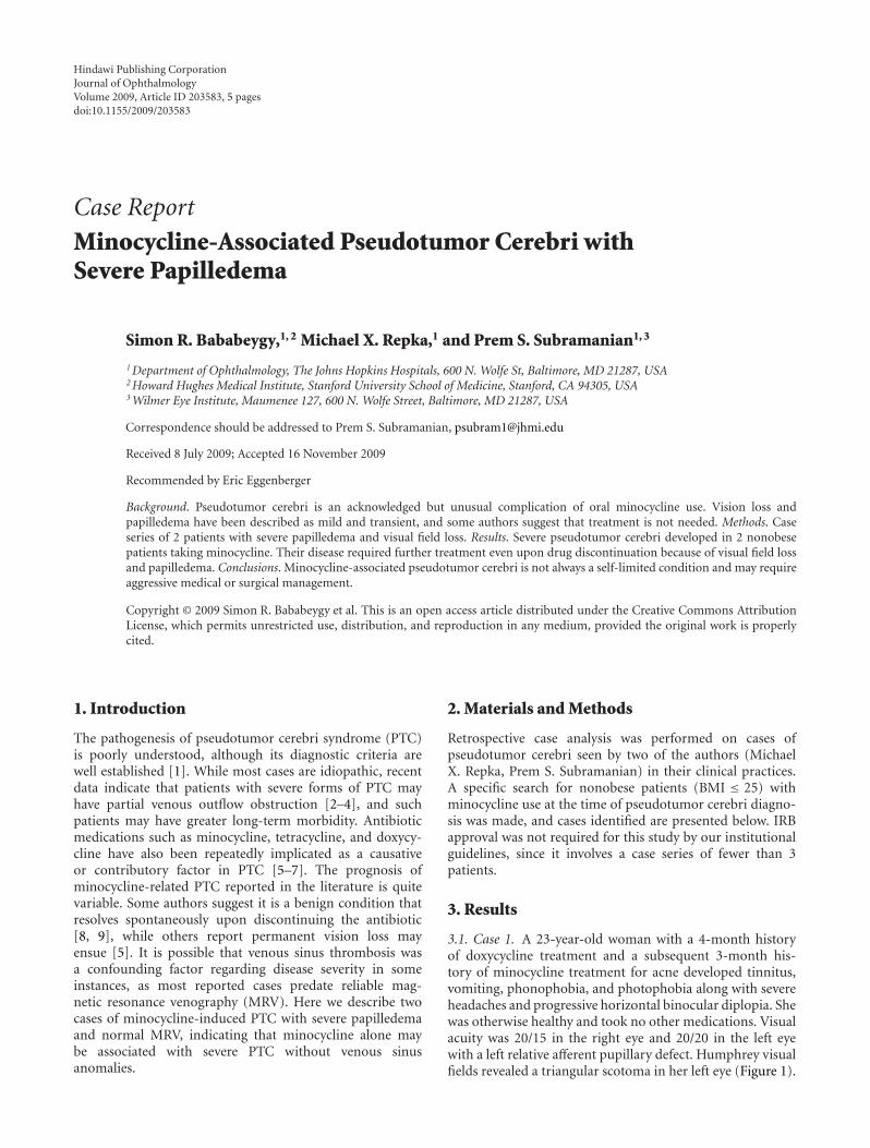
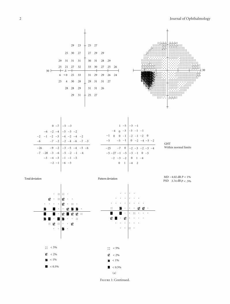
3.1. Case 1. A 23-year-old woman with a 4-month historyof doxycycline treatment and a subsequent 3-month his-tory of minocycline treatment for acne developed tinnitus,vomiting, phonophobia, and photophobia along with severeheadaches and progressive horizontal binocular diplopia. Shewas otherwise healthy and took no other medications. Visualacuity was 20/15 in the right eye and 20/20 in the left eyewith a left relative afferent pupillary defect. Humphrey visualfields revealed a triangular scotoma in her left eye (Figure 1).
2 Journal of Ophthalmology
29 23 25 27
27
27
27 27
25 30 27 27 29
29
2929
29
29
29
29
29
31 31 31 31
31
31 31
31
31 31
30
3030 30
32
28
28
2828
6
25 25
25
26
26
26
33
330 25
25
24
304
21
0
0
0
−7 −5 −3
−6 −2 −4 −5 −3 −2
−2 −1 −2 −3 −4 −2 −4 −2
−6 −7 −2 −2 −4 −6 −7 −3
−2 −1 −6 −3
−3 −4 −3 −1 −1 −5
−7 −28 −3 −6 −5 −2 −1 −4
−26 −9 −2 −3 −5 −4 −5 −6
1 −5 −3 −1
−3 −3 −1 −1
0
0
0
−1−1
−1
−2
−2 −1
−5 −5 −2 −4 −5
−2
0 1 −4 2
−2 −3 1 −4
−5 −27 −1 −5 −3
−25 −2−7 −3 −2
−1
−3
GHTWithin normal limits
MDPSD
− dBdB
P < 1%
P <
< 5%
< 2%
<
<
<
Δ
< 5%
< 2%
<
<
1%
0.5%
.5%
4.023.74
1%
0.5%
Pattern deviationTotal deviation
−2
−3
4−
00
0
−4
(a)
Figure 1: Continued.
Journal of Ophthalmology 3
27
31
31
31
31
31
31
31
3030
30
30
30 30
30
32
3232
32
3232
32
33
33
33
33
33
33
33 33
33 33 33
24
24
24
24
3232
34
34
3 20
35 2
−1 −5 −2 2
2 0 1 −1 1 0
−4 0 0 0 0 0 −2 −3
−2 −7 −1 −1 −1 −1 −1 −11
−1 −2 0 −2 0 0 −3 −7−7−2 0 −1 −2 0 −2 −1
−3 0 −1 −2 −1 −2
−3 −2 −3 0
−1 −6 −2 2
2 0 0 −1 0 −1
−4 0 0 0 −1 0 −2 −3
−2 −7 −1 −1 −1 −1 −1 −11
−1 −2 0 −2 0 0 −3 −7
−7−2 0 −1 −2 0 −2 −1
−3 0 −1 −2 −1 −2
−3 −2 −3 0
Δ
29
27
28
28 29
29
28
29 28
29 28
Pattern deviationTotal deviation
< 5%
< 2%
<
<
1%
0.5%
< 5%
< 2%
<
<
1%
0.5%
GHTWithin normal limits
MDPSD
− dBdB
P < 1%
P < .5%
1.532.35
(b)
Figure 1: Automated perimetry results, case 1. In the left eye (a), the mean deviation was −4.02 with blind spot enlargement as well as somedepression extending toward fixation from the blind spot. In the right eye (b), there was a mean deviation of −1.53 and slight enlargementof the blind spot.
4 Journal of Ophthalmology
(a) (b)
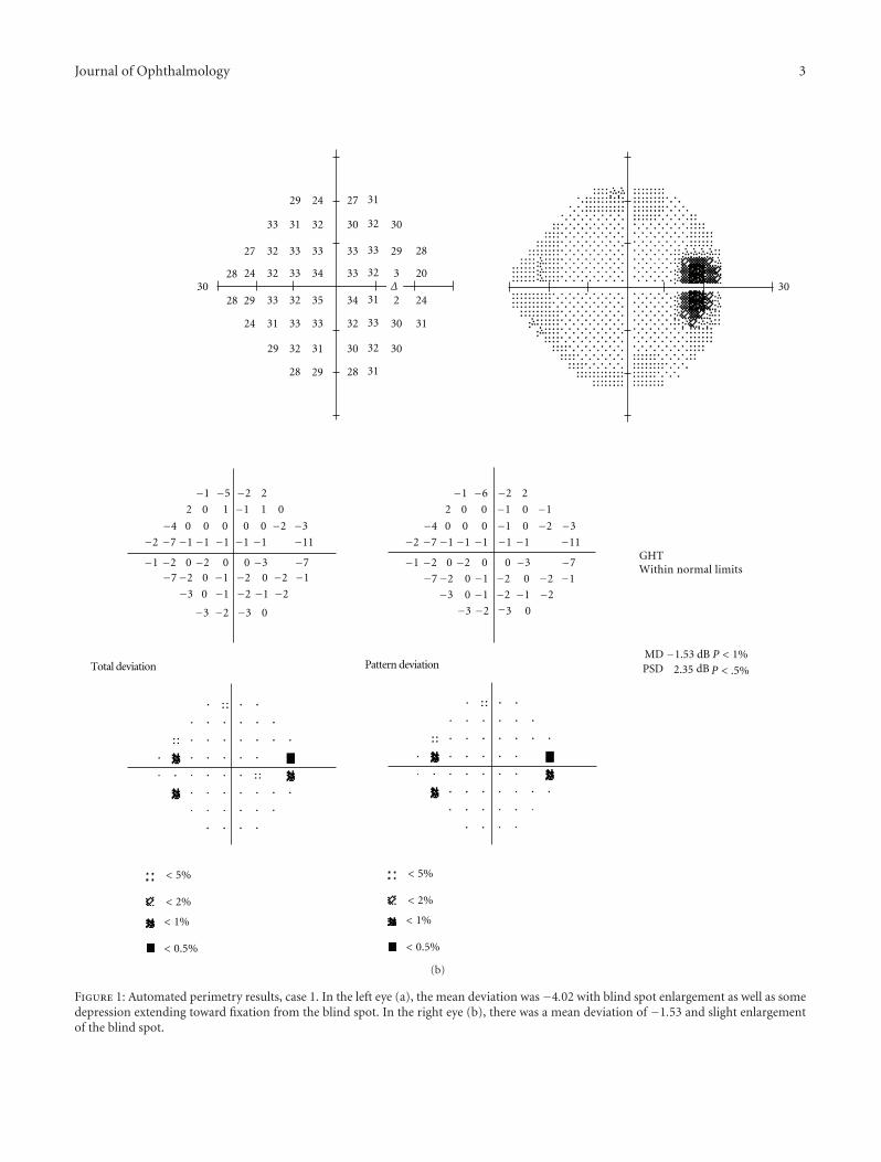
Figure 2: Optic disc appearance, case 2. Fundoscopy of the left eye (a) and right eye (b) reveals grade IV papilledema as evidenced by severeelevation and hemorrhages.
Severe bilateral papilledema was present, greater onthe left than the right. Lumbar puncture demonstrated anopening pressure of 41 cm H2O with normal cerebrospinalfluid. Minocycline use was discontinued. Head CT andcontrast-enhanced MRI were both normal and an MRVshowed no venous thrombosis or stasis. The patient’s symp-toms of diplopia improved 3 weeks later, but papilledemaand headache persisted with worsening of visual fields.Repeat MRV was normal. The intracranial pressure remainedelevated at 33 cm H2O despite treatment with 2 g aceta-zolamide/day. Ventriculoperitoneal shunting was performedfor intracranial pressure control.
3.2. Case 2. A 15-year-old girl with a 6-week history ofminocycline treatment for acne vulgaris developed progres-sive bifrontal headache with pain around the right eye,vertical binocular diplopia, nausea, neck pain, and musclespasm. Visual acuity was 20/20-2 in the right eye and 20/15-1 in the left eye. Threshold visual fields revealed enlargedblind spots in both eyes, but otherwise normal findings.Dilated ophthalmoscopy disclosed florid papilledema inboth eyes with peripapillary hemorrhages and choroidalfolds (Figure 2). Head CT and enhanced MRI were bothnormal, and an MRV showed no evidence of venous throm-bosis. The opening pressure was 55 cm H2O with normalCSF formula. Minocycline was discontinued. Acetazolamidetreatment was prescribed, then discontinued 5 months laterafter resolution of the papilledema, headaches, visual fielddefects, and diplopia.
4. Discussion
We present 2 cases of minocycline-associated PTC innonobese patients in whom symptoms were severe, thoughthere were no signs of venous sinus thrombosis on MRV.In one case, papilledema and elevated ICP persisted formore than 3 months despite stopping minocycline. Visionloss ensued in one patient despite maximal medical therapy.Duration of PTC can never be known for sure, and it
is possible that both patients had disc swelling for sometime before diagnosis and thus suffered vision loss as aresult. However, vision loss and papilledema in PTC foundincidentally are usually mild. There is conflicting evidencein the literature regarding the prognosis of minocycline-induced PTC. In the largest published series of minocycline-treated patients, consisting of 12 patients, 6 had severepapilledema at diagnosis and 3 had residual visual fieldloss [5]. Kesler et al. reported 18 patients with a history oftetracycline use associated with pseudotumor cerebri andfound that 6 patients had a relapsing course, while 12 patientshad rapid recovery from the disease upon stopping theantibiotic agent [10]. The authors suggested that tetracyclineplayed only a minor role in the patients who relapsed;however, a number of their patients were obese [10], incontrast to the patients we present in this report.
Others have stated that minocycline-induced PTC usu-ally has minimal visual consequence and resolves rapidlyupon discontinuing the medication [8]. Normalization ofintracranial pressure within 1 month of stopping tetracyclinehas been reported as well [9]. However, the mechanism bywhich tetracycline or minocycline may induce pseudotumorcerebri has not been determined. Our patient in case 1was treated with both doxycycline and then minocycline,although her symptoms did not arise until the latter drugwas used. Our findings suggest that minocycline alone mayinduce severe PTC with persistently elevated intracranialpressure, and patients with this condition may requiremedical and surgical treatment beyond discontinuation ofthe medication. We propose that minocycline-associatedpseudotumor cerebri is not a benign condition and mustbe followed closely with aggressive interventions to preventvision loss.
Acknowledgment
This work is supported in part by an unrestricted grant tothe Wilmer Eye Institute from Research to Prevent BlindnessFoundation. The authors have no financial interest in thematerials or products discussed in this manuscript.
Journal of Ophthalmology 5
References
[1] D. I. Friedman and D. M. Jacobson, “Diagnostic criteria foridiopathic intracranial hypertension,” Neurology, vol. 59, no.10, pp. 1492–1495, 2002.
[2] B. K. Owler, R. Allan, G. Parker, and M. Besser, “Pseudotu-mour cerebri, CSF rhinorrhoea and the role of venous sinusstenting in treatment,” British Journal of Neurosurgery, vol. 17,no. 1, pp. 79–83, 2003.
[3] J. N. P. Higgins, G. Tipper, M. Varley, and J. D. Pickard,“Transverse sinus stenoses in benign intracranial hypertensiondemonstrated on CT venography,” British Journal of Neuro-surgery, vol. 19, no. 2, pp. 137–140, 2005.
[4] J. O. King, P. J. Mitchell, K. R. Thomson, and B. M.Tress, “Cerebral venography and manometry in idiopathicintracranial hypertension,” Neurology, vol. 45, no. 12, pp.2224–2228, 1995.
[5] A. M. Chiu, W. L. Chuenkongkaew, W. T. Cornblath, et al.,“Minocycline treatment and pseudotumor cerebri syndrome,”American Journal of Ophthalmology, vol. 126, no. 1, pp. 116–121, 1998.
[6] K. Mochizuki, T. Takahashi, M. Kano, K. Terajima, and N.Hori, “Pseudotumor cerebri induced by minocycline therapyfor acne vulgaris,” Japanese Journal of Ophthalmology, vol. 46,no. 6, pp. 668–672, 2002.
[7] F. Monaco, V. Agnetti, and R. Mutani, “Benign intracranialhypertension after minocycline therapy,” European Neurology,vol. 17, no. 1, pp. 48–49, 1978.
[8] D. I. Friedman, L. K. Gordon, R. A. Egan, et al., “Doxycyclineand intracranial hypertension,” Neurology, vol. 62, no. 12, pp.2297–2299, 2004.
[9] B. J. Winn, Y. J. Liao, and J. C. Horton, “Intracranial pressurereturns to normal about a month after stopping tetracyclineantibiotics,” Archives of Ophthalmology, vol. 125, no. 8, pp.1137–1138, 2007.
[10] A. Kesler, Y. Goldhammer, A. Hadayer, and P. Pianka, “Theoutcome of pseudotumor cerebri induced by tetracyclinetherapy,” Acta Neurologica Scandinavica, vol. 110, no. 6, pp.408–411, 2004.

Submit your manuscripts athttp://www.hindawi.com
Stem CellsInternational
Hindawi Publishing Corporationhttp://www.hindawi.com Volume 2014
Hindawi Publishing Corporationhttp://www.hindawi.com Volume 2014
MEDIATORSINFLAMMATION
of
Hindawi Publishing Corporationhttp://www.hindawi.com Volume 2014
Behavioural Neurology
EndocrinologyInternational Journal of
Hindawi Publishing Corporationhttp://www.hindawi.com Volume 2014
Hindawi Publishing Corporationhttp://www.hindawi.com Volume 2014
Disease Markers
Hindawi Publishing Corporationhttp://www.hindawi.com Volume 2014
BioMed Research International
OncologyJournal of
Hindawi Publishing Corporationhttp://www.hindawi.com Volume 2014
Hindawi Publishing Corporationhttp://www.hindawi.com Volume 2014
Oxidative Medicine and Cellular Longevity
Hindawi Publishing Corporationhttp://www.hindawi.com Volume 2014
PPAR Research
The Scientific World JournalHindawi Publishing Corporation http://www.hindawi.com Volume 2014
Immunology ResearchHindawi Publishing Corporationhttp://www.hindawi.com Volume 2014
Journal of
ObesityJournal of
Hindawi Publishing Corporationhttp://www.hindawi.com Volume 2014
Hindawi Publishing Corporationhttp://www.hindawi.com Volume 2014
Computational and Mathematical Methods in Medicine
OphthalmologyJournal of
Hindawi Publishing Corporationhttp://www.hindawi.com Volume 2014
Diabetes ResearchJournal of
Hindawi Publishing Corporationhttp://www.hindawi.com Volume 2014
Hindawi Publishing Corporationhttp://www.hindawi.com Volume 2014
Research and TreatmentAIDS
Hindawi Publishing Corporationhttp://www.hindawi.com Volume 2014
Gastroenterology Research and Practice
Hindawi Publishing Corporationhttp://www.hindawi.com Volume 2014
Parkinson’s Disease
Evidence-Based Complementary and Alternative Medicine
Volume 2014Hindawi Publishing Corporationhttp://www.hindawi.com


![RESEARCH ARTICLE Open Access Minocycline reduces …also been reported in juvenile and adult models of hydrocephalus [9,10]. As a second generation tetracycline-based molecule, minocycline](https://img.pdfslide.us/doc/110x75/60c061b518e21034410441e9/research-article-open-access-minocycline-reduces-also-been-reported-in-juvenile.jpg)