Embed Size (px)
Citation preview
Advanced Biology Human Anatomy
Mink Dissection of Muscles
In this portion of our study of the Mink, we will be focusing on the muscular system. These instructions tell you what muscles you are to identify and provide instructions on information you are expected to learn about those muscles. Additionally, these instructions will reference passages in the dissection manual that explain how to locate the muscles listed here.
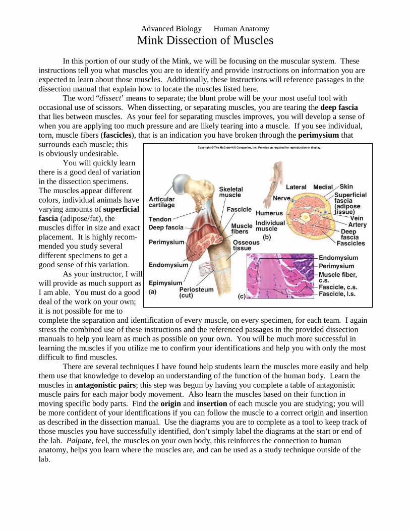
The word “dissect’ means to separate; the blunt probe will be your most useful tool with occasional use of scissors. When dissecting, or separating muscles, you are tearing the deep fascia that lies between muscles. As your feel for separating muscles improves, you will develop a sense of when you are applying too much pressure and are likely tearing into a muscle. If you see individual, torn, muscle fibers (fascicles), that is an indication you have broken through the perimysium that surrounds each muscle; this is obviously undesirable.
You will quickly learn there is a good deal of variation in the dissection specimens. The muscles appear different colors, individual animals have varying amounts of superficial fascia (adipose/fat), the muscles differ in size and exact placement. It is highly recom- mended you study several different specimens to get a good sense of this variation. As your instructor, I will will provide as much support as I am able. You must do a good deal of the work on your own; it is not possible for me to complete the separation and identification of every muscle, on every specimen, for each team. I again stress the combined use of these instructions and the referenced passages in the provided dissection manuals to help you learn as much as possible on your own. You will be much more successful in learning the muscles if you utilize me to confirm your identifications and help you with only the most difficult to find muscles. There are several techniques I have found help students learn the muscles more easily and help them use that knowledge to develop an understanding of the function of the human body. Learn the muscles in antagonistic pairs; this step was begun by having you complete a table of antagonistic muscle pairs for each major body movement. Also learn the muscles based on their function in moving specific body parts. Find the origin and insertion of each muscle you are studying; you will be more confident of your identifications if you can follow the muscle to a correct origin and insertion as described in the dissection manual. Use the diagrams you are to complete as a tool to keep track of those muscles you have successfully identified, don’t simply label the diagrams at the start or end of the lab. Palpate, feel, the muscles on your own body, this reinforces the connection to human anatomy, helps you learn where the muscles are, and can be used as a study technique outside of the lab.
Advanced Biology Human Anatomy
Mink Dissection of Muscles
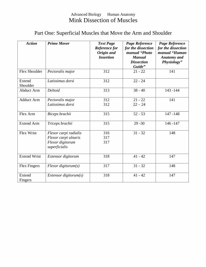
Part One: Superficial Muscles that Move the Arm and Shoulder
Action Prime Mover Text Page Reference for Origin and Insertion
Page Reference for the dissection manual “Photo
Manual Dissection
Guide”
Page Reference for the dissection manual “Human
Anatomy and Physiology”
Flex Shoulder Pectoralis major 312 21 - 22 141
Extend Shoulder
Latissimus dorsi 312 22 - 24
Abduct Arm Deltoid 313 38 - 40 143 -144
Adduct Arm Pectoralis major Latissimus dorsi
312 312
21 - 22 22 – 24
141
Flex Arm Biceps brachii 315 52 - 53 147 -148
Extend Arm Triceps brachii 315 29 -30 146 -147
Flex Wrist Flexor carpi radialis Flexor carpi ulnaris Flexor digitorum superficialis
316 317 317
31 - 32 148
Extend Wrist Extensor digitorum
318 41 - 42 147
Flex Fingers Flexor digitorum(s)
317 31 - 32 148
Extend Fingers
Extensor digitorum(s) 318 41 - 42 147
Advanced Biology Human Anatomy
Mink Dissection of Muscles
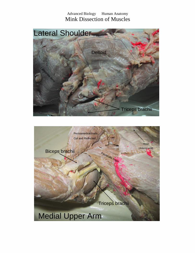
Pectoralis major
Pectoantebrachialis
Cut and Reflected
Medial Upper Arm
Biceps brachii
Triceps brachii
Head
(Anterior end)
Deltoid
Triceps brachii
Latissimus dorsi
Lateral Shoulder
Advanced Biology Human Anatomy
Mink Dissection of Muscles
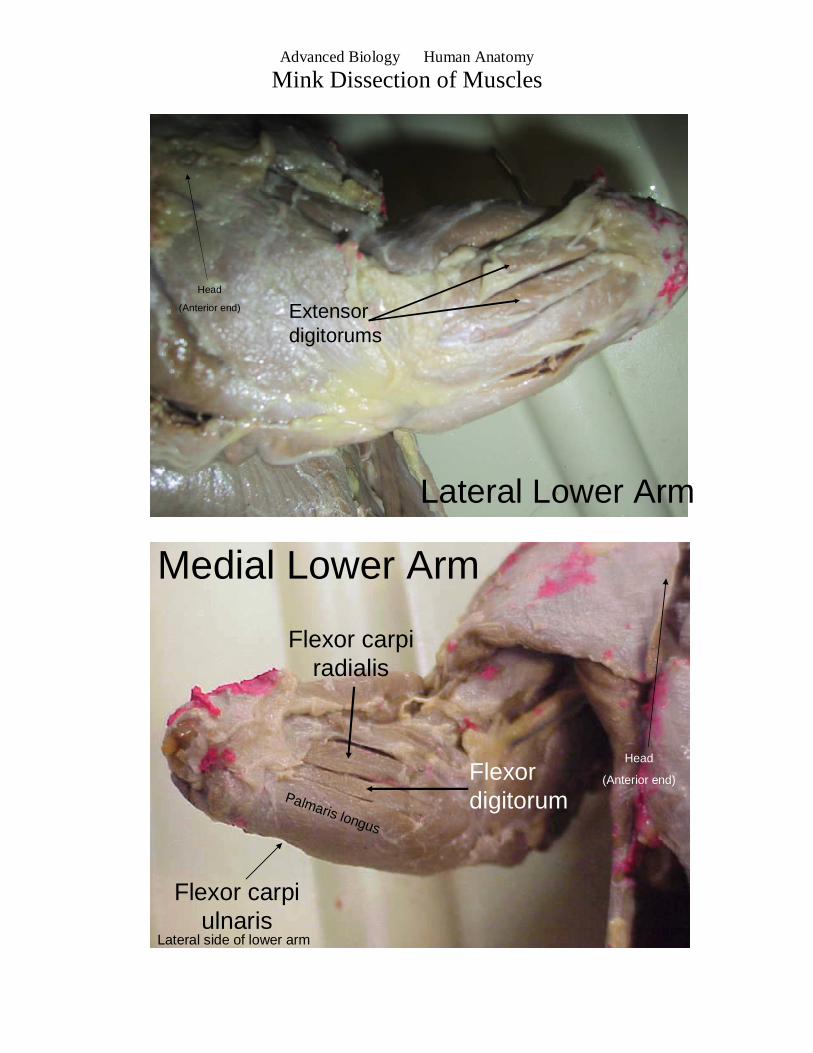
Medial Lower Arm
Palmaris longus
Flexor digitorum
Flexor carpiradialis
Flexor carpiulnaris
Lateral side of lower arm
Head
(Anterior end)
Lateral Lower Arm
Extensor digitorums
Head
(Anterior end)
Advanced Biology Human Anatomy
Mink Dissection of Muscles
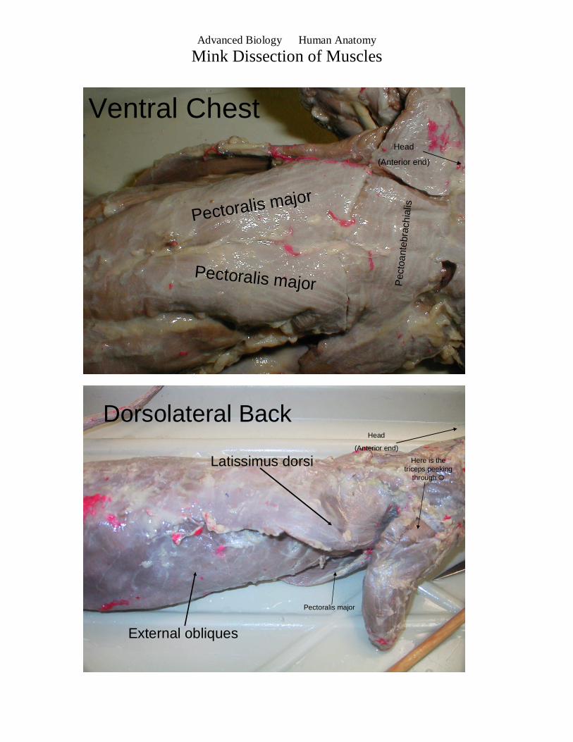
Dorsolateral Back
Latissimus dorsi
External obliques
Here is the triceps peeking
through ☺
Pectoralis major
Head
(Anterior end)
Pectoralis major
Pectoralis major Pec
toan
tebr
achi
ali s
Head
(Anterior end)
Ventral Chest
Advanced Biology Human Anatomy
Mink Dissection of Muscles
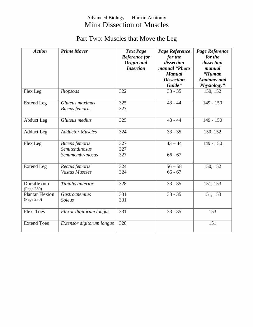
Part Two: Muscles that Move the Leg
Action Prime Mover Text Page Reference for Origin and Insertion
Page Reference for the
dissection manual “Photo
Manual Dissection
Guide”
Page Reference for the
dissection manual “Human
Anatomy and Physiology”
Flex Leg Iliopsoas
322 33 - 35 150, 152
Extend Leg Gluteus maximus Biceps femoris
325 327
43 - 44 149 - 150
Abduct Leg Gluteus medius
325 43 - 44 149 - 150
Adduct Leg Adductor Muscles
324 33 - 35 150, 152
Flex Leg Biceps femoris Semitendinosus Semimembranosus
327 327 327
43 – 44
66 - 67
149 - 150
Extend Leg Rectus femoris Vastus Muscles
324 324
56 – 58 66 - 67
150, 152
Dorsiflexion (Page 230)
Tibialis anterior 328 33 - 35 151, 153
Plantar Flexion (Page 230)
Gastrocnemius Soleus
331 331
33 - 35 151, 153
Flex Toes Flexor digitorum longus
331 33 - 35 153
Extend Toes Extensor digitorum longus
328 151
Advanced Biology Human Anatomy
Mink Dissection of Muscles
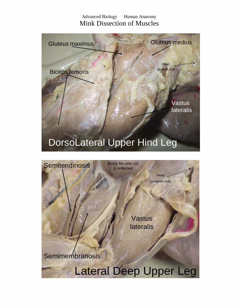
Lateral Deep Upper Leg
Bicep femoris cut & reflected
Vastuslateralis
Semitendinosis
Semimembranosis
Head
(Anterior end)
DorsoLateral Upper Hind Leg
Gluteus maximus Gluteus medius
Biceps femoris
Head
(Anterior end)
Vastuslateralis
Advanced Biology Human Anatomy
Mink Dissection of Muscles
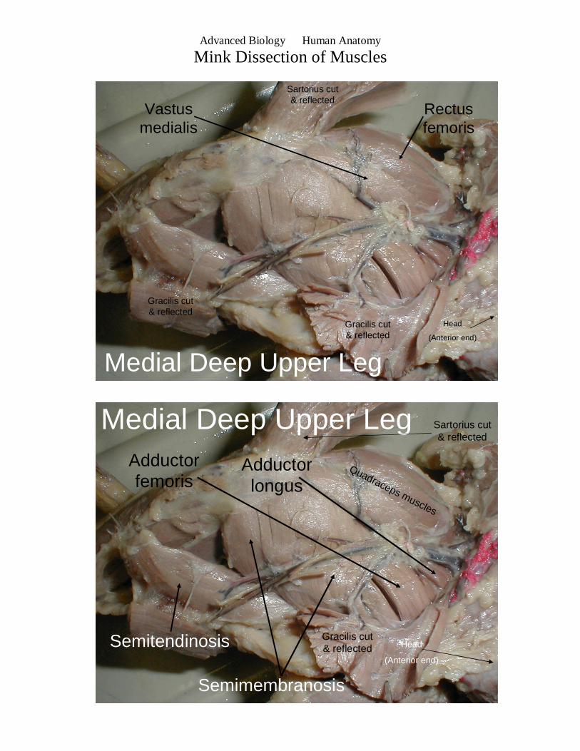
Medial Deep Upper Leg
Rectusfemoris
Vastusmedialis
Head
(Anterior end)
Sartorius cut & reflected
Gracilis cut & reflected
Gracilis cut & reflected
Head
(Anterior end)
Semitendinosis
Semimembranosis
Medial Deep Upper LegAdductor femoris
Adductor longus
Quadraceps muscles
Gracilis cut & reflected
Sartorius cut & reflected
Advanced Biology Human Anatomy
Mink Dissection of Muscles
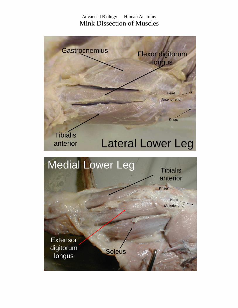
Lateral Lower Leg
Gastrocnemius Flexor digitorumlongus
Tibialisanterior
Head
(Anterior end)
Knee
Medial Lower LegTibialisanterior
Extensor digitorum
longusSoleus
Head
(Anterior end)
Knee
Advanced Biology Human Anatomy
Mink Dissection of Muscles
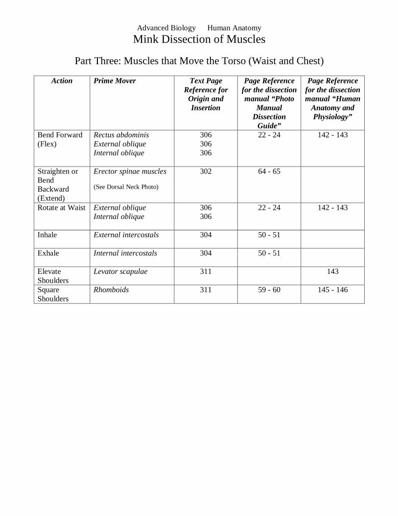
Part Three: Muscles that Move the Torso (Waist and Chest)
Action Prime Mover Text Page Reference for Origin and Insertion
Page Reference for the dissection manual “Photo
Manual Dissection
Guide”
Page Reference for the dissection manual “Human
Anatomy and Physiology”
Bend Forward (Flex)
Rectus abdominis External oblique Internal oblique
306 306 306
22 - 24 142 - 143
Straighten or Bend Backward (Extend)
Erector spinae muscles (See Dorsal Neck Photo)
302 64 - 65
Rotate at Waist External oblique Internal oblique
306 306
22 - 24 142 - 143
Inhale External intercostals
304 50 - 51
Exhale Internal intercostals
304 50 - 51
Elevate Shoulders
Levator scapulae 311 143
Square Shoulders
Rhomboids 311 59 - 60 145 - 146
Advanced Biology Human Anatomy
Mink Dissection of Muscles
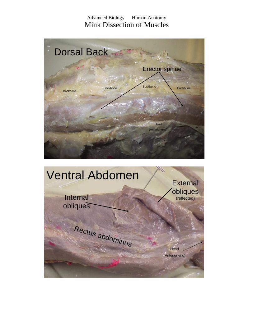
Dorsal Back
Erector spinae
BackboneBackbone Backbone Backbone
Head
(Anterior end)
Ventral Abdomen
Rectus abdominus
Internal obliques
External obliques
(reflected)
Head
(Anterior end)
Advanced Biology Human Anatomy
Mink Dissection of Muscles
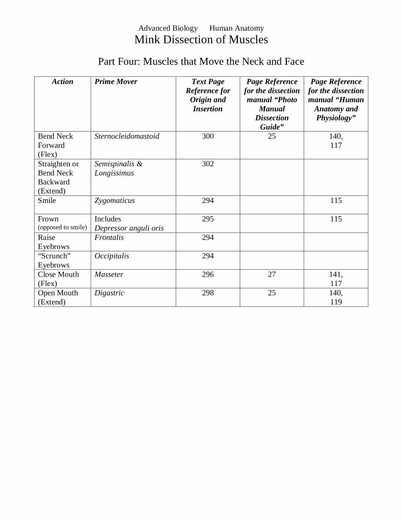
Part Four: Muscles that Move the Neck and Face
Action Prime Mover Text Page Reference for Origin and Insertion
Page Reference for the dissection manual “Photo
Manual Dissection
Guide”
Page Reference for the dissection manual “Human
Anatomy and Physiology”
Bend Neck Forward (Flex)
Sternocleidomastoid 300 25 140, 117
Straighten or Bend Neck Backward (Extend)
Semispinalis & Longissimus
302
Smile
Zygomaticus 294 115
Frown (opposed to smile)
Includes Depressor anguli oris
295 115
Raise Eyebrows
Frontalis 294
“Scrunch” Eyebrows
Occipitalis 294
Close Mouth (Flex)
Masseter 296 27 141, 117
Open Mouth (Extend)
Digastric 298 25 140, 119
Advanced Biology Human Anatomy
Mink Dissection of Muscles
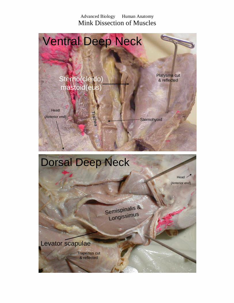
Ventral Deep Neck
Platysma cut & reflected
Trachea
Sterno(cleido)mastoid(eus)
Sternohyoid
Head
(Anterior end)
Dorsal Deep Neck
Trapezius cut & reflected
Levator scapulae
Head
(Anterior end)
Semispinalis &
Longissimus