Embed Size (px)
Citation preview
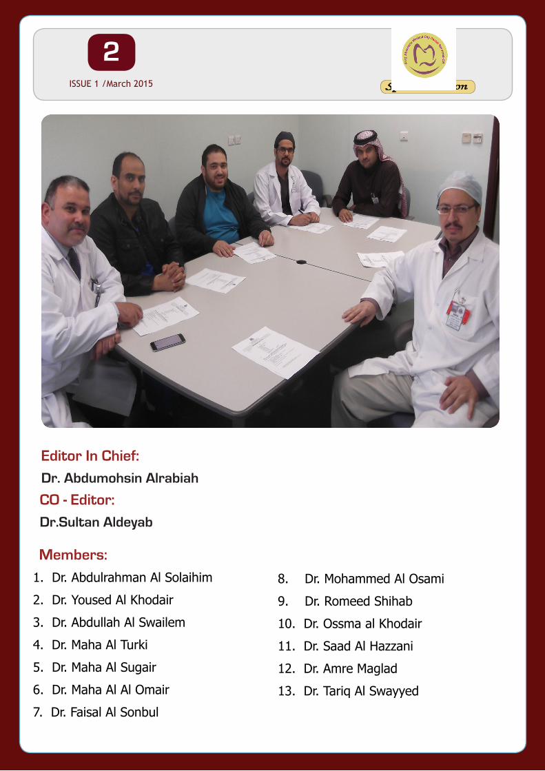
1Special Edition ISSUE 1 /March 2015
MINISTRY OF NATIONAL GUARD HEALTH AFFAIRS
Restorative Dental Newsletter
ISSUE 1 /March 2015
2Special Edition ISSUE 1 /March 2015
I N
T H I
S I S
S U E
Editor In Chief:
Dr. Abdumohsin Alrabiah
CO - Editor:
Dr.Sultan Aldeyab
Members:
1. Dr. Abdulrahman Al Solaihim
2. Dr. Yoused Al Khodair
3. Dr. Abdullah Al Swailem
4. Dr. Maha Al Turki
5. Dr. Maha Al Sugair
6. Dr. Maha Al Al Omair
7. Dr. Faisal Al Sonbul
8. Dr. Mohammed Al Osami
9. Dr. Romeed Shihab
10. Dr. Ossma al Khodair
11. Dr. Saad Al Hazzani
12. Dr. Amre Maglad
13. Dr. Tariq Al Swayyed
3Special Edition ISSUE 1 /March 2015
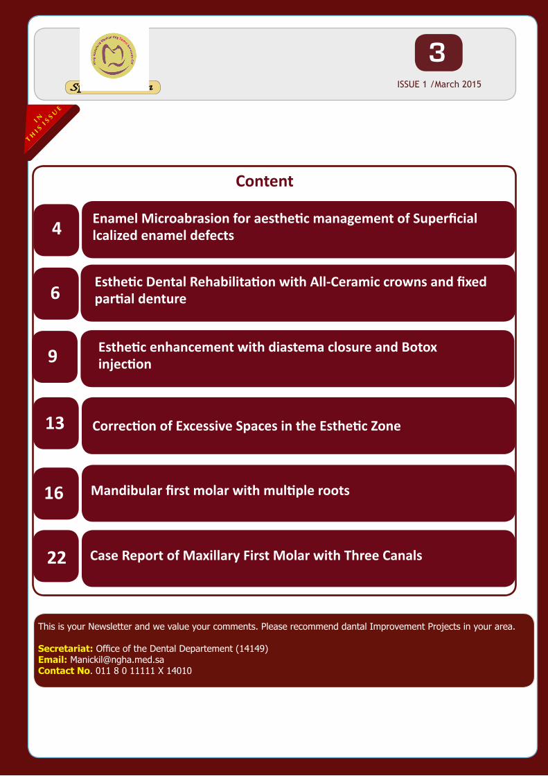
Content
4
Esthetic Dental Rehabilitation with All-Ceramic crowns and fixed partial denture
Enamel Microabrasion for aesthetic management of Superficial lcalized enamel defects
6
Esthetic enhancement with diastema closure and Botox injection
Mandibular first molar with multiple roots
9
16
Correction of Excessive Spaces in the Esthetic Zone13
Case Report of Maxillary First Molar with Three Canals22
This is your Newsletter and we value your comments. Please recommend dantal Improvement Projects in your area.
Secretariat: Office of the Dental Departement (14149) Email: [email protected] No. 011 8 0 11111 X 14010
I N
T H I
S I S
S U E
4Special Edition ISSUE 1 /March 2015
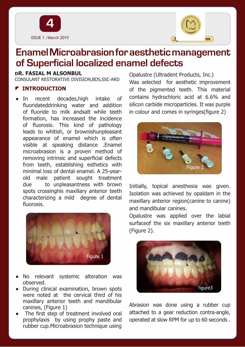
●● In recent decades,high intake of fluoridateddrinking water and addition of fluoride to milk andsalt while teeth formation, has increased the incidence of fluorosis. This kind of pathology leads to whitish, or brownishunpleasant appearance of enamel which is often visible at speaking distance .Enamel microabrasion is a proven method of removing intrinsic and superficial defects from teeth, establishing esthetics with minimal loss of dental enamel. A 25-year-old male patient sought treatment due to unpleasantness with brown spots crossinghis maxillary anterior teeth characterizing a mild degree of dental fluorosis.
●● No relevant systemic alteration was observed.
●● During clinical examination, brown spots were noted at the cervical third of his maxillary anterior teeth and mandibular canines, (Figure 1)
●● The first step of treatment involved oral prophylaxis by using prophy paste and rubber cup.Microabrasion technique using
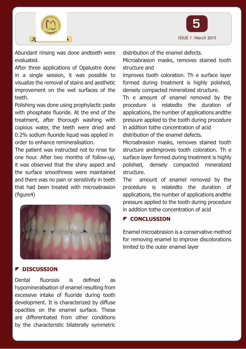
Opalustre (Ultradent Products, Inc.) Was selected for aesthetic improvement of the pigmented teeth. This material contains hydrochloric acid at 6.6% and silicon carbide microparticles. It was purple in colour and comes in syringes(figure 2)
Initially, topical anesthesia was given. Isolation was achieved by opaldam in the maxillary anterior region(canine to canine)and mandibular canines. Opalustre was applied over the labial surfaceof the six maxillary anterior teeth (Figure 2).
Abrasion was done using a rubber cup attached to a gear reduction contra-angle, operated at slow RPM for up to 60 seconds .
Enamel Microabrasion for aesthetic management of Superficial localized enamel defectsDR. FASIAL M ALSONBULCONSULANT RESTORATIVE DIVISION,BDS,SSC-ARD
zINTRODUCTION
Figure 2
figure3
Figure 1
5Special Edition ISSUE 1 /March 2015
Abundant rinsing was done andteeth were evaluated.After three applications of Opalustre done in a single session, it was possible to visualize the removal of stains and aesthetic improvement on the wet surfaces of the teeth. Polishing was done using prophylactic paste with phosphate fluoride. At the end of the treatment, after thorough washing with copious water, the teeth were dried and 0.2% sodium fluoride liquid was applied inorder to enhance remineralisation.The patient was instructed not to rinse for one hour. After two months of follow-up, it was observed that the shiny aspect and the surface smoothness were maintained and there was no pain or sensitivity in teeth that had been treated with microabrasion (figure4)
Dental fluorosis is defined as hypomineralisation of enamel resulting from excessive intake of fluoride during tooth development. It is characterized by diffuse opacities on the enamel surface. These are differentiated from other conditions by the characteristic bilaterally symmetric
distribution of the enamel defects.Microabrasion masks, removes stained tooth structure andimproves tooth coloration. Th e surface layer formed during treatment is highly polished, densely compacted mineralized structure.Th e amount of enamel removed by the procedure is relatedto the duration of applications, the number of applications andthe pressure applied to the tooth during procedure in addition tothe concentration of aciddistribution of the enamel defects.Microabrasion masks, removes stained tooth structure andimproves tooth coloration. Th e surface layer formed during treatment is highly polished, densely compacted mineralized structure.The amount of enamel removed by the procedure is relatedto the duration of applications, the number of applications andthe pressure applied to the tooth during procedure in addition tothe concentration of acid
Enamel microabrasion is a conservative method for removing enamel to improve discolorations limited to the outer enamel layer
zDISCUSSION
zCONCLUSSION
6Special Edition ISSUE 1 /March 2015
Dental Treatment today shifted toward dental esthetics & more cosmetic procedures. All ceramic inlay , onlays , veneers and crowns can provide some of the most esthetically pleasing restorations currently available. They can be made to match natural tooth structure accurately in terms of color, surface texture, and translucency .
A 24-year-old female medically fit was referred because of appearance of her maxillary anterior teeth. The patient was not satisfied with her smile . (fig.1) She has poor oral hygiene and has undergone multiple treatments . she has multiple unpleasant composite restoration in anterior teeth with recurrent caries, bulky connected crowns in 14, 15 teeth with unsatisfied root canal treatment , necrotic pulp of tooth 11& 25, unpleasing cantilever FPD on 23 to replace22 . 15 & 14 has broken endo-files inside the canals (fig.2)
●● During the treatment planning session, the patient was given the option of porcelain-fused-to-metal or metal-free restorations. The patient chose to have all of the teeth restored with ceramic. Occlusion was analyzed preoperatively , both clinically and with the aid of mounted study models on a semi adjustable articulator. A diagnostic wax-up was completed and modified at chair side with the patient’s input, until the final form of the new restorations was deemed esthetically satisfactory.
●● The patient was given oral hygiene instructions, caries control was done , The FPD was removed followed by temporization
Root canal treatment was Figure 2: pre-operative OPG show over all teeth situation before treatment
Figure 1: Pre-operati ve view showing a, the smile of the patient b, frontal view show multiple caries teeth ,multiple restoration with recurrent caries , PFM crowns . Patient was unhappy with esthetic
Esthetic Dental Rehabilitation with All-Ceramic crowns and fixed partial denture: A Case ReportAbdulmohsen Al Rabiah, BDS,SSC-ARD Sahar Al Mansouri,BDS
zCASE PRESENTATION
7Special Edition ISSUE 1 /March 2015
done to 15,14,13,21,23,24,25 (fig.3) then cast post was done to tooth 15 &
●● prefabricated fiber post for 14,13,21, 24,25 teeth. At the stage of tooth preparation the abutment teeth were prepared using modified shoulder diamond burs (coarse and superfine) before an impression was taken for final restorations .
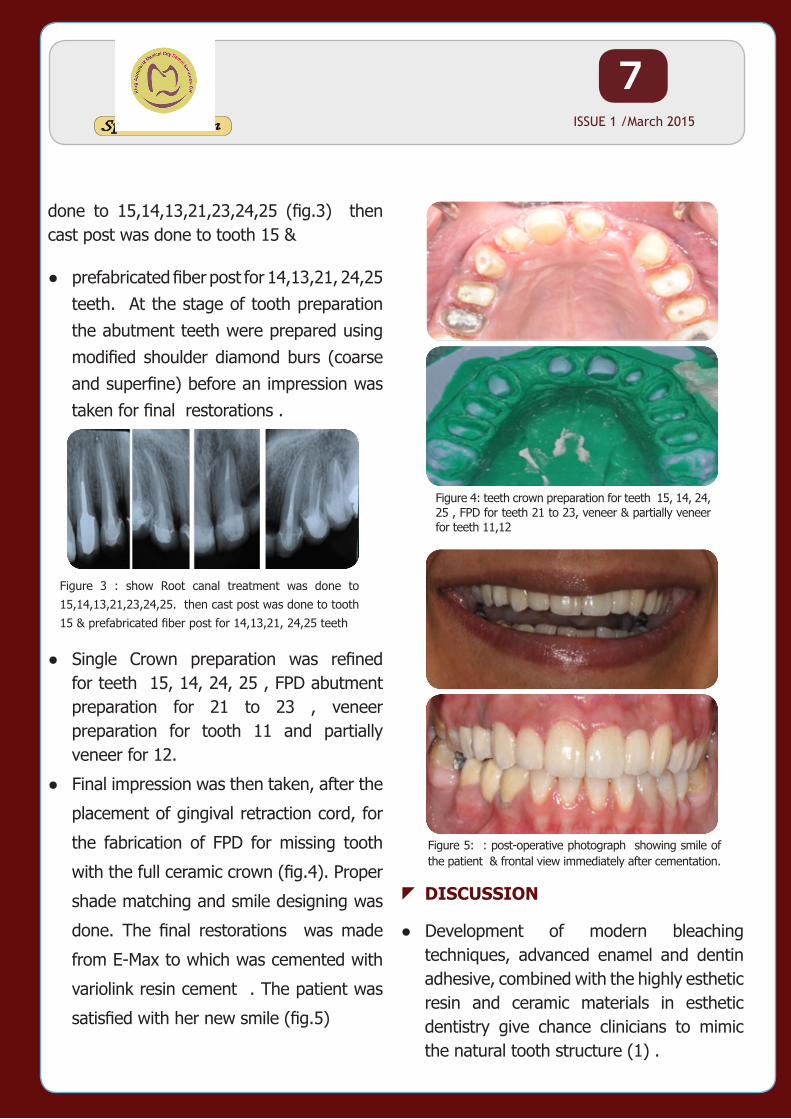
●● Single Crown preparation was refined for teeth 15, 14, 24, 25 , FPD abutment preparation for 21 to 23 , veneer preparation for tooth 11 and partially veneer for 12.
●● Final impression was then taken, after the
placement of gingival retraction cord, for
the fabrication of FPD for missing tooth
with the full ceramic crown (fig.4). Proper
shade matching and smile designing was
done. The final restorations was made
from E-Max to which was cemented with
variolink resin cement . The patient was
satisfied with her new smile (fig.5)
●● Development of modern bleaching techniques, advanced enamel and dentin adhesive, combined with the highly esthetic resin and ceramic materials in esthetic dentistry give chance clinicians to mimic the natural tooth structure (1) .
Figure 3 : show Root canal treatment was done to 15,14,13,21,23,24,25. then cast post was done to tooth 15 & prefabricated fiber post for 14,13,21, 24,25 teeth
Figure 5: : post-operative photograph showing smile of the patient & frontal view immediately after cementation.
Figure 4: teeth crown preparation for teeth 15, 14, 24, 25 , FPD for teeth 21 to 23, veneer & partially veneer for teeth 11,12
zDISCUSSION
8Special Edition ISSUE 1 /March 2015
●● Greater success for anterior teeth has been the trend for IPS Empress crowns. Fradeani and Redemagni reported an overall survival rate of 95.2 percent at 11 years for 125 IPS Empress crowns, which represents 98.9 percent survival in the anterior segment and 84.4 percent survival in the posterior segment (2) and the authors reported a veneer success rate of 98.8 percent after six years (3).
●● Two manufacturers have recommended their all-ceramic systems for anterior three-unit pros-theses: a glass-infiltrated alumina (In-Ceram Alumina) and a lithium disilicate–based glass-ceramic (IPS Empress 2 [now IPS e.max Press]) (4).
●● In a three-year study of 61 three-unit FPDs (In-Ceram Alumina), Sorensen and colleagues reported survival rates of 100 percent for anterior teeth and 83 percent for posterior teeth (5).
●● current evidence suggesting that all-ceramic restorations have an acceptable clinical longevity that accompanies their long-lasting esthetic advantages (6).
Currently The material’s strength and optical properties of All- ceramic restorations offer dental professionals multiple options for achieving highly durable and esthetically pleasing restorations.
●● Türkaslan S, Utku Ulusoy V: Esthetic rehabilitation of crowded maxillary anterior teeth utilizing ceramic veneers. Cases
Journal 2009, 2:8329●● Fradeani M, Redemagni M. An 11-year
clinical evaluation of leucite-reinforced glass-ceramic crowns: a retrospective study. Quintessence Int 2002;33(7): 503–510
●● Fradeani M. Six-year follow-up with Empress veneers. Int J Periodontics Restorative Dent 1998;18(3):216–225.
●● Kelly JR. Dental ceramics: current thinking and trends. Dent Clin North Am 2004;48(2):viii, 513–530.
●● Sorensen JA, Kang SK, Torres TJ, Knode H. In-Ceram fixed partial dentures: three-year clinical trial results. J Calif Dent Assoc 1998;26(3):207–214.
●● 6. Della Bona A, and Kelly JR : The Clinical Success Of All-Ceramic Restorations . J Am Dent Assoc. 2008 sep; 139.
zCONCLUSSION
zREFERENCE
9Special Edition ISSUE 1 /March 2015
Unattractive smile due to short upper lip, excessive gingival exposure , and Diastema can be self-conscious or even psychologically affected and hence could be the main reason to seek orthodontic intervention.1,3 Where orthodontic treatments can not be applied, it is inevitable to carry out the restorative treatments to accomplish the function and the aesthetics.4 , 5This clinical report presents the rehabilitation of a midline maxillary diastemas and gummy smile using IPS e-max ceramic crowns and Botox Injections. All ceramic crowns and Botox injection were successfully applied to correct esthetic problems and achieve improved esthetic and functional outcomes.
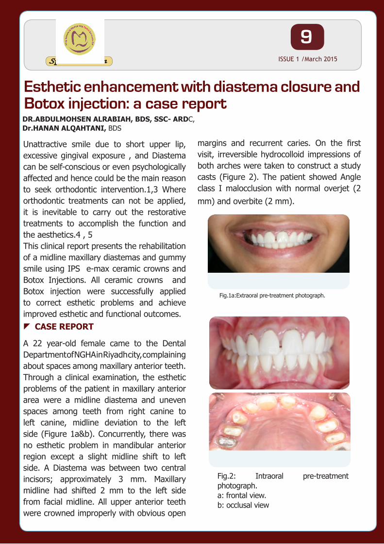
A 22 year-old female came to the Dental Department of NGHA in Riyadh city, complaining about spaces among maxillary anterior teeth. Through a clinical examination, the esthetic problems of the patient in maxillary anterior area were a midline diastema and uneven spaces among teeth from right canine to left canine, midline deviation to the left side (Figure 1a&b). Concurrently, there was no esthetic problem in mandibular anterior region except a slight midline shift to left side. A Diastema was between two central incisors; approximately 3 mm. Maxillary midline had shifted 2 mm to the left side from facial midline. All upper anterior teeth were crowned improperly with obvious open
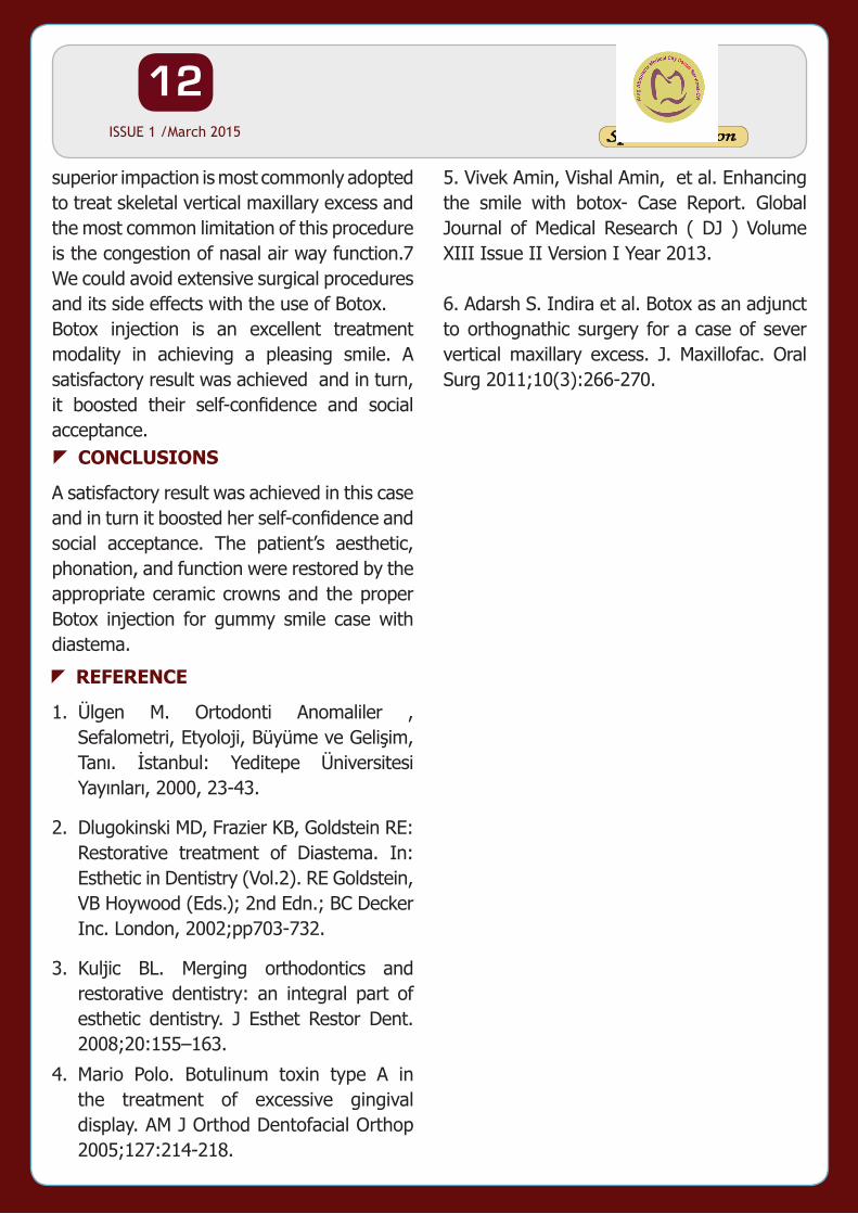
margins and recurrent caries. On the first visit, irreversible hydrocolloid impressions of both arches were taken to construct a study casts (Figure 2). The patient showed Angle class I malocclusion with normal overjet (2 mm) and overbite (2 mm).
Esthetic enhancement with diastema closure and Botox injection: a case reportDR.ABDULMOHSEN ALRABIAH, BDS, SSC- ARDC, Dr.HANAN ALQAHTANI, BDS
zCASE REPORT
Fig.1a:Extraoral pre-treatment photograph.
Fig.2: Intraoral pre-treatment photograph. a: frontal view. b: occlusal view
10Special Edition ISSUE 1 /March 2015
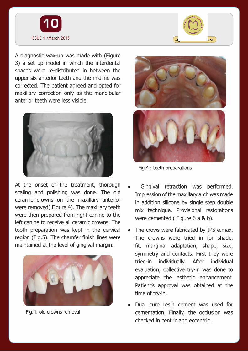
A diagnostic wax-up was made with (Figure 3) a set up model in which the interdental spaces were re-distributed in between the upper six anterior teeth and the midline was corrected. The patient agreed and opted for maxillary correction only as the mandibular anterior teeth were less visible.
At the onset of the treatment, thorough scaling and polishing was done. The old ceramic crowns on the maxillary anterior were removed( Figure 4). The maxillary teeth were then prepared from right canine to the left canine to receive all ceramic crowns. The tooth preparation was kept in the cervical region (Fig.5). The chamfer finish lines were maintained at the level of gingival margin.
●● Gingival retraction was performed. Impression of the maxillary arch was made in addition silicone by single step double mix technique. Provisional restorations were cemented ( Figure 6 a & b).
●● The crows were fabricated by IPS e.max. The crowns were tried in for shade, fit, marginal adaptation, shape, size, symmetry and contacts. First they were tried-in individually. After individual evaluation, collective try-in was done to appreciate the esthetic enhancement. Patient’s approval was obtained at the time of try-in.
●● Dual cure resin cement was used for cementation. Finally, the occlusion was checked in centric and eccentric.
Fig.3: Diagnostic wax up
Fig.4: old crowns removal
Fig.4 : teeth preparations
11Special Edition ISSUE 1 /March 2015
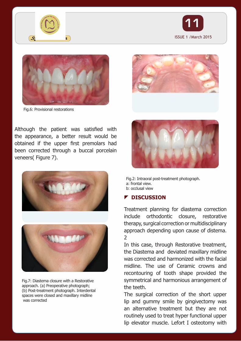
Although the patient was satisfied with the appearance, a better result would be obtained if the upper first premolars had been corrected through a buccal porcelain veneers( Figure 7).
Fig.6: Provisional restorations
Fig.7: Diastema closure with a Restorative approach. (a) Preoperative photograph; (b) Post-treatment photograph. Interdental spaces were closed and maxillary midline was corrected
Treatment planning for diastema correction include orthodontic closure, restorative therapy, surgical correction or multidisciplinary approach depending upon cause of distema. 2In this case, through Restorative treatment, the Diastema and deviated maxillary midline was corrected and harmonized with the facial midline. The use of Ceramic crowns and recontouring of tooth shape provided the symmetrical and harmonious arrangement of the teeth.The surgical correction of the short upper lip and gummy smile by gingivectomy was an alternative treatment but they are not routinely used to treat hyper functional upper lip elevator muscle. Lefort I osteotomy with
Fig.2: Intraoral post-treatment photograph. a: frontal view. b: occlusal view
zDISCUSSION
12Special Edition ISSUE 1 /March 2015
5. Vivek Amin, Vishal Amin, et al. Enhancing the smile with botox- Case Report. Global Journal of Medical Research ( DJ ) Volume XIII Issue II Version I Year 2013.
6. Adarsh S. Indira et al. Botox as an adjunct to orthognathic surgery for a case of sever vertical maxillary excess. J. Maxillofac. Oral Surg 2011;10(3):266-270.
superior impaction is most commonly adopted to treat skeletal vertical maxillary excess and the most common limitation of this procedure is the congestion of nasal air way function.7 We could avoid extensive surgical procedures and its side effects with the use of Botox.Botox injection is an excellent treatment modality in achieving a pleasing smile. A satisfactory result was achieved and in turn, it boosted their self-confidence and social acceptance.
A satisfactory result was achieved in this case and in turn it boosted her self-confidence and social acceptance. The patient’s aesthetic, phonation, and function were restored by the appropriate ceramic crowns and the proper Botox injection for gummy smile case with diastema.
1. Ülgen M. Ortodonti Anomaliler , Sefalometri, Etyoloji, Büyüme ve Gelisim, Tanı. Istanbul: Yeditepe Üniversitesi Yayınları, 2000, 23-43.
2. Dlugokinski MD, Frazier KB, Goldstein RE: Restorative treatment of Diastema. In: Esthetic in Dentistry (Vol.2). RE Goldstein, VB Hoywood (Eds.); 2nd Edn.; BC Decker Inc. London, 2002;pp703-732.
3. Kuljic BL. Merging orthodontics and restorative dentistry: an integral part of esthetic dentistry. J Esthet Restor Dent. 2008;20:155–163.
4. Mario Polo. Botulinum toxin type A in the treatment of excessive gingival display. AM J Orthod Dentofacial Orthop 2005;127:214-218.
zCONCLUSIONS
zREFERENCE
13Special Edition ISSUE 1 /March 2015
The use of porcelain crowns and veneers to solve esthetic problems has been shown to be a valid management option especially in the anterior esthetic zone. This case report discusses a patient having diastema in the anterior region. The patient was treated with orthodontic treatment and porcelain crowns & veneers in the maxillary arch for the closure of diastema.
A 35 year old male patient with a chief complaint of discolored anterior teeth and gaps between the teeth. The patient was unhappy with the appearance of his teeth. After thorough examination, impressions for diagnostic models were made in irreversible hydrocolloid. The models were studied to decide the shape and size of the restorations with help of a diagnostic wax up. Before proceeding for
Correction of Excessive Spaces in the Esthetic ZoneSultan Aldeyab BDS,AEGD,SSC-ARDMinistry of national gauard,king abdulaziz medical city
zINTRODUCTION tooth preparation, shade was selected using Classical shade guide (Vita Zahnfabrik, Germany). The maxillary teeth were then prepared from right 2nd premolar to the left 2nd premolar to receive porcelain crowns and laminate veneers. Impression of the maxillary arch was made in addition silicone.The laminates were etched with 4 % Hydrofluoric acid, After etching, they were washed thoroughly using liberal amount of water. On drying, a coat of Silane coupling agent ( Porcelain Primer, Bisco,USA ). performed on two teeth at a time starting at the midline.The prepared teeth were etched using 37% Phosphoric Acid for 15 seconds. On air drying bonding agent was applied & light cured for 10 seconds. Dual cure composite luting agent was used for cementation. The laminates were spot cured for 5 seconds initially. Excess cement was removed with
explorer and then complete curing was done for 20 seconds. On completion of the cementation procedure, the occlusion was checked in centric and eccentric positions for interferences. The high points were removed and polished .
The etiology of diastema may be attributed to the following factors: (a) Hereditary- congenitally missing teeth, tooth and jaw size discrepancy, supernumerary teeth & frenum attachments; (b) Developmental problems- habits, periodontal disease,tooth loss, posterior bite collapse (Oesterle & Shellhart, 1999). Treatment planning for diastema correction includes orthodontic closure, restorative therapy, surgical correction or multidisciplinary approach depending upon the cause of diastema (Dlugokinski et al, 2002).
zCLINICAL REPORT
zDISCUSSION
14Special Edition ISSUE 1 /March 2015
Orthodontic treatment, bonded porcelain crowns & veneers can provide successful esthetic and functional long-term service for patients.
zCONCLUSION
Orthodontic treatment
Office bleaching
Mounted diagnostic cast
Smile of the patient before and after
15Special Edition ISSUE 1 /March 2015
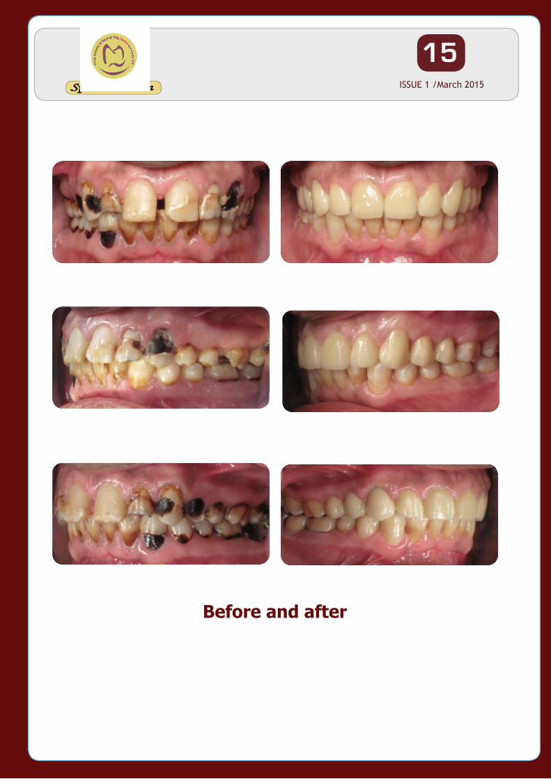
Before and after
16Special Edition ISSUE 1 /March 2015
A mandibular first molar with two distal roots is an interesting example of anatomic variation. This paper describes case report of mandibular first molar with four roots (two mesial and two distal).The canals were shaped with K3 instrument (Sybron Endo, West Collins, CA, USA) and irrigated with 2.5% sodium hypochlorite. The canals were then obturated with gutta- percha and AH 26 sealer. This case report shows an anatomic variation of external morphology of the tooth.
●● Mandibular first molar, two mesial and two distal roots.
●● The purpose of this article is to report the successful treatment of a mandibular first molar with four-rooted, two mesial and two distal roots.
Endodontic therapy involves treating vital and necrotic dental pulps so that patients can retain their natural teeth in function and esthetics. The main reasons for endodontic failure are apical percolation, incomplete canal obturation, and the presence of untreated canals (Ingle et al. 1976). Failure to recognise any unusual canal configuration would eventually lead to unsuccessful treatment outcome. Although successful therapy depends on many factors, one of the
most important is missing canal which can lead to an infection years after treatment and cause the tooth to require further treatment.
Thus, a thorough knowledge of the root and root canal morphology, along with the various anatomical variations, is essential in order to reach this goal (Vertucci 2005). All dental clinicians could benefit from regular continuing education that focuses on anatomy. A thorough knowledge of root and root canal morphology and a good anticipation of their possible morphological variations will help reduce endodontic failure caused by incomplete debridement and obturation. The mandibular first molar can display several anatomical variations. The common morphology that first mandibular molars exhibit is two-rooted with two mesial and one distal canal (Barker et al. 1974, Vertucci 1984). Recent studies (Table 1) reported a higher incidence of second canals in distal roots of mandibular permanent first molar than earlier studies (Al Nazhan 1999, Gulabival 2001 &2002, Sert et al.2004). The identification and external morphology of these root complexes, containing a lingual or buccal supernumerary root, are described by Carlsen and Alexandersen (1990 & 1991).
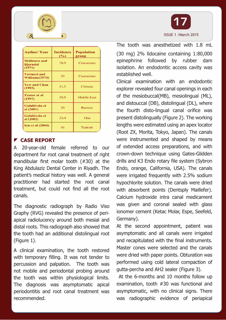
Table 1: Incidence of two canals in distal root of mandibular first molar(Parolia et al.2009)
Mandibular First Molar With Multiple RootsAziza Al TurkiBDS,MSc.
zKeywords:
zABSTRACT
zINTRODUCTION
17Special Edition ISSUE 1 /March 2015
A 20-year-old female referred to our department for root canal treatment of right mandibular first molar tooth (#30) at the King Abdulaziz Dental Center in Riyadh. The patient’s medical history was well. A general practitioner had started the root canal treatment, but could not find all the root canals.
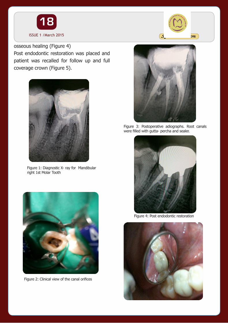
The diagnostic radiograph by Radio Viso Graphy (RVG) revealed the presence of peri-apical radiolucency around both mesial and distal roots. This radiograph also showed that the tooth had an additional distolingual root (Figure 1).
A clinical examination, the tooth restored with temporary filling. It was not tender to percussion and palpation. The tooth was not mobile and periodontal probing around the tooth was within physiological limits. The diagnosis was asymptomatic apical periodontitis and root canal treatment was recommended.
The tooth was anesthetized with 1.8 mL
(30 mg) 2% lidocaine containing 1:80,000 epinephrine followed by rubber dam isolation. An endodontic access cavity was established well.Clinical examination with an endodontic explorer revealed four canal openings in each of the mesiobuccal(MB), mesiolingual (ML), and distouccal (DB), distolingual (DL), where the fourth disto-lingual canal orifice was present distolingually (Figure 2). The working lengths were estimated using an apex locator (Root ZX, Morita, Tokyo, Japan). The canals were instrumented and shaped by means of extended access preparations, and with crown-down technique using Gates-Glidden drills and K3 Endo rotary file system (Sybron Endo, orange, California, USA). The canals were irrigated frequently with 2.5% sodium hypochlorite solution. The canals were dried with absorbent points (Dentsply Maillefer). Calcium hydroxide intra canal medicament was given and coronal sealed with glass ionomer cement (Ketac Molar, Espe, Seefeld, Germany). At the second appointment, patient was asymptomatic and all canals were irrigated and recapitulated with the final instruments. Master cones were selected and the canals were dried with paper points. Obturation was performed using cold lateral compaction of gutta-percha and AH2 sealer (Figure 3). At the 6-months and 10 months follow up examination, tooth #30 was functional and asymptomatic, with no clinical signs. There was radiographic evidence of periapical
zCASE REPORT
18Special Edition ISSUE 1 /March 2015
osseous healing (Figure 4)Post endodontic restoration was placed and patient was recalled for follow up and full coverage crown (Figure 5).
Figure 1: Diagnostic X- ray for Mandibular right 1st Molar Tooth
Figure 2: Clinical view of the canal orifices
Figure 3: Postoperative adiographs. Root canals were filled with gutta- percha and sealer.
Figure 4: Post endodontic restoration
19Special Edition ISSUE 1 /March 2015
Based on the literature and this clinical case, it is evident that knowledge of the anatomical variations of the mandibular molars is extremely important for the success of endodontic treatment. Most dentists are used to treating normal roots with similar traits; as a result, many failures can occur. However it must be noticed that abnormalities are rare, but it is possible that a patient referred may have one of these rare anatomic variations.An awareness and understanding of the presence of unusual root canal morphology can thus contribute to the successful outcome of root canal treatment (Filip et al. 2007). This report describes success
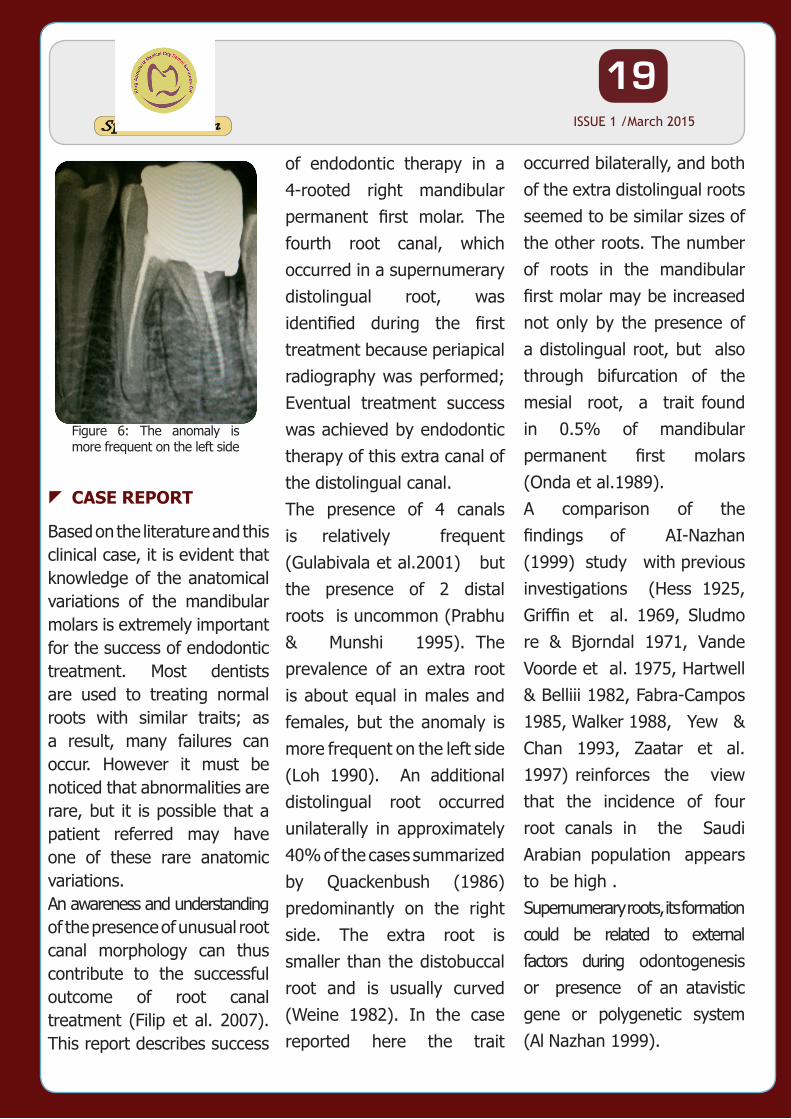
Figure 6: The anomaly is more frequent on the left side
zCASE REPORT
of endodontic therapy in a 4-rooted right mandibular permanent first molar. The fourth root canal, which occurred in a supernumerary distolingual root, was identified during the first treatment because periapical radiography was performed; Eventual treatment success was achieved by endodontic therapy of this extra canal of the distolingual canal.The presence of 4 canals is relatively frequent (Gulabivala et al.2001) but the presence of 2 distal roots is uncommon (Prabhu & Munshi 1995). The prevalence of an extra root is about equal in males and females, but the anomaly is more frequent on the left side (Loh 1990). An additional distolingual root occurred unilaterally in approximately 40% of the cases summarized by Quackenbush (1986) predominantly on the right side. The extra root is smaller than the distobuccal root and is usually curved (Weine 1982). In the case reported here the trait
occurred bilaterally, and both of the extra distolingual roots seemed to be similar sizes of the other roots. The number of roots in the mandibular first molar may be increased not only by the presence of a distolingual root, but also through bifurcation of the mesial root, a trait found in 0.5% of mandibular permanent first molars (Onda et al.1989).A comparison of the findings of AI-Nazhan (1999) study with previous investigations (Hess 1925, Griffin et al. 1969, Sludmo re & Bjorndal 1971, Vande Voorde et al. 1975, Hartwell & Belliii 1982, Fabra-Campos 1985, Walker 1988, Yew & Chan 1993, Zaatar et al. 1997) reinforces the view that the incidence of four root canals in the Saudi Arabian population appears to be high .Supernumerary roots, its formation could be related to external factors during odontogenesis or presence of an atavistic gene or polygenetic system (Al Nazhan 1999).
20Special Edition ISSUE 1 /March 2015
zCONCLUSION
zREFERENCES
A clinician should have complete knowledge of anatomic variation of macrostructure and internal and external root canal anatomy. The possibility of an extra root should also be considered and looked for carefully. An accurate diagnosis of these supernumerary roots can avoid complications that arise during canal negotiation and enlargement. This case has been reported to share our experience and increase the awareness of clinicians on tooth morphology of mandibular first molar teeth for a more predictable treatment outcome.
1. Al Nazhan S. Incidence of fourth canal in root canal treated mandibular fi rst molars in a Saudi Arabian sub-population. Intl Endod J. 1999; 32: 49–52.
2. Barker BC, parson KC, Mills PR,Williams GL. Anatomy of root canals, III. Permanent mandibular molars. Aust Dent J 1974;19:403–13.
3. Carlsen O, Alexandesen V.Radix entomolaris: identification and morphology Scan JDentRes 1990;98:363–73.
4. Carlsen O, Alexandersen V. Radix paramolaris in permanent mandibular molars: identification and morphology. Scan J Dent Res 1991;99:6189 –95.
5. Fabra-Campos H ( 1 9 8 5) Unusual root anatomy of mandibular first molars. Journal of Bndodontics
12 , 568-72.
6. Filip L. Calberson, Roeland J. De Moor, Christophe A. Deroose. The Radix Entomolaris and Paramolaris: Clinical approach in endodontics. Journal of Endod 2007; 33:58-63.
7. Grfin J , Skidmore A, Alberico C (1969) The determination of the frequency of occurrence of four canals in maxillary and mandubular first molars. GRS, West Virginia University, School ofDentistry, Morgantown, West Viiginia. Unpublished study.
8. Gulabivala K, Aung TH, Alavi A, Ng YL. Root and canal morphology of Burmese mandibular molars. Int
Endod J. 2001; 34:359–70.9. Gulabivala K, Opasanon A,
Ng YL, Alavi A. Root and canal morphology of Thai mandibular molars. Int Endod J. 2002; 35:56–62.
10. Hartwell G , Belliii R(1982) Clinical investigation of in vivo endodontically treated mandibular and maxillary molars. Journal oJBndoduntics 8 , 555-7.
11. Hess W (1925 ) Anatomy of the root canals of the teeth of the permanent dentition, Part I. New York: William Wood & CO, 1-35.
12. Ingle JI, Beveridge EE, Glick DH, Weichman JA. Modern endodontic therapy. In: Ingle JI, Beveridge EE, editors. Endodontics. 2nd ed. Philadelphia: Lea & Febiger; 1976. p. 1–57.
13. Onda S, Minemura R, Masaki T, Funatsu S. Shape and number of the roots of the permanent molar teeth. Bull Tokyo Dent Coll 1989; 30(4):221-31.
14. Loh HS. Incidence and features of three-rooted permanent mandibular molars. Aust Dent J 1990; 35(5):434-7.
21Special Edition ISSUE 1 /March 2015
15. Parolia A, Kundabala M2, Thomas MS, Mohan M, Joshi N. Three rooted, four canalled mandibular first molar (Radix
16. Entomolaris). Kathmandu University Medical Journal (2009), Vol. 7, No. 3, Issue 27, 289-292.
17. Prabhu NT, Munshi AK. Additional distal root in permanent mandibular first molars: report of a case. Quintessence Int 1995; 26(8):567-9.
18. Quackenbush LE. Mandibular molar with three distal root canals. Dent traumatol. 1986; 2:48–9
19. Sert S, Aslanalp V, Tanalp J. Investigation of the root canal confi gurations of mandibular permanent teeth in the Turkish population. Int Endod J. 2004; 37: 494–
20. Slcidmore A, Bjorndal A (1971) Root canal morphology of the human mandibular fist molar. Oral Surgery, Oral Medicine. Oral Pathology 32, 778-84. 10.
21. Vande Voorde H, Odendahl D, Davis J(1975 ) Molar 4th canals: frequent cause of endodonlic failure? Illinois Dental Journal 44, 779-86.
22. Vertucci FJ, Root canal anatomy of thehuman permanent teeth.
22Special Edition ISSUE 1 /March 2015
Case Report of Maxillary First Molar with Three CanalsDr. Tariq Suliman AlswayyedAdvance Restorative DentistDental Center
The major goals of root canal treatment are to 1) remove irritants from the root canal system; 2) fill or obturate the cleaned and shaped system; and3) prevent future recontamination of sealed root canals.
Therefore The knowledge of the root canal morphology and the possible anatomical variations are important for the successful endodontic treatment.
Maxillary first premolar has a wide variation in root anatomy and root canal morphology.
Many researcher couldn’t report three canal in maxillary first premolars. While the incidence of having three canals were ranging between 0.5% and 5% reported by other researchers.(1-3)In Saudi population Atieh (10) report a 1.2% incidence of
maxillary first premolars with three canal while Al-Nazhan et al in 2012(11) shows and incidence of 2.3%.(4-9)This article is reporting a successful endodontic management of a three rooted maxillary first premolar with 18 months recall.
A 42 years old Saudi Male patient was referred from screening clinic at Riyadh Dental Center- king Saud Medical Complex for comprehensive management of sever tooth loss.Patient chief complaint was esthetic and pain with cold and hot drinks related to all his teeth specially lower posterior teeth.
Patient complained from Gastroesophageal reflux disease (GRED) for 5 years and his condition was treated successfully by medication since 4 years.
After diagnostic tests were done including
( Orthopantogram , Lateral
Cephalometric , complete mouth survey radiographs ,cold test, percussion and palpation tests , mounting of diagnostic casts, saliva test ,diet analysis).
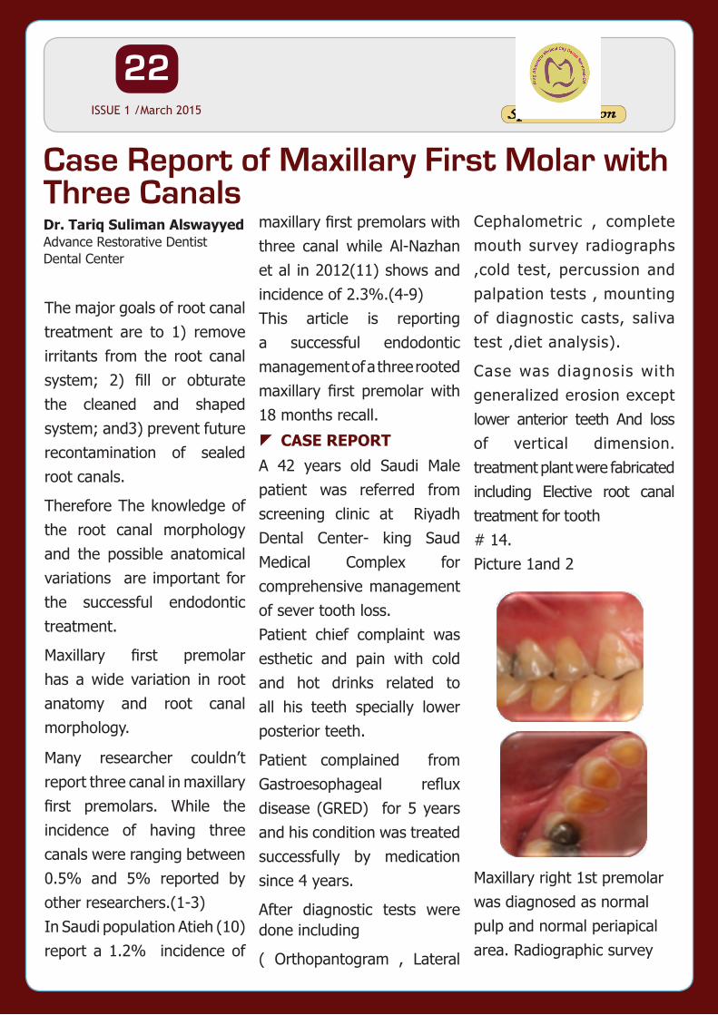
Case was diagnosis with generalized erosion except lower anterior teeth And loss of vertical dimension. treatment plant were fabricated including Elective root canal treatment for tooth # 14.Picture 1and 2
Maxillary right 1st premolar was diagnosed as normal pulp and normal periapical area. Radiographic survey
zCASE REPORT
23Special Edition ISSUE 1 /March 2015
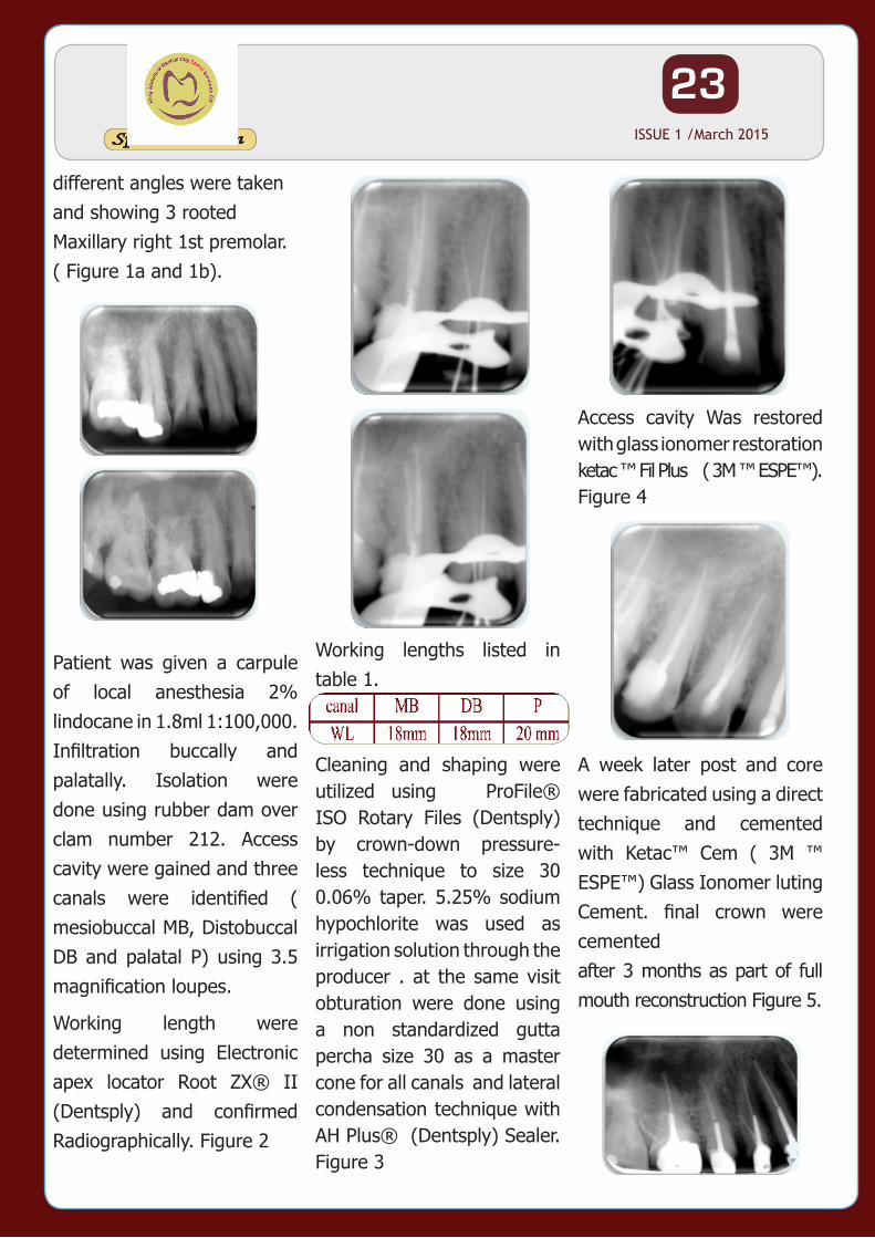
Access cavity Was restored with glass ionomer restoration ketac ™ Fil Plus ( 3M ™ ESPE™).Figure 4
A week later post and core were fabricated using a direct technique and cemented with Ketac™ Cem ( 3M ™ ESPE™) Glass Ionomer luting Cement. final crown were cementedafter 3 months as part of full mouth reconstruction Figure 5.
Working lengths listed in table 1.
Cleaning and shaping were utilized using ProFile® ISO Rotary Files (Dentsply) by crown-down pressure-less technique to size 30 0.06% taper. 5.25% sodium hypochlorite was used as irrigation solution through the producer . at the same visit obturation were done using a non standardized gutta percha size 30 as a master cone for all canals and lateral condensation technique with AH Plus® (Dentsply) Sealer. Figure 3
different angles were taken and showing 3 rooted Maxillary right 1st premolar.( Figure 1a and 1b).
Patient was given a carpule of local anesthesia 2% lindocane in 1.8ml 1:100,000. Infiltration buccally and palatally. Isolation were done using rubber dam over clam number 212. Access cavity were gained and three canals were identified ( mesiobuccal MB, Distobuccal DB and palatal P) using 3.5 magnification loupes.
Working length were determined using Electronic apex locator Root ZX® II (Dentsply) and confirmed Radiographically. Figure 2
24Special Edition ISSUE 1 /March 2015
a recall after 18 month tooth was asymptomatic tooth to percussion and palpation , periodontium was healthy and periapical radiograph shows a sealed coronal restoration and normal periapical area figure 6 & 7
picture 3 & 4
1. Caliskan MK, Pehlivan Y, Sepetcioglu F, Turkun M, Tuncer SS: Root canal morphology of human permanent teeth in a Turkish population. J Endod 21(4):200, 1995.
2. Walker RT: Root form and canal anatomy of maxillary first
premolars in a southern Chinese population. Dent Traumatol 3:130, 1987.
3. Green D: Double canals in single roots. Oral Surg Oral Med Oral Pathol 35:689, 1973.
4. Vertucci FJ: Root canal anatomy of the human permanent teeth. Oral Surg Oral Med Oral Pathol 58:589, 1984.
5. Pineda F, Kuttler Y: Mesiodistal and buccolingual roentgenographic
investigation of 7275 root canals. Oral Surg Oral Med Oral Pathol 33:101, 1972.
zREFERENCES 6. Pecora JD, Sousa Neto MD, Saquy PC: Internal anatomy,direction and number of roots and size of mandibular canines. Braz Dent J 4:53, 1993.
7. Kuttler Y: Microscopic investigation of root apexes. J AmDent Assoc 50:544, 1955.
8. Kerekes K, Tronstad L: Morphometric observations on root canals of human premolars. J Endod 3:417, 1998.
9. Zaatar EI, Al-Kandari AM, Alhomaidah S, Al Yasin IM: Frequency of endodontic treatment in Kuwait: radiographic evaluation of 846 endodontically treated teeth. J Endod 23:453, 1997.
10. Atieh MA: Root and canal morphology of maxillary first premolars in a Saudi population.J Contemp Dent Pract. 2008 Jan 1;9 (1):46-53.
11. S. Al-Nazhan, A. Al-Daafas, N. Al Maflehi: R a d i o g r a p h i c
25Special Edition ISSUE 1 /March 2015
investigation of in vivo endodontically treated maxillary premolars in a Saudi Arabian sub-population. Saudi Endo. J. 2012 vol 2 pp1-5.

26Special Edition ISSUE 1 /March 2015