Embed Size (px)
Citation preview
Medical Coverage Policy
Page 1 of 31 Medical Coverage Policy: 0139
Effective Date ............................................06/15/2017 Next Review Date ......................................06/15/2018 Coverage Policy Number .................................. 0139
Minimally Invasive Intradiscal/ Annular Procedures and Trigger Point Injections Table of Contents Coverage Policy .................................................. 1 Overview.............................................................. 3 General Background ........................................... 3 Coding/Billing Information ................................. 16 References ........................................................ 21
Related Coverage Resources Acupuncture Bone Graft Substitutes for Use in Bone Repair Botulinum Therapy Discography Intervertebral Disc (IVD) Prostheses Lumbar Fusion for Spinal Instability and Degenerative
Disc Conditions, Including Sacroiliac Fusion Mechanical Devices for the Treatment of Back Pain Percutaneous Vertebroplasty, Kyphoplasty, and
Sacroplasty Spinal Orthoses
INSTRUCTIONS FOR USE The following Coverage Policy applies to health benefit plans administered by Cigna Companies. Certain Cigna Companies and/or lines of business only provide utilization review services to clients and do not make coverage determinations. References to standard benefit plan language and coverage determinations do not apply to those clients. Coverage Policies are intended to provide guidance in interpreting certain standard benefit plans administered by Cigna Companies. Please note, the terms of a customer’s particular benefit plan document [Group Service Agreement, Evidence of Coverage, Certificate of Coverage, Summary Plan Description (SPD) or similar plan document] may differ significantly from the standard benefit plans upon which these Coverage Policies are based. For example, a customer’s benefit plan document may contain a specific exclusion related to a topic addressed in a Coverage Policy. In the event of a conflict, a customer’s benefit plan document always supersedes the information in the Coverage Policies. In the absence of a controlling federal or state coverage mandate, benefits are ultimately determined by the terms of the applicable benefit plan document. Coverage determinations in each specific instance require consideration of 1) the terms of the applicable benefit plan document in effect on the date of service; 2) any applicable laws/regulations; 3) any relevant collateral source materials including Coverage Policies and; 4) the specific facts of the particular situation. Coverage Policies relate exclusively to the administration of health benefit plans. Coverage Policies are not recommendations for treatment and should never be used as treatment guidelines. In certain markets, delegated vendor guidelines may be used to support medical necessity and other coverage determinations.
Coverage Policy INJECTION THERAPY: TRIGGER POINT Diagnostic/Stabilization Phase Trigger-point injection(s) of anesthetic and/or corticosteroid (CPT codes 20552, 20553) for diagnosis/stabilization of subacute or chronic back, or neck pain, or subacute or chronic myofascial pain syndrome is considered medically necessary when pain has persisted despite appropriate conservative treatment, including pharmacological therapy, physical therapy, and/or a home exercise program. A maximum of four injection sessions for diagnosis and stabilization may be performed at minimum intervals of one week when provided to determine whether injections provide therapeutic benefit. Therapeutic Phase
Page 2 of 31 Medical Coverage Policy: 0139
Therapeutic trigger-point injections of anesthetic and/or corticosteroid (CPT codes 20552, 20553) are considered medically necessary when prior diagnostic/stabilization injections resulted in a beneficial clinical response (e.g., improvement in pain, functioning, activity tolerance) and BOTH of the following criteria are met:
• subacute or chronic back pain, neck pain, or myofascial pain syndrome persists • injections are provided in conjunction with an active treatment program, which may include pain
management, physical therapy, and/or a home exercise program A maximum of six treatment sessions for injection of the same muscle may be performed at a minimum interval of two months, if the preceding therapeutic injection resulted in more than 50% relief for at least six weeks. Long-term repeated or maintenance therapeutic trigger point injections for any indication are considered experimental, investigational or unproven. Repeat therapeutic trigger point injections provided for 12 months or longer may result in medical necessity review. When performed for any indication each of the following is considered experimental, investigational, or unproven:
• dry needling of trigger points (CPT Code 64999) • ultrasound guidance (CPT code 76942) for trigger point injections
INJECTION THERAPY: INTRADISCAL STEROID INJECTION Intradiscal steroid injection for the treatment of acute, subacute, or chronic back or neck pain is considered experimental, investigational, or unproven. PERCUTANEOUS AND ENDOSCOPIC LAMINECTOMY AND DISC DECOMPRESSION PROCEDURES of the CERVICAL, THORACIC, OR LUMBAR SPINE A percutaneous or endoscopic laminectomy or disc decompression procedure, including but not limited to ANY of the following, is considered experimental, investigational or unproven (this list may not be all-inclusive):
• automated percutaneous lumbar discectomy (APLD)/automated percutaneous nucleotomy (CPT code 62287, HCPCS codes C2614)
• endoscopic anterior spinal surgery/Yeung endoscopic spinal system (YESS)/percutaneous endoscopic diskectomy (PELD)/arthroscopic microdiscectomy, selective endoscopic discectomy (SED) (CPT code 62287)
• endoscopic disc decompression (CPT code 62380), ablation, or annular modulation using the DiscFX™ System (CPT code 62287)
• percutaneous laminotomy/laminectomy, percutaneous spinal decompression (e.g., mild® procedure) (CPT codes 22899, 64999, 0274T, 0275T)
• percutaneous laser discectomy /decompression, laser-assisted disc decompression (LADD) (CPT code 62287)
• endoscopic, anterior cervical disc decompression (e.g., Cervical Deuk Laser Disc Repair) (CPT code 22899)
THERMAL INTRADISCAL PROCEDURES Each of the following procedures is considered experimental, investigational or unproven (this list may not be all-inclusive):
• intervertebral disc biacuplasty (CPT code 22899)
Page 3 of 31 Medical Coverage Policy: 0139
• intradiscal electrothermal annuloplasty (e.g., intradiscal electrothermal therapy [IDET™]) (CPT codes 22526, 22527)
• percutaneous intradiscal radiofrequency thermocoagulation (PIRFT), intradiscal radiofrequency thermomodulation or percutaneous radiofrequency thermomodulation (CPT code 22899, HCPCS code S2348)
• Coblation® Nucleoplasty™, disc nucleoplasty, decompression nucleoplasty plasma disc decompression (CPT code 62287)
• intraosseous radiofrequency nerve ablation of basivertebral nerve (e.g., INTRACEPT® Intraosseous Nerve Ablation System) (CPT code 64999)
OTHER PROCEDURES The following procedures are each considered experimental, investigational or unproven (this list may not be all-inclusive):
• devices for annular repair (e.g., Inclose™ Surgical Mesh System, Xclose™ Tissue Repair System (Anulex Technologies, Inc., Minnetonka, MN)
• epiduroscopy, epidural myeloscopy, epidural spinal endoscopy (CPT code 64999) • intradiscal and/or paravertebral oxygen/ozone injection • spinal decompression using Baxano iO-Flex® System (e.g., Baxano Device)
Overview This Coverage Policy addresses injection therapy and other minimally invasive intradiscal and/or annular procedures for treatment of back pain conditions. General Background Back pain is a frequent cause of chronic pain and disability, affecting approximately 15% of the U.S. population during their lifetime. Most episodes of low back pain improve substantially within a month without formal medical intervention. In a small minority of patients, back pain may be persistent and disabling. Conservative treatment may include pharmacological therapy (e.g., analgesics, anti-inflammatory drugs, muscle relaxants), exercise, spinal manipulation, acupuncture, cognitive-behavioral therapy, and physical therapy. If these measures are unsuccessful, a number of interventional techniques and procedures may be considered that attempt to target specific structures or spinal abnormalities considered to be potential sources of pain, including back muscles and soft tissues, degenerated facet or sacroiliac joints, spinal canal stenosis, and degenerated or herniated intervertebral discs (Chou et al., 2009). Surgery may be appropriate for medical conditions with remediable underlying pathology (e.g. herniated disc) when confirmed and correlated with imaging findings. There is evidence that surgical discectomy provides significant pain relief in selected patients with lumbar disc prolapse with sciatica that fails to improve with conservative treatment. Discectomy was originally performed in an open operation over the spine called hemilaminectomy, in which the muscles are dissected away from the spine and access to the intervertebral disc is obtained by cutting away a piece of spinal bone (i.e., lamina). This technique remains the treatment of choice in some patients, including those with severe pain or weakness and complicated herniation. Microsurgical discectomy (i.e., microdiscectomy) is a less invasive technique that evolved in an effort to decrease postoperative morbidity and recovery time. Microdiscectomy employs direct visualization but is performed through a smaller (15–25 mm) central incision with the use of an operating microscope. Microdiscectomy outcomes are similar to outcomes seen with open discectomy, and microdiscectomy is considered the standard treatment by which to compare other minimally invasive therapies. Management of back pain that is persistent and disabling despite the use of recommended conservative treatment is challenging. Numerous diagnostic and therapeutic injections and other interventional and surgical treatments have therefore been proposed for the treatment back pain.
Page 4 of 31 Medical Coverage Policy: 0139
Choosing Wisely: The North American Spine Society (NASS) Choosing Wisely recommendations state when treating low back pain bed rest for more than 48 hours is not recommended; in patients with low back pain, bed rest exceeding 48 hours in duration has not been shown to be of benefit.
Injection Therapy: Trigger Point Trigger point injection therapy involves the injection of anesthetic or corticosteroids into distinct, focal hyper-irritable spots (i.e., trigger points) located in a tight band of skeletal muscle. Myofascial pain syndrome is a chronic form of muscle pain centered around trigger points. Palpable nodules may be present in the taut band of the muscle which become painful when the tender zone is stimulated. Pain may be perceived at the site of the trigger point or can be referred to other parts of the body, including the back and neck. Fluoroscopic or computed tomography guidance is performed with other types of injections used to diagnose and treat back and neck pain (e.g., epidural steroid injections, facet joint injections) to identify the surrounding structures and to ensure accurate needle placement to the target area. Guidance has been performed with trigger point injections. Although there are no standard criteria, a common method of identifying a trigger point is through manual examination using a palpation technique; palpating the band leads to a local twitch response (LTR) where contraction of the muscle fibers in the taut band is observed. The diagnostic reliability of this method however is inconsistent. As a result, use of ultrasound has been investigated to identify the trigger point and to visualize the twitch response resulting from the injection. Particularly for deep muscles, such as the lower back, it has been purported the use of ultrasound is clinically useful to identify the LTR and therefore improve the efficacy of the injection (Rha, et al., 2011).Evidence in the published medical literature evaluating the efficacy of adding ultrasound or other guidance to trigger point injections is limited to primarily pilot studies, case reports, case series, case control studies and literature reviews (Khumbare, et al., 2016; Shin, et al., 2014; Shankar, Reddy, 2012; Rha, et al., 2011; Sikdar, et al., 2009; Botwin, et al., 2008; Lewis and Tehan, 1999). Sample populations are small and reported clinical outcomes are inconsistent. A majority of comparative trials compare ultrasound guided trigger point injections to other non-trigger point forms of treatment. While some professional societies have published recommended guidelines for trigger point injections, they do not include the use of guidance for the trigger point injection. In the absence of well-designed comparative clinical trials evaluating the efficacy of trigger point injection with and without guidance, strong evidence based conclusions cannot be made. Further clinical validation is necessary to support improved health outcomes with the use of ultrasound guidance for trigger point injections. A Cochrane systematic review was conducted to determine if injection therapy is more effective than placebo or other treatments for patients with subacute or chronic low back pain (Staal et al., 2008). This updated review evaluated 18 randomized controlled trials (n=1179) of injection therapy involving epidural, facet or local sites (i.e., tender or trigger points) in patients with non-radicular pain. The injected drugs included corticosteroids, local anesthetics, and a variety of other drugs. Overall, the results indicated that there was no strong evidence for or against the use of any type of injection therapy. The authors concluded that there is insufficient evidence to support the use of injection therapy in subacute and chronic low back pain, but it cannot be ruled out that specific subgroups of patients may respond to a specific type of injection therapy. Peloso et al. (2007) conducted a Cochrane systematic review to determine the effects of medication and injections on primary outcomes (e.g., pain) for adults with mechanical neck disorders and whiplash. The review evaluated 36 trials that examined the effects of steroid injections, anesthetic agents, psychotropic agents, and NSAIDs. The authors stated that lidocaine injection into myofascial trigger points appeared effective in two trials. Guidelines on injection therapies, low-back pain, and lumbar fusion published by the American Association of Neurological Surgeons (AANS)/Congress of Neurological Surgeons (Watters, et al., 2014; Resnick et al., 2005), based on a systematic review of studies evaluating trigger point injections, facet joint injections, and epidural steroid injections, concluded that there is conflicting evidence suggesting that the use of local trigger point injections can be effective for the short-term relief of low-back pain. There are no data to suggest that trigger point injections with either steroids or anesthetics alone provide lasting benefit for patients suffering from chronic low-back pain. American College of Occupational and Environmental Medicine (ACOEM) evidence-based practice guidelines on low back disorders, updated in 2011, state that trigger and/or tender point injections are not recommended for
Page 5 of 31 Medical Coverage Policy: 0139
treatment of acute low back pain because there are other more efficacious treatment strategies available. These injections may be reasonable as second or tertiary options for subacute or chronic low back pain that is not resolving with conservative treatment (e.g., NSAID, progressive aerobic exercises, and other exercises). The guideline states that injections should consist solely of topical anesthetic (e.g., bupivacaine), and that there is no evidence that steroid is required for efficacy of these injections. Repeat injections should be linked to subjective and objective improvements and be a component of an active therapy program. The ACOEM guideline recommends an interval of at least three to four weeks between injections. If the results are unsatisfactory after the first set, the injections may be repeated. If subjective and objective improvements are not seen, further injections are not recommended. An American Society of Interventional Pain Physicians (ASIPP) Practice Guideline, Interventional Techniques in the Management of Chronic Pain, Part 2.0 (Manchikanti et al., 2001) includes the following recommendations for trigger point injections:
• In the diagnostic or stabilization phase, a patient may receive trigger point injections at intervals of no sooner than one week and preferably two weeks.
• In the treatment or therapeutic phase (after the stabilization is completed), the frequency should be two months or longer between each injection provided that at least >50% relief is obtained for six weeks.
• In the diagnostic or stabilization phase, the number of trigger point injections should be limited to no more than four times per year.
• In the treatment or therapeutic phase, the trigger point injections should be repeated only as necessary judging by the medical necessity criteria and these should be limited to a maximum of six times for local anesthetic and steroid injections.
• Under unusual circumstances with a recurrent injury or cervicogenic headache trigger point injections may be repeated at intervals of six weeks after stabilization in the treatment phase.
Based on the available evidence and specialty society recommendations and guidelines, trigger point injections may be appropriate for selected patients with persistent chronic back, neck or myofascial pain despite appropriate conservative treatment. These injections may provide short-term improvement and allow a determination as to whether conservative treatment will be successful. Dry Needling of trigger points has been proposed as a treatment of myofascial pain in various parts of the body, including low back pain. Dry-needling involves the insertion of a needle (acupuncture needle or other type of needle) into a trigger point without injecting any medication in an effort to deactivate the trigger point. The needle is not left in place; it is removed and is often followed by stretching exercises. A Cochrane systematic review of acupuncture and dry needling (Furlan, et al., 2003, updated 2011) concluded that there is insufficient evidence to make any recommendation regarding acupuncture or dry needling for acute low back pain. For chronic low back pain, acupuncture and dry needling may be useful adjuncts to other therapies. Because most studies were of poor methodological quality, however, there is a need for higher quality trials in this area. There is insufficient evidence in the peer-reviewed published scientific literature to demonstrate the efficacy of dry needling for the treatment of acute or chronic back pain. Injection Therapy: Intradiscal Steroid Intradiscal steroid injection, in which glucocorticoids are injected directly into the intervertebral disc under fluoroscopy, has been proposed as a method to reduce the degree of disc herniation and/or produce an inflammatory response. According to The ACOEM evidence-based practice guidelines on low back disorders (2011) intradiscal steroid injections are not recommended for the management of acute low back pain. The available evidence indicates that intradiscal steroid injections are not effective. There is no quality evidence that these injections improve the natural history of the condition, or that they provide a treatment benefit compared to no treatment or treatment with epidural steroids. In addition, these injections may cause discitis, progression of disc degeneration, and
Page 6 of 31 Medical Coverage Policy: 0139
calcification of the intervertebral disc. The guideline also states that intradiscal steroids are moderately not recommended for subacute or chronic low back pain. There is insufficient evidence in the published medical literature to determine the safety and efficacy of intradiscal steroid injection for the treatment of back pain. Percutaneous and Endoscopic Laminectomy and Disc Decompression Procedures of the Cervical, Thoracic, and /or Lumbar Spine Minimally invasive techniques have been developed which utilize small incisions and employ the use of a multitude of instruments to decompress and/or remove herniated intervertebral disc material under endoscopic or radiologic view. The instruments used for these procedures include arthroscopic instruments, endoscopes, lasers, or other specially designed devices. Percutaneous Disc Decompression: Percutaneous disc decompression involves surgical procedures performed to relieve pressure at the site of a herniated disc (e.g., chemical, thermal or mechanical). Hayes, Inc. published a technology directory report (Hayes, 2014, reviewed 2016, 2017a) evaluating percutaneous disc decompression for cervical disc herniation. A total of 14 studies met inclusion criteria for the review with sample size ranging from 17 to 176 subjects, undergoing five types of PDD interventions (laser, no laser, nuceloplasty, Coblation, and full endoscopic laminotomy) for cervical disc herniation. Follow-up ranged from four weeks to approximately five years. A majority of the studies were limited by lack of controls. Hayes concluded there was insufficient evidence to draw conclusions regarding efficacy of percutaneous disc decompression for cervical disc herniation. Manchikanti et al. (2013) conducted a systematic review to evaluate the evidence for percutaneous disc decompression (PDD) with Dekompressor in the management of chronic low back and lower extremity pain. The primary outcome was pain relief; secondary outcome measures included functional improvement, improvement of psychological status, opioid intake, and return to work. The authors stated that the evidence of effectiveness is limited, but the procedure may be recommended for patients with persistent pain after failure of other intervention techniques when microdiscectomy is not indicated. Automated Percutaneous Lumbar Discectomy (APLD)/Automated Percutaneous Nucleotomy: Automated percutaneous lumbar discectomy (APLD), also referred to as automated percutaneous nucleotomy, is a minimally-invasive surgical procedure employing the use of an automated tissue removal instrument and is used for the removal of herniated lumbar intervertebral discs. In this procedure, a cannula is placed in the center of the disc under fluoroscopic guidance using a posterolateral approach. A probe connected to an automated cutting and aspiration device is then introduced through the cannula. The disc is then aspirated until no more nuclear material is obtained. The goal of treatment is to remove herniated disc material that may be pressing on the nerve root resulting in pain and other symptoms (Hayes, 2016). Hayes, Inc. published a technology directory report (Hayes, 2014, reviewed 2015, 2016) evaluating automated percutaneous lumbar discectomy (APLD). The authors reviewed 16 peer-reviewed studies, including five comparison and 11 uncontrolled trials. According to the report, although APLD was determined to be a safe procedure that may improve symptoms of herniated disc, the quality of evidence was low and was insufficient to draw conclusions regarding efficacy of APLD for lumbar disc herniation. A systematic review published by Manchikanti et al. (2013) evaluated the use of automated percutaneous mechanical lumbar discectomy for treatment of contained herniated lumbar discs. The primary outcome was pain relief; secondary outcome measures were functional improvement, improvement of psychological status, opioid intake, and return to work. Nineteen observation studies were included; of the three randomized trials reviewed, none met inclusion criteria for methodological quality assessment. The evidence is limited for automated percutaneous mechanical lumbar discectomy, but the procedure may provide appropriate relief in properly selected patients with contained lumbar disc herniation. ASIPP 2013 Practice Guidelines for the Management of Chronic Spinal Pain, state that the evidence is limited to fair for APLD, and that the procedure is recommended in select cases.
Page 7 of 31 Medical Coverage Policy: 0139
The North American Spine Society (NASS) published evidence based guidelines for the diagnosis and treatment of lumbar disc herniation (NASS, 2012). Within these guidelines APLD is defined as “a procedure in which a cannula is inserted into the intervertebral disc space, usually with fluoroscopic guidance, and nuclear material is removed without direct visualization by nucleotome, laser or radiofrequency heat. This is an indirect visualization technique using the endoscope and fluoroscopic guidance.” NASS recommends APLD as a treatment of lumbar disc herniation with radiculopathy. However, NASS noted the available evidence is poor (C recommendation) and that there is insufficient evidence to recommend for or against APLD compared with open discectomy in the treatment of subjects with lumbar disc herniation and radiculopathy. American College of Occupational and Environmental Medicine (ACOEM) evidence-based practice guidelines on low back disorders, surgical considerations (2011) states that there is no quality evidence that automated percutaneous discectomy is an effective treatment for any back or radicular pain problem. Hirsch et al. (2009) conducted a systematic evaluation of the literature to determine the effectiveness of APLD. The primary outcome measure was pain relief; short term effectiveness was defined as significant (>50%) pain relief at six months, and long term effectiveness was defined as significant pain relief at one year. Other outcome measures included functional improvement, improvement in psychological status, and return to work. The authors concluded that this systematic review indicates Level II-2 evidence for APLD; APLD may provide appropriate relief in properly selected patients with contained lumbar disc prolapse. (Level II-2 evidence, as defined by the U.S. Preventive Services Task Force as evidence obtained from well-designed cohort or case-control analytic studies, preferably from more than one center or research group.). The authors acknowledged the paucity of randomized controlled trials in the literature as a limitation. A Cochrane review of surgery for lumbar disc prolapse, published in 2003 and updated in 2007 (Gibson and Waddell), assessed the effects of available surgical interventions and states that trials of APLD suggest that clinical outcomes are at best fair and certainly worse than microdiscectomy, although the importance of patient selection is acknowledged. The authors stated that there is a need for high-quality randomized controlled trials on APLD and for long-term studies into the effects of surgery on the lifetime natural history of disc disease. The Cochrane review concluded that unless or until better scientific evidence is available, APLD should be regarded as a research technique. There is insufficient evidence in the peer-reviewed medical literature to support the safety and efficacy of APLD. Results of published studies are inconsistent and do not demonstrate long-term improvement. There is no evidence that APLD is as effective as discectomy/microdiscectomy. Endoscopic Anterior Spinal Surgery / Yeung Endoscopic Spinal Surgery (YESS)/ Selective Endoscopic Discectomy (SED): The Yeung Endoscopic Spinal System (Richard Wolf Surgical Instrument Corporation) is a specialized endoscope developed for percutaneous spinal endoscopy and discectomy. This endoscope has multi-channel inflow and outflow ports, allowing visualization through one port and suction or other therapeutic services through the working port. The YESS is also used for other spinal procedures, including arthroscopic microdiscectomy, radiofrequency ablation, injection of intraoperative steroids, and laser disc decompression and ablation. Selective Endoscopic Discectomy™ (SED), performed with the YESS endoscope, is used to shrink and remove herniated discs. Percutaneous Endoscopic Diskectomy (PELD): PELD is a minimally invasive procedure in which indirect access to the herniated disc is made under fluoroscopic guidance using an endoscope and specialized instruments; removal of the disc occurs using laser or other mechanical means. Within a Health Technology Brief document published by Hayes, eight studies were reviewed evaluating safety and efficacy of PELD as treatment of primary lumbar disc herniation were reviewed (Hayes, 2017b). Hayes concluded although overall that the body of evidence was low-quality the evidence consistently suggests PELD performs similarly to other surgical alternatives for decompression when there was failure of conservative management. However, Hayes acknowledged “substantial uncertainty exists due to the overall quality of the body of evidence and additional studies are needed to evaluate comparative effectiveness and determine patient selection criteria when employed for primary disc herniation”. In a second Health Technology Brief document Hayes evaluated PELD as treatment of recurrent lumbar disc herniation. A total of six studies were included in the review. According to the report, overall a low quality body of evidence suggests PELD may be inferior to comparison treatments for
Page 8 of 31 Medical Coverage Policy: 0139
decreasing back pain and that PELD may have higher recurrence rates than comparison treatments (Hayes, 2017c). Percutaneous Endoscopic/Arthroscopic Microdiscectomy: Percutaneous endoscopic/arthroscopic microdiscectomy is a procedure that involves the use of an endoscopic or arthroscopic guided approach to removing herniated disc material. The herniated disc is accessed and removed through small incisions using cannulas and other instruments. Endoscopic Disc Decompression/Ablation/Annular Modulation using Disc-FX™ System (Elliquence LLC, Baldwin, NY): The Disc-FX™ system is a single-use disposable kit used to perform minimally invasive lumbar disc procedures, including endoscopic disc decompression, nucleus ablation and annulus modulation. There is a steep learning curve for procedures used to access and treat lesions with endoscopic guidance. The purported advantages of endoscopic discectomy or its superiority over microsurgical discectomy have not been demonstrated in the medical literature. There are no prospective controlled clinical trials of the YESS or the Disc FX system, nor are there any prospective studies with long-term follow-up. The efficacy of endoscopic spinal surgery and surgery with the YESS or Disc FX System has not been established in the peer-reviewed medical literature. Percutaneous Laminotomy/Laminectomy/Percutaneous Spinal Decompression (e.g., mild® Procedure): The mild® Device Kit (Vertos Medical, Inc., Aliso Viejo, CA) received U.S. Food and Drug Administration (FDA) approval on February 4, 2010. The device kit is a set of specialized arthroscopic surgical instruments intended to be used to perform lumbar decompressive procedures for the treatment of various spinal conditions. The mild device is used for image-guided minimally invasive lumbar decompression, referred to as the mild (minimally invasive lumbar decompression) procedure. The procedure is performed under fluoroscopic guidance through a dorsal approach to the spine. The instruments are inserted and positioned on the posterior spinal lamina, to the left or right of the spinous process. The tools are used to cut and remove tissue and bone from the posterior side of the lumbar spine to create a space inside the spine that can help decompress some of the spinal nerves. The mild® procedure has been proposed as a minimally invasive alternative to conservative treatment or surgical decompression for the treatment of lumbar spinal stenosis. Staats et al. (2016) reported the six month results of a randomized controlled trial comparing the treatment outcomes of the MILD procedure (n=149) and epidural steroid injection (n=153) for lumbar spinal stenosis. Outcomes were measured using ODI, numeric pain rating scale (NPRS), and Zurich Claudication questionnaire. Primary efficacy was the proportion of ODI responders, tested for statistical superiority of the MILD group versus the active control grou with secondary efficacy proportion of NPRS and ZCQ responders using validated MIC thresholds. At 6 months, all primary and secondary efficacy results provided statistically significant evidence that MILD is superior to the active control of epidural steroid injection. The authors are continuing to obtain outcomes extending to two years post procedure. Limitations of the study noted by the authors included lack of blinding and the possibility of a higher non-responder rate versus standard of care in both groups due to restrictions of the study for use of adjunctive therapies. Hayes, Inc. published a Health Technology Brief evaluating minimally invasive lumbar decompression for lumbar spinal stenosis (Hayes, 2015). One randomized controlled trial, six prospective cohort studies, and two retrospective studies were included in the review. The average follow-up was 24 months and the range of subjects was reported at 27-78. All subjects had symptomatic lumbar spinal stenosis, and the majority had failed previous nonsurgical conservative therapy for lumbar spinal stenosis. Most subjects were treated bilaterally, at 2 lumbar (L) levels, principally at L4 to L5 and/or L3 to L4. According to the published report the mild procedure was safe over the short-term, relieved pain, reduced disability, and improved quality of life in most subjects. However Hayes acknowledged there is insufficient evidence to support long-term safety and effectiveness. Chopko (2013) reported two-year outcomes of mild lumbar decompression in the treatment of patients with neurogenic claudication associated lumbar spinal stenosis. The study included 45 of 58 patients included in an earlier analysis of one-year results Of the 13 patients unavailable at two years and not included in the two-year cohort, 3 underwent lumbar spine surgery, one died of unrelated causes, and nine did not respond or withdrew from the study. Outcome measures included the Visual Analog Scale (VAS), Oswestry Disability Index (ODI),
Page 9 of 31 Medical Coverage Policy: 0139
and Zurich Claudication Questionnaire (ZCQ). At two years, VAS improved from an average of 7.2 at baseline to a mean of 4.8 (p<0.0001); 79% reported an improvement in VAS scores and 29% reported lack of improvement or no improvement. Improvement in physical function and mobility was significant, as measured by the ZCQ and ODI. There were no major adverse events or device-related complications. Limitations of the study include lack of a control group or blinding, and significant numbers of patients lost to follow-up. Brown (2012) conducted a double-blind randomized study of epidural steroid injections (ESI) vs. the mild procedure in patients with symptomatic lumbar spinal stenosis (n=38). The included patients had painful lower limb neurogenic claudication, with hypertrophic ligamentum flavum as a contributing factor, and had failed conservative treatment. Patients were randomized to the mild procedure (n=21) or ESI (n=17). At six weeks, 76% of the patients in the mild group reported a two point improvement in VAS scores in compared to 35% of patients in the ESI group. There was a significant improvement in Oswestry disability scores in the mild group at six weeks (p<0.05), while in the ESI group improvement was not statistically significant. There were no procedure-related or device-related complications in either group. At six weeks, 17 of 21 patients in the ESI group crossed over to the mild procedure. Comparative 12 week outcome data was therefore not available. It is difficult to draw conclusions from this study due to the small number of participants and lack of data on long term outcomes. In addition, patients in the ESI group were treated with a single interlaminar injection; which is generally not typical of ESI treatment. An observational study conducted by Mekhail et al. (2011) at 11 sites reported one year outcome data on 58 patients treated for spinal stenosis with the mild procedure, with statistically significant improvement in VAS scores and ODI. A single-site case series conducted by Mekhail et al. in 2012 reported 12-month outcomes for 40 consecutive patients treated for spinal stenosis with the mild procedure. There was significant functional improvement and decreased disability as measured by the Pain disability index (PDI), Roland-Morris Disability Questionnaire, walking distance, standing time, and VAS scores. Deer and Kapurai (2010) published a retrospective review to evaluate the acute safety of the mild procedure. Charts of 90 consecutive patients who underwent the mild procedure for decompression of central lumbar stenosis were reviewed. No major adverse events or complications related to the devices or procedure were reported. There were no incidents of dural puncture or tear, blood transfusion, nerve injury, epidural bleeding or hematoma. Because the review did not include outcome data, no determination as to clinical efficacy can be made. The authors stated that prospective randomized studies have been initiated to collect patient outcomes data regarding post-treatment pain and functional capacity. Chopka and Caraway (2010) published a preliminary report of MiDAS I (mild Decompression Alternative to Open Surgery, a multi-center prospective case series to evaluate the mild procedure for treatment of symptomatic lumbar spinal stenosis. The procedure was offered as an alternative to surgery or continued medical management. No major device or procedure-related complications were reported. At six weeks, statistically significant reduction of pain as measured by the Visual Analog Scale, Oswestry Disability Index, and Zurich Claudication Questionnaire, and Standard Form -12. (SF-12). Percutaneous/Laparoscopic Laser Discectomy/Decompression/ Laser-Assisted Decompression (LADD): Laser-assisted discectomy, also called laser-assisted disc decompression (LADD) or laser disc decompression, is a minimally-invasive procedure proposed as an alternative to discectomy/microdiscectomy. It is intended to provide symptomatic relief of pain cause by a contained herniated intervertebral disc. Laser light energy is used to vaporize part of the nucleus pulposus, resulting in a reduction in intradiscal pressure. Several approaches may be used, depending on the location of the disc and type of laser being used. With one method, a needle is inserted percutaneously into the disc approximately one centimeter (cm) posterior to the disc center, and a flexible optical quartz fiber is threaded through the needle into the disc, delivering laser energy to vaporize and coagulate the nucleus pulposus. In the laparoscopic approach, a trocar is inserted periumbilically and the abdomen is inflated with carbon dioxide. Additional trocars are placed above the pelvic brim. The large and small bowels are retracted, and the iliac bifurcation is identified. The posterior peritoneum is opened and retracted. The L5-S1 interspace is identified and its margins confirmed by x-ray. The annulus of the disc is opened and excised with the neodymium: yttrium-aluminum-garnet (Nd: YAG) laser.
Page 10 of 31 Medical Coverage Policy: 0139
Updated ASIPP Practice Guidelines for the Management of Chronic Spinal Pain (2013) state that the evidence for percutaneous lumbar laser disc decompression is limited. ACOEM evidence-based practice guidelines on low back disorders, surgical considerations (2011) states that there is no quality evidence that laser discectomy is an effective treatment for any back or radicular pain problem. A review of the literature published by Schenck et al. (2006) evaluated 16 clinical trials representing a total of 1579 patients. Most were case series with small sample sizes, making interpretation of success rates difficult. Generalization of the results into general clinical practice remains difficult due to different inclusion and exclusion criteria, laser types, and outcome measures as well as the variation in duration of follow-up. These shortcomings prevent a valid comparison to studies evaluating the outcome of conventional surgical treatment for lumbar disc herniation. The authors concluded that well-designed research of sufficient scientific strength comparing percutaneous laser disc decompression to both conventional surgery and conservative management is needed to determine whether this procedure has a role in the treatment of lumbar disc herniation. A Cochrane systematic review of surgery for lumbar disc prolapse, published in 2003 and updated in 2007 (Gibson and Waddell), assessed the effects of available surgical interventions and states that trials of laser discectomy suggest that clinical outcomes are at best fair and certainly worse than microdiscectomy, although the importance of patient selection is acknowledged. The authors stated that there is a need for high-quality, randomized controlled trials on laser discectomy and for long-term studies into the effects of surgery on the lifetime natural history of disc disease. The Cochrane Review further concluded that unless or until further scientific evidence is available, laser discectomy should be regarded as a research technique. There is insufficient evidence in the published medical literature to demonstrate the safety, efficacy and long-term outcome of laser discectomy. There are no randomized controlled trials that evaluate laser discectomy and compare this procedure to established treatment methods. Endoscopic Anterior Cervical Disc Decompression: Cervical Deuk Laser Disc repair is an endoscopic anterior cervical transdiscal surgical procedure under investigation for treatment of symptomatic cervical disc disease (e.g., spondylosis, stenosis, herniations). The repair involves three procedures, a selective partial discectomy, foraminoplasty, and annular debridement. The procedure may be performed as an alternative to anterior cervical discectomy and fusion for treatment of cervical degenerative disc disease. In theory, the endoscopic approach does not require the removal of the intervertebral disc to reach the posterior disc complex, as a result there is no postoperative iatrogenic instability or deformity. In addition, it is not necessary to stabilize the spine with interbody devices, fusion, implants or biologics. At present, evidence in the peer-reviewed published scientific literature is limited to few uncontrolled case series and is insufficient to support the safety and efficacy of endoscopic anterior cervical disc decompression (i.e., Cervical Deuk Laser Disc repair). There is insufficient evidence in the medical literature to demonstrate the safety and efficacy percutaneous laminotomy/laminectomy approaches, including the mild procedure. Additional well designed trials with long-term outcome data are needed to determine how this procedure compares to available alternative treatments for lumbar stenosis. Thermal Intradiscal Procedures Intraosseous Radiofrequency Nerve Ablation: Radiofrequency ablation of intraosseous nerves is an emerging technology intended for treatment of chronic low back pain. Intraosseous nerves are reportedly found within the vertebrae, are referred to as basiverterbal nerves and are present in the basiverterbral foramen. Authors contend the nerves may be a source of intraosseous back pain and that interruption of the nerve pathway using radiofrequency will relieve the associated pain. One device under investigation, The INTERCEPT® System (Relievant MedSystems, Inc, Redwood City, CA) recently received FDA approval for use as a minimally invasive radiofrequency system for treatment of chronic lumbar back pain at one or more levels (i.e., L3 to S1), when back pain is present despite at least six months of conservative care and is accompanied by either Type I or Type 2 Modic changes on MRI (FDA K153272). Evidence in the peer reviewed, published scientific literature evaluating ablation of basiverterbal nerves consists mainly of pilot studies and is insufficient to support safety and efficacy at this time; additional studies are needed to support strong evidence-based conclusions.
Page 11 of 31 Medical Coverage Policy: 0139
Intradiscal Electrothermal Annuloplasty (e.g., intradiscal electrothermal therapy [IDET™]): Intradiscal electrothermal annuloplasty (IEA), also referred to as intradiscal electrothermal therapy (IDET™), intradiscal electrothermal percutaneous annuloplasty, intradiscal thermal annuloplasty, or targeted intradiscal thermal therapy. is a minimally invasive procedure that has been proposed as an alternative to spinal fusion for the treatment of chronic discogenic low back pain. Following a provocative discogram, IEA is performed by inserting a catheter into the annulus and threading a flexible electrode through the catheter and around the inside of the disc, pressing against the posterior edge of the annulus. The electrode is then heated to a temperature of 90º F for up to 17 minutes. Analgesics and/or antibiotics are then injected and the catheter is withdrawn. The heating of the electrode denatures the collagen of the annulus and coagulates the nerve endings, with the ultimate goal of relieving back pain. A systematic review of percutaneous thermocoagulation intradiscal techniques for discogenic low back pain (Urrutia, et al., 2007) included six studies (283 patients) of IEA and percutaneous intradiscal radiofrequency thermocoagulation (PIRFT). The studies included in the review of IEA consisted of two randomized controlled trials (Freeman and Pauza, discussed above), and two nonrandomized trials. One of the nonrandomized trials assessed the effectiveness of IEA vs. a rehabilitation program consisting of physical therapy, exercise, education and counseling, and the other compared IEA to PIRFT. In both randomized controlled trials that assessed IEA vs. placebo, pain, disability, and quality of life were assessed for six months. There was a small difference in favor of IEA in one study (Pauza), although the difference in disability was clinically irrelevant, while there was no difference in the higher-quality, more recent study (i.e., Freeman). The Freeman study also assessed depression, sitting and work tolerance, medication and neurologic deficit, and found no difference between IEA and placebo. In the nonrandomized trial comparing IEA and a rehabilitation program, the proportion of patients with a ≥ 50% reduction in pain was higher in the IEA group at both 12 and 24 months. The authors concluded that the available evidence does not support the efficacy or effectiveness of percutaneous thermocoagulation intradiscal techniques for the treatment of discogenic low back pain. The authors noted that previous case reports suggested that the procedure might be effective, but these reports, derived from data registries, could not take into account the effect of regression to the mean, the natural history of the condition, the placebo effect, and other potential confounders such as co-interventions and other mechanical and psychosocial factors. Freeman (2006) conducted a systematic review of the evidence of the efficacy of IEA. The review included 11 prospective cohort studies, five retrospective studies, and two randomized controlled trials. The prospective cohort studies reported on a total of 256 patients with a mean follow-up of 17.1 months (range 12–28 months). The mean improvement in the VAS for back pain was 3.4 points (range 1.4–6.5), and the mean improvement in ODI was 5.2 points (range 4.0–6.4) The five retrospective studies included 379 patients and reported that between 13 and 23% of patients subsequently underwent surgery for low back pain within the study period. The two randomized controlled trials, Pauza, 2004 and Freeman, 2005, provided inconsistent evidence, as described above. The author concluded that the evidence for efficacy of IEA remains weak and has not passed the standard of scientific proof. A randomized, double-blind controlled trial was conducted by Freeman et al. (2005) to test the safety and efficacy of IEA compared with placebo for treatment of chronic discogenic low back pain. Patients with one- or two-level symptomatic disc degeneration with posterior or posterolateral annular tears who failed to improve after conservative therapy were considered for the study. Patients were randomized on a 2:1 ratio to IEA (n=38) or a sham procedure (n=19). An independent technician connected the catheter to the generator and delivered electrothermal energy to only the treatment group. Surgeon, patient, and independent outcome assessor were all blinded to the treatment. Low Back Outcome Score (LBOS), Oswestry Disability Index, SF-36, the Zung Depression Index (ZDI) and Modified Somatic Perceptions Questionnaire (MSPQ) were measured at baseline and at six months. Successful outcome was defined as no neurological deficit, improvement in LBOS of greater than seven points, and improvement in SF-36 subsets (i.e., physical function and bodily pain) of greater than one standard deviation. No patient in either group showed improvement of greater than seven points in LBOS or greater than one standard deviation in the specified SF-36 domains. Mean ODI was 41.42 at baseline and 39.77 at six months for the IEA group compared with 40.74 at baseline and 41.58 at six months for the placebo group. There was no significant change in ZDI or MSPQ for either group. The authors concluded that there was no significant benefit from IEA over placebo.
Page 12 of 31 Medical Coverage Policy: 0139
Pauza et al. (2004) conducted a prospective, randomized controlled trial comparing IEA with placebo. Sixty-four patients were randomized to receive IEA or sham treatment. The subjects were not aware of which treatment they received. Outcome tools used were the VAS, the SF-36, and the Oswestry Disability Scale. It is unclear whether the post-procedure outcome examiners were blinded regarding which patients received true IEA. The modest success rates reported in this trial were much less compelling than those from previously published uncontrolled studies. The investigators reported that both groups showed improvement, with mean improvements higher in the active treatment arm. Using the VAS, IEA demonstrated a 2.4-point decrease in the mean pain score. An 11-point decrease was reported in the mean Oswestry score. The baseline disability level of most of the patients was low, and recruitment methods may have led to patient selection bias. The sample size was insufficient to achieve adequate statistical power, and follow-up was limited to six months. In addition, eight patients who dropped out of the study were not included in the data analysis. While the results of this study suggest that IEA may improve outcomes for patients with discogenic low back pain, these methodological flaws make it impossible to draw valid conclusions about the efficacy of this technology.
ASA 2010 Practice Guidelines for Chronic Pain Management states that Thermal intradiscal procedures: intervertebral disc annuloplasty (IDET) may be considered for young, active patients with early single-level degenerative disc disease with well-maintained disc height.
ACOEM evidence-based practice guidelines on low back disorders (2011) states that IDET is not recommended for treatment of acute, subacute, or chronic low back pain or any other back-related disorder. Updated American Society of Interventional Pain Physicians (ASIPP) Evidence-Based Practice Guidelines in the Management of Chronic Spinal Pain (Manchicanti, et al., 2013).state that the evidence for IDET is limited to fair, and that the procedure may be performed in a select group of patients with discogenic pain non-responsive to conservative modalities, including epidural injections. The safety, efficacy, and long-term outcomes of intradiscal electrothermal annuloplasty in the treatment of patients with chronic discogenic low back pain have not been established in the published medical literature. This procedure has not been proven to achieve equivalent or improved patient outcomes compared to available and established alternatives. In addition, the long-term effect of thermal coagulation of intervertebral discs has not been determined. Percutaneous Intradiscal Radiofrequency Thermocoagulation (PIRFT)/ Intradiscal Radiofrequency Thermomodulation/Percutaneous Radiofrequency Thermomodulation: PIRFT may also be referred to as intradiscal radiofrequency thermomodulation or percutaneous radiofrequency thermomodulation. This procedure, used to treat chronic discogenic low back pain, is similar to intradiscal electrothermal therapy (IDET). With IDET, a catheter with a temperature-controlled, thermal-resistive coil is inserted under fluoroscopic guidance into the posterior annular wall of the affected disc, causing annular denervation. With PIRFT, the catheter is placed into the center of the disc rather than the annulus. The mechanism of reported clinical improvement with PIRFT is unclear, since the temperature at the annulus has been found to be well below the temperature required for annular denervation (Davis, 2003). More recently bipolar radiofrequency thermocoagulation has been investigated as treatment of discogenic low back pain (Zhang, et al., 2016). During bipolar radiofrequency thermocoagulation two cannulas are heated simultaneously in contrast to a single cannula as in PIRFT. Urrutia et al. (2007) conducted a systematic review to evaluate the evidence for the percutaneous thermocoagulation intradiscal techniques IDET and PIRFT in the treatment of discogenic low back pain. Six studies with a total of 283 patients were included. Two randomized controlled trials, including the Barendse trial described below, showed no differences between PIRFT and placebo and between different PIRFT techniques. The authors stated that, although previous case reports and nonrandomized trials suggested positive results, results from randomized clinical trials show that PIRFT is not effective for the treatment of discogenic low back pain. Barendse et al. (2001) conducted a randomized, double-blind, placebo-controlled trial of PIRFT using the Radionics discTRODE™ RF annuloplasty system. The Radionics system was approved by the U.S. Food and Drug Administration (FDA) through the 510(k) process in October 2000. A total of 28 patients were selected who had a history of at least one year of chronic low back pain, evidence of radiculopathy on neurological
Page 13 of 31 Medical Coverage Policy: 0139
examination and a positive response to discography. Patients were randomly assigned to one of two treatment groups. Patients in the radiofrequency group (n=13) received a 90-second 70 degree centigrade (C) lesion of the intervertebral disc. Patients in the control group (n=15) underwent the same procedure but without the use of radiofrequency current. The treating physician and patients were blinded to group assignment. Patients were assessed by a blinded investigator before treatment and eight weeks after treatment. There was no difference between the two groups based on visual analog scores for pain, global perceived effect and the Oswestry disability scale. The treatment was considered a success in one patient in the radiofrequency group and two patients in the control group. The authors concluded that PIRFT is not effective in reducing chronic discogenic low back pain. Updated American Society of Interventional Pain Physicians (ASIPP) Evidence-Based Practice Guidelines in the Management of Chronic Spinal Pain (Manchicanti, et al., 2013) state that the evidence is limited for discTRODE (PIRFT). According to the evidence-based clinical practice guideline from the American Pain Society, Interventional Therapies, Surgery, and Interdisciplinary Rehabilitation for Low Back Pain (Chou et al., 2009), the level of evidence for PIRFT is poor. The authors were unable to estimate the net benefit of the procedure in the treatment of patients with nonradicular low back pain. American College of Occupational and Environmental Medicine (ACOEM) practice guidelines on low back disorders, (2011) states that PIRFT is strongly not recommended for treatment of acute, subacute, or chronic low back pain, particularly including discogenic low back pain. There is insufficient evidence in the published medical literature to demonstrate the safety, efficacy and long-term outcomes of PIRFT. There is no evidence that this procedure is as effective as established alternatives for the treatment of back pain. Intervertebral Disc Biacuplasty/Cooled Radiofrequency: The Baylis TransDiscal™ system (Baylis Medical Inc., Montreal Canada) is used to perform intervertebral biacuplasty. The TransDiscal system received FDA approval through the 510(k) process on December 19, 2006. The system is designed to deliver controlled RF energy via two electrodes. Two TransDiscal Probes and the Pain Management Pump Unit, connected to the Baylis Pain Management Generator, work in concert to deliver RF energy. The system is intended to be used to create RF lesions in nervous tissue, including that which is situated in intervertebral disc material. Separate components of the system had previously received FDA approval; the 2006 approval combined the indications of the predicate devices. (U.S. FDA website). Intervertebral biacuplasty using the TransDiscal system has been investigated in the treatment of lumbar discogenic pain. The procedure is performed using a bipolar approach in conjunction with internally water-cooled RF probes to coagulate and decompress disc material. Two introducers are placed bilaterally in the posterolateral discs and the TransDiscal probes are then inserted into the introducers. RF energy is applied and directed through the disc between the two probe electrodes. The cooling system is designed to maintain and balance the temperature in each probe, allowing RF energy to be delivered with greater power to heat a larger volume of disc tissue, while avoiding overheating of adjacent tissue. Desai et al (2016) conducted a prospective randomized clinical trial to compare outcomes of intradiscal biacuplasty and conventional medical management (n=29) with subjects who received conventional medical management alone (n=34). At six months following treatment, subjects were allowed to cross-over to the experiment group and were subsequently followed for an additional six months. The initial experimental group was followed for 12 months. The primary outcome measured was pain level change using VAS with secondary outcomes that included assessments of function, disability, mental health, quality of life and use of opioids. At 12 months post procedure pain reduction, and improvement in function and disability scores were reported to be statistically significant and clinically meaningful in the original experimental group. The authors reported 50% of the cross over group responded to the intervention, with mean outcomes similar to the original group. Daily opioid intake was reduced in both the original and cross-over group. In the authors opinion the study demonstrated long-term effectiveness of intradiscal biacuplasty combined with conventional medical
Page 14 of 31 Medical Coverage Policy: 0139
management. Limitations of the study included small sample populations, one-year outcomes, and inconsistent follow-up as reported by the authors. Kapural et al. (2013) conducted a randomized controlled trial to evaluate transdiscal radiofrequency biacuplasty (IDB) for discogenic lower back pain (n=59). Twenty nine patients were randomized to IDB and 30 to a sham procedure. All had a history of chronic low back pain for longer than six months. The primary outcome measures were physical function, pain, disability, and opioid usage. At six months, there were statistically significant improvements in the treated group compared to the control group in physical function (p=0.129), pain (p=0.006), and disability (p=0.037). There was no significant difference between groups in opioid usage. Limitations of the study include lack of long-term follow-up and small sample size. Of 1894 patients screened, only 59 were included. Kapural et al. (2015) reported in follow-up that the improvements initially reported at 6 months were maintained at nine and 12 months. Kapural et al. (2008) conducted a pilot study (n=15) of intervertebral disc biacuplasty in the treatment of lumbar discogenic pain. Included patients had a history of chronic low back pain unresponsive to nonoperative care for greater than six months, back pain exceeding leg pain, concordant pain on provocative discography, disc height > 50% of control, and evidence of single-or tow-level degenerative disc disease without evidence of additional changes on MRI. Outcomes were evaluated by questionnaire at one, three and six months. Median VAS pain score decreased from 7 cm at baseline to 4 cm at one month and 3 cm at six months. The Oswestry score improved from 23.3 to 16.5 at one month, with similar results at six months. The SF-36 physical functioning scores improved from 51 to 70 points at six month, and the Bodily Pain score improved from 38 to 54. There was no significant change from baseline in daily opioid use. No procedure-related complications were reported. Updated ASIPP guideline referenced above (Manchicanti, et al., 2013) state that the evidence for biacuplasty is limited to fair, and that the procedure may be performed in a select group of patients with discogenic pain non-responsive to conservative modalities, including epidural injections. There is insufficient evidence in the published medical literature to demonstrate the safety, efficacy and long-term outcomes of intervertebral disc biacuplasty. Coblation® Nucleoplasty™/Disc Nucleoplasty/Decompression Nucleoplasty/Plasma Disc Decompression: Coblation Nucleoplasty, also referred to as disc nucleoplasty, decompression nucleoplasty, or plasma disc decompression, is a minimally invasive technique for decompression of contained herniated discs using the Arthrocare Perc-D Coblation Spine Wand. The Spine Wand is a bipolar radiofrequency device designed to decompress the disc nucleus with energy and heat. The tip of the wand is slightly curved to allow channeling. Nucleoplasty uses Coblation technology, which generates a low temperature plasma field intended to allow precise ablation with minimal risk of thermal injury. The tip temperature is 50–70 degrees C. A plasma field, a millimicron-thick layer of highly energized particles, causes molecular dissociation of the disc material directly in front of the tip. This creates a channel from the posterolateral annulus to the anteromedial annulus. During withdrawal, the coagulation mode is used. Six separate channels are typically created. The thermal effect is reported to result in denaturization of the Type II collagen, causing shrinkage of the surrounding collagen and widening of the channel (Sharps, et al., 2002; Singh, et al., 2003; Davis, 2003) There are no published randomized controlled trials evaluating nucleoplasty in the medical literature; studies consist primarily of uncontrolled case series (Sharp and Isaac, 2002; Singh et al., 2003; Bhagia et al., 2006; Cincu, et al., 2015; Ren, et al., 2015). Studies evaluating long-term outcomes supporting clinical efficacy are lacking. A Cochrane review of surgery for lumbar disc prolapse (Gibson and Waddell, 2007) states that, unless or until better scientific evidence is available, Coblation therapy should be regarded as a research technique. Updated ASIPP Practice Guidelines for the Management of Chronic Spinal Pain (2013) state that the evidence is limited to fair for nucleoplasty, and that the procedure is recommended in select cases. The evidence-based clinical practice guideline from the American Pain Society, Interventional Therapies, Surgery, and Interdisciplinary Rehabilitation for Low Back Pain (Chou et al., 2009), states that there are no trials
Page 15 of 31 Medical Coverage Policy: 0139
evaluating Coblation nucleoplasty. The authors were unable to estimate the net benefit of the procedure in the treatment of patients with back pain, with or without radiculopathy. ACOEM evidence-based practice guidelines on low back disorders, surgical considerations (2011) state that there is no quality evidence that Coblation therapy is an effective treatment for any back or radicular pain problem. The safety, efficacy and long-term outcomes of this Coblation nucleoplasty have not been demonstrated in the published medical literature. In addition, the long-term consequences of thermal denervation and tissue damage associated with this procedure are unknown. Other Minimally Invasive Procedures Baxano iO-Flex® System: The Baxano iO-Flex® System (Baxano, Inc., San Jose, California) is a method of decompression that employs an “inside-out” approach according to the manufacturer. The system consists of a microblade shaver and several accessories which can be used in either minimally invasive or open procedures and according to the manufacturer instead of cutting through healthy pieces of the spine, the iO-Flex® System uses a fine surgical wire to guide the thin iO-Flex® shaver instrument to the location of the overgrown bone and tissue to shave away the stenosis from the inside out. Use of this method is purported to preserve facet joint integrity/lamina, thus maintaining stability and minimizing muscle trauma by allowing decompression of up to 4 nerve roots through a single-point access and unlike traditional rigid instruments used for lumbar decompression the Baxano iO-Flex System utilizes thin flexible instruments. The FDA approvals for these devices suggests the devices are designed for accessing, cutting, and biting soft tissue and bone during surgery involving the spinal column. Nevertheless, evidence in the peer-reviewed scientific literature evaluating these emerging technologies is lacking, therefore evidence based conclusions cannot be made. Oxygen/Ozone Injection: Intradiscal oxygen-ozone injection has been proposed as a minimally invasive treatment of lumbar disc herniation. Ozone is reported to be a strong oxidizer that rapidly reacts and oxidizes the proteoglycans in the nucleus pulposus. The procedure is based on the premise that a small reduction in disc volume may result in a significant reduction in pain. The technique is similar to discography and other percutaneous disc procedures. Under image guidance, a needle is positioned into the nucleus pulposus, 1-3 ml of oxygen/ozone from a medical ozone generator is injected into the disc, and 7-9 ml is injected into the paravertebral muscle surrounding the disc. A pain suppressant (e.g., bupivacaine) and/or corticosteroid may also be injected. Oxygen/ozone injection is primarily practiced in Europe and Asia. No medical ozone generators for use in intradiscal injection have received U.S. Food and Drug Administration (FDA) approval. A meta-analysis of the effectiveness and safety of ozone treatments for herniated lumbar discs conducted by Steppan et al. (2010) reported a mean improvement of 3.9 for Visual Analog Scale (VAS) and 25.7 for Oswestry Disability Index (ODI). The likelihood for showing improvement on the Modified McNab outcome scale was reported as 79.7%, and the likelihood of complications, 0.064%. It is difficult to draw firm conclusions from this analysis due to the quality of included studies. Of 11 included studies, 9 were retrospective, 2 were prospective, and one consisted of unpublished data. In some studies data required for meta-analysis was not reported, and was estimated by the authors. There is insufficient evidence in the published medical literature to demonstrate the safety and efficacy of ozone injection or to determine how this treatment compares to other available treatment options for disc herniation. In addition, no medical ozone generators have received FDA approval. Epiduroscopy/Epidural Myeloscopy/Epidural Spinal Endoscopy: Epiduroscopy, also referred to epidural myeloscopy or epidural spinal endoscopy, is a technique that uses an epiduroscope to visualize the epidural space. It is used in the diagnosis and treatment of intractable low back pain, especially in patients with radiculopathy. Scarring of the epidural space occurs in approximately 50% of patients who have undergone multiple surgeries for back pain. This may lead to formation of epidural fibrosis, adhesions of the nerve root, causing recurrence of pain. In epiduroscopy, a needle is advanced into the sacral canal through which a guide-wire is inserted and advanced. The needle is replaced with an introducer sheath through which an endoscope is inserted. Saline is flushed through the system to expand the sacral space, which can then be examined through the endoscope. Although epiduroscopy may be performed as a diagnostic procedure, it is usually performed in
Page 16 of 31 Medical Coverage Policy: 0139
conjunction with the Racz procedure or epidural adhesiolysis. There is no evidence in the published medical literature to support the use of epiduroscopy as a diagnostic procedure. There is no evidence that this invasive technique provides clinically useful information not available with current noninvasive diagnostic methods. There is insufficient evidence in the published medical literature to support the use of epiduroscopy in the diagnosis or treatment of back pain. There are no published, well-designed, prospective clinical trial of adequate size that evaluates these procedures nor is there information available regarding long-term outcomes. The safety, efficacy and long-term outcomes of these procedures have not been established. Devices for Annular Repair Following Spinal Surgery: Discectomy procedures involve removal of a bony portion of the vertebral body to access the posterior side of the disc space, and removal of the impinging fragment from the disc. This fragment may be within the wall of the anulus, requiring incision into the anulus to remove it. Sutures may be placed to seal the annular defect to reduce recurrent herniation following discectomy. The Inclose™ Surgical Mesh System and the Xclose™ Tissue Repair System (Anulex Technologies, Inc., Minnetonka, MN) have been proposed for annular repair following discectomy as an alternative method to re-approximate the compromised tissue of the anulus fibrosus. Use of the Xclose system for this indication, however is beyond the scope of the FDA 510 (k) clearance, detailed below. The Inclose Surgical Mesh System received FDA approval through the 510(k) process on August 18, 2005. According to the 510(k) summary, the device is comprised of a mesh implant and two suture assemblies (anchor bands). The mesh implant is an expandable braided patch that is inserted through the aperture of the tissue defect and affixed to surrounding soft tissue with the anchor bands. The product may be used to support soft tissue where weakness exists, or for the repair of hernias requiring the addition of a reinforcing, or bridging material, such as the repair of groin hernias. The Xclose Tissue Repair System received FDA approval through the 510(k) process on August 7, 2006. The system is described in the 510(k) summary as consisting of two non-absorbable braided surgical 3-0 suture and T-anchor assemblies connected with a loop of green 2-0 suture. The 2-0 suture loop is used to facilitate tightening, drawing the 3-0 suture assemblies together and re-approximating the tissue. The system is indicated for use in soft tissue approximation for procedures such as general and orthopedic surgery. There is inadequate evidence to demonstrate the safety and efficacy of these devices or to determine the impact on patient outcomes compared to standard surgical techniques. In addition to the procedures described above, several recently introduced techniques combine established surgical approaches for disc removal with additional procedures for which safety and efficacy has not been established, including radiofrequency, laser or other disc ablation and modulation procedures (e.g., Disc-Fx [Elliquency Innovations, Oceanside NY]), selective endoscopic discectomy (SED). Use Outside the U.S. Guidance, National Institute for Health and Clinical Excellence (NICE) (United Kingdom): Interventional procedural guidance issued by NICE for the following procedures states that in view of uncertainty about the efficacy of these procedures each should not be done without special arrangements for consent and for audit or research:
• Automated percutaneous mechanical lumbar discectomy (2005, IPG141) • Percutaneous endoscopic laser thoracic discectomy (2004, IPG61) • Percutaneous endoscopic laser cervical discectomy (2009, IPG303) • Epiduroscopic lumbar discectomy through the sacral hiatus for sciatica (2016, IPG570) • Percutaneous electrothermal treatment of the intervertebral disc annulus for low back pain and sciatica
(2016, IPG544) • Percutaneous intradiscal radiofrequency treatment of the intervertebral disc nucleus for low back pain
(2016, IPG545) • Percutaneous coblation of the intervertebral disc for low back pain and sciatica (2016, IPG543)
Coding/Billing Information
Page 17 of 31 Medical Coverage Policy: 0139
Note: 1) This list of codes may not be all-inclusive. 2) Deleted codes and codes which are not effective at the time the service is rendered may not be eligible for reimbursement. Injection Therapy: Trigger Point Considered Medically Necessary when criteria in the applicable policy statements listed above are met: CPT®* Codes
Description
20552† Injection(s); single or multiple trigger point(s), 1 or 2 muscle(s) 20553† Injection(s); single or multiple trigger point(s), 3 or more muscle(s)
†Note: Considered Experimental/Investigational/Unproven when used to report dry needling of trigger points ICD-9-CM Diagnosis Codes
Description
723.1 Cervicalgia 723.8 Other syndromes affecting cervical region 724.1 Pain in thoracic spine 724.2 Lumbago 724.5 Backache, unspecified 724.8 Other symptoms referable to back 724.9 Other unspecified back disorders
ICD-10-CM Diagnosis Codes
Description
M43.8X9 Other specified deforming dorsopathies, site unspecified M53.80 Other specified dorsopathies, site unspecified M53.81 Other specified dorsopathies, occipito-atlanto-axial region M53.82 Other specified dorsopathies, cervical region M53.83 Other specified dorsopathies, cervicothoracic region M53.84 Other specified dorsopathies, thoracic region M53.85 Other specified dorsopathies, thoracolumbar region M53.9 Dorsopathy, unspecified M54.2 Cervicalgia M54.5 Low back pain M54.6 Pain in thoracic spine M54.81 Occipital neuralgia M54.89 Other dorsalgia M54.9 Dorsalgia, unspecified
Considered Experimental/Investigational/Unproven: ICD-9-CM Diagnosis Codes
Description
All other codes ICD-10-CM Description
Page 18 of 31 Medical Coverage Policy: 0139
Diagnosis Codes All other codes
Dry Needling of Trigger Points Considered Experimental/Investigational/Unproven: CPT®* Codes
Description
64999 Unlisted procedure, nervous system ICD-9-CM Diagnosis Codes
Description
All codes ICD-10-CM Diagnosis Codes
Description
All codes Ultrasound Guidance for Trigger Point Injections Considered Experimental/Investigational/Unproven: CPT®* Codes
Description
76942 Ultrasonic guidance for needle placement (eg, biopsy, aspiration, injection, localization device), imaging supervision and interpretation
ICD-9-CM Diagnosis Codes
Description
All codes ICD-10-CM Diagnosis Codes
Description
All codes Injection Therapy: Intradiscal Steroid Injection Considered Experimental/Investigational/Unproven: CPT®* Codes
Description
22899 Unlisted procedure, spine 64999 Unlisted procedure, nervous system
ICD-9-CM Diagnosis Codes
Description
All codes
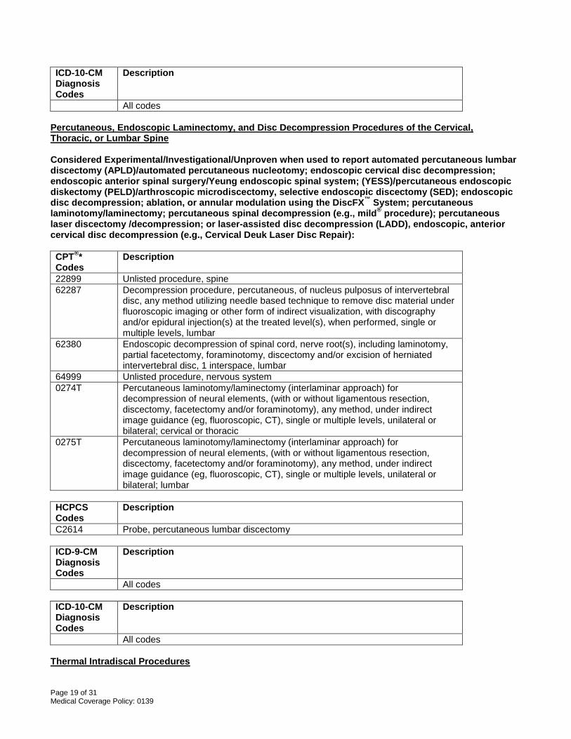
Page 19 of 31 Medical Coverage Policy: 0139
ICD-10-CM Diagnosis Codes
Description
All codes Percutaneous, Endoscopic Laminectomy, and Disc Decompression Procedures of the Cervical, Thoracic, or Lumbar Spine Considered Experimental/Investigational/Unproven when used to report automated percutaneous lumbar discectomy (APLD)/automated percutaneous nucleotomy; endoscopic cervical disc decompression; endoscopic anterior spinal surgery/Yeung endoscopic spinal system; (YESS)/percutaneous endoscopic diskectomy (PELD)/arthroscopic microdiscectomy, selective endoscopic discectomy (SED); endoscopic disc decompression; ablation, or annular modulation using the DiscFX™ System; percutaneous laminotomy/laminectomy; percutaneous spinal decompression (e.g., mild® procedure); percutaneous laser discectomy /decompression; or laser-assisted disc decompression (LADD), endoscopic, anterior cervical disc decompression (e.g., Cervical Deuk Laser Disc Repair): CPT®* Codes
Description
22899 Unlisted procedure, spine 62287 Decompression procedure, percutaneous, of nucleus pulposus of intervertebral
disc, any method utilizing needle based technique to remove disc material under fluoroscopic imaging or other form of indirect visualization, with discography and/or epidural injection(s) at the treated level(s), when performed, single or multiple levels, lumbar
62380 Endoscopic decompression of spinal cord, nerve root(s), including laminotomy, partial facetectomy, foraminotomy, discectomy and/or excision of herniated intervertebral disc, 1 interspace, lumbar
64999 Unlisted procedure, nervous system 0274T
Percutaneous laminotomy/laminectomy (interlaminar approach) for decompression of neural elements, (with or without ligamentous resection, discectomy, facetectomy and/or foraminotomy), any method, under indirect image guidance (eg, fluoroscopic, CT), single or multiple levels, unilateral or bilateral; cervical or thoracic
0275T Percutaneous laminotomy/laminectomy (interlaminar approach) for decompression of neural elements, (with or without ligamentous resection, discectomy, facetectomy and/or foraminotomy), any method, under indirect image guidance (eg, fluoroscopic, CT), single or multiple levels, unilateral or bilateral; lumbar
HCPCS Codes
Description
C2614 Probe, percutaneous lumbar discectomy ICD-9-CM Diagnosis Codes
Description
All codes ICD-10-CM Diagnosis Codes
Description
All codes Thermal Intradiscal Procedures
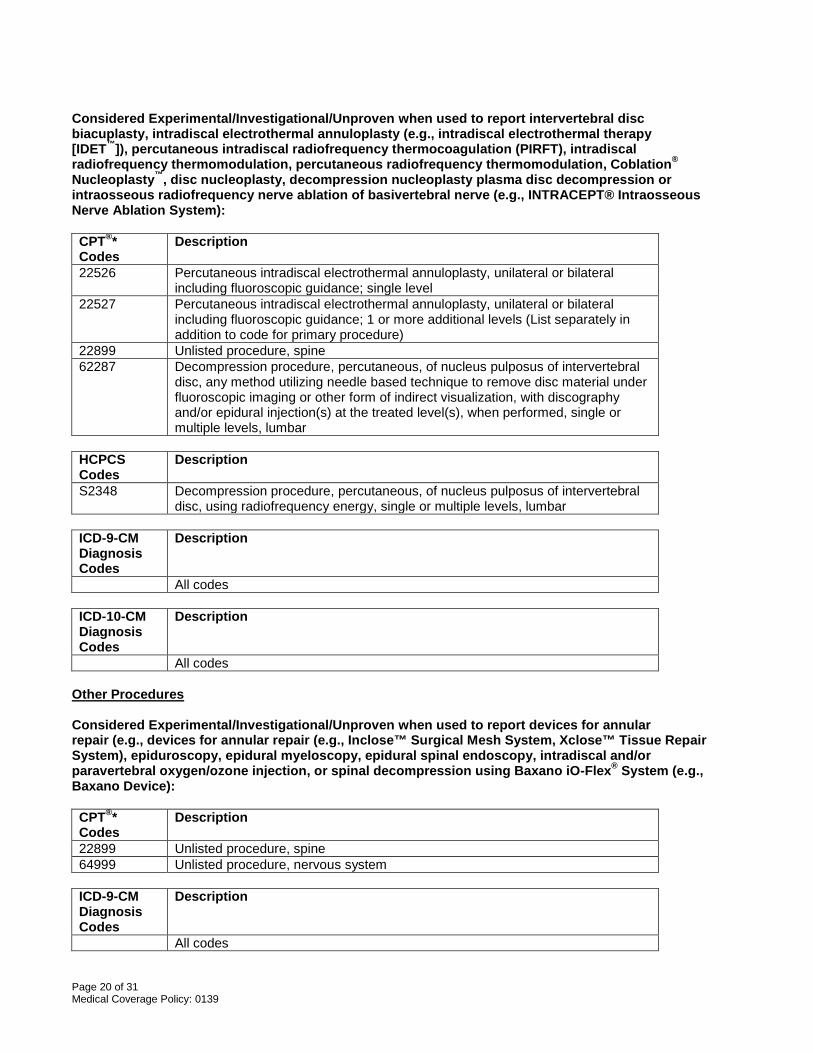
Page 20 of 31 Medical Coverage Policy: 0139
Considered Experimental/Investigational/Unproven when used to report intervertebral disc biacuplasty, intradiscal electrothermal annuloplasty (e.g., intradiscal electrothermal therapy [IDET™]), percutaneous intradiscal radiofrequency thermocoagulation (PIRFT), intradiscal radiofrequency thermomodulation, percutaneous radiofrequency thermomodulation, Coblation®
Nucleoplasty™, disc nucleoplasty, decompression nucleoplasty plasma disc decompression or intraosseous radiofrequency nerve ablation of basivertebral nerve (e.g., INTRACEPT® Intraosseous Nerve Ablation System): CPT®* Codes
Description
22526 Percutaneous intradiscal electrothermal annuloplasty, unilateral or bilateral including fluoroscopic guidance; single level
22527 Percutaneous intradiscal electrothermal annuloplasty, unilateral or bilateral including fluoroscopic guidance; 1 or more additional levels (List separately in addition to code for primary procedure)
22899 Unlisted procedure, spine 62287 Decompression procedure, percutaneous, of nucleus pulposus of intervertebral
disc, any method utilizing needle based technique to remove disc material under fluoroscopic imaging or other form of indirect visualization, with discography and/or epidural injection(s) at the treated level(s), when performed, single or multiple levels, lumbar
HCPCS Codes
Description
S2348 Decompression procedure, percutaneous, of nucleus pulposus of intervertebral disc, using radiofrequency energy, single or multiple levels, lumbar
ICD-9-CM Diagnosis Codes
Description
All codes ICD-10-CM Diagnosis Codes
Description
All codes Other Procedures Considered Experimental/Investigational/Unproven when used to report devices for annular repair (e.g., devices for annular repair (e.g., Inclose™ Surgical Mesh System, Xclose™ Tissue Repair System), epiduroscopy, epidural myeloscopy, epidural spinal endoscopy, intradiscal and/or paravertebral oxygen/ozone injection, or spinal decompression using Baxano iO-Flex® System (e.g., Baxano Device): CPT®* Codes
Description
22899 Unlisted procedure, spine 64999 Unlisted procedure, nervous system
ICD-9-CM Diagnosis Codes
Description
All codes
Page 21 of 31 Medical Coverage Policy: 0139
ICD-10-CM Diagnosis Codes
Description
All codes *Current Procedural Terminology (CPT®) ©2016 American Medical Association: Chicago, IL. References 1. Abejon D, Garcia-del-Valle S, Fuentes ML, Gomez-Arnau JI, Reig E, van Zundert J. Pulsed radiofrequency
in lumbar radicular pain: clinical effects in various etiological groups. Pain Pract. 2007 Mar;7(1):21-6. 2. American Society of Anesthesiologists Task Force on Chronic Pain Management, American Society of
Regional Anesthesia and Pain Medicine. Practice guidelines for chronic pain management: an updated report by the American Society of Anesthesiologists Task Force on Chronic Pain Management and the American Society of Regional Anesthesia and Pain Medicine. Anesthesiology. 2010 Apr;112(4):810-33.
3. Arts MP, Brand R, van den Akker ME, Koes BW, Bartels RH, Peul WC; Leiden-The Hague Spine
Intervention Prognostic Study Group (SIPS). Tubular diskectomy vs conventional microdiskectomy for sciatica: a randomized controlled trial. JAMA. 2009 Jul 8;302(2):149-58.
4. Arts MP, Brand R, van den Akker ME, Koes BW, Bartels RH, Peul WC;Tubular diskectomy vs conventional
microdiskectomy for the treatment of lumbar disk herniation: 2-year results of a double-blind randomized controlled trial. Neurosurgery. 2011 Jul;69(1):135-44; discussion 144.
5. Aydin SM, Gharibo CG, Mehnert M, Stitik TP. The role of radiofrequency ablation for sacroiliac joint pain: a
meta-analysis. PM R. 2010 Sep;2(9):842-51.
6. Becker S, Hadjipavlou A, Heggeness MH. Ablation of the basivertebral nerve for treatment of back pain: a clinical study. Spine J. 2017 Feb;17(2):218-223.
7. Benyamin RM, Manchikanti L, Parr AT, Diwan S, Singh V, Falco FJ, et al. The effectiveness of lumbar
interlaminar epidural injections in managing chronic low back and lower extremity pain. Pain Physician. 2012 Jul-Aug;15(4):E363-404.
8. Benyamin RM, Wang VC, Vallejo R, Singh V, Helm Ii S. A systematic evaluation of thoracic interlaminar
epidural injections. Pain Physician. 2012 Jul-Aug;15(4):E497-514. 9. Bhagia SM, Slipman CW, Nirschi M, Isaac Z, El-Abd, Sharps LS, Garvin C. Side effects and complications
after percutaneous disc decompression using coblation technology. Am J Phys Med Rehabil. 2006 Jan;85(1):6-13.
10. Bogduk N. Evidence-informed management of chronic low back pain with facet injections and radiofrequency
neurotomy. Spine J. 2008 Jan-Feb;8(1):56-64. 11. Boswell MV, Shah RV, Everett CR, Sehgal N, Mckenzie-Brown AM, Abdl S, et al. Interventional techniques
in the management of chronic spinal pain: evidence-based practice guidelines. Pain Physician. 8(1); 2005. 12. Boswell MV, Trescot AM, Datta S, Schulz DM, Hansen HC, Abdi S, et al.; American Society of Interventional
Pain Physicians. Interventional techniques: evidence-based practice guidelines in the management of chronic spinal pain. Pain Physician. 2007 Jan;10(1):7-111.
13. Botwin KP, Sharma K, Saliba R, Patel BC. Ultrasound-guided trigger point injections in the cervicothoracic
musculature: a new and unreported technique. Pain Physician. 2008 Nov-Dec;11(6):885-9.
Page 22 of 31 Medical Coverage Policy: 0139
14. Brown LL. A double-blind, randomized, prospective study of epidural steroid injection vs. the mild®
procedure in patients with symptomatic lumbar spinal stenosis. Pain Pract. 2012 Jun;12(5):333-41.
15. Bubnov RV, Wang J. Clinical Comparative Study for Ultrasound-Guided Trigger-Point Needling for Myofascial Pain Medical Acupuncture. Dec 2013: 437-443.
16. Cahana A, Van Zundert J, Macrea L, van Kleef M, Sluijter M. Pulsed radiofrequency: current clinical and
biological literature available. Pain Med. 2006 Sep-Oct;7(5):411-23. 17. Carragee EJ. Clinical practice. Persistent low back pain. N Engl J Med. 2005 May 5;352(18):1891-8. 18. Casal-Moro R, Castro-Menéndez M, Hernández-Blanco M, Bravo-Ricoy JA, Jorge-Barreiro FJ. Long-term
outcome after microendoscopic diskectomy for lumbar disk herniation: a prospective clinical study with a 5-year follow-up. Neurosurgery. 2011 Jun;68(6):1568-75; discussion 1575.
19. Centers for Medicare & Medicaid Services. National Coverage Determination (NCD) for Percutaneous
Image-Guided Lumbar decompression for Lumbar Spinal Stenosis. 2014 Jan 9. Accessed May 10, 2016. Available at URL address: http://www.cms.gov/medicare-coverage-database/details/ncd-details.aspx?NCDId=358&ncdver=1&CoverageSelection=Both&ArticleType=All&PolicyType=Final&s=All&KeyWord=Spinal+stenosis&KeyWordLookUp=Title&KeyWordSearchType=And&from2=search.asp&bc=gAAAABAAAAAAAA%3D%3D&
20. Cervical and thoracic spine disorders. In: Hegmann, KT, editor. Occupational medicine practice guidelines:
evaluation and management of common health problems and functional recovery in workers, 3rd ed. Elk Grove Village, IL. American College of Occupational and Environmental Medicine (ACOEM); 2011.
21. Chen Y, Derby R, Lee, S. Percutaneous disc decompression in the management of chronic low back pain.
Orthop Clin North Am 2004 Jan;35(1):17-23. 22. Chen H, Kelling J. mild procedure for lumbar decompression: a review. Pain Pract. 2013 Feb;13(2):146-53. 23. Childers MK, Feldman JB, Guo HM. Myofascial pain syndrome. In: Frontera W, editor. Essentials of
Physical Medicine and Rehabilitation, 2nd ed. Saunders, an imprint of Elsevier; 2008. 24. Chopko BW. Long-term results of percutaneous lumbar decompression for LSS: two-year outcomes. Clin J
Pain. 2013 Nov;29(11):939-43.
25. Chou R, Atlas SJ, Stanos SP, Rosenquist RW. Nonsurgical interventional therapies for low back pain: a review of the evidence for an American Pain Society Clinical Practice Guideline. Spine. 2009 May 1;34(10):1078-93.
26. Chou R, Loeser JD, Owens DK, Rosenquist RW, Atlas SJ, Baisden J, et al. for the American Pain Society
Low Back Pain Guideline Panel. Interventional therapies, surgery, and interdisciplinary rehabilitation for low back pain. Spine. 2009 14(10): 1066-1077.
27. Chopko B, Caraway DL. MiDAS I (mild Decompression Alternative to Open Surgery): a preliminary report of
a prospective, multi-center clinical study. Pain Physician. 2010 Jul;13(4):369-78. 28. Chun-jing H, Hao-xiong N, jia-xiang N. The application of percutaneous lysis of epidural adhesions in
patients with failed back surgery syndrome. Acta Cir Bras. 2012 Apr;27(4):357-62.
29. Cincu R, Lorente Fde A, Gomez J, Eiras J, Agrawal A. One decade follow up after nucleoplasty in the management of degenerative disc disease causing low back pain and radiculopathy. Asian J Neurosurg. 2015 Jan-Mar;10(1):21-5.
Page 23 of 31 Medical Coverage Policy: 0139
30. Cohen SP, Bicket MC, Jamison D, Wilkinson I, Rathmell JP. Epidural steroids: a comprehensive, evidence-based review. Reg Anesth Pain Med. 2013 May-Jun;38(3):175-200.
31. Cojocaru MC, Cojocaru IM, Voiculescu VM, Cojan-Carlea NA, Dumitru VL, Berteanu M. Trigger points--
ultrasound and thermal findings. J Med Life. 2015 Jul-Sep;8(3):315-8.
32. Davis TT, Sra P, Fuller N, Bae H. Lumbar intervertebral thermal therapies. Orthop Clin North Am. 2003 Apr;34(2):255-62.
33. Deer TR, Kapurai L. New image-guided ultra-minimally invasive lumbar decompression method: the mild
procedure. Pain Physician. 2010 Jan;13(1):35-41.
34. Desai MJ, Kapural L, Petersohn JD, et al. Twelve-month follow-up of a randomized clinical trial comparing intradiscal biacuplasty to conventional medical management for discogenic lumbar pain. Pain Med. Aug 2016;0:1-13.
35. Deukmedjian AJ, Cianciabella A, Cutright J, Deukmedjian A. Cervical Deuk Laser Disc Repair(®): A novel,
full-endoscopic surgical technique for the treatment of symptomatic cervical disc disease. Surg Neurol Int. 2012;3:142. doi: 10.4103/2152-7806.
36. Deyo RA, Weinstein JN. Low back pain. N Engl J Med 2001;344(5):363-370. 37. Diwan S, Manchikanti L, Benyamin RM, Bryce DA, Geffert S, Hameed H, et al. Effectiveness of cervical
epidural injections in the management of chronic neck and upper extremity pain. Pain Physician. 2012 Jul-Aug;15(4):E405-34.
38. Eskander MS, Aubin ME, Drew JM, Eskander JP, Balsis SM, Eck J, Lapinsky AS, Connolly PJ. Is there a
difference between simultaneous or staged decompressions for combined cervical and lumbar stenosis? J Spinal Disord Tech. 2011 Aug;24(6):409-13.
39. Falco FJ, Datta S, Manchikanti L, Sehgal N, Geffert S, Singh V, Smith HS, Boswell MV. An updated review
of the diagnostic utility of cervical facet joint injections. Pain Physician. 2012 Nov-Dec;15(6):E807-38. 40. Falco FJ, Manchikanti L, Datta S, Wargo BW, Geffert S, Bryce DA, Atluri S, et al. Systematic review of the
therapeutic effectiveness of cervical facet joint interventions: an update. Pain Physician. 2012 Nov-Dec;15(6):E839-68.
41. Fessler RG, O’Toole JE, Eicholz KM, Perez-Cruet MJ. The development of minimally invasive spine surgery.
Neurosurg Clin N Am. 2006 Oct;17(4):401-9. 42. Freeman BJC. IDET: a critical appraisal of the evidence. Eur Spine J. 2006 Aug;15 (Supplement 15):448-
457. 43. Freeman BJC, Fraser RD, Cain, CMJ, Hall, DJ, Chapple, DCL. A randomized, double-blind, controlled trial:
intradiscal electrothermal therapy versus placebo for the treatment of chronic discogenic low back pain. Spine. 2005 Nov 1;30(21):2369-77; discussion 2378.
44. Frontera: Essentials of physical medicine and rehabilitation. Saunders, an imprint of Elsevier; 2008. 45. Furlan AD, van Tulder M, Cherkin D, Tsukayama H, Lao L, Koes B, Berman B. Acupuncture and dry-
needling for low back pain. Cochrane Database Syst Rev. 2005 Jan 25;(1):CD001351. 46. Gardocki RJ, Park AL. Lower back pain and disorders of intervertebral discs. In: Canale & Beaty: Campbell’s
operative orthopaedics, 12th ed. Mosby, an imprint of Elsevier; 2012.
Page 24 of 31 Medical Coverage Policy: 0139
47. Garg B, Nagraja UB, Jayaswal A. Microendoscopic versus open discectomy for lumbar disc herniation: a prospective randomised study. J Orthop Surg (Hong Kong). 2011 Apr;19(1):30-4.
48. Gerdesmeyer L, Wagenpfeil S, Birkenmaier C, Veihelmann A, Hauschild M, Wagner K, et al. Percutaneous
epidural lysis of adhesions in chronic lumbar radicular pain: a randomized, double-blind, placebo-controlled trial. Pain Physician. 2013 May-Jun;16(3):185-96.
49. Gibson JNA, Grant IC, Waddell G. Surgical interventions for lumbar disc prolapse. Cochrane Database Syst
Rev. 2007 Apr 18;(2):CD001350. 50. Goroll AH, Mulley AG. Approach to the patient with back pain. In: Primary care medicine, office evaluation
and management of the adult patient, 6th ed. Philadelphia, PA: Lippincott Williams & Wilkins; 2009.
51. Gupta L, Singh SP. Ultrasound-Guided Trigger Point Injection for Myofascial Trigger Points in the Subscapularis and Pectoralis Muscles. Yonsei Med J. 2016 Mar;57(2):538.
52. Haines SJ, Jordan N, Boen JR, Nyman JA, Oldridge NB, Lindgren BR. Discectomy strategies for lumbar disc
herniation: results of the LAPDOG trial. J Clin Neurosci. 2002 Jul;9(4):440-6. 53. Han PP, Kenny K, Dickman CA. Thoracoscopic approaches to the thoracic spine: experience with 241
surgical procedures. Neurosurgery. 2002 Nov;51(5 Suppl):S88-95. 54. Hansen H, Manchikanti L, Simopoulos TT, Christo PJ, Gupta S, Smith HS, Hameed H, Cohen SP. A
systematic evaluation of the therapeutic effectiveness of sacroiliac joint interventions. Pain Physician. 2012 May;15(3):E247-78.
55. Hansen HC, McKenzie-Brown, AM, Cohen SP, Swicegood JR, Colson JD, Manchikanti L. Sacroiliac joint
interventions: a systematic review. Pain Physician. 2007 Jan;10(1):165-84.
56. Hayes, Inc. Medical Technology Directory Report. Automated percutaneous lumbar discectomy. Lansdale, Pa. Hayes, Inc.; published December 4, 2013; annual review 2014, 2015, 2016.
57. Hayes, Inc. Medical Technology Directory Report. Percutaneous Disc Decompression for Cervical Disc
Herniation. Lansdale, Pa. Hayes, Inc. published 2014, annual review 2014, 2017a. 58. Hayes, Inc. Health Technology Brief. Minimally invasive lumbar decompression (mild; Vertos Medical Inc.)
for lumbar spinal stenosis. Hayes, Inc.; published October 2, 2014, reviewed August 10, 2015. 59. Hayes, Inc. Health Technology Brief. Percutaneous Endoscopic Lumbar Discectomy for Primary Lumbar
Disc Herniation. Lansdale, Pa. Hayes, Inc. March 2, 2017b.
60. Hayes, Inc. Health Technology Brief. Percutaneous Endoscopic Lumbar Discectomy for Recurrent Lumbar Disc Herniation. Lansdale, Pa. Hayes, Inc. March 23, 2017c.
61. Hayes, Inc. Search and Summary. Ultrasound-guided trigger point injections for treatment of pain in adults.
Lansdale, Pa. Hayes, Inc.; March 31, 2016. 62. Helm Ii S, Benyamin RM, Chopra P, Deer TR, Justiz R. Percutaneous adhesiolysis in the management of
chronic low back pain in post lumbar surgery syndrome and spinal stenosis: a systematic review. Pain Physician. 2012 Jul-Aug;15(4):E435-62.
63. Helm II S, Deer TR, Manchikanti L, Datta S, Chopra P, Singh V, Hirsch JA. Effectiveness of thermal annular
procedures in treating discogenic low back pain. Pain Physician. 2012 May-Jun;15(3):E279-304.
Page 25 of 31 Medical Coverage Policy: 0139
64. Hirsch JA, Singh V, Falco FJE, Benyamin RM, Manchikanti L. Automated percutaneous lumbar discectomy for the contained herniated lumbar disc:a systematic assessment of evidence. Pain Physician. 2009 May-Jun;12(3):601-20.
65. Igarashi t, Hirahayashi Y, Seo N, Suitoh K, Fuuda H, Suzuki H. Lysis of adhesions and epidural injection of
steroid/local anaesthetic during epiduroscopy potentially alleviate low back and leg pain in elderly patients with lumbar spinal stenosis. Br J Anaesth. 2004 Aug;93(2):181-7.
66. Institute for Clinical Systems Improvement (ICSI). Health care guideline: adult acute and subacute low back
pain. 2012 Jan. Accessed May 10, 2016. Available at URL address: http://www.icsi.org/search/?q=back+pain+
67. Ito K, Yukawa Y, Machino M, Inoue T, Ouchida J, Tomita K, Kato F. Treatment outcomes of intradiscal
steroid injection/selective nerve root block for 161 patients with cervical radiculopathy. Nagoya J Med Sci. 2015 Feb;77(1-2):213-9.
68. Iwatsuki K, Yoshimine T, Awash K. Alternative denervation using laser irradiation in lumbar facet syndrome.
Lasers Surg Med. 2007 Mar;39(3):225-9. 69. Kandarpa K, Machan L. Spinal facet procedures and sacroiliac joint injections. In: Handbook of interventional
radiologic procedures, 4th ed. Lippincott, Williams & Wilkins, 2011. 70. Kapural L, Ng A, Dalton J, Mascha E, Kapural M, de La Garza M, Mekhail N. Intervertebral disc biacuplasty
for the treatment of lumbar discogenic pain: results of a six-month follow-up. Pain Med. 2008 Jan-Feb;9(1):60-7.
71. Kapural L, Vrooman B, Sarwar S, Krizanac-Bengez L, Rauck R, Gilmore C, North J, Girgis G, Mekhail N. A
randomized, placebo-controlled trial of transdiscal radiofrequency, biacuplasty for treatment of discogenic lower back pain. Pain Med. 2013 Mar;14(3):362-73.
72. Kapural L, Vrooman B, Sarwar S, Krizanac-Bengez L, Rauck R, Gilmore C, North J, Mekhail N.
Radiofrequency intradiscal biacuplasty for treatment of discogenic lower back pain: a 12-month follow-up. Pain Med. 2015 Mar;16(3):425-31.
73. Kikuike K, Miyamoto K, Hosoe H, Shimizu K. One-staged combined cervical and lumbar decompression for
patients with tandem spinal stenosis on cervical and lumbar spine: analyses of clinical outcomes with minimum 3 years follow-up. J Spinal Disord Tech. 2009 Dec;22(8):593-601.
74. Kumbhare DA, Elzibak AH, Noseworthy MD. Assessment of Myofascial Trigger Points Using Ultrasound. Am
J Phys Med Rehabil. 2016 Jan;95(1):72-80. 75. Levin KH. Nonsurgical interventions for spine pain. Neurol Clin. 2007 May;25(2):495-505.
76. Lewis J, Tehan P. A blinded pilot study investigating the use of diagnostic ultrasound for detecting active
myofascial trigger points. Pain. 1999 Jan;79(1):39-44. 77. Lindner R, Sluijter ME, Schleinzer W. Pulsed radiofrequency treatment of the lumbar medial branch for facet
pain: a retrospective analysis. Pain Med. 2006 Sep-Oct;7(5):435-9.
78. Liu L, Huang QM, Liu QG, Ye G, Bo CZ, Chen MJ, Li P. Effectiveness of dry needling for myofascial trigger points associated with neck and shoulder pain: a systematic review and meta-analysis. Arch Phys Med Rehabil. 2015 May;96(5):944-55.
79. Low back disorders. In: Hegmann KT, editor(s). Occupational medicine practice guidelines. Evaluation and
management of common health problems and functional recovery in workers. 3rd ed. Elk Grove Village (IL): American College of Occupational and Environmental Medicine (ACOEM); 2011.
Page 26 of 31 Medical Coverage Policy: 0139
80. Manchikanti L, Abdi S, Atluri S, Benyamin RM, Boswell MV, Buenaventura RM, et al. An update of
comprehensive evidence-based guidelines for interventional techniques in chronic spinal pain. Part II: guidance and recommendations. Pain Physician. 2013 Apr;16(2 Suppl):S49-283.
81. Manchikanti KN, Atluri S, Singh V, Geffert S, Sehgal N, Falco FJ. An update of evaluation of therapeutic
thoracic facet joint interventions. Pain Physician. 2012 Jul-Aug;15(4):E463-81. 82. Manchikanti L, Boswell MV, Singh V, Benyamin RM, Fellows B, Abdi S, Buenaventura RM, et al.
Comprehensive evidence-based guidelines for interventional techniques in the management of chronic spinal pain. Pain Physician. 2009 Jul-Aug;12(4):699-802.
83. Manchikanti L, Boswell MV, Rivera JJ, Pampati VS, Damron KS, McManus CD, et al. A randomized,
controlled trial of spinal endoscopic adhesiolysis in chronic refractory low back and lower extremity pain. BMC Anesthesiol. 2005 Jul 6;5:10.
84. Manchikanti L, Boswell MV, Singh V, Pampati V, Damrom KS, Beyer CD. Prevalence of facet joint pain in
chronic spinal pain of cervical, thoracic, and lumbar regions. BMC Musculoskelet Disord. 2004 May 28;5(1):15.
85. Manchikanti L, Cash KA, McManus CD, Pampati V, Benyamin RM. A randomized, double-blind, active-
controlled trial of fluoroscopic lumbar interlaminar epidural injections in chronic axial or discogenic low back pain: results of 2-year follow-up. Pain Physician. 2013 Sep-Oct;16(5):E491-504.
86. Manchikanti L, Pampati V, Fellows B, Rivera J, Beyer CD, Damron KS. Role of one day epidural
adhesiolysis in management of chronic low back pain: a randomized clinical trial. Pain Physicain. 2001; 4(2):153-66.
87. Manchikanti L, Cash KA, Pampati V, Wargo BW, Malla Y. A randomized, double-blind, active control trial of
fluoroscopic cervical interlaminar epidural injections in chronic pain of cervical disc herniation: results of a 2-year follow-up. Pain Physician. 2013 Sep-Oct;16(5):465-78.
88. Manchikanti L, Rivera JJ, Pampati V, Damron KS, Beyer CD, Brandon, DE. Spinal endoscopic adhesiolysis
in the management of chronic low back pain: a preliminary report of a randomized double-blind trial. Pain Physician. 2003; 6(3):259-67.
89. Manchikanti L, Rivera JJ, Pampati V, Damron KS, McManus CD, Brandon, DE. One day lumbar epidural
adhesiolysis and hypertonic saline neurolysis in treatment of chronic low back pain: a randomized double-blind trial. Pain Physician. 2004; 7(2):177-86.
90. Manchikanti L, Singh V, Cash KA, Pampati V, Damron KS, Boswell MV. Effect of fluoroscopically guided
caudal epidural steroid or local anesthetic injections in the treatment of lumbar disc herniation and radiculitis: a randomized, controlled, double blind trial with a two-year follow-up. Pain Physician. 2012 Jul-Aug;15(4):273-86.
91. Manchikanti L, Singh V, Calodney AK, Helm S 2nd, Deer TR, Benyamin RM, Falco FJ, Hirsch JA.
Percutaneous lumbar mechanical disc decompression utilizing Dekompressor®: an update of current evidence. Pain Physician. 2013 Apr;16(2 Suppl):SE1-24.
92. Manchikanti L, Singh V, Falco FJ, Calodney AK, Onyewu O, Helm S 2nd, Benyamin RM, Hirsch JA. An
updated review of automated percutaneous mechanical lumbar discectomy for the contained herniated lumbar disc. Pain Physician. 2013 Apr;16(2 Suppl):SE151-84.
93. Manchikanti L, Singh V, Falco FJ, Cash VP. Evaluation of lumbar facet joint nerve blocks in managing
chronic low back pain: a randomized, double-blind, controlled trial with a 2-year follow-up. Int J Med Sci. 2010 May 28;7(3):124-35.
Page 27 of 31 Medical Coverage Policy: 0139
94. Manchikanti L, Singh V, Kloth D, Slipman CW, Jasper JF, Trescot AM, et al. Interventional techniques in the
management of chronic pain: part 2.0. ASIPP Practice Guidelines. Pain Physician. 4(1):24-98. 95. Manchikanti L, Singh V, Falco FJ, Cash KA, Fellows B. Comparative outcomes of a 2-year follow-up of
cervical medial branch blocks in management of chronic neck pain: a randomized, double-blind controlled trial. Pain Physician. 2010 Sep-Oct;13(5):437-50.
96. Martin DC, Willis ML, Mullinax A, Clarke NL, Homburger JA, Berger IH. Pulsed radiofrequency application in
the treatment of chronic pain. Pain Pract. 2007 Mar;7(1):31-5. 97. Mekhail N, Costandi S, Abraham B, Samuel SW. Functional and patient-reported outcomes in symptomatic
lumbar spinal stenosis following percutaneous decompression. Pain Pract. 2012 Jul;12(6):417-25. 98. Mekhail N, Vallejo R, Coleman MH, Benyamin RM. Long-term results of percutaneous lumbar
decompression mild(®) for spinal stenosis. Pain Pract. 2012 Mar;12(3):184-93. doi: 10.1111/j.1533-2500.2011.00481.x. Epub 2011 Jun 16.
99. Murphy K, Muto M, Steppan J, Meaders T, Boxley C. Treatment of Contained Herniated Lumbar Discs With
Ozone and Corticosteroid: A Pilot Clinical Study. Can Assoc Radiol J. 2015 Nov;66(4):377-84.
100. Muto M, Ambrosanio G, Guarnieri G, Capobianco E, Piccolo G, Annunziata G, Rotondo A. Low back pain and sciatica: treatment with intradiscal-intraforaminal O(2)-O (3) injection. Our experience. Radiol Med. 2008 Aug;113(5):695-706.
101. National Institute for Clinical Excellence (NICE). Percutaneous intradiscal radiofrequency treatment of the
intervertebral disc nucleus for low back pain. Interventional procedures guidance [IPG545]. January 2016). Accessed April 25, 2017. Available at URL address: https://www.nice.org.uk
102. National Institute for Health and Clinical Excellence. Percutaneous coblation of the intervertebral disc for low
back pain and sciatica (2016, IPG543). Accessed April 25, 2017. Available at URL address: https://www.nice.org.uk
103. National Institute for Clinical Excellence (NICE). Percutaneous intradiscal radiofrequency treatment of the
intervertebral disc nucleus for low back pain (2016, IPG545). Accessed April 25, 2017. Available at URL address: https://www.nice.org.uk
104. National Institute for Clinical Excellence (NICE). Epiduroscopic lumbar discectomy through the sacral hiatus
for sciatica (2016, IPG570). Accessed April 25, 2017. Available at URL address: https://www.nice.org.uk
105. National Institute for Clinical Excellence (NICE). Percutaneous endoscopic laser thoracic discectomy. Interventional Procedures Guidance 61. London, UK: NICE; 2004 May. Accessed April 25, 2017. Available at URL address: https://www.nice.org.uk
106. National Institute for Health and Clinical Excellence. Percutaneous endoscopic laser cervical discectomy.
Interventional Procedures Guidance 303. London, UK: National Institute for Health and Clinical Excellence; 2009. Accessed April 25, 2017. Available at URL address: https://www.nice.org.uk
107. National Institute for Clinical Excellence (NICE). Therapeutic endoscopic division of epidural adhesions.
Interventional Procedures Guidance 333. London, UK: NICE; 2010 Feb. Accessed May 10, 2016. Available at URL address: http://guidance.nice.org.uk/IPG333
108. Nelemans PJ, de Bie RA, de Vet HCW, Sturmans F. Injection therapy for subacute and chronic low-back
pain. Cochrane Database Syst Rev. 2000;(2):CD001824; amended 2005 Feb 23.
Page 28 of 31 Medical Coverage Policy: 0139
109. Nellensteijn J, Ostelo R, Bartels R, Peul W, van Royen B, van Tulder M. Transforaminal endoscopic surgery for lumbar stenosis: a systematic review. Eur Spine J. 2010 Jun;19(6):879-86.
110. North American Spine Society. Choosing Wisely: Five things physicians and patients should question.
Accessed May 10, 2016. Available at URL address: http://www.choosingwisely.org/wp-content/uploads/2013/10/NASS-5things-List_102013.pdf
111. North American Spine Society (NASS). Clinical Guidelines for Diagnosis and Treatment of Lumbar Disc
Herniation with Radiculopathy. 2012. Accessed November 10, 2015. Available at URL address: http://www.spine.org/Documents/LumbarDiscHerniation.pdf
112. North American Spine Society (NASS). Evidence based clinical guidelines for multidisciplinary spine care.
Diagnosis and Treatment of Cervical Radiculopathy from Degenerative Disorders. 2012. Accessed October 23, 2015. Available at URL address: https://www.spine.org/Documents/ResearchClinicalCare/Guidelines/CervicalRadiculopathy.pdf
113. North American Spine Society (NASS). Evidence based clinical guidelines for multidisciplinary spine care.
Diagnosis and Treatment of Degenerative Lumbar Spinal Stenosis. Revised 2011. Accessed May 10, 2016. Available at URL address: https://www.spine.org/Documents/ResearchClinicalCare/Guidelines/LumbarStenosis.pdf
114. Novak S, Nemeth WC. The basis for recommending repeating epidural steroid injections for radicular low
back pain: a literature review. Arch Phys Med Rehabil. 2008 Mar;89(3):543-52.
115. Obernauer J, Galiano K, Gruber H, Bale R, Obwegeser AA, Schatzer R, Loizides A. Ultrasound-guided versus Computed Tomography-controlled facet joint injections in the middle and lower cervical spine: a prospective randomized clinical trial. Med Ultrason. 2013 Mar;15(1):10-5.
116. Park Y, Lee JH, Park KD, Ahn JK, Park J, Jee H. Ultrasound-Guided vs. Fluoroscopy-Guided Caudal
Epidural Steroid Injection for the Treatment of Unilateral Lower Lumbar Radicular Pain: A Prospective, Randomized, Single-Blind Clinical Study. Am J Phys Med Rehabil. 2013 Apr 29.
117. Parr AT, Manchikanti L, Hameed H, Conn A, Manchikanti KN, Benyamin RM, et al. Caudal epidural
injections in the management of chronic low back pain: a systematic appraisal of the literature. Pain Physician. 2012 May;15(3):E159-98.
118. Patel N, Gross A, Brown L, Gekht G. A randomized, placebo-controlled study to assess the efficacy of lateral
branch neurotomy for chronic sacroiliac joint pain. Pain Med. 2012 Mar;13(3):383-98.
119. Ren DJ, Liu XM, Du SY, Sun TS, Zhang ZC, Li F. Percutaneous Nucleoplasty Using Coblation Technique for the Treatment of Chronic Nonspecific Low Back Pain: 5-year Follow-up Results. Chin Med J (Engl). 2015 Jul 20;128(14):1893-7.
120. Resnick DK, Choudhri TF, Dailey AT, Groff MW, Khoo L, Matz PG, et al. American Association of
Neurological Surgeons/Congress of Neurological Surgeons. Guidelines for the performance of fusion procedures for degenerative disease of the lumbar spine. Part 13: injection therapies, low-back pain, and lumbar fusion. J Neurosurg Spine. 2005 Jun;2(6):707-15.
121. Rha DW, Shin JC, Kim YK, Jung JH, Kim YU, Lee SC. Detecting local twitch responses of myofascial trigger
points in the lower-back muscles using ultrasonography. Arch Phys Med Rehabil. 2011 Oct;92(10):1576-1580.e1.
122. Rhighesso O, Falavigna A, Avanzi O. Comparison of open discectomy with microendoscopic discectomy in
lumbar disc herniations: results of a randomized controlled trial. Neurosurgery. 2007 Sep;61(3):545-9; discussion 549.
Page 29 of 31 Medical Coverage Policy: 0139
123. Ribeiro LH, Vilar Furtado IN, Konai MS, Rosenfeld A, Andreo AB, Natour J. Effect of Facet Joint Injection Versus Systemic Steroids in Low Back Pain: A Randomized Controlled Trial. Spine (Phila Pa 1976). 2014 Feb 27.
124. Schenk B, Brouwer PA, Paul WC, van Buchem MA. Percutaneous laser disk decompression: a review of the
literature. AJNR Am J Neuroradiol. 2006 Jan;27(1):232-5..
125. Scott A, Guo B. Health Technology Assessment. Trigger point injections for chronic non-malignant musculoskeletal pain. 2005; Alberta Heritage Foundation for Medical Research. Accessed May 10, 2016. Available at URL address: http://www.ihe.ca/advanced-search/trigger-point-injections-for-chronic-non-malignant-musculoskeletal-pain
126. Shankar H, Reddy S. Two- and three-dimensional ultrasound imaging to facilitate detection and targeting of
taut bands in myofascial pain syndrome. Pain Med. 2012 Jul;13(7):971-5.
127. Sharps LS, Isaac Z. Percutaneous disc decompression using nucleoplasty. Pain Physician. 2002: 5(2):121-26.
128. Sherk HH, Vangsness CT, Thabit G, Jackson RW. Electromagnetic surgical devices in orthopaedics. Lasers
and radiofrequency. J Bone Joint Surg Am. 2002 Apr;84-A(4):675-81.
129. Shin HJ, Shin JC, Kim WS, Chang WH, Lee SC. Yonsei Med J. Application of ultrasound-guided trigger point injection for myofascial trigger points in the subscapularis and pectoralis muscles to post-mastectomy patients: a pilot study. 2014 May;55(3):792-9.
130. Sikdar S, Shah JP, Gebreab T, Yen RH, Gilliams E, Danoff J, Gerber LH. Novel applications of ultrasound
technology to visualize and characterize myofascial trigger points and surrounding soft tissue. Arch Phys Med Rehabil. 2009 Nov;90(11):1829-38
131. Singh V, Piryani C, Liao K. Evaluation of percutaneous disc decompression using coblation in chronic back
pain with or without leg pain. Pain Physician. 2003; 6(3): 273-80.
132. Slipman CW, Bhat AL, Gilchrist RV, Issac Z, Chou L, Lenrow DA. A critical review of the evidence for the use of zygapophysial injections and radiofrequency denervation in the treatment of low back pain. Spine J. 2003 Jul-Aug;3(4):310-6.
133. Smuck M, Crisostomo RA, Trivedi K, Agrawal D. Success of initial and repeated medial branch neurotomy
for zygapophysial joint pain: a systematic review. PM R. 2012 Sep;4(9):686-92.
134. Staal JB, de Bie R, de Vet HCW, Hildebrandt J, Nelemans P. Injection therapy for subacute and chronic low-back pain. Cochrane Database Syst Rev. 2011 Issue 3.
135. Staats PS, Benyamin RM. AS ENCORE: Randomized Controlled Clinical Trial Report of 6-Month Results.
Pain Physician. 2016 Feb;19(2):25-38.
136. Steppan J, Meaders T, Muto M, Murphy KJ. A metaanalysis of the effectiveness and safety of ozone treatments for herniated lumbar discs. J Vasc Interv Radiol. 2010 Apr;21(4):534-48.
137. Suh MR, Chang WH, Choi HS, Lee SC. Ultrasound-guided myofascial trigger point injection into brachialis
muscle for rotator cuff disease patients with upper arm pain: a pilot study. Ann Rehabil Med. 2014 Oct;38(5):673-81.
138. Tekin I, Mirzai H, Ok G, Erbuyun K, Vatansever D. A comparison of conventional and pulsed radiofrequency
denervation in the treatment of chronic facet joint pain. Clin J Pain. 2007 Jul-Aug;23(6):524-9.
Page 30 of 31 Medical Coverage Policy: 0139
139. Trescott, AM, Chopra P, Abdi S, Datta S, Schultz. Systematic review of effectiveness and complications of adhesiolysis in the management of chronic spinal pain: an update. Pain Physician. 2007 Jan;10(1):129-46.
140. Urrutia G, Kovacs F, Nishishinya MB, Olabe J. Percutaneous thermocoagulation intradiscal techniques for
discogenic low back pain. Spine. 2007 May 1;32(10):1146-54.
141. U.S. Food and Drug Administration. Baxano MicroBlade Shaver and Accessories. 510(k) approval K100958. July 2010. Accessed April 13, 2017. Available at URL address: https://www.accessdata.fda.gov/cdrh_docs/pdf10/K100958.pdf
142. U.S. Food and Drug Administration. Neuro Check Device with IO-Flex Wire. 510(k) approval K113533. April
2012. Accessed April 13, 2017. Available at URL address: https://www.accessdata.fda.gov/scripts/cdrh/cfdocs/cfpmn/pmn.cfm?id=k113533
143. U.S. Food and Drug Administration. Intercept Intraosseous Nerve Ablation System. 510(k) approval
K153272. Accessed April 17, 2017. Available at URL address: https://www.accessdata.fda.gov/cdrh_docs/pdf15/k153272.pdf
144. U.S. Food and Drug Administration Drug Safety Communication. FDA requires label changes to warn of rare
but serious neurologic problems after epidural corticosteroid injections for pain. Accessed May 10, 2016. Available at URL address: http://www.fda.gov/Drugs/DrugSafety/ucm394280.htm
145. Vallejo R, Benyamin RM, Kramer J, Stanton G, Joseph NJ. Pulsed radiofrequency denervation for the
treatment of sacroiliac joint syndrome. Pain Med. 2006 Sep-Oct;7(5):429-34.
146. van Kleef M, Barendse AM, Kessels A, Voets HM, Weber WEJ, de Lane S. Randomized trial of radiofrequency lumbar facet denervation for chronic low back pain. Spine. 1999 Sep 15;24(18):1937-42.
147. van Kleef M, Liem L, Lousberg R, Barendse G, Kessels F, Sluijter M. Radiofrequency lesion adjacent to the
dorsal root ganglion for cervicobrachial pain: a prospective double blind randomized study. Neurosurgery. 1996;38(6):1127-32.
148. van Wijk RMAW, Geurts JW, Wynne HJ, Hammink E, Buskens E, Lousberg R, et al. Radiofrequency
denervation of lumbar facet joints in the treatment of chronic low back pain: a randomized, double-blind, sham lesion-controlled trial. Clin J Pain. 2005 Jul-Aug;21(4):335-44. Erratum in: Clin J Pain. 2005 Sep-Oct;21(5):462.
149. Van Zundert J, Patihn J, Kessels A, Lame I, van Suijekom H, van Kleef M. Pulsed radiofrequency adjacent to
the cervical dorsal root ganglion in chronic cervical radicular pain: a double-blind sham controlled randomized clinical trial. Pain. 2007;127:173-82.
150. Veihelmann A, Devens C, Trouillier H, Birkenmaier C, Gerdesmeyer L, Refior HJ. Epidural neuroplasty
versus physiotherapy to relieve pain in patients with sciatica: a prospective randomized blinded clinical trial. J Orthop Sci. 2006 Jul;11(4):365-9.
151. Watters WC 3rd, Resnick DK, Eck JC, Ghogawala Z, Mummaneni PV, Dailey AT, Choudhri TF, Sharan A,
Groff MW, Wang JC, Dhall SS, Kaiser MG. Guideline update for the performance of fusion procedures for degenerative disease of the lumbar spine. Part 13: injection therapies, low-back pain, and lumbar fusion. J Neurosurg Spine. 2014 Jul;21(1):79-90.
152. Wong CS1, Wong SH. A new look at trigger point injections. Anesthesiol Res Pract. 2012;2012:492452.
153. Wu Z, Wei LX, Li J, Wang Y, Ni D, Yang P, Zhang Y. Percutaneous treatment of non-contained lumbar disc
herniation by injection of oxygen-ozone combined with collagenase. Eur J Radiol. 2009 Dec;72(3):499-504.
Page 31 of 31 Medical Coverage Policy: 0139
154. Yeung AT, Tsou PM. Posterolateral endoscopic excision for lumbar disc herniation: Surgical technique, outcome, and complications in 307 consecutive cases. Spine. 2002 Apr 1;27(7):722
155. Zhang L, Ding XL, Zhao XL, Wang JN, Li YP, Tian M. Fluoroscopy-guided Bipolar Radiofrequency
Thermocoagulation Treatment for Discogenic Low Back Pain. Chin Med J (Engl). 2016 Oct 5;129(19):2313-8.
“Cigna Companies” refers to operating subsidiaries of Cigna Corporation. All products and services are provided exclusively by or through such operating subsidiaries, including Cigna Health and Life Insurance Company, Connecticut General Life Insurance Company, Cigna Behavioral Health, Inc., Cigna Health Management, Inc., QualCare, Inc., and HMO or service company subsidiaries of Cigna Health Corporation. The Cigna name, logo, and other Cigna marks are owned by Cigna Intellectual Property, Inc. © 2017 Cigna.