Embed Size (px)
Citation preview
92
Minimal apical preparation … Srikanth P et al Journal of International Oral Health 2015; 7(6):92-96
Original ResearchReceived: 28th January 2015 Accepted: 20th April 2015 Conflicts of Interest: None
Source of Support: Nil
Minimal Apical Enlargement for Penetration of Irrigants to the Apical Third of Root Canal System: A Scanning Electron Microscope StudyP Srikanth1, Amaravadi Gopi Krishna2, Siva Srinivas3, E Sujayeendranatha Reddy4, Someshwar Battu5, Swathi Aravelli1
Contributors:1Senior Lecturer, Department of Department of Conservative Dentistry and Endodontics, SVS Institute of Dental Sciences, Mahaboobnagar, Telangana, India; 2Reader, Department of Department of Conservative Dentistry and Endodontics, Saraswati Dhanwantari Dental College & Hospital, Parbhani, Maharashtra, India; 3Proffesor, Department of Department of Conservative Dentistry and Endodontics, VYWS Dental College and Hospital, Amaravathi, Maharashtra, India; 4Reader, Department of Department of Conservative Dentistry and Endodontics, SVS Institute of Dental Sciences, Mahaboobnagar, Telangana, India; 5Reader, Department of Department of Conservative Dentistry and Endodontics, Anil Neerukonda Institute of Dental Sciences, Vishakapatnam, Andhra Pradesh, India.Correspondence:Dr. Srikanth P. SVS Institute of Dental Sciences, Mahaboobnagar, Telangana, India. Phone: +91-9246901750. Email: [email protected] to cite the article:Srikanth P, Krishna AG, Srinivas S, Reddy ES, Battu S, Reddy N. Minimal apical enlargement for penetration of irrigants to the apical third of root canal system: A scanning electron microscope study. J Int Oral Health 2015;7(6):92-96.Abstract:Background: The aim of this study was to determine minimal apical enlargement for irrigant penetration into apical third of root canal system using scanning electron microscope (SEM).Materials and Methods: Distobuccal canals of 40 freshly extracted human maxillary first molar teeth were instrumented using crown-down technique. The teeth were divided into four test groups according to size of their master apical file (MAF) (#20, #25, #30, #35 0.06% taper), and two control groups. After final irrigation, removal of debris and smear layer from the apical third of root canals was determined under a SEM. Data was analyzed using Kruskal–Wallis and Mann–Whitney tests.Results: Smear layer removal in apical third for MAF size #30 was comparable with that of the control group (size #40).Conclusion: Minimal apical enlargement for penetration of irrigants to the apical third of root canal system is #30 size.
Key Words: Master apical file, minimal apical enlargement, smear layer
IntroductionThe aim of endodontic treatment is to eliminate microorganism from the root canal system and prevention of reinfection. To achieve this objective, root canals were cleaned before filling using mechanical instrumentation, supplemented with irrigants and intracanal medication.1 Morphology of root canals is very complex, some organic tissue and bacteria are left inside the canal system
despite using various chemomechanical preparations. Thus, root canal irrigation solution needed to aid canal debridement.2
Studies have shown that the mechanical instrumentation of root canals leave a smear layer covering the instrumented wall.3 The smear layer has been shown to hinder the penetration of intracanal disinfectants and sealer into dentinal tubules and has potential to compromise the seal of root canal filling.4
Sodium hypochlorite is most popularly used chemical solution in the chemo-mechanical preparation of the root canal system, and it has been systematically used in endodontics in a concentration ranging from 0.5% to 5.25%. Although it has excellent antimicrobial activity and capacity of dissolving organic materials, this solution alone does not effectively remove the smear layer, because its physiochemical action is limited to the removal of organic particles. Therefore, combination of NaOCl and EDTA are capable of removing the smear layer.5
The use of chemicals, ultrasonics, and lasers in combination or alone has been evaluated for removal of the smear layer with varying results. It has been reported that smear layer removal is less predictable in the apical region as compared with coronal and middle third of the root. This could be attributed to comparatively smaller apical canal dimensions hindering the penetration of irrigants and resulting in limited contact between canal wall and irrigants.1
During canal preparation, apical size has been crucial, in defining successful debridement of the root canal system.6 The irrigant penetration into the apical one-third of canal and removal of debris is dependent on the final size of the instrument used in the canals. The master apical file (MAF) size has been related to the initial apical size in many studies. Historically, the “three sizes up from the first file to bind,” rule was still being used in modified forms.7
Possible methods of increasing the penetration of irrigating solution into the apical third of root canal and dentinal tubule include the use of ultrasonics and addition of surfactants to reduce surface tension of irrigating solution.1
Materials and MethodsForty freshly extracted human maxillary first molar teeth with distobuccal root length of 19- 21 mm were used in this study. The access cavity was prepared, working length (WL) of the

93
Minimal apical preparation … Srikanth P et al Journal of International Oral Health 2015; 7(6):92-96
distobuccal canal was determined by #10 K-file 0.5 mm short of the apical foramen. Then the distobuccal root end was covered with melted wax to disable the operator from seeing root canal instrumentation during cleaning and shaping.
The teeth were divided into four experimental groups of eight teeth each, and two control groups with four teeth in each. The distobuccal canals were instrumented by crown down technique using hand files (DENTSPLY) and rotary files (K3 Sybron Endo).
The teeth in the four experimental groups were enlarged to a #20 size file (0.06 taper) in Group 1; #25 size file (0.06 taper) in Group 2; #30 size file (0.06 taper) in Group 3; and # 35 size file (0.06 taper) in Group 4. The two control groups were enlarged to # 40 size file (0.06 taper).
During the process of instrumentation, all groups were irrigated with 2 ml of 5% sodium hypochlorite using a 27-gauge needle. In the four experimental groups and positive control group each root canal received a final irrigation of 5 ml of smear clear for 1 min followed by 5 ml of 5% NaOCl. Final irrigation in the negative control group was only with 5 ml of 5% NaOCl. Root canals in all groups were irrigated with 5 ml of saline to remove any residue of irrigants and dried with paper points.
The distobuccal root was separated from each tooth using a high-speed handpiece. Each root was split longitudinally in a buccolingual direction with a chisel and mallet. One-half of each root was randomly selected and placed in 2% glutaraldehyde solution for 24 h.
The fixed specimens were rinsed two times with a sodium cacodylate buffered solution (0.1 M. pH 7.2), incubated in osmium tetraoxide for 1 h, dehydrated with ascending concentration of ethyl alcohol (30-100%) and placed in a dessicator for at least 24 h.
Each specimen was then mounted on a aluminium stub, coated with 25 µm of gold palladium, and examined under a scanning electron microscope. Photographs of the apical third of each canal were taken for final evaluation with a magnification of ×2500.
In a blind manner, three investigators scored the presence or absence of smear layer on the surface of a root canal or in the dentinal tubules from the coded photomicrographs.
A score 1 through 8 was used for the evaluation of photomicrographs.2
Score 1: The surface is devoid of debris and smear layer.
Score 2: The surface is devoid of smear layer, but little of debris is observed.
Score 3: The surface has been cleaned, but both smear layer and debris are dispersedly Observed.
Score 4: The surface has been cleaned, but the level of smear layer and debris is also noticeable.
Score 5: The clean surface is bit greater than the un-clean surface.
Score 6: Almost half of the debris and smear layer have been removed.
Score 7: The greater part of smear layer and debris are left.
Score 8: The surface is completely covered with smear layer and debris.
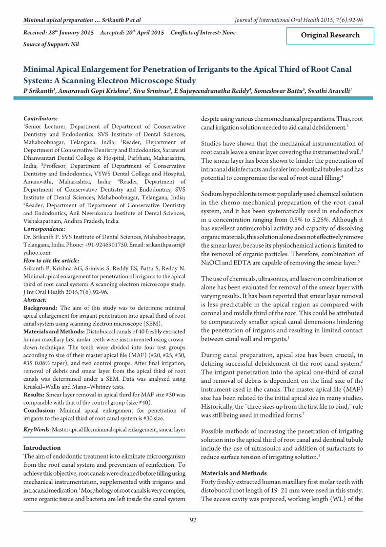
ResultsAll of the four specimens in the positive control group were free of smear layer and had significant erosion at the orifices of the dentinal tubules (Figure 1). All the specimens in the negative control group were covered with smear layer and debris (Table 1 and Figure 2). All of the specimens in the experimental Group 1 were covered with smear layer and debris (Figure 3). The mean score for specimens in the experimental Group 2 (Figure 4) (instrumented at the WL to a #25 size file) was 2.45 (Figure 5). The mean score for experimental Group 3 was 1.06 indicating that 90% of debris and smear layer was removed in this group. Similar observations were made in all the specimens
Table 1: Average scores of the smear layer and debris removal for control groups.
Sample Positive control (Size #40)
Negative control (Size #40)
1 1 82 1 83 1 84 1 8Mean 1 8
Figure 1: Scanning electron microscope image of positive control.
94
Minimal apical preparation … Srikanth P et al Journal of International Oral Health 2015; 7(6):92-96
Table 2: Average scores of the smear layer and debris removal for four groups.
Sample Group 1 (Size #20)
Group 2 (Size #25)
Group 3 (Size #30)
Group 4 (Size #35)
1 8 3 1 12 8 2.2 1 13 8 2 1 14 8 2.4 1 15 8 3 1.3 16 8 3 1.1 17 8 2 1 18 8 2 1.1 1Mean 8 2.45 1.06 1
Table 3: Inter group comparison for smear layer removal
Comparison between groups Z PGroup 1 versus Group 2 −3.614 0.000Group 2 versus Group 3 −3.435 0.000Group 3 versus Group 4 −1.852 0.234Group 1 versus Group 2 −1.924 0.322
Figure 2: Scanning electron microscope image of negative control.
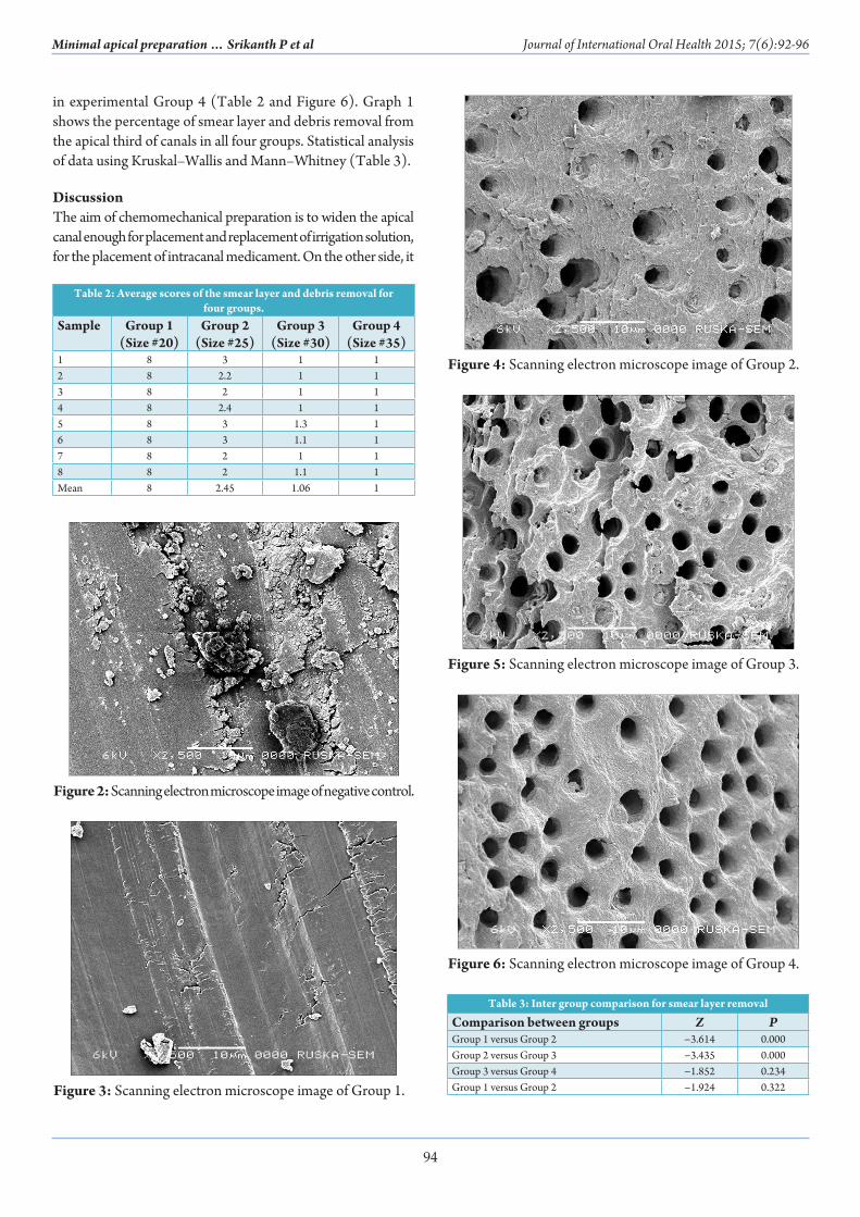
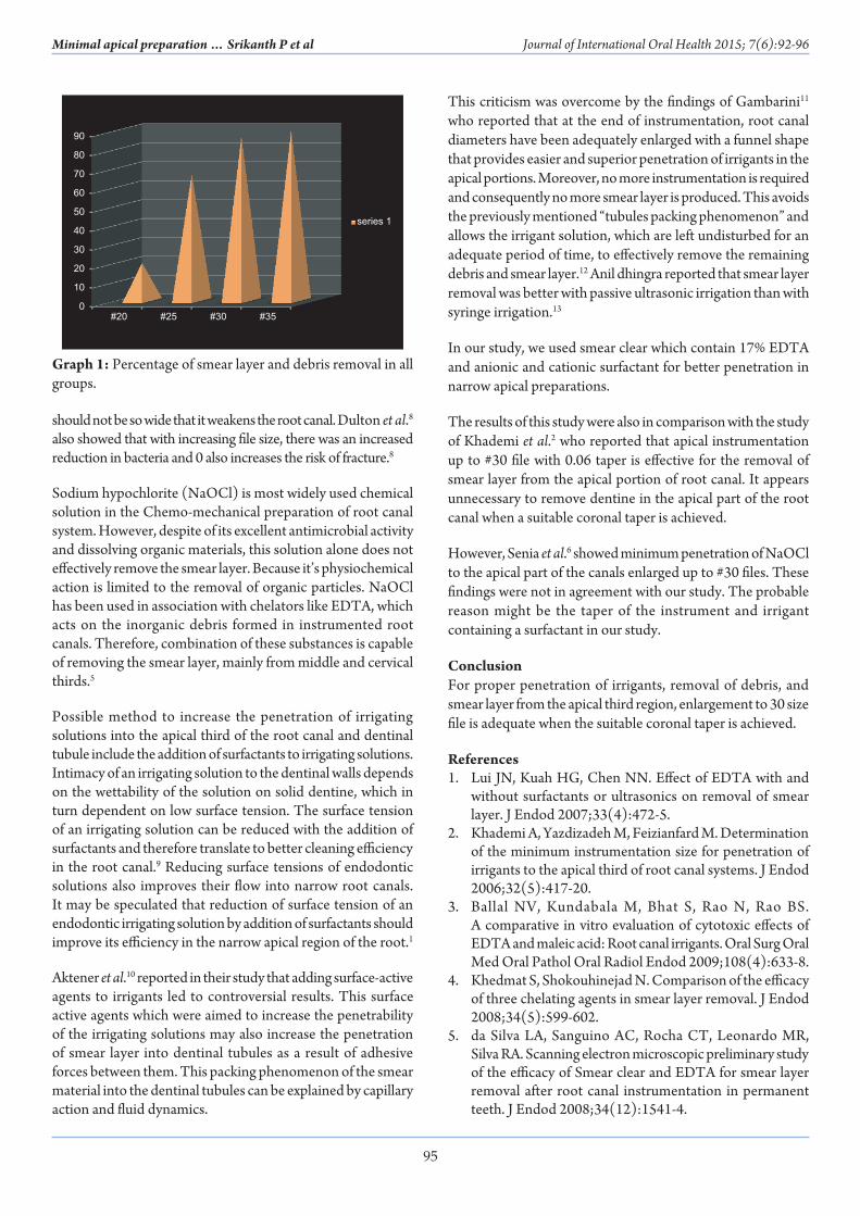
in experimental Group 4 (Table 2 and Figure 6). Graph 1 shows the percentage of smear layer and debris removal from the apical third of canals in all four groups. Statistical analysis of data using Kruskal–Wallis and Mann–Whitney (Table 3).
DiscussionThe aim of chemomechanical preparation is to widen the apical canal enough for placement and replacement of irrigation solution, for the placement of intracanal medicament. On the other side, it
Figure 3: Scanning electron microscope image of Group 1.
Figure 4: Scanning electron microscope image of Group 2.
Figure 5: Scanning electron microscope image of Group 3.
Figure 6: Scanning electron microscope image of Group 4.
95
Minimal apical preparation … Srikanth P et al Journal of International Oral Health 2015; 7(6):92-96
should not be so wide that it weakens the root canal. Dulton et al.8 also showed that with increasing file size, there was an increased reduction in bacteria and 0 also increases the risk of fracture.8
Sodium hypochlorite (NaOCl) is most widely used chemical solution in the Chemo-mechanical preparation of root canal system. However, despite of its excellent antimicrobial activity and dissolving organic materials, this solution alone does not effectively remove the smear layer. Because it’s physiochemical action is limited to the removal of organic particles. NaOCl has been used in association with chelators like EDTA, which acts on the inorganic debris formed in instrumented root canals. Therefore, combination of these substances is capable of removing the smear layer, mainly from middle and cervical thirds.5
Possible method to increase the penetration of irrigating solutions into the apical third of the root canal and dentinal tubule include the addition of surfactants to irrigating solutions. Intimacy of an irrigating solution to the dentinal walls depends on the wettability of the solution on solid dentine, which in turn dependent on low surface tension. The surface tension of an irrigating solution can be reduced with the addition of surfactants and therefore translate to better cleaning efficiency in the root canal.9 Reducing surface tensions of endodontic solutions also improves their flow into narrow root canals. It may be speculated that reduction of surface tension of an endodontic irrigating solution by addition of surfactants should improve its efficiency in the narrow apical region of the root.1
Aktener et al.10 reported in their study that adding surface-active agents to irrigants led to controversial results. This surface active agents which were aimed to increase the penetrability of the irrigating solutions may also increase the penetration of smear layer into dentinal tubules as a result of adhesive forces between them. This packing phenomenon of the smear material into the dentinal tubules can be explained by capillary action and fluid dynamics.
This criticism was overcome by the findings of Gambarini11 who reported that at the end of instrumentation, root canal diameters have been adequately enlarged with a funnel shape that provides easier and superior penetration of irrigants in the apical portions. Moreover, no more instrumentation is required and consequently no more smear layer is produced. This avoids the previously mentioned “tubules packing phenomenon” and allows the irrigant solution, which are left undisturbed for an adequate period of time, to effectively remove the remaining debris and smear layer.12 Anil dhingra reported that smear layer removal was better with passive ultrasonic irrigation than with syringe irrigation.13
In our study, we used smear clear which contain 17% EDTA and anionic and cationic surfactant for better penetration in narrow apical preparations.
The results of this study were also in comparison with the study of Khademi et al.2 who reported that apical instrumentation up to #30 file with 0.06 taper is effective for the removal of smear layer from the apical portion of root canal. It appears unnecessary to remove dentine in the apical part of the root canal when a suitable coronal taper is achieved.
However, Senia et al.6 showed minimum penetration of NaOCl to the apical part of the canals enlarged up to #30 files. These findings were not in agreement with our study. The probable reason might be the taper of the instrument and irrigant containing a surfactant in our study.
ConclusionFor proper penetration of irrigants, removal of debris, and smear layer from the apical third region, enlargement to 30 size file is adequate when the suitable coronal taper is achieved.
References1. Lui JN, Kuah HG, Chen NN. Effect of EDTA with and
without surfactants or ultrasonics on removal of smear layer. J Endod 2007;33(4):472-5.
2. Khademi A, Yazdizadeh M, Feizianfard M. Determination of the minimum instrumentation size for penetration of irrigants to the apical third of root canal systems. J Endod 2006;32(5):417-20.
3. Ballal NV, Kundabala M, Bhat S, Rao N, Rao BS. A comparative in vitro evaluation of cytotoxic effects of EDTA and maleic acid: Root canal irrigants. Oral Surg Oral Med Oral Pathol Oral Radiol Endod 2009;108(4):633-8.
4. Khedmat S, Shokouhinejad N. Comparison of the efficacy of three chelating agents in smear layer removal. J Endod 2008;34(5):599-602.
5. da Silva LA, Sanguino AC, Rocha CT, Leonardo MR, Silva RA. Scanning electron microscopic preliminary study of the efficacy of Smear clear and EDTA for smear layer removal after root canal instrumentation in permanent teeth. J Endod 2008;34(12):1541-4.
0
10
20
30
40
50
60
70
80
90
#20 #25 #30 #35
series 1
Graph 1: Percentage of smear layer and debris removal in all groups.
96
Minimal apical preparation … Srikanth P et al Journal of International Oral Health 2015; 7(6):92-96
6. Khabiri M, Jahromi MZ, Feizianfard M, Kachooi RA. Comparison of irrigation penetration into the apical part of canals in hand and rotary instrumentations. Dent Res J 2007;4:26-9.
7. Mickel AK, Chogle S, Liddle J, Huffaker K, Jones JJ. The role of apical size determination and enlargement in the reduction of intracanal bacteria. J Endod 2007;33(1):21-3.
8. Wu MK, Barkis D, Roris A, Wesselink PR. Does the first file to bind correspond to the diameter of the canal in the apical region. Int Endod J 2002;35(3):264-7.
9. Kuah HG, Lui JN, Tseng PS, Chen NN. The effect of EDTA with and without ultrasonics on removal of the smear layer. J Endod 2009;35(3):393-6.
10. Aktener BO, Cengiz T, Piskin B. The penetration of smear material into dentinal tubules during instrumentation with surface-active reagents: A scanning electron microscopic
study. J Endod 1989;15:588-90.11. Gambarini G. Shaping and cleaning the root canal
system: A scanning electron microscopic evaluation of a new instrumentation and irrigation technique. J Endod 1999;25(12):800-3.
12. Spanó JC, Silva RG, Guedes DF, Sousa-Neto MD, Estrela C, Pécora JD. Atomic absorption spectrometry and scanning electron microscopy evaluation of concentration of calcium ions and smear layer removal with root canal chelators. J Endod 2009;35(5):727-30.
13. Dhingra A, Mangat P, Miglani A, Kalkhande S, Bhullar HK. To evaluate the effect of two passive ultrasonic irrigation methods on removal of dentin debris from root canal systems using computational fluid dynamics study model. Int J Contemp Dent Med Rev 2014;2014:Article ID: 011214. doi: 10.15713/ins.ijcdmr.20.