Embed Size (px)
Citation preview
Bone Conserving Hip ReplacementSurgical technique
MiniHip™
a
MiniHip™
2 |
Contents
Operative summary 4
Overview 5
Pre-operative templating 6
Operative technique 7
Neck resection 7
Femoral canal preparation 7
Trial reduction 8
Implantation of the stem 8
Final reduction 9
Appendix: Stem extraction 9
Sizing guide 10
Description 11
Indications 11
Contraindications 11
Ordering information 11
a
| 3
Activity | Versatility | Efficiency
The bone conserving hip for active patients
a
MiniHip™
4 |
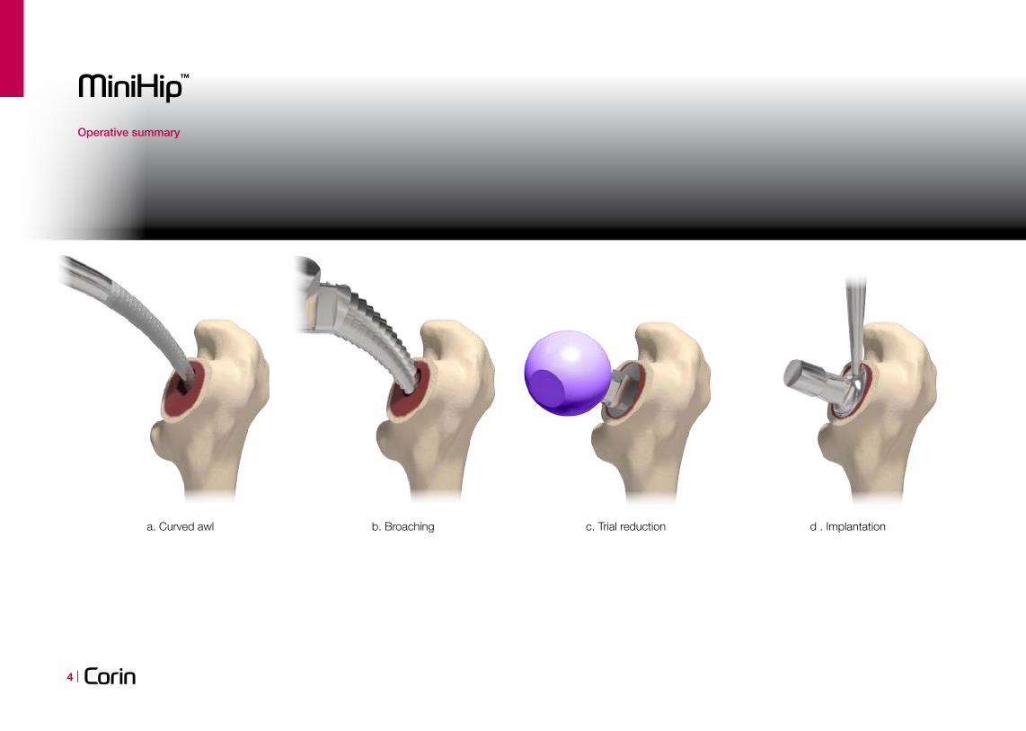
Operative summary
a. Curved awl b. Broaching c. Trial reduction d . Implantation
a
| 5
Overview
The MiniHip bone preserving, physiological hip replacement has been specifically designed to address the clinical needs of active men and women of all ages who would usually be appropriate for a standard uncemented hip replacement.
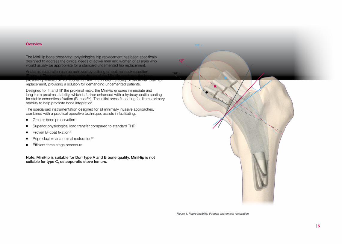
Anatomic restoration can be achieved by utilising an optimal neck resection (Figure 1). The dedicated short stem design of the MiniHip combines the bone preserving benefits of hip resurfacing with the inherent stability of traditional total hip replacement, providing a solution for demanding uncemented patients.
Designed to ‘fit and fill’ the proximal neck, the MiniHip ensures immediate and long-term proximal stability, which is further enhanced with a hydroxyapatite coating for stable cementless fixation (Bi-coat™). The initial press fit coating facilitates primary stability to help promote bone integration.
The specialised instrumentation designed for all minimally invasive approaches, combined with a practical operative technique, assists in facilitating:
■ Greater bone preservation
■ Superior physiological load transfer compared to standard THR1
■ Proven Bi-coat fixation2
■ Reproducible anatomical restoration3,4
■ Efficient three stage procedure
Note: MiniHip is suitable for Dorr type A and B bone quality. MiniHip is not suitable for type C, osteoporotic stove femurs.
Figure 1. Reproducibility through anatomical restoration
138º ≤
127º
118º ≥
a
MiniHip™
6 |
Pre-operative templating
When using the MiniHip system, pre-operative planning is critical to determine the optimal size, neck angle and offset of the implant. Templating will help determine the level of neck resection and optimal cup position. The MiniHip X-ray templates are available in four different magnifications (100%, 110%, 115% and 120%). The 115% magnification is provided as standard.
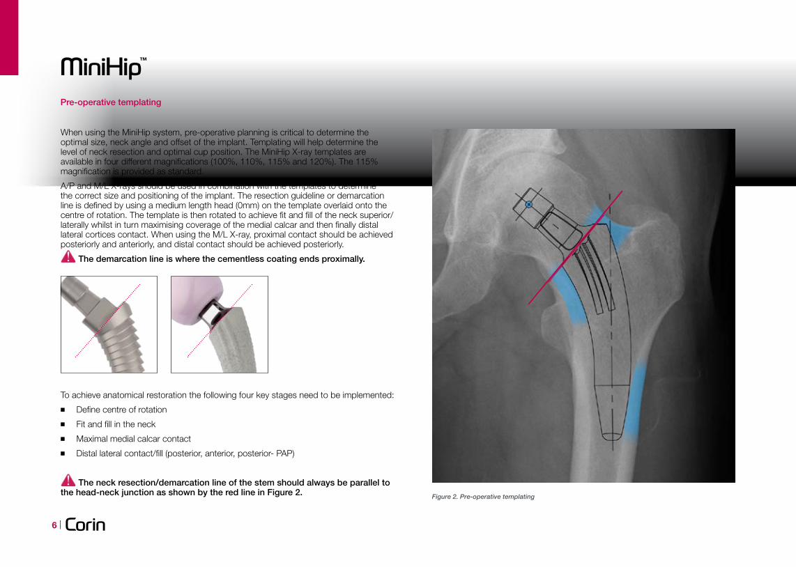
A/P and M/L X-rays should be used in combination with the templates to determine the correct size and positioning of the implant. The resection guideline or demarcation line is defined by using a medium length head (0mm) on the template overlaid onto the centre of rotation. The template is then rotated to achieve fit and fill of the neck superior/laterally whilst in turn maximising coverage of the medial calcar and then finally distal lateral cortices contact. When using the M/L X-ray, proximal contact should be achieved posteriorly and anteriorly, and distal contact should be achieved posteriorly.
The demarcation line is where the cementless coating ends proximally.
To achieve anatomical restoration the following four key stages need to be implemented:
■ Define centre of rotation
■ Fit and fill in the neck
■ Maximal medial calcar contact
■ Distal lateral contact/fill (posterior, anterior, posterior- PAP)
The neck resection/demarcation line of the stem should always be parallel to the head-neck junction as shown by the red line in Figure 2. Figure 2. Pre-operative templating
a
| 7
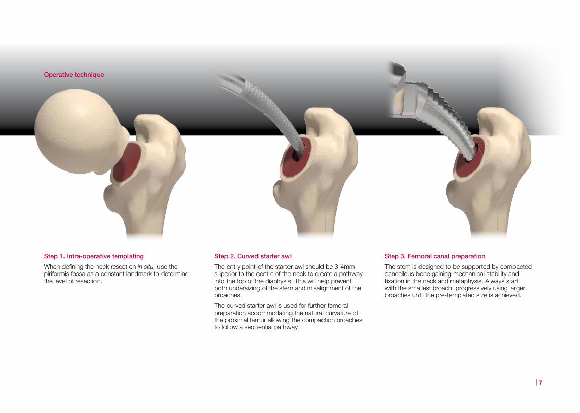
Step 3. Femoral canal preparation
The stem is designed to be supported by compacted cancellous bone gaining mechanical stability and fixation in the neck and metaphysis. Always start with the smallest broach, progressively using larger broaches until the pre-templated size is achieved.
Step 1. Intra-operative templating
When defining the neck resection in situ, use the piriformis fossa as a constant landmark to determine the level of resection.
Operative technique
Step 2. Curved starter awl
The entry point of the starter awl should be 3-4mm superior to the centre of the neck to create a pathway into the top of the diaphysis. This will help prevent both undersizing of the stem and misalignment of the broaches.
The curved starter awl is used for further femoral preparation accommodating the natural curvature of the proximal femur allowing the compaction broaches to follow a sequential pathway.

a
MiniHip™
8 |
A stable fit is achieved when the broach fits and fills the proximal femur and the face of the final broach sits flush with the resection line, as detailed in the pre-operative templating on page 6.
In order to ensure that the femur will accommodate the planned size of implant, a 1mm cancellous ring should be maintained around the broach for sizes 1-4 and a 2mm ring for sizes 5-9. Usually a 2-4mm ring of cancellous bone around the medial calcar is also observed.
Step 4. Trial reduction
A trial reduction may then be performed using a trial head and the broach.
Intraoperative X-ray
To assess the trial orientation and sizing, it is recommended that an intraoperative X-ray using a C-arm be taken whilst the broach, trial neck and head are in still in situ prior to implantation of the definitive implants.
Step 5. Implantation of the stem
The final broach indicates the size of the definitive implant, which is inserted by hand.
If the definitive stem seats by more than 10mm proud above the resection line, remove the stem and re-broach with the finishing broach 2-4mm below the resection line.
a
| 9
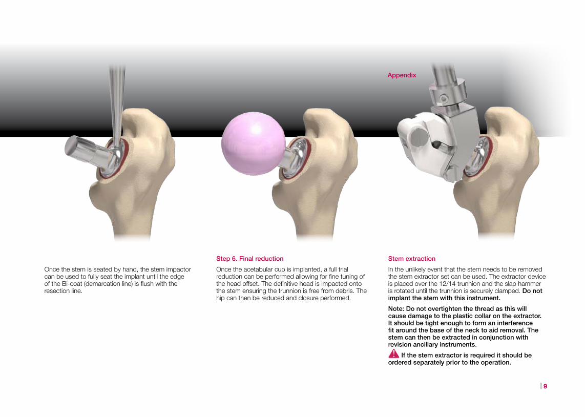
Stem extraction
In the unlikely event that the stem needs to be removed the stem extractor set can be used. The extractor device is placed over the 12/14 trunnion and the slap hammer is rotated until the trunnion is securely clamped. Do not implant the stem with this instrument.
Note: Do not overtighten the thread as this will cause damage to the plastic collar on the extractor. It should be tight enough to form an interference fit around the base of the neck to aid removal. The stem can then be extracted in conjunction with revision ancillary instruments.
If the stem extractor is required it should be ordered separately prior to the operation.
Once the stem is seated by hand, the stem impactor can be used to fully seat the implant until the edge of the Bi-coat (demarcation line) is flush with the resection line.
Step 6. Final reduction
Once the acetabular cup is implanted, a full trial reduction can be performed allowing for fine tuning of the head offset. The definitive head is impacted onto the stem ensuring the trunnion is free from debris. The hip can then be reduced and closure performed.
Appendix
a
MiniHip™
10 |
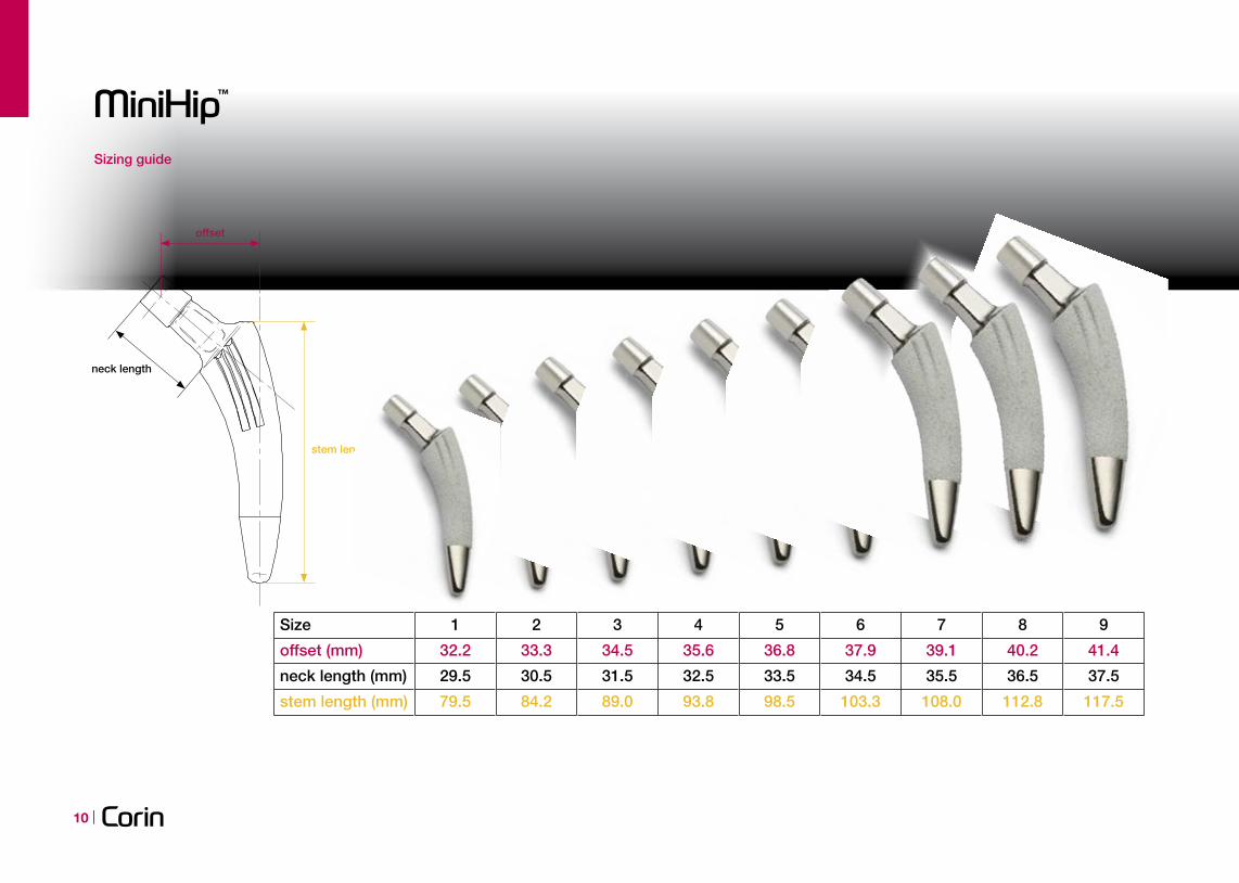
stem length
offset
neck length
stem length (mm)
neck length (mm)
offset (mm)
Size
79.5
29.5
32.2
1
84.2
30.5
33.3
2
89.0
31.5
34.5
3
93.8
32.5
35.6
4
98.5
33.5
36.8
5
103.3
34.5
37.9
6
108.0
35.5
39.1
7
112.8
36.5
40.2
8
117.5
37.5
41.4
9
Sizing guide

a
| 11
Description
The MiniHip is a titanium femoral stem (Ti-6Al-4V) coated with a layer of hydroxyapatite applied over a layer of pure titanium. The distal section of the stem is polished. The device is available in a range of nine sizes each providing a 130o CCD neck angle. The device is intended to be used with 12/14 modular taper heads.
The MiniHip is intended to provide increased patient mobility and reduce pain by replacing the damaged hip joint articulation in patients where there is evidence of sufficient sound bone to seat and support the components.
Indications
The indications for the MiniHip stem as a total hip arthroplasty include:
■ Non-inflammatory degenerative joint disease including osteoarthritis and avascular necrosis
■ Rheumatoid arthritis
■ Correction of functional deformity
■ Developmental dysplasia/congenital dislocation of the hip
The MiniHip stem is indicated for cementless use only.
Contraindications
■ Infection
■ Osteomyelitis
■ Sepsis
■ Osteomalacia
■ Distant foci of infections
■ Osteoporosis
■ Marked bone loss or bone resorption
■ Metabolic disorders which may impair bone formation
■ Vascular insufficiency
■ Muscular atrophy or neuromuscular disease
■ Allergy to implant material
■ Severe deformity
Ordering information
Implants
580.0001 Size 1 Standard Stem
580.0002 Size 2 Standard Stem
580.0003 Size 3 Standard Stem
580.0004 Size 4 Standard Stem
580.0005 Size 5 Standard Stem
580.0006 Size 6 Standard Stem
580.0007 Size 7 Standard Stem
580.0008 Size 8 Standard Stem
580.0009 Size 9 Standard Stem
Instruments
580.9991 MiniHip Cementless Stem System
X-Ray templates
580.9190 MiniHip X-ray template 115%
580.9090 MiniHip X-ray template 100%
Stem extractor set
580.9410 Stem Extractor Set

©2012 Corin P No I920 Rev2 02/2012 ECR 11672
Printed on 9lives 80 which contains 80% total recycled fibre and is produced at a mill which holds the ISO 14001 for Environmental Management Systems. The pulp is bleached using Elemental Chlorine Free processes.
The Corinium CentreCirencester, GL7 1YJ, UKt: +44 (0)1285 659 866f: +44 (0)1285 658 960e: [email protected]
www.coringroup.com
References:
1. Short stems are less likely to lead to bone resorption; bone remodeling following THR. Yeoman M, Cizinauskas A, Lowry C, Vincent G, Collins SN, Simpson DJ, Continuum Blue, UK; 2 – Corin Ltd, UK; 3 – Imorphics, UK. Data held on file at Corin
2. Collier CG. The assessment of early osteointegration, as a function of coating. August 2002. Report held on file.
3. Short stem total hip replacement: are you being conservative enough? Simpson DJ, Lowry C, Yeoman M, Cizinauskas A, Vincent G, Collins SN, Corin, UK; 2 - Continuum Blue, UK; 3 - Imorphics, UK. Data held on file at Corin
4. Presented data at the Baden Baden 58th Annual VSO meeting, (Tetnang, Germany) 2010. Dr Christian Grasselli discussed the multicentre clinical data for 250 bone conserving MiniHips.