Embed Size (px)
Citation preview
www.rsc.org/loc Volume 9 | Number 8 | 21 April 2009 | Pages 1021–1152
ISSN 1473-0197
Miniaturisation for chemistry, physics, biology, & bioengineering
WangDroplet PCR array
PanDIY 3D microfluidics
GroismanCell culture under oxygen controlled conditions
Neil and CesProteomics with smart droplets
PAPER www.rsc.org/loc | Lab on a Chip
DNA methylation analysis on a droplet-in-oil PCR array
Yi Zhang,a Vasudev Bailey,ab Christopher M. Puleo,a Hariharan Easwaran,b Elizabeth Griffiths,b
James G. Herman,b Stephen B. Baylinb and Tza-Huei Wang*ac
Received 4th December 2008, Accepted 18th February 2009
First published as an Advance Article on the web 6th March 2009
DOI: 10.1039/b821780g
We performed on-chip DNA methylation analysis using methylation-specific PCR (MSP) within an
arrayed micro droplet-in-oil platform that is designed for more practical application of microfluidic
droplet technologies in clinical applications. Unique features of this ready-to-use device include arrayed
primers that are pre-deposited into open micro-reaction chambers and use of the oil phase as
a companion fluid for both sample actuation and compartmentalization. These technical advantages
allow for infusion of minute amounts of sample for arrayed MSP analysis, without the added
complexities inherent in microfluidic droplet-based studies. Ease of use of this micro device is
exemplified by analysis of two tumor suppressor promoters, p15 and TMS1 using an on-chip
methylation assay. These results were consistent with standard MSP protocols, yet the simplicity of the
droplet-in-oil microfluidic PCR platform provides an easy and efficient tool for DNA methylation
analysis in a large-scale arrayed manner.
Introduction
Although various methods are available for analyzing nucleic
acids, polymerase chain reaction (PCR) remains one of the most
widely used techniques. Many molecular biology protocols today
still heavily rely on PCR or modified PCR. For example, allele-
specific PCR is used for identifying single nucleotide poly-
morphisms,1 asymmetric PCR is used for generating single
stranded DNA hybridization probes or targets,2 real-time
quantitative PCR is used for DNA/RNA quantification,3
BEAMing PCR is incorporated in next-generation sequencing,4,5
and methylation-specific PCR (MSP) is used for DNA methyl-
ation detection.6 Currently, one of the major trends in biological
and chemical analysis has been the development of micro-
fabricated miniaturized platforms.7–12 In particular, great effort
has been put into the miniaturization of genetic tests13–15 as they
provide fast analysis, reduced reagent and sample consumption,
and high portability. The most straightforward approach used
involves fabricating an array of reaction chambers of micro,
nano or even picoliter volumes for parallel PCR reactions.16–21
Alternatively, miniaturization is realized by performing PCR in
free micro droplets on a modified surface. The droplets function
as the virtual reaction chambers by providing fluidic confine-
ment.22–24 However, many current array based devices are
designed for performing multiple PCR reactions in identical
conditions with the same primers and DNA templates, thus
limiting their use to singleplex analysis. Multiple gene screening
with different primer sets still relies on separate sample loading
via complex fluidic networks. Furthermore, most designs
aDepartment of Biomedical Engineering, Johns Hopkins University,Baltimore, MD, 21218, USA. E-mail: [email protected]; Tel: +1-410-516-7086bCancer Biology Program, The Sidney Kimmel Comprehensive CancerCenter at Johns Hopkins, Baltimore, MD, 21210, USAcDepartment of Mechanical Engineering, Johns Hopkins University,Baltimore, MD, 21218, USA
This journal is ª The Royal Society of Chemistry 2009
concentrate on engineering perspectives, focusing on the minia-
turization and system integration, while often overlooking
practicality issues. As a result, very complicated microfluidic
control modules are usually incorporated in order to cope with
pressurization and micro bubble formation and expansion
during thermal cycling.25,26 Unfortunately, such delicate devices
often require operation by experienced microfluidic personnel
and thus are difficult to fit into routine biological laboratories
and clinical settings. Hence they are not commonly adopted for
use in PCR based diagnostic assays such as MSP.
MSP (Fig. 1) is most extensively used for analyzing DNA
methylation that is closely associated with tumorigenesis,
a multi-step process resulting from gain-of-function (oncogenes)
or loss-of-function (tumor suppressor) gene alterations. These
changes could be caused either by genetic or epigenetic changes.
One of the most well studied epigenetic changes is the heritable
transcriptional silencing of tumor suppressor genes (TSG) by
aberrant CpGDNA hypermethylation of their promoters.27–31 In
higher order eukaryotes, DNA methylation only occurs at 50
cytosines in CpG dinucleotides, which when observed in high
frequency at many transcription promoter regions are termed
CpG islands.32 There is clear evidence that the down regulation
of tumor suppressor genes in cancer is tightly associated with
DNA hypermethylation in the promoter regions, where the
transcription of DNA to RNA is initiated33 Therefore, the
assessment of DNA methylation status has great clinical impli-
cations, offering another important parameter for early cancer
diagnosis and prognosis, as well as responsiveness to cancer
therapy.34–36 MSP takes advantage of the change in DNA
sequence after bisulfite modification, wherein only unmethylated
cytosines are converted to uracils, while methylated cytosines
remain unaltered. Primers specific to the modified DNA
sequences are then introduced to amplify and distinguish meth-
ylated from unmethylated DNA. Generally, each bisulfite treated
DNA sample is run in two separate MSP reactions. Each
reaction contains either a methylated specific primer set or an
Lab Chip, 2009, 9, 1059–1064 | 1059
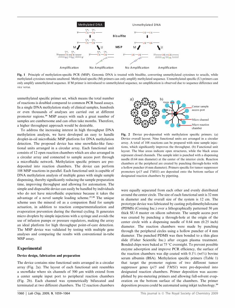
Fig. 1 Principle of methylation-specific PCR (MSP). Genomic DNA is treated with bisulfite, converting unmethylated cytosines to uracils, while
methylated cytosines remains unaltered. Methylated specific (M) primers can only amplify methylated sequence. Unmethylated specific (U) primers can
only amplify unmethylated sequence. If M primer is introduced to unmethylated sequence, no amplification is observed due to sequence difference and
vice versa.
Fig. 2 Device pre-deposited with methylation specific primers. (a)
Device overall layout. Nine functional units are arranged in a circular
array. A total of 108 reactions can be prepared with nine sample injec-
tions, which significantly improves the throughput. (b) Functional unit
layout. The blue areas indicate open structures, while the black areas
represent closed channels. The sample inlet is punched with a dispensing
needle (0.64 mm diameter) at the center of the interior circle. Reaction
chambers at the peripheral are created by punching through-holes with
a hollow puncher (4 mm diameter). Primers specific for tumor suppressor
promoters (p15 and TMS1) are deposited onto the bottom surface of
designated reaction chambers by pipetting.
unmethylated specific primer set, which means the total number
of reactions is doubled compared to common PCR based assays.
In a single DNA methylation study of clinical samples, hundreds
or even thousands of analyses are carried out at different
promoter regions.34 MSP assays with such a great number of
samples are cumbersome and can often take months. Therefore,
a higher throughput approach would be desirable.
To address the increasing interest in high throughput DNA
methylation analysis, we have developed an easy to handle
droplet-in-oil microfluidic MSP platform for DNA methylation
detection. The proposed device has nine snowflake-like func-
tional units arranged in a circular array. Each functional unit
consists of 12 open reaction chambers which are also arranged in
a circular array and connected to sample access port through
a microfluidic network. Methylation specific primers are pre-
deposited into reaction chambers. The device can perform
108 MSP reactions in parallel. Each functional unit is capable of
DNA methylation analysis of multiple genes with single sample
dispensing, thereby significantly reducing the sample preparation
time, improving throughput and allowing for automation. The
simple and disposable device can easily be handled by individuals
who do not have microfluidic experience because it takes the
advantage of a novel sample loading scheme.37,38 The unique
scheme uses the mineral oil as a companion fluid for sample
actuation, in addition to reaction compartmentalization and
evaporation prevention during the thermal cycling. It generates
micro droplets by simple injections with a syringe and avoids the
use of infusion pumps or pressure regulators, making the array
an ideal platform for high throughput point-of-care detection.
The MSP device was validated by testing with multiple gene
analyses and comparing the results with conventional in-tube
MSP assay.
Experimental
Device design, fabrication and preparation
The device contains nine functional units arranged in a circular
array (Fig. 2a). The layout of each functional unit resembles
a snowflake where six channels of 500 mm width extend from
a center sample input port to peripheral reaction chambers
(Fig. 2b). Each channel was symmetrically bifurcated and
terminated at two different chambers. The 12 reaction chambers
1060 | Lab Chip, 2009, 9, 1059–1064
were equally separated from each other and evenly distributed
around the center circle. The size of each functional unit is 32 mm
in diameter and the overall size of the system is 12 cm. The
prototype device was fabricated by casting polydimethylsiloxane
(PDMS) (Corning Inc.) over a lithographically patterned 50 mm
thick SU-8 master on silicon substrate. The sample access port
was created by punching a through-hole at the origin of the
center circle with a dispensing needle of 0.64 mm (0.25 inch)
diameter. The reaction chambers were made by punching
through the peripheral circles using a hollow puncher of 4 mm
diameter. The punched PDMS was then bonded to a thin glass
slide (Fisher Scientific Inc.) after oxygen plasma treatment.
Bonded chips were baked at 75 �C overnight. To prevent possible
enzyme adsorption and improve PCR efficiency, the surface of
the reaction chambers was dip coated with 0.1% (wt%) bovine
serum albumin (BSA). Methylation specific primers (Table 1)
that target the promoter regions of two different tumor
suppressor genes (p15 and TMS1) were pre-deposited into
designated reaction chambers. Primer deposition was accom-
plished by pre-metering primers and allowing full-solvent evap-
oration on the bottom surface of the chambers. The primer
deposition process could be automated using inkjet technology.39
This journal is ª The Royal Society of Chemistry 2009
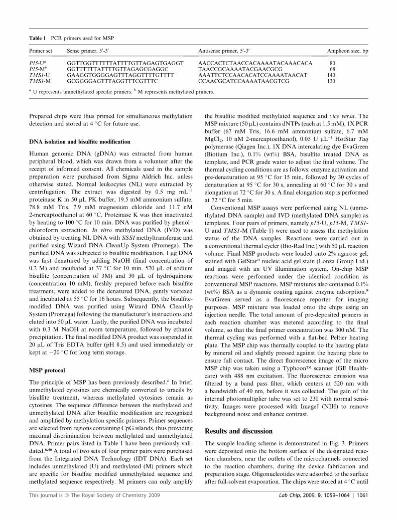
Table 1 PCR primers used for MSP
Primer set Sense primer, 50-30 Antisense primer, 50-30 Amplicon size, bp
P15-Ua GGTTGGTTTTTTATTTTGTTAGAGTGAGGT AACCACTCTAACCACAAAATACAAACACA 80P15-Mb GGTTTTTTATTTTGTTAGAGCGAGGC TAACCGCAAAATACGAACGCG 68TMS1-U GAAGGTGGGGAGTTTAGGTTTTGTTTT AAATTCTCCAACACATCCAAAATAACAT 140TMS1-M GCGGGGAGTTTAGGTTTCGTTTC CCAACGCATCCAAAATAACGTCG 130
a U represents unmethylated specific primers. b M represents methylated primers.
Prepared chips were thus primed for simultaneous methylation
detection and stored at 4 �C for future use.
DNA isolation and bisulfite modification
Human genomic DNA (gDNA) was extracted from human
peripheral blood, which was drawn from a volunteer after the
receipt of informed consent. All chemicals used in the sample
preparation were purchased from Sigma Aldrich Inc. unless
otherwise stated. Normal leukocytes (NL) were extracted by
centrifugation. The extract was digested by 0.5 mg mL�1
proteinase K in 50 mL PK buffer, 19.5 mM ammonium sulfate,
78.8 mM Tris, 7.9 mM magnesium chloride and 11.7 nM
2-mercaptoethanol at 60 �C. Proteinase K was then inactivated
by heating to 100 �C for 10 min. DNA was purified by phenol–
chloroform extraction. In vitro methylated DNA (IVD) was
obtained by treating NL DNA with SSSI methyltransferase and
purified using Wizard DNA CleanUp System (Promega). The
purified DNA was subjected to bisulfite modification. 1 mg DNA
was first denatured by adding NaOH (final concentration of
0.2 M) and incubated at 37 �C for 10 min. 520 mL of sodium
bisulfite (concentration of 3M) and 30 mL of hydroquinone
(concentration 10 mM), freshly prepared before each bisulfite
treatment, were added to the denatured DNA, gently vortexed
and incubated at 55 �C for 16 hours. Subsequently, the bisulfite-
modified DNA was purified using Wizard DNA CleanUp
System (Promega) following the manufacturer’s instructions and
eluted into 50 mL water. Lastly, the purified DNA was incubated
with 0.3 M NaOH at room temperature, followed by ethanol
precipitation. The final modified DNA product was suspended in
20 mL of Tris EDTA buffer (pH 8.5) and used immediately or
kept at �20 �C for long term storage.
MSP protocol
The principle of MSP has been previously described.6 In brief,
unmethylated cytosines are chemically converted to uracils by
bisulfite treatment, whereas methylated cytosines remain as
cytosines. The sequence difference between the methylated and
unmethylated DNA after bisulfite modification are recognized
and amplified by methylation specific primers. Primer sequences
are selected from regions containing CpG islands, thus providing
maximal discrimination between methylated and unmethylated
DNA. Primer pairs listed in Table 1 have been previously vali-
dated.6,40 A total of two sets of four primer pairs were purchased
from the Integrated DNA Technology (IDT DNA). Each set
includes unmethylated (U) and methylated (M) primers which
are specific for bisulfite modified unmethylated sequence and
methylated sequence respectively. M primers can only amplify
This journal is ª The Royal Society of Chemistry 2009
the bisulfite modified methylated sequence and vice versa. The
MSPmixture (50 mL) contains dNTPs (each at 1.5 mM), 1X PCR
buffer (67 mM Tris, 16.6 mM ammonium sulfate, 6.7 mM
MgCl2, 10 nM 2-mercaptoethanol), 0.05 U mL�1 HotStar Taq
polymerase (Qiagen Inc.), 1X DNA intercalating dye EvaGreen
(Biotium Inc.), 0.1% (wt%) BSA, bisulfite treated DNA as
template, and PCR grade water to adjust the final volume. The
thermal cycling conditions are as follows: enzyme activation and
pre-denaturation at 95 �C for 15 min, followed by 30 cycles of
denaturation at 95 �C for 30 s, annealing at 60 �C for 30 s and
elongation at 72 �C for 30 s. A final elongation step is performed
at 72 �C for 5 min.
Conventional MSP assays were performed using NL (unme-
thylated DNA sample) and IVD (methylated DNA sample) as
templates. Four pairs of primers, namely p15-U, p15-M, TMS1-
U and TMS1-M (Table 1) were used to assess the methylation
status of the DNA samples. Reactions were carried out in
a conventional thermal cycler (Bio-Rad Inc.) with 50 mL reaction
volume. Final MSP products were loaded onto 2% agarose gel,
stained with GelStar� nucleic acid gel stain (Lonza Group Ltd.)
and imaged with an UV illumination system. On-chip MSP
reactions were performed under the identical condition as
conventional MSP reactions. MSP mixtures also contained 0.1%
(wt%) BSA as a dynamic coating against enzyme adsorption.9
EvaGreen served as a fluorescence reporter for imaging
purposes. MSP mixture was loaded onto the chips using an
injection needle. The total amount of pre-deposited primers in
each reaction chamber was metered according to the final
volume, so that the final primer concentration was 300 nM. The
thermal cycling was performed with a flat-bed Peltier heating
plate. The MSP chip was thermally coupled to the heating plate
by mineral oil and slightly pressed against the heating plate to
ensure full contact. The direct fluorescence image of the micro
MSP chip was taken using a Typhoon� scanner (GE Health-
care) with 488 nm excitation. The fluorescence emission was
filtered by a band pass filter, which centers at 520 nm with
a bandwidth of 40 nm, before it was collected. The gain of the
internal photomultiplier tube was set to 230 with normal sensi-
tivity. Images were processed with ImageJ (NIH) to remove
background noise and enhance contrast.
Results and discussion
The sample loading scheme is demonstrated in Fig. 3. Primers
were deposited onto the bottom surface of the designated reac-
tion chambers, near the outlets of the microchannels connected
to the reaction chambers, during the device fabrication and
preparation stage. Oligonucleotides were adsorbed to the surface
after full-solvent evaporation. The chips were stored at 4 �C until
Lab Chip, 2009, 9, 1059–1064 | 1061
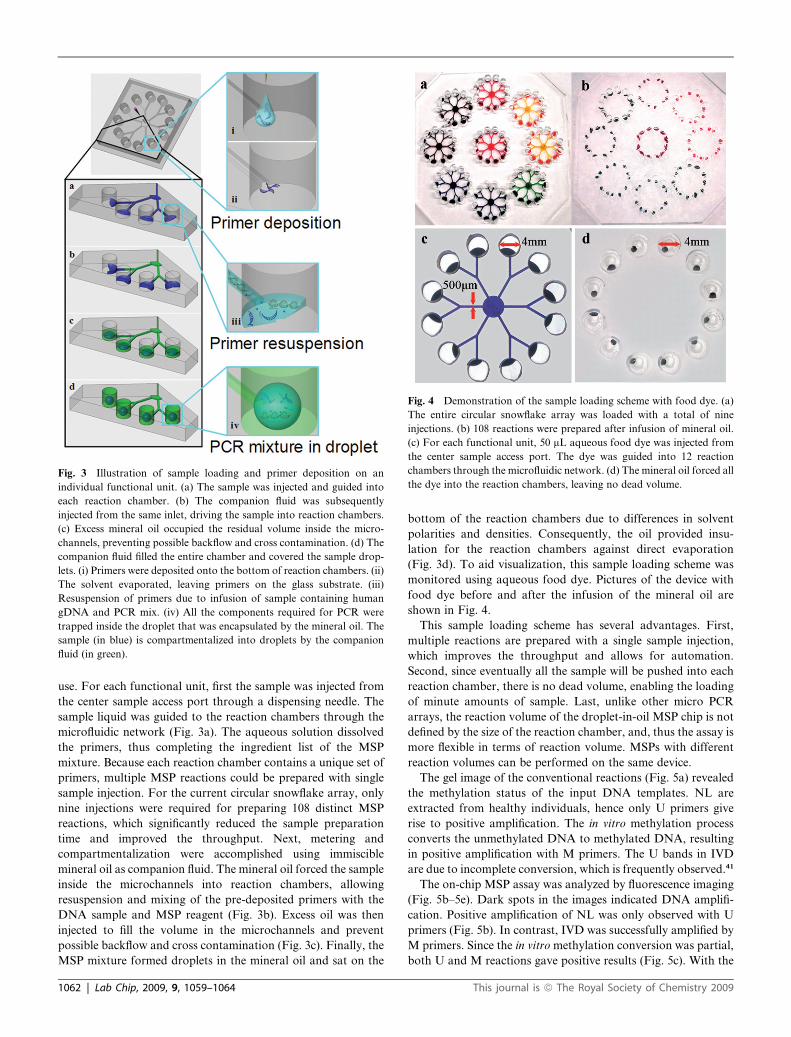
Fig. 3 Illustration of sample loading and primer deposition on an
individual functional unit. (a) The sample was injected and guided into
each reaction chamber. (b) The companion fluid was subsequently
injected from the same inlet, driving the sample into reaction chambers.
(c) Excess mineral oil occupied the residual volume inside the micro-
channels, preventing possible backflow and cross contamination. (d) The
companion fluid filled the entire chamber and covered the sample drop-
lets. (i) Primers were deposited onto the bottom of reaction chambers. (ii)
The solvent evaporated, leaving primers on the glass substrate. (iii)
Resuspension of primers due to infusion of sample containing human
gDNA and PCR mix. (iv) All the components required for PCR were
trapped inside the droplet that was encapsulated by the mineral oil. The
sample (in blue) is compartmentalized into droplets by the companion
fluid (in green).
Fig. 4 Demonstration of the sample loading scheme with food dye. (a)
The entire circular snowflake array was loaded with a total of nine
injections. (b) 108 reactions were prepared after infusion of mineral oil.
(c) For each functional unit, 50 mL aqueous food dye was injected from
the center sample access port. The dye was guided into 12 reaction
chambers through the microfluidic network. (d) The mineral oil forced all
the dye into the reaction chambers, leaving no dead volume.
use. For each functional unit, first the sample was injected from
the center sample access port through a dispensing needle. The
sample liquid was guided to the reaction chambers through the
microfluidic network (Fig. 3a). The aqueous solution dissolved
the primers, thus completing the ingredient list of the MSP
mixture. Because each reaction chamber contains a unique set of
primers, multiple MSP reactions could be prepared with single
sample injection. For the current circular snowflake array, only
nine injections were required for preparing 108 distinct MSP
reactions, which significantly reduced the sample preparation
time and improved the throughput. Next, metering and
compartmentalization were accomplished using immiscible
mineral oil as companion fluid. The mineral oil forced the sample
inside the microchannels into reaction chambers, allowing
resuspension and mixing of the pre-deposited primers with the
DNA sample and MSP reagent (Fig. 3b). Excess oil was then
injected to fill the volume in the microchannels and prevent
possible backflow and cross contamination (Fig. 3c). Finally, the
MSP mixture formed droplets in the mineral oil and sat on the
1062 | Lab Chip, 2009, 9, 1059–1064
bottom of the reaction chambers due to differences in solvent
polarities and densities. Consequently, the oil provided insu-
lation for the reaction chambers against direct evaporation
(Fig. 3d). To aid visualization, this sample loading scheme was
monitored using aqueous food dye. Pictures of the device with
food dye before and after the infusion of the mineral oil are
shown in Fig. 4.
This sample loading scheme has several advantages. First,
multiple reactions are prepared with a single sample injection,
which improves the throughput and allows for automation.
Second, since eventually all the sample will be pushed into each
reaction chamber, there is no dead volume, enabling the loading
of minute amounts of sample. Last, unlike other micro PCR
arrays, the reaction volume of the droplet-in-oil MSP chip is not
defined by the size of the reaction chamber, and, thus the assay is
more flexible in terms of reaction volume. MSPs with different
reaction volumes can be performed on the same device.
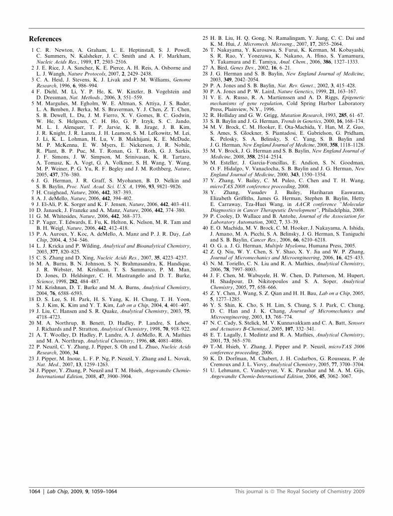
The gel image of the conventional reactions (Fig. 5a) revealed
the methylation status of the input DNA templates. NL are
extracted from healthy individuals, hence only U primers give
rise to positive amplification. The in vitro methylation process
converts the unmethylated DNA to methylated DNA, resulting
in positive amplification with M primers. The U bands in IVD
are due to incomplete conversion, which is frequently observed.41
The on-chip MSP assay was analyzed by fluorescence imaging
(Fig. 5b–5e). Dark spots in the images indicated DNA amplifi-
cation. Positive amplification of NL was only observed with U
primers (Fig. 5b). In contrast, IVD was successfully amplified by
M primers. Since the in vitromethylation conversion was partial,
both U and M reactions gave positive results (Fig. 5c). With the
This journal is ª The Royal Society of Chemistry 2009
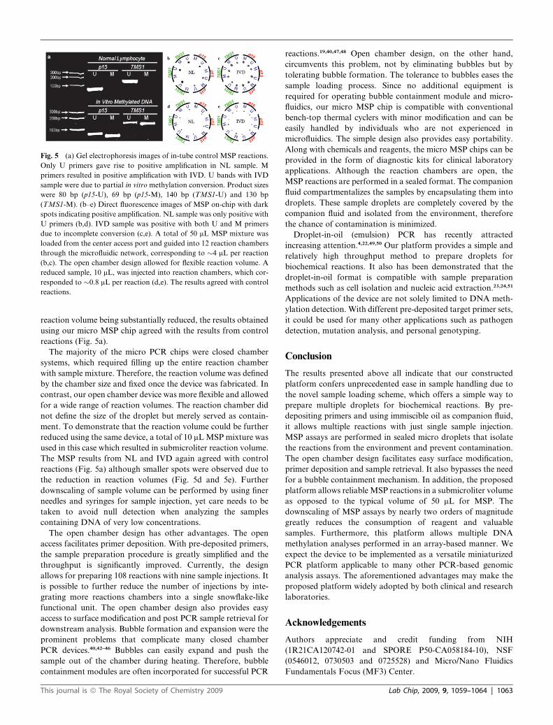
Fig. 5 (a) Gel electrophoresis images of in-tube control MSP reactions.
Only U primers gave rise to positive amplification in NL sample. M
primers resulted in positive amplification with IVD. U bands with IVD
sample were due to partial in vitro methylation conversion. Product sizes
were 80 bp (p15-U), 69 bp (p15-M), 140 bp (TMS1-U) and 130 bp
(TMS1-M). (b–e) Direct fluorescence images of MSP on-chip with dark
spots indicating positive amplification. NL sample was only positive with
U primers (b,d). IVD sample was positive with both U and M primers
due to incomplete conversion (c,e). A total of 50 mL MSP mixture was
loaded from the center access port and guided into 12 reaction chambers
through the microfluidic network, corresponding to �4 mL per reaction
(b,c). The open chamber design allowed for flexible reaction volume. A
reduced sample, 10 mL, was injected into reaction chambers, which cor-
responded to �0.8 mL per reaction (d,e). The results agreed with control
reactions.
reaction volume being substantially reduced, the results obtained
using our micro MSP chip agreed with the results from control
reactions (Fig. 5a).
The majority of the micro PCR chips were closed chamber
systems, which required filling up the entire reaction chamber
with sample mixture. Therefore, the reaction volume was defined
by the chamber size and fixed once the device was fabricated. In
contrast, our open chamber device was more flexible and allowed
for a wide range of reaction volumes. The reaction chamber did
not define the size of the droplet but merely served as contain-
ment. To demonstrate that the reaction volume could be further
reduced using the same device, a total of 10 mLMSP mixture was
used in this case which resulted in submicroliter reaction volume.
The MSP results from NL and IVD again agreed with control
reactions (Fig. 5a) although smaller spots were observed due to
the reduction in reaction volumes (Fig. 5d and 5e). Further
downscaling of sample volume can be performed by using finer
needles and syringes for sample injection, yet care needs to be
taken to avoid null detection when analyzing the samples
containing DNA of very low concentrations.
The open chamber design has other advantages. The open
access facilitates primer deposition. With pre-deposited primers,
the sample preparation procedure is greatly simplified and the
throughput is significantly improved. Currently, the design
allows for preparing 108 reactions with nine sample injections. It
is possible to further reduce the number of injections by inte-
grating more reactions chambers into a single snowflake-like
functional unit. The open chamber design also provides easy
access to surface modification and post PCR sample retrieval for
downstream analysis. Bubble formation and expansion were the
prominent problems that complicate many closed chamber
PCR devices.40,42–46 Bubbles can easily expand and push the
sample out of the chamber during heating. Therefore, bubble
containment modules are often incorporated for successful PCR
This journal is ª The Royal Society of Chemistry 2009
reactions.19,40,47,48 Open chamber design, on the other hand,
circumvents this problem, not by eliminating bubbles but by
tolerating bubble formation. The tolerance to bubbles eases the
sample loading process. Since no additional equipment is
required for operating bubble containment module and micro-
fluidics, our micro MSP chip is compatible with conventional
bench-top thermal cyclers with minor modification and can be
easily handled by individuals who are not experienced in
microfluidics. The simple design also provides easy portability.
Along with chemicals and reagents, the micro MSP chips can be
provided in the form of diagnostic kits for clinical laboratory
applications. Although the reaction chambers are open, the
MSP reactions are performed in a sealed format. The companion
fluid compartmentalizes the samples by encapsulating them into
droplets. These sample droplets are completely covered by the
companion fluid and isolated from the environment, therefore
the chance of contamination is minimized.
Droplet-in-oil (emulsion) PCR has recently attracted
increasing attention.4,22,49,50 Our platform provides a simple and
relatively high throughput method to prepare droplets for
biochemical reactions. It also has been demonstrated that the
droplet-in-oil format is compatible with sample preparation
methods such as cell isolation and nucleic acid extraction.23,24,51
Applications of the device are not solely limited to DNA meth-
ylation detection. With different pre-deposited target primer sets,
it could be used for many other applications such as pathogen
detection, mutation analysis, and personal genotyping.
Conclusion
The results presented above all indicate that our constructed
platform confers unprecedented ease in sample handling due to
the novel sample loading scheme, which offers a simple way to
prepare multiple droplets for biochemical reactions. By pre-
depositing primers and using immiscible oil as companion fluid,
it allows multiple reactions with just single sample injection.
MSP assays are performed in sealed micro droplets that isolate
the reactions from the environment and prevent contamination.
The open chamber design facilitates easy surface modification,
primer deposition and sample retrieval. It also bypasses the need
for a bubble containment mechanism. In addition, the proposed
platform allows reliable MSP reactions in a submicroliter volume
as opposed to the typical volume of 50 mL for MSP. The
downscaling of MSP assays by nearly two orders of magnitude
greatly reduces the consumption of reagent and valuable
samples. Furthermore, this platform allows multiple DNA
methylation analyses performed in an array-based manner. We
expect the device to be implemented as a versatile miniaturized
PCR platform applicable to many other PCR-based genomic
analysis assays. The aforementioned advantages may make the
proposed platform widely adopted by both clinical and research
laboratories.
Acknowledgements
Authors appreciate and credit funding from NIH
(1R21CA120742-01 and SPORE P50-CA058184-10), NSF
(0546012, 0730503 and 0725528) and Micro/Nano Fluidics
Fundamentals Focus (MF3) Center.
Lab Chip, 2009, 9, 1059–1064 | 1063
References
1 C. R. Newton, A. Graham, L. E. Heptinstall, S. J. Powell,C. Summers, N. Kalsheker, J. C. Smith and A. F. Markham,Nucleic Acids Res., 1989, 17, 2503–2516.
2 J. E. Rice, J. A. Sanchez, K. E. Pierce, A. H. Reis, A. Osborne andL. J. Wangh, Nature Protocols, 2007, 2, 2429–2438.
3 C. A. Heid, J. Stevens, K. J. Livak and P. M. Williams, GenomeResearch, 1996, 6, 986–994.
4 F. Diehl, M. Li, Y. P. He, K. W. Kinzler, B. Vogelstein andD. Dressman, Nat. Methods., 2006, 3, 551–559.
5 M. Margulies, M. Egholm, W. E. Altman, S. Attiya, J. S. Bader,L. A. Bemben, J. Berka, M. S. Braverman, Y. J. Chen, Z. T. Chen,S. B. Dewell, L. Du, J. M. Fierro, X. V. Gomes, B. C. Godwin,W. He, S. Helgesen, C. H. Ho, G. P. Irzyk, S. C. Jando,M. L. I. Alenquer, T. P. Jarvie, K. B. Jirage, J. B. Kim,J. R. Knight, J. R. Lanza, J. H. Leamon, S. M. Lefkowitz, M. Lei,J. Li, K. L. Lohman, H. Lu, V. B. Makhijani, K. E. McDade,M. P. McKenna, E. W. Myers, E. Nickerson, J. R. Nobile,R. Plant, B. P. Puc, M. T. Ronan, G. T. Roth, G. J. Sarkis,J. F. Simons, J. W. Simpson, M. Srinivasan, K. R. Tartaro,A. Tomasz, K. A. Vogt, G. A. Volkmer, S. H. Wang, Y. Wang,M. P. Weiner, P. G. Yu, R. F. Begley and J. M. Rothberg, Nature,2005, 437, 376–380.
6 J. G. Herman, J. R. Graff, S. Myohanen, B. D. Nelkin andS. B. Baylin, Proc. Natl. Acad. Sci. U.S. A, 1996, 93, 9821–9826.
7 H. Craighead, Nature, 2006, 442, 387–393.8 A. J. deMello, Nature, 2006, 442, 394–402.9 J. El-Ali, P. K. Sorger and K. F. Jensen, Nature, 2006, 442, 403–411.10 D. Janasek, J. Franzke and A. Manz, Nature, 2006, 442, 374–380.11 G. M. Whitesides, Nature, 2006, 442, 368–373.12 P. Yager, T. Edwards, E. Fu, K. Helton, K. Nelson, M. R. Tam and
B. H. Weigl, Nature, 2006, 442, 412–418.13 P. A. Auroux, Y. Koc, A. deMello, A. Manz and P. J. R. Day, Lab
Chip, 2004, 4, 534–546.14 L. J. Kricka and P. Wilding, Analytical and Bioanalytical Chemistry,
2003, 377, 820–825.15 C. S. Zhang and D. Xing, Nucleic Acids Res., 2007, 35, 4223–4237.16 M. A. Burns, B. N. Johnson, S. N. Brahmasandra, K. Handique,
J. R. Webster, M. Krishnan, T. S. Sammarco, P. M. Man,D. Jones, D. Heldsinger, C. H. Mastrangelo and D. T. Burke,Science, 1998, 282, 484–487.
17 M. Krishnan, D. T. Burke and M. A. Burns, Analytical Chemistry,2004, 76, 6588–6593.
18 D. S. Lee, S. H. Park, H. S. Yang, K. H. Chung, T. H. Yoon,S. J. Kim, K. Kim and Y. T. Kim, Lab on a Chip, 2004, 4, 401–407.
19 J. Liu, C. Hansen and S. R. Quake, Analytical Chemistry, 2003, 75,4718–4723.
20 M. A. Northrup, B. Benett, D. Hadley, P. Landre, S. Lehew,J. Richards and P. Stratton, Analytical Chemistry, 1998, 70, 918–922.
21 A. T. Woolley, D. Hadley, P. Landre, A. J. deMello, R. A. Mathiesand M. A. Northrup, Analytical Chemistry, 1996, 68, 4081–4086.
22 P. Neuzil, C. Y. Zhang, J. Pipper, S. Oh and L. Zhuo, Nucleic AcidsResearch, 2006, 34.
23 J. Pipper, M. Inoue, L. F. P. Ng, P. Neuzil, Y. Zhang and L. Novak,Nat. Med., 2007, 13, 1259–1263.
24 J. Pipper, Y. Zhang, P. Neuzil and T. M. Hsieh, Angewandte Chemie-International Edition, 2008, 47, 3900–3904.
1064 | Lab Chip, 2009, 9, 1059–1064
25 H. B. Liu, H. Q. Gong, N. Ramalingam, Y. Jiang, C. C. Dai andK. M. Hui, J. Micromech. Microeng., 2007, 17, 2055–2064.
26 T. Nakayama, Y. Kurosawa, S. Furui, K. Kerman, M. Kobayashi,S. R. Rao, Y. Yonezawa, K. Nakano, A. Hino, S. Yamamura,Y. Takamura and E. Tamiya, Anal. Chem., 2006, 386, 1327–1333.
27 A. Bird, Genes Dev., 2002, 16, 6–21.28 J. G. Herman and S. B. Baylin, New England Journal of Medicine,
2003, 349, 2042–2054.29 P. A. Jones and S. B. Baylin, Nat. Rev. Genet., 2002, 3, 415–428.30 P. A. Jones and P. W. Laird, Nature Genetics, 1999, 21, 163–167.31 V. E. A. Russo, R. A. Martienssen and A. D. Riggs, Epigenetic
mechanisms of gene regulation, Cold Spring Harbor LaboratoryPress, Plainview, N.Y., 1996.
32 R. Holliday and G. W. Grigg, Mutation Research, 1993, 285, 61–67.33 S. B. Baylin and J. G. Herman, Trends in Genetics, 2000, 16, 168–174.34 M. V. Brock, C. M. Hooker, E. Ota-Machida, Y. Han, M. Z. Guo,
S. Ames, S. Glockner, S. Piantadosi, E. Gabrielson, G. Pridham,K. Pelosky, S. A. Belinsky, S. C. Yang, S. B. Baylin andJ. G. Herman,New England Journal ofMedicine, 2008, 358, 1118–1128.
35 M. V. Brock, J. G. Herman and S. B. Baylin, New England Journal ofMedicine, 2008, 358, 2514–2514.
36 M. Esteller, J. Garcia-Foncillas, E. Andion, S. N. Goodman,O. F. Hidalgo, V. Vanaclocha, S. B. Baylin and J. G. Herman, NewEngland Journal of Medicine, 2000, 343, 1350–1354.
37 Y. Zhang, V. Bailey, C. M. Puleo, C. Chen and T. H. Wang,microTAS 2008 conference proceeding, 2008.
38 Y. Zhang, Vasudev J. Bailey, Hariharan Easwaran,Elizabeth Griffiths, James G. Herman, Stephen B. Baylin, HettyE. Carraway, Tza-Huei Wang, in AACR conference ‘‘MolecularDiagnostics in Cancer Therapeutic Development’’, Philadelphia, 2008.
39 P. Cooley, D. Wallace and B. Antohe, Journal of the Association forLaboratory Automation, 2002, 7, 33–39.
40 E. O. Machida, M. V. Brock, C. M. Hooker, J. Nakayama, A. Ishida,J. Amano, M. A. Picchi, S. A. Belinsky, J. G. Herman, S. Taniguchiand S. B. Baylin, Cancer Res., 2006, 66, 6210–6218.
41 O. G. a. J. G. Herman, Multiple Myeloma, Humana Press, 2005.42 Z. Q. Niu, W. Y. Chen, S. Y. Shao, X. Y. Jia and W. P. Zhang,
Journal of Micromechanics and Microengineering, 2006, 16, 425–433.43 N. M. Toriello, C. N. Liu and R. A. Mathies, Analytical Chemistry,
2006, 78, 7997–8003.44 J. F. Chen, M. Wabuyele, H. W. Chen, D. Patterson, M. Hupert,
H. Shadpour, D. Nikitopoulos and S. A. Soper, AnalyticalChemistry, 2005, 77, 658–666.
45 Z. Y. Chen, J. Wang, S. Z. Qian and H. H. Bau, Lab on a Chip, 2005,5, 1277–1285.
46 Y. S. Shin, K. Cho, S. H. Lim, S. Chung, S. J. Park, C. Chung,D. C. Han and J. K. Chang, Journal of Micromechanics andMicroengineering, 2003, 13, 768–774.
47 N. C. Cady, S. Stelick, M. V. Kunnavakkam and C. A. Batt, Sensorsand Actuators B-Chemical, 2005, 107, 332–341.
48 E. T. Lagally, I. Medintz and R. A. Mathies, Analytical Chemistry,2001, 73, 565–570.
49 T.-M. Hsieh, Y. Zhang, J. Pipper and P. Neuzil, microTAS 2006conference proceeding, 2006.
50 K. D. Dorfman, M. Chabert, J. H. Codarbox, G. Rousseau, P. deCremoux and J. L. Viovy, Analytical Chemistry, 2005, 77, 3700–3704.
51 U. Lehmann, C. Vandevyver, V. K. Parashar and M. A. M. Gijs,Angewandte Chemie-International Edition, 2006, 45, 3062–3067.
This journal is ª The Royal Society of Chemistry 2009