Embed Size (px)
Citation preview
IntroductionThe epithelium of the Drosophila wing undergoes aconspicuous cytoskeletal reorganisation during differentiation.Between 32 hours and 60 hours APF (after pupariumformation) a columnar epithelium of roughly hexagonal cellsexpands in surface area four-fold, as each cell flattens andinterdigitates with its neighbours. By 60 hours, each cell hasalso elaborated a complex apical structure – the hair pedestalthat supports the growth of actin-filled apical extensionstermed wing hairs (Mitchell et al., 1983; Fristrom et al., 1993).Remarkably, this entire morphological transformation occursafter the first layer of cuticle has already been assembled abovethe epidermal cells (Mitchell et al., 1983).
This first cuticle layer – known as the cuticulin envelope –is only 120-175 Å deep. It is structurally different from othercuticle layers, being mainly composed of crosslinked proteinsand lipids (see Locke, 2001). In Drosophila wings, thisprotective envelope is secreted at about 32-34 hours APF, itcovers the epidermis while changes in cell shape are takingplace and eventually forms the outer-most surface of themature cuticle, once the bulk of the chitinous cuticle has beensecreted (Mitchell et al., 1983). The cuticulin envelope definesan enclosed extracellular compartment above the apicalmembrane, where the assembly of the cuticle can proceed(Locke, 1998). The formation of a cuticulin envelope is acommon feature of all arthropods cuticles and has long beenrecognised as critical for defining the final form of arthropodepithelia (Locke, 2001).
Nothing is known about the genes required to specify theform and function of the cuticulin envelope in Drosophila. We
have studied mutants in two genes, dusky(dy) and miniature(m), that have been known for a long time to affect themorphology of adult wing cells [m was isolated in 1910 by T.H. Morgan and dy in 1916 by C. Bridges (reviewed in Lindsleyand Zimm, 1992)]. In mutants for either gene, the size of thewhole wing is significantly reduced and the cuticle is darkerthan in the wildtype (hence the names of the genes).Dobzhansky showed that the reduced size of m wings isbecause of a reduction in the size of individual wing epidermalcells (Dobzhansky, 1929): these wings have a normal numberof cells and are correctly patterned (see also Newby et al.,1991).
One of these two genes, dusky, has recently been shown toencode a transmembrane protein containing a ZP (zonapellucida) domain (DiBartolomeis et al., 2002), a motifcommon to a large family of vertebrate and invertebrateextracellular matrix components (Bork and Sander, 1992;Wassarman et al., 2001). One member of this family, theproduct of the C. elegans cuticulin-1gene (cut-1), has beenidentified as a structural component of the most external partof the worm cuticle (Sebastiano et al., 1991). Here we showthat the m gene also encodes a ZP protein. Min and Dyproteins, together with one other encoded by a previouslyunidentified Drosophila gene revealed by the genomesequence, define a novel subfamily of dusky-related ZPproteins. We show that all three genes of this subfamily areexpressed in cuticle-secreting epithelia. We also describethe cellular behaviour of mutants of both m and dy duringwing morphogenesis. We argue that these proteins may becomponents of the cuticulin envelope itself or of a specialised
1199
We have characterised the function of two Drosophilagenes, miniature and dusky, that are required for themorphological reorganisation of the apical membraneduring wing epidermis differentiation. These genes encodetransmembrane proteins containing a ZP (zona pellucida)domain and are homologous to several vertebrate andinvertebrate apical matrix components. miniature andduskyare only expressed in tissues secreting a cuticle, andthe Min protein localises to the apical membrane during
the early stages of cuticle formation. We propose that Minand Dusky form a novel subfamily within the ZP domainproteins and are specifically involved in the interactionsbetween the apical membrane, the cytoskeleton and theforming cuticle.
Key words: ZP proteins, Cuticulin envelope, Epithelialmorphogenesis, Cuticule formation, Drosophila
Summary
Drosophila miniature and dusky encode ZP proteinsrequired for cytoskeletal reorganisation during wingmorphogenesisFernando Roch 1,2,*, Claudio R. Alonso 1 and Michael Akam 1
1Laboratory for Development and Evolution, University Museum of Zoology, Department of Zoology, University of Cambridge, Downing Street,Cambridge CB2 3EJ, UK2Centre de Biologie du Développement, CNRS UMR5547, Université Paul Sabatier, Bât. 4R3, 118 Route de Narbonne 31062, Toulouse, France*Author for correspondence (e-mail: [email protected])
Accepted 29 November 2002Journal of Cell Science 116, 1199-1207 © 2003 The Company of Biologists Ltddoi:10.1242/jcs.00298
Research Article
1200
apical matrix that is necessary for the organisation of the apicalmembrane and its interaction with cytoskeletal componentsduring cell shape reorganisation.
Materials and Methods Strains and characterisation of m1
We used the following strains: Oregon-R, Df(1)MR (Roberts andJackson, 1997) and m1 and dy1 (Lindsley and Zimm, 1992). UASα-cateninGFP(Oda and Tsukida, 1999) expression was driven in pupalwings in the posterior compartment by means of the engrailedGAL4driver (Brand and Perrimon, 1994). The entire CG9369coding region(miniature) from wild-type and m1 adult flies was sequenced fromthree independent clones after PCR amplification of genomic DNA.
MicroscopyFor scanning electron microscopy (SEM), adult whole animals weredehydrated in absolute ethanol and then in a critical point dryer andsubsequently coated with gold in a Polaron sputter coater. They werevisualised with a Philips XL30 FEG scanning electron microscope.Samples for transmission electron microscopy (TEM) were fixedovernight at 4°C by immersion in 4% glutaraldehyde in 0.1 M PIPES
buffer at pH 7.4 containing 2 mM CaCl2 and 0.3% H2O2. Afterfixation, samples were rinsed twice in PIPES buffer, treated with 1%osmium ferricyanide for 1 hour at 4°C, rinsed in distilled water, bulkstained in 2% uranyl acetate, dehydrated in ethanol and finallyembedded in Spur’s epoxy resin. Thin sections (50 nm) were cut witha Leica Ultracut-UCT microtome, double stained with uranyl acetateand lead citrate and viewed in a Philips CM100 transmission electronmicroscope operated at 80 KV. For confocal microscopy, pupae werecollected at puparium formation (0 hours APF), aged until the desiredstage and fixed overnight in 4% formaldehyde in PEM (0.1 M PIPES,1 mM EGTA, 2 mM MgSO4, pH 6.9) after removal of the operculum.The following day, the pupal case was removed and the wings handpeeled before staining with 2 mM rhodamin phalloidin (MolecularProbes) in PBT. The wings were mounted in Vectashield (Vector) forvisualisation on a Leica confocal microscope.
In situ hybridisationExonic fragments of 1.5 kb and 1.8 kb corresponding respectively toCG9369and CG15013were cloned from genomic DNA by PCR inthe pGEMT vector (Promega). Sense and antisense DIG-labelledriboprobes were synthesised using the Boehringer kit from thesevectors and from the dy complete cDNA cloned in Bluescript (a giftof R. Jackson). In situ hybridisation in embryos and pupae were
Journal of Cell Science 116 (7)
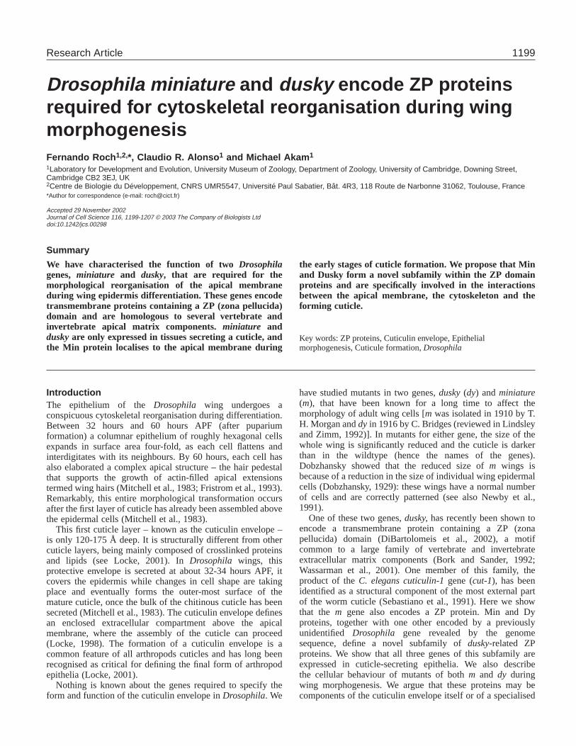
Fig. 1.Cuticle morphology is disrupted in Df(1)MR adultwings. Wild-type and Df(1)MRwings dissected 2 hours afteradult eclosion. (A-D) Light microscopy. The mutant wing inB is reduced in size, although veins (L2-L5) and wingmargin (WM) are correctly patterned. (C,D) A closer view ofthe same wings reveals that hairs are much closer in themutant than in the wildtype (arrowheads). (D) The picture ofthe mutant corresponds to a slightly lower focus plane,showing the cell outlines. Note the ring of cuticlesurrounding each hair in the mutant. (E,F) SEMmicrographs. The mutant hairs appear twisted and branched(open arrowheads), in contrast to the long and slender wild-type epidermal hairs (E). Some cuticle blobs appear bulgingout of the main wing surface in the mutant (arrow). (G-J) TEM sections. The two wing surfaces (dorsal and ventral)are separated in the mutant (H,J), and the resulting spacecontains cuticle invaginations (small arrows) and cell debris(arrows). The presence of cuticle defects is indicated byarrowheads and at a higher magnification in the lower panels(I,J). The position of some epidermal hairs is also indicated(open arrowheads in G,H).
1201ZP proteins in Drosophila epidermis
performed following standard protocols (Sturtevant et al., 1993) withsome modifications, all available from F.R. upon request.
Immunostainings and generation of the Min antibodyThe fragment coding for amino acids 346 to 547 of the CG9369predicted protein was amplified by PCR from wild-type genomicDNA and cloned in-frame in the pRSETB vector (Invitrogen). Theresulting 6× His-tagged protein was purified with the Pharmacia kitfrom the soluble fraction obtained after bacterial sonication. Thefraction containing the purified protein was loaded in a SDS-PAGEgel, and the resulting band was cut and used to immunise rabbitsfollowing standard protocols (Eurogentec, Belgium). The rabbit anti-Min serum fails to recognise any specific signal in immunostainingsperformed on m1 mutant embryos and is capable of specificallyrecognising the Min protein produced in vivo with a UAS-miniaturetransgene (F.R. and C.R.A., unpublished). Stainings were carried outusing rabbit anti-Min (1:100) and rat anti-E-Cadherin (1:50) (a giftfrom O. Renaud). Secondaries were 1:200 FITC anti-rabbit and 1:200Cy5 anti-rat (Jackson Labs). Cell nuclei were labelled with theTOPRO nuclear dye (Molecular Probes).
ResultsAll miniature and dusky mutations map very close to oneanother in the 10E1-2 region of the X chromosome (Dorn andBurdick, 1962). Although both classes of mutation affect cellmorphology in a similar way, the fact that they fall into twodistinct complementation groups suggests that they affectdistinct genes (Newby et al., 1991).
In this work we have studied the phenotypes caused byDf(1)MR, a small deletion that fails to complement both dyandm mutations (Roberts and Jackson, 1997). Males hemizygousfor this deficiency are viable, but the size of their wings issignificantly smaller than in either wild-type (Fig. 1A,B), or indyor msingle mutants (data not shown). Df(1)MRremoves thedy coding sequence and is thus a dy-null allele (DiBartolomeiset al., 2002). Comparison of the molecular maps of the m-dyregion (DiBartolomeis et al., 2002) with the genome sequence(Adams et al., 2000) reveals that Df(1)MR also removes mostof the coding region of an adjacent predicted gene, CG9369.This gene, which we have identified unequivocally asminiature (see below), encodes a transmembrane proteinclearly related to that encoded by the dy locus. This, and thefact that dy and m point alleles have similar phenotypes,suggests that these two proteins may have a similar function.
miniature and dusky mutant wings secrete abnormalcuticlesTo understand the cellular basis for the observed reduction incell size, we characterised in detail the phenotype of adultmales hemizygous for Df(1)MRand hence lacking both m anddy functions. Light and scanning electron microscopy (SEM)show that Df(1)MRwings dissected 2 hours after adult eclosionhave pronounced defects in their cuticle. In both wild-type andDf(1)MRwings, each epidermal cell makes a hair on its apicalsurface. However, in Df(1)MR, wings hairs are much closer to
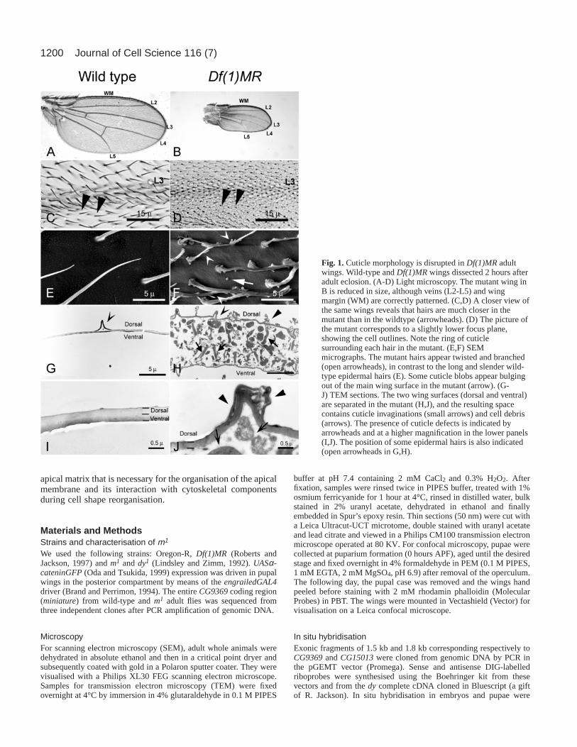
Fig. 2.Apical membrane reorganisation fails to occur in Df(1)MRmutants. Confocal images of wild-type and Df(1)MRpupal wings of differentstages stained as indicated. Cartoons show cell outlines (grey) and the position of the apical junctions (black) in a transverse section of theepithelium at each stage. At 28-32 hours APF, wild-type and mutant epithelia are indistinguishable. By 46-50 hours APF, the formation of hairpedestals accumulating large amounts of actin (arrowheads) is hindered in the mutant. Note also the presence of an actin-ring around each cellin the mutant, visible also in the transverse sections (open arrowheads). The mutant cells also fail to acquire a star-like cell contour at the levelof their apical junctions. At 62-66 hours APF, the rhodamin-phalloidin binds the apical membrane that appears folded inside the wing. Note thatin the mutant a space between the apical membrane folds is present, where cuticle is secreted (open arrowheads). The aberrant apical junctionsof the mutants also accumulate large amounts of α-catenin–GFP. Bar, 5 µm.
1202
each other than in the wildtype, suggesting that the size ofindividual cells is significantly reduced (Fig. 1C-F). Df(1)MRhairs are also shorter than in the wildtype and their structure isabnormal: they are twisted and often branched (Fig. 1F). Wealso observe localised distensions of the cuticle surface thatbulge out of the main wing surface, a phenotype that indicatesdefects in cuticle formation (Fig. 1F). Similar defects are alsoobserved in both dy and m single mutants, but they are lesssevere (data not shown).
Transmission electron microscopy (TEM) of thin sections ofDf(1)MRadult wings confirms that cuticle structure is severelyaffected in the mutants. In wild-type animals, wing epidermalcells have undergone apoptosis by 2 hours after eclosion, andthe dorsal and ventral cuticle surfaces forming the wing areclosely apposed by their basal sides (Johnson and Milner, 1987)(Fig. 1G,I). In the mutants, cuticular material present inside thewing prevents the apposition of dorsal and ventral surfaces.These indentations of cuticle form a honeycomb-like pattern,marking the cell outlines that are visible in adult mutant wingsunder light microscopy (Fig. 1D). The indentations appear toobstruct clearance of the cellular debris produced afterapoptosis of the epidermal cells (Johnson and Milner, 1987), aswe observe degenerating cell fragments between the two wingsurfaces (Fig. 1H,J). In contrast, wild-type wings of similar ageare totally clean of cell debris (Fig. 1G,I).
The effects of duskyand miniature mutations are obviousonly in the wings: the cuticle covering the epidermis of otherparts of the body (haltere, notum, legs and abdominal plates)appears completely normal. To determine if subtle defects maybe present elsewhere, we also performed TEM studies on thecuticle covering the adult haltere, a dorsal appendage that ishomologous to the wing. Despite the high resolution of thisanalysis, we could detect no defects in the haltere cuticle (datanot shown).
miniature and dusky mutants disrupt cell morphogenesisduring differentiationTo investigate the origin of the defects observed in Df(1)MRadult wing cells, we compared the morphology of wild-typeand mutant cells at different stages during pupal developmentusing confocal microscopy. We employed two cytoskeletonmarkers: rhodamine-phalloidin, a fluorescent compound thatbinds specifically to actin filaments and stains cell outlines andepidermal hairs (Fristrom et al., 1993) and an α-catenin–GFPfusion protein (Oda and Tsukida, 1999). This GFP-taggedprotein localises in the apical junctions between epidermalcells where the apical and basolateral membrane compartmentsmeet.
We detect no obvious abnormalities in the morphology ofDf(1)MR mutant cells at 28-32 hours APF, a time when wingcells are columnar and still hexagonal in outline (Fig. 2).Between 42 and 48 hours APF, wild-type cells flatten, expandand become star shaped (Fristrom et al., 1993). They alsodevelop prominent actin-filled extensions of their apicalsurface, called hair pedestals, that can be visualised in wingsstaged 46-50 hours APF (Fig. 2, arrowheads). In mutants of thesame age the apical cell contours remain hexagonal, and theaccumulation of actin at the hair pedestals is impaired (Fig. 2).They also accumulate actin abnormally, forming a ring in theapical part of the mutant cells (Fig. 2, open arrowheads).
In wild-type wings staged 62-66 hours APF, phalloidinstrongly stains the apical cell membrane, which by then issecreting the adult chitinous cuticle (Fristrom et al., 1993) (Fig.2). In Df(1)MRmutants, the apical membrane folds deeply intothe cleft between adjacent cells, almost forming a capsulearound the apical side of each cell (Fig. 2). The walls of thesecapsules are apposed to those of the neighbouring cells, leavinga narrow space in between (Fig. 2, open arrowheads). Belowthese structures, the cell maintains its hexagonal contour at the
Journal of Cell Science 116 (7)
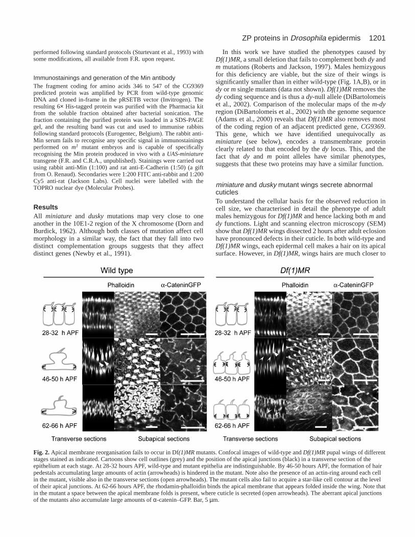
Fig. 3.mand dyencode transmembrane proteins with a ZP domain.(A) Genomic organisation of the m/dy region. The position of exonsis indicated by black boxes. The regions deleted in the differentdeficiencies are shown below. The position of the smallrearrangement present in the m1 mutant is indicated (arrow).(B) Similarity diagrams based on a ClustalW multiple sequencealignment of the ZP domains from the indicated Drosophila,nematode and mammal proteins. (C) Predicted modular structure ofMin, Dy and Dyl proteins (protein accession numbers areAAF48088, AAF48089 and AAF47884, respectively). The positionof the transmembrane domain is highlighted by a vertical box.Tetrabasic RRR/AR motifs are indicated by triangles. Signal peptidesare indicated by black boxes (SP). (D) Alignment of Dy, Dyl, Minand Cut-1 ZP domains. Black and shaded boxes indicate identicaland similar residues, respectively. The position of the ZP structuraldomain is shown between brackets, and the eight conserved cysteinescharacteristic of this domain are marked with asterisks (see text fordetails).
1203ZP proteins in Drosophila epidermis
level of the apical junctions, which accumulate larger amountsof α-catenin–GFP than in the wildtype (Fig. 2).
miniature, like dusky, encodes a transmembrane proteinwith a ZP domainThe dy gene has recently been identified (DiBartolomeis et al.,2002). It encodes a putative transmembrane protein with anextracellular motif called a ZP domain, a conserved domainpresent in transmembrane proteins described in bothvertebrates and invertebrates as components of variousextracellular matrices (Bork and Sander, 1992; Wassarman etal., 2001; Wilkin et al., 2000; Chung et al., 2001).
Genetic data indicate that m mutations lie just to the left(i.e. telomeric) of dy (Dorn and Burdick, 1962). A survey ofthe Drosophilagenome sequence (Adams et al., 2000) revealsthat the gene immediately to the left of dy in the chromosome(CG9369) also encodes a transmembrane protein with a ZPdomain. We sequenced the complete predicted coding regionof this gene in the m1 mutant and found a deletion of 33nucleotides associated with an insertion of 13 nucleotides,370 amino acids downstream of the CG9369 predicted startcodon (Fig. 3A). This small rearrangement causes aframeshift and a premature stop codon within the codingsequence, indicating that CG9369is the m gene and that m1
is probably a null allele for m. This is consistent withdeficiency mapping data published by the F. R. Jackson group(DiBartolomeis et al., 2002). Df(1)m259, which fails tocomplement m alleles but not dy alleles, removes onlysequences upstream of m (presumably regulatory sequencesnecessary for m expression), whereas Df(1)MR, Df(1)m30 andDf(1)KA6, which fail to complement both m and dy alleles,
delete sequences from both coding regions (DiBartolomeis etal., 2002) (Fig. 3A).
A BLAST search of the whole genome of Drosophila,usingeither the dy or m ZP domains as probes, reveals that there aremore than a dozen putative proteins encoded by the fly genomethat contain a ZP domain. Sequence comparison of theDrosophilaZP domains among themselves and with other non-fly homologues indicates that three of these Drosophila ZPgenes encode a distinct subfamily of ZP proteins (Fig. 3B anddata not shown). These three genes are dy, m and CG15013,which is a predicted open reading frame located at 64B1(Adams et al., 2000) (Fig. 3B). The ZP domains of Dy andCG15013 are most similar (70% identity), with that of Minbeing more divergent (45% identity with Dy, Fig. 3D). Theirsimilarity to other DrosophilaZP domains and C. elegansCut-1 is largely confined to eight key cysteine residues, landmarksof a ZP domain (Fig. 3D and data not shown).
We have studied the structure of the Dy, Min and CG15013proteins using SMART software to predict the positionof architectural domains from protein primary sequences(Schultz et al., 1998). The three proteins include a putativetransmembrane domain separating a short intracellular C-terminus and a large extracellular N-terminus containing theZP domain (Fig. 3C). In addition, Dy, Min and CG15013contain an ER import signal peptide in their N-terminus(DiBartolomeis et al., 2002) (Fig. 3C), which is consistent withthem being single-pass transmembrane proteins.
They also have a basic tetrapeptide RRRR (RRAR in thecase of Dy) located in between the ZP domain and thetransmembrane domain, within the extracellular part of theprotein. This small motif is common to many proteinprecursors cleaved by endopeptidases of the secretory pathway
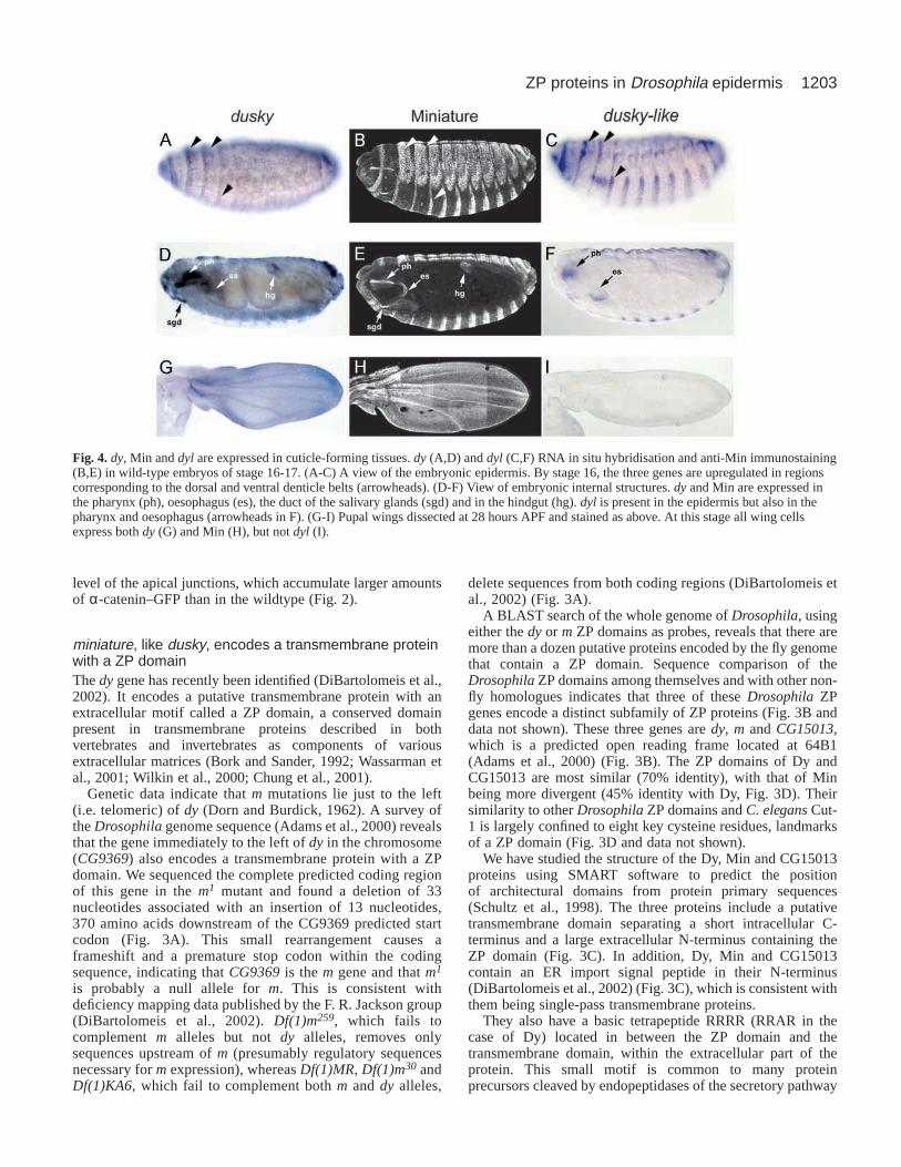
Fig. 4. dy, Min and dyl are expressed in cuticle-forming tissues.dy (A,D) and dyl (C,F) RNA in situ hybridisation and anti-Min immunostaining(B,E) in wild-type embryos of stage 16-17. (A-C) A view of the embryonic epidermis. By stage 16, the three genes are upregulated in regionscorresponding to the dorsal and ventral denticle belts (arrowheads). (D-F) View of embryonic internal structures. dyand Min are expressed inthe pharynx (ph), oesophagus (es), the duct of the salivary glands (sgd) and in the hindgut (hg). dyl is present in the epidermis but also in thepharynx and oesophagus (arrowheads in F). (G-I) Pupal wings dissected at 28 hours APF and stained as above. At this stage all wing cellsexpress both dy (G) and Min (H), but not dyl (I).
1204
(Hosaka et al., 1997) and could be a target in Drosophilafor afurin type endopeptidase, which releases the ZP-domain-containing region. Because of the similarities betweenCG15013 and Dy proteins (see also below), we name the geneCG15013 dusky-like(dyl).
miniature, dusky and dusky-like are expressed in tissuesinvolved in cuticle secretion To determine how dy, m and dyl are expressed, we carried outRNA in situ hybridisations at different stages with probes forthese genes. We also generated an antibody against the Minprotein to study its subcellular localisation.
The three gene products are expressed in partiallyoverlapping domains during embryogenesis and pupaldevelopment. In the embryo, they are only expressed in tissuesthat will secrete cuticle, including the epidermis, foregut andhindgut (Hillman and Lesnick, 1970) (Fig. 4A-F). In theepidermis, expression starts at about stage 14, and RNA levelsincrease until at least stage 17, when formation of the cuticleprevents the penetration of our probes. In each segment,transcripts of all three genes are more abundant in cells formingthe dorsal and ventral denticle belts than in other parts of theepidermis (Fig. 4A,C). These cells also contain high levels ofMin protein (Fig. 4B). dy and Min are also expressed in thecells forming the duct linking the salivary glands with the
oesophagus (Fig. 4D,E), and Min protein is detected at lowlevels in the cells forming the embryonic tracheae (data notshown).
RNA in situ hybridisation and antibody staining show thatdyand Min are also expressed in pupal wings by 28 hours APF,which is consistent with their genetic requirement in this tissue(Fig. 4G,H). However, we could not detect expression of dylin pupal wings of the same stage (Fig. 4I), indicating that thisgene could have an embryo-specific role.
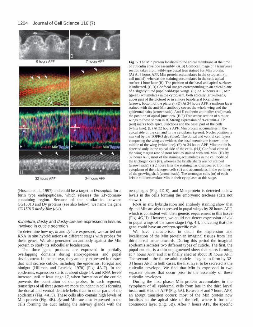
We have characterised in detail the expression andlocalisation of the Min protein in imaginal tissues from latethird larval instar onwards. During this period the imaginalepidermis secretes two different types of cuticle. The first, thepupal cuticle, is a thin unpigmented sheet that starts formingat 7 hours APF, and it is finally shed at about 18 hours APF.The second – the future adult cuticle – begins to form by 32-34 hours APF. In both cases, the first layer to be secreted is thecuticulin envelope. We find that Min is expressed in twoseparate phases that occur prior to the assembly of thesecuticulin envelopes.
During the first phase, Min protein accumulates in thecytoplasm of all epidermal cells from late in the third larvalinstar until 6 hours APF (Fig. 5A). Between 6 and 7 hours APF,an abrupt transition occurs; most of the Min protein nowlocalises to the apical side of the cell, where it forms acontinuous layer (Fig. 5B). After 7 hours APF, the specific
Journal of Cell Science 116 (7)
Fig. 5.The Min protein localises to the apical membrane at the timeof cuticulin envelope assembly. (A,B) Confocal image of a transversesection taken from wild-type pupal legs stained for Min protein.(A) At 6 hours APF, Min protein accumulates in the cytoplasm (n,cell nuclei), whereas the staining accumulates in the cells apicalsurface 1 hour later (B). The position of the basal and apical surfacesis indicated. (C,D) Confocal images corresponding to an apical planeof a slightly tilted pupal wild-type wings. (C) At 32 hours APF, Min(green) accumulates in the cytoplasm, both apically (arrowheads,upper part of the picture) or in a more basolateral focal plane(arrows, bottom of the picture). (D) At 34 hours APF, a uniform layerstained with the anti-Min antibody covers the whole wing and theepidermal hairs (arrowheads). Anti E-cadherin antibodies (red) markthe position of apical junctions. (E-F) Transverse section of similarwings to those shown in B. Strong expression of α-catenin–GFP(red) marks both apical junctions and the basal part of the cells(white line). (E) At 32 hours APF, Min protein accumulates in theapical side of the cell and in the cytoplasm (green). Nuclei position ismarked by the TOPRO dye (blue). The dorsal and ventral cell layerscomposing the wing are evident; the basal membrane is now in themiddle of the wing (white line). (F) At 34 hours APF, Min protein isdetected only in the apical side of the cells. (H,I) Confocal view ofthe wing margin row of stout bristles stained with anti-Min. (H) By32 hours APF, most of the staining accumulates in the cell body ofthe trichogen cells (tr), whereas the bristle shafts are not stained(arrowheads). (I) 2 hours later the staining has disappeared from thecytoplasm of the trichogen cells (tr) and accumulates in the peripheryof the growing shaft (arrowheads). The tormogen cells (to) of eachbristle still accumulate Min in their cytoplasm at this stage.
1205ZP proteins in Drosophila epidermis
staining disappears, probably because Min protein has becomechemically crosslinked to itself or to other components of theforming cuticle.
We observe a similar pattern in late pupal wings, before thesecretion of the adult cuticle. Min protein first appears in thewing at about 28 hours APF (Fig. 5C-F). It accumulates to highlevels in the cell cytoplasm until, by 32-34 hours APF, itlocalises to the apical membrane, forming a continuous layercovering the epidermal cells (Fig. 5D,F) and the developingbristles (Fig. 5H,I).
The pattern of accumulation of Min protein in the haltereepidermis is similar to that in the wing (data not shown), eventhough no mutant phenotype is visible in this tissue.
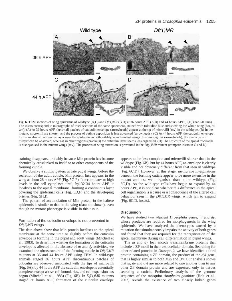
Formation of the cuticulin envelope is not prevented inDf(1)MR wingsThe data above show that Min protein localises to the apicalmembrane at the same time or slightly before the cuticulinenvelope is forming in the pupal and adult wings (Mitchell etal., 1983). To determine whether the formation of the cuticulinenvelope is affected in the absence of m and dy activities, weexamined the ultrastructure of the forming cuticle in Df(1)MRmutants at 36 and 44 hours APF using TEM. In wild-typeanimals staged 36 hours APF, discontinuous patches ofcuticulin are observed associated with the tips of microvilli(Fig. 6A); by 44 hours APF the cuticulin envelope is essentiallycomplete, except above cell boundaries, and cell expansion hasbegun (Mitchell et al., 1983) (Fig. 6B). In Df(1)MR mutantsstaged 36 hours APF, formation of the cuticulin envelope
appears to be less complete and microvilli shorter than in thewildtype (Fig. 6B), but by 44 hours APF, an envelope is clearlyvisible and not obviously different from that seen in wildtype(Fig. 6C,D). However, at this stage, membrane invaginationsbeneath the forming cuticle appear to be more extensive in themutant and less well organised than in the wildtype (Fig.6C,D). As the wild-type cells have begun to expand by 44hours APF, it is not clear whether this difference in the apicalcell organisation is a cause or a consequence of the altered cellbehaviour seen in the Df(1)MR wings, which fail to expand(Fig. 6C,D, insets).
DiscussionWe have studied two adjacent Drosophila genes, m and dy,whose products are required for morphogenesis in the wingepidermis. We have analysed the phenotypes caused by amutation that simultaneously impairs the activity of both genesand found that they are required for the reorganisation of theapical membrane during cell differentiation in pupal wings.
The m and dy loci encode transmembrane proteins thatinclude a ZP motif in their extracellular domain. Searching forother related proteins in Drosophilawe have identified a thirdprotein containing a ZP domain, the product of the dyl gene,that is highly similar to both Min and Dy. Our analysis showsthat m, dy and dyl are more related to one another than to anyother ZP domain protein and are expressed only in tissuessecreting a cuticle. Preliminary analysis of the genomesequence of the mosquito Anopheles gambiae(Holt et al.,2002) reveals the existence of two closely linked genes
Fig. 6.TEM sections of wing epidermis of wildtype (A,C) and Df(1)MR(B,D) at 36 hours APF (A,B) and 44 hours APF (C,D) (bar, 500 nm).The insets correspond to micrographs of thick sections of the same specimens, stained with toloudine blue and showing the whole wing (bar, 50µm). (A) At 36 hours APF, the small patches of cuticulin envelope (arrowheads) appear at the tip of microvilli (mv) in the wildtype. (B) In themutant, microvilli are shorter, and the process of cuticle deposition is less advanced (arrowheads). (C) At 44 hours APF, the cuticulin envelopeforms an almost continuous layer over the epidermis in both wild-type and mutant wings. In some regions (arrowheads), the characteristictrilayer can be observed, whereas in other regions (brackets) the cuticulin layer seems less organised. (D) The structure of the apical microvilliis disorganised in the mutant wings (mv). The process of wing extension is prevented in the Df(1)MRmutant (compare insets in C and D).
1206
mapping in the X chromosome coding for proteins highlysimilar to Min and Dy. This suggests that this specific genefamily has a long history within the Dipterans. Outside thisgroup, no specific orthologues of the min/dy/dyl family haveyet been defined. However, there are suggestions that thefunction of ZP proteins in cuticle synthesis might be widelyconserved. One of the C. elegansproteins containing a ZPdomain, Cut-1, has been identified as a structural componentof the nematode cuticle. Cut-1 protein is apically secreted andlocalises in the most external cuticle layer, the cuticulin(Sebastiano et al., 1991). In the Drosophilawing, the epidermalcells secrete the cuticulin envelope prior to the reorganisationof the apical membrane that takes place during differentiation.One attractive hypothesis is that m, dy and dyl, like the Cut-1protein, could be structural components of the fly cuticulinenvelope. We observe that Min accumulates in the cytoplasmand is then transported to the apical membrane by the time ofcuticulin envelope assembly. Min protein then forms acontinuous layer covering the whole epidermal surface,consistent with the idea that Min is an envelope component.Unfortunately, our antibodies fail to detect Min protein shortlyafter secretion, so we cannot determine whether it becomesintegrated into the cuticle or not. Nevertheless, we observe thatin Df(1)MRmutants the formation of the cuticulin envelope isnot prevented, indicating that neither Min nor Dy are essentialfor the formation of this structure. Instead, we see that theformation of the cuticulin envelope is delayed and that theapical membrane is disorganised. Whether this is the primarycause or is simply correlated with the failure of epidermal wingcells to undergo changes in cell shape is difficult to establish.
A second possibility consistent with our observations is thatm, dy and dyl are components of a specialised apical matrixsecreted at the time of the cuticulin envelope formation andnecessary for the reorganisation of apical membranes duringdifferentiation. ZP-domain-containing proteins characterisedin flies and vertebrates seem to participate in the formation ofapical extracellular matrices in different cellular contexts. Forinstance ZP1, ZP2 and ZP3 proteins are the main componentsof the mammalian oocyte zona pellucida and form a specialisedapical matrix that is required for oocyte maturation (Rankinand Dean, 2000). Another example is the DrosophilaNompAprotein, which is a component of an extracellular structurecalled the dendritic cap of mechanosensory receptors (Chunget al., 2001). This structure mediates the interaction betweenthe neuronal sensory processes and the external components ofthe sensory organ, where mechanical stimuli are transducedinto neuronal membrane potentials (Chung et al., 2001).However, the NompA protein contains other conserveddomains in its extracellular portion (Chung et al., 2001), andthe role of its ZP domain remains to be elucidated. Perhapsproteins of the m/dy family, containing only a conserved ZPextracellular domain, are specialised in mediating interactionsbetween the forming cuticle, the cell membrane andcytoskeleton components involved in cell shape reorganisation.
We have shown that m, dy and dyl are expressed throughoutembryogenesis in many cuticle-forming tissues. It is thusparadoxical that the phenotype we observe in the m/dymutantis specific to the wings. Indeed, the Df(1)MRmutant is viable,and we have not detected defects elsewhere in the adult or inthe embryonic cuticle. One possible explanation for thisspecificity is that loss of the ZP proteins activity is most
deleterious in tissues undergoing extensive rearrangement oftheir apical membranes, like the wing cells. We note thatnormal haltere cells do not undergo major changes in shapeafter the assembly of the cuticulin envelope (Roch and Akam,2000), and even though Min is expressed in this tissue in asimilar way to the wing, the haltere cells differentiate normally.
Functional redundancy is another possible explanation forthe wing specificity of the phenotypes. The product of the dylgene is a good candidate for fulfilling the roles of dy and m intheir absence. All three proteins are closely related, and all areexpressed in the embryonic epidermis during differentiation.Interestingly, dyl is not detectably expressed in the wing,providing a simple explanation for the sensitivity of this tissueto m and dy mutations. Unfortunately, there are at present nospecific mutations affecting the dyl gene. Other more distantlyrelated Drosophila ZP proteins may also contribute to theformation of the cuticulin envelope or to the apical matrix. Inregard to the functional redundancy, a clear parallelism can beestablished with the proteins forming the mammalian zonapellucida. In mice, the presence of ZP3 and ZP2 is absolutelyrequired for the formation of this matrix, whereas loss-of-function mutants for ZP1 present milder defects in its structure(Rankin et al., 2001).
We particularly thank F. R. Jackson for sharing stocks, plasmidsand results prior to publication. We are grateful to F. Payre, in whoselaboratory part of this work was carried out, and to W. Lee, H. López-Shier, O. Renaud, J. Roote, J. Skepper and I. Palacios for strains,antibodies and precious advice. We thank also two anonymousreferees for constructive criticisms. F. R. was in part funded by aEMBO Long Term Fellowship. This work was supported by theWellcome Trust.
ReferencesAdams, M. D., Celniker, S. E., Holt, R. A., Evans, C. A., Gocayne, J. D.,
Amanatides, P. G., Scherer, S. E., Li, P. W., Hoskins, R. A., Galle, R. F.et al. (2000). The genome sequence of Drosophila melanogaster. Science287, 2185-2195.
Bork, P. and Sander, C.(1992). A large domain common to sperm receptor(Zp2 and Zp3) and TGF-B type III receptor. FEBS Lett.300, 237-240.
Brand, A. H. and Perrimon, N. (1993). Targeted gene expression as a meansof altering cell fates and generating dominant phenotypes. Development118,401-415.
Chung, Y. D., Zhu, J., Han, Y. and Kernan, M. J.(2001). nompAencodesa PNS-specific, ZP domain protein required to connect mechanosensorydendrites to sensory structures. Neuron29, 415-428.
DiBartolomeis, S. M., Akten, B., Genova, G., Roberts, M. A. and Jackson,F. R. (2002). Molecular analysis of the Drosophila miniature-dusky(m-dy)gene complex: m-dymRNAs encode transmembrane proteins with similarityto C. eleganscuticulin. Mol. Genet. Genomics 267, 564-576.
Dobzhansky, T. (1929). The influence of the quantity and quality ofchromosomal material on the size of the cells in Drosophila melanogaster.Roux. Arch. Entw. Mech. Organ115, 363-379.
Dorn, G. L. and Burdick, A. L. (1962). On the recombinational structure andcomplementation relationships in them-dy complex of Drosophilamelanogaster. Genetics47, 503-518.
Fristrom, D., Wilcox, M. and Fristrom, J. (1993). The distribution of PSintegrins, laminin A and F-actin during key stages in Drosophila wingdevelopment. Development117, 509-523.
Hillman, R. and Lesnick, L. H. (1970). Cuticle formation in the embryo ofDrosophila melanogaster. J. Morph.131, 383-396.
Holt, R. A., Subramanian, G. M., Halpern, A., Sutton, G. G., Charlab, R.,Nusskern, D. R., Wincker, P., Clark, A. G., Ribeiro, J. M., Wides, R. etal. (2002). The genome of the malaria mosquito Anopheles gambiae.Science295, 129-149.
Hosaka, M., Nagahama, M., Kim, W. S., Watanabe, T., Hatsuzawa, K.,Ikemizu, J., Murakami, K. and Nakayama, K. (1991). Arg-X-Lys/Arg-
Journal of Cell Science 116 (7)
1207ZP proteins in Drosophila epidermis
Arg motif is a signal for precursor cleavage catalyzed by furin within theconstitutive secretory pathway. J. Biol. Chem.266, 12127-12132.
Johnson, S. A. and Milner, M. J. (1987). The final stages of wingdevelopment in Drosophila melanogaster. Tissue Cell19, 505-513.
Lindsley, D. L. and Zimm, G. G. (1992). In The genome of Drosophilamelanogaster.San Diego: Academic Press.
Locke, M. (1966). The structure and formation of the cuticulin layer in theepicuticle of an insect Calpodes ethlius(Lepidoptera, Hesperidae). J.Morph. 118, 461-494.
Locke, M. (1998). Epidermis. In Microscopic anatomy of invertebrates, vol.11A, Insecta(eds F. W. Harrison and M. Locke), pp. 75-138. New York:Wiley-Liss, John Wiley & Sons, Inc.
Locke, M. (2001). The Wigglesworth lecture: insects for studying fundamentalproblems in biology. J. Insect Physiol. 47, 495-507.
Mitchell, H. K., Roach, J. and Petersen, N. S.(1983). The morphogenesisof cell hairs on Drosophilawings. Dev. Biol.95, 387-398.
Newby, L. M., White, L., DiBartolomeis, S. M., Walker, B. J., Dowse, H.B., Ringo, J. M., Khuda, N. and Jackson, F. R.(1991). Mutational analysisof the Drosophila miniature-dusky(m-dy) locus: effects on cell size andcircadiam rhythms. Genetics128, 571-582.
Oda, H. and Tsukida, S. (1999). Dynamic features of adherens junctionsduring Drosophilaembryonic epithelial morphogenesis revealed by a Dα-catenin-GFP fusion protein. Dev. Genes Evol.209, 218-225.
Rankin, T. L., O’Brien, M., Lee, E., Wigglesworth, K., Eppig, J. and Dean,
J. (2001). Defective zonae pellucidae in Zp2-null mice disruptfolliculogenesis, fertility and development. Development128, 1119-1226.
Rankin, T. and Dean, J.(2000). The zona pellucida: using molecular geneticsto study the mammalian egg coat. Rev. Reprod.5, 114-121.
Roberts, M. and Jackson, F. R.(1997). A new miniature-duskyallele.Drosophila Information Service80, 104-105.
Roch, F. and Akam, M. (2000). Ultrabithorax and the control of cellmorphology in Drosophilahalteres. Development127, 97-107.
Schultz, J., Milpetz, F., Bork, P. and Ponting, C. P.(1998). SMART, a simplemodular architecture research tool: identification of signaling domains.Proc. Natl. Acad. Sci USA95, 5857-5864.
Sebastiano, M., Lassandro, F. and Bazzicalupo, P.(1991). cut-1 aCaenorhabditis elegansgene coding for a Dauer-specific noncollagenouscomponent of the cuticle. Dev. Biol.146, 519-530.
Sturtevant, M. A., Roark, M. and Bier, E. (1993). The Drosophila rhomboidgene mediates the localized formation of wing veins and interactsgenetically with components of the EGF-R signalling pathway. Genes Dev.7, 961-973.
Wassarman, P. M., Jovine, L. and Litscher, E. S.(2001). A profile offertilization in mammals. Nat. Cell Biol. 3, E59-E64.
Wilkin, M. B., Becker, M. N., Mulvey, D., Phan, I., Chao, A., Cooper, K.,Chung, H. J., Campbell, I. D., Baron, M. and MacIntyre, R.(2000).DrosophilaDumpy is a gigantic extracellular protein required to maintaintension at epidermal-cuticle attachment sites. Curr. Biol. 10, 559-567.