Embed Size (px)
Citation preview

Mini-Review
Protein degradation and aging
Marta Martinez-Vicente, Guy Sovak, Ana Maria Cuervo*
Department of Anatomy and Structural Biology, Marion Bessin Liver Research Center, Albert Einstein College of Medicine,
Ullmann Building Room 611, 1300 Morris Park Avenue, Bronx, NY 10461, USA
Received 31 March 2005; received in revised form 8 July 2005; accepted 8 July 2005
Available online 24 August 2005
Abstract
Continuous turnover of intracellular proteins is essential for the maintenance of cellular homeostasis and for the regulation of multiple
cellular functions. The first reports showing a decrease in total rates of protein degradation with age are dated more than 50 years ago, when
the major players in protein degradation where still to be discovered. The current advances in the molecular characterization of the two main
intracellular proteolytic systems, the lysosomal and the ubiquitin proteasome system, offer now the possibility of a systematic search for the
defect(s) that lead to the declined activity of these systems in old organisms. We discuss here, in light of the current findings, how
malfunctioning of these two proteolytic systems can contribute to different aspects of the phenotype of aging and to the pathogenesis of some
age-related diseases.
q 2005 Elsevier Inc. All rights reserved.
Keywords: Proteolysis; Proteases; Lysosomes; Autophagy; Proteasome; Ubiquitin
1. Intracellular protein degradation: constant
destruction for continuous rejuvenation
All intracellular proteins undergo continuous synthesis
and degradation (Mortimore et al., 1989; Schimke, 1970).
This constant protein turnover, among other functions, helps
reduce, to a minimum, the time a particular protein is
exposed to the hazardous cellular environment, and
consequently, the probability of being damaged or altered.
At a first sight, this constant renewal of cellular components
before they lose functionality may appear a tremendous
waste of cellular resources. However, it is well justified
considering the detrimental consequences that the accumu-
lation of damaged intracellular components has on cell
function and survival (Goldberg, 2003). Furthermore,
protein degradation rather than mere destruction is indeed
a recycling process, as the constituent amino acids of the
degraded protein are reutilized for the synthesis of new
proteins (Mortimore et al., 1989; Schimke, 1970). The rates
at which different proteins are synthesized and degraded
inside cells are different and can change in response to
0531-5565/$ - see front matter q 2005 Elsevier Inc. All rights reserved.
doi:10.1016/j.exger.2005.07.005
* Corresponding author. Tel.: C1 718 430 2689; fax: C1 718 430 8975.
E-mail address: [email protected] (A.M. Cuervo).
different stimuli or under different conditions. This balance
between protein synthesis and degradation also allows cells
to rapidly modify intracellular levels of proteins to adapt to
changes in the extracellular environment. Proper protein
degradation is also essential for cell survival under
conditions resulting in extensive cellular damage. In fact,
activation of the intracellular proteolytic systems occurs
frequently as part of the cellular response to stress (recently
reviewed in Cuervo, 2004b; Goldberg, 2003). In this role as
‘quality control’ systems, the proteolytic systems are
assisted by molecular chaperones, which ultimately
determine the fate of the damaged/unfolded protein
(Fig. 1). Damaged proteins are first recognized by molecular
chaperones, which facilitate protein refolding/repairing. If
the damage is too extensive, or under conditions unfavor-
able for protein repair, damaged proteins are targeted for
degradation. Protein degradation is also essential during
major cellular remodeling (i.e. embryogenesis, morphogen-
esis, cell differentiation), and as a defensive mechanism
against harmful agents and pathogens (recently reviewed in
Cuervo, 2004a; Klionsky, 2005).
Two major proteolytic systems are responsible for most
intracellular protein turnover: the lysosomal system and the
ubiquitin-proteasome system (reviewed in Ciechanover,
2005; Cuervo, 2004b; Goldberg, 2003). Although lysosomes
were the first proteolytic system discovered, the recent
Experimental Gerontology 40 (2005) 622–633
www.elsevier.com/locate/expgero

Fig. 1. Intracellular fate of altered proteins. In normally functioning cells (Young) damaged proteins are recognized by molecular chaperones that attempt to
repair (refold) them. If refolding is not possible, chaperones target the damaged protein for degradation by the intracellular proteolytic systems. As cells age
(Old) the defective activity of the major proteolytic systems leads to chaperone ‘overload’ and to the intracellular accumulation of damaged/unfolded protein
products. Missfolded proteins often aggregate, probably as a defensive mechanism, sequestering in this aggregates chaperones, proteases and other neighboring
proteins. Protein aggregates slowly accumulate in all cells through the life, but their formation can be precipitate under particular cellular conditions or in
certain pathologies.
M. Martinez-Vicente et al. / Experimental Gerontology 40 (2005) 622–633 623
advances on the molecular characterization of this system
have motivated their come back to the spot light (Cuervo,
2004b; Klionsky, 2005). The entry of the ubiquitin-
proteasome system into the scene, back in the early 80s,
resulted in a complete change of mind, expanding the role of
protein degradation from mere housekeeping to regulator of
major intracellular processes, such as cell cycle and cell
division (Ciechanover, 2005).
The first reports showing a decrease in total rates of
protein degradation with age are dated more than 30
years ago, when the major players in protein degradation
where still to be discovered (Makrides, 1983). Since
then, this age-related decline in proteolytic activity has
been observed in almost all organisms analyzed, and
specific defects in the different proteolytic systems with
age have been reported. Keeping in mind the myriad of
intracellular functions in which protein degradation
participates, it is not surprising that the consequences
of the age-related alterations in the proteolytic systems
are widespread and contribute to a broad variety of
pathologies (reviewed in Cuervo, 2004a; Keller et al.,
2004; Shintani and Klionsky, 2004; Ward, 2002). We
briefly recapitulate here some of the main characteristics
of these two major proteolytic systems, highlighting
recent findings that have contributed to our current
understanding of their functioning, and discuss the major
changes described in these systems with age and their
consequences in aging and in some age-related
pathologies.
2. The lysosomal/autophagic system: the return
of the big giant
The term autophagy refers to any process resulting in
the degradation of intracellular components inside
lysosomes or the vacuole (the equivalent to lysosomes
in yeast) (reviewed in Cuervo, 2004a,b; Klionsky, 2005).
Lysosomes are single membrane organelles, which
contain a large assortment of hydrolases capable of
degrading any kind of macromolecules. Extracellular
macromolecules can also be internalized and degraded in
lysosomes through what is known as heterophagy (details
about the main forms of heterophagy—endocytosis and
phagocytosis—can be found in D’Hondt et al., 2000).
Lastly, some extracellular proteins, such as secretory
proteins, can undergo lysosomal degradation in the cells
in which they were synthesized, by fusion of secretory
vesicles with lysosomes instead of the plasma membrane.
This process, known as crinophagy, is a common
mechanism used by secretory cells to modulate secretion
rates (reviewed in (Cuervo, 2004b). The lysosome
becomes thus the ‘end terminal’ of a variety of pathways
that carry intra- or extracellular components for complete

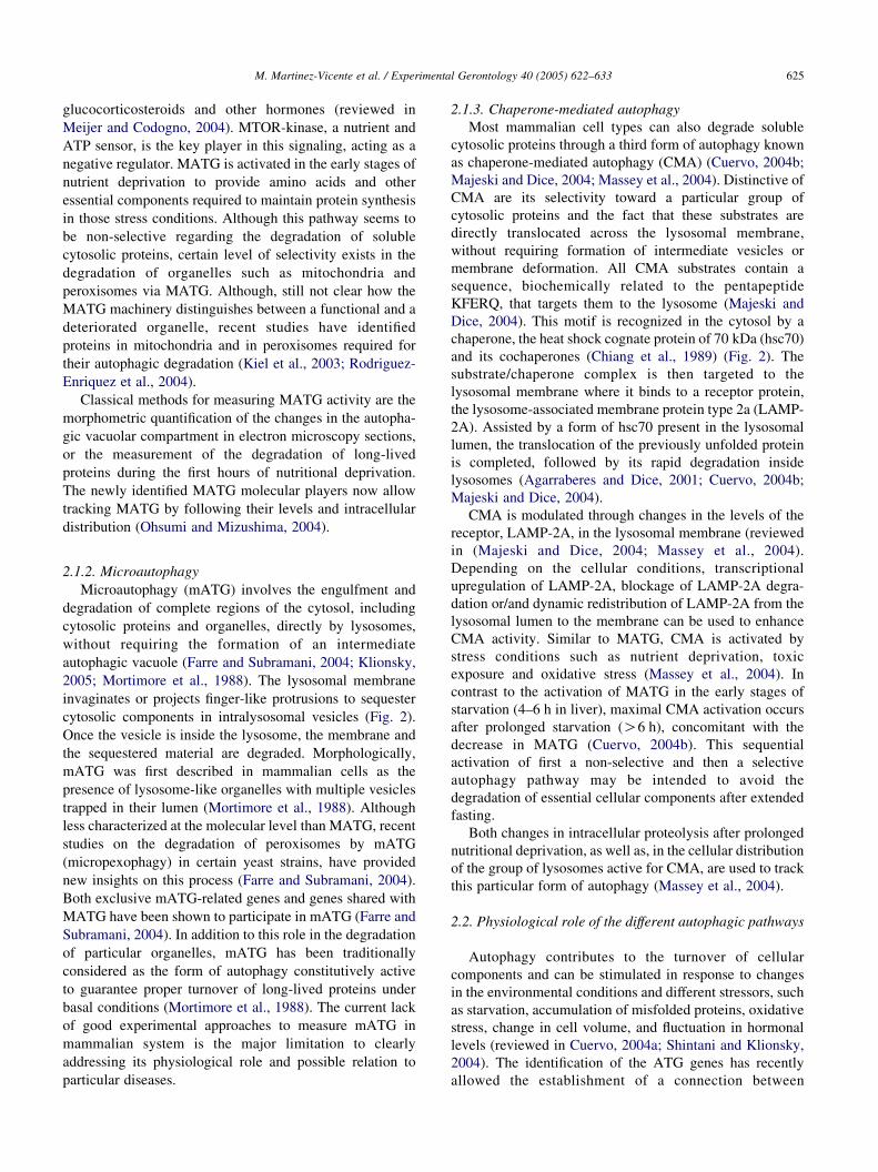
Fig. 2. Schematic model of the main forms of autophagy in mammalian cells and their changes in aging. Internalization of complete regions of cytosol first into
autophagosomes that fuse then with lysosomes (macroautophagy) or directly by the lysosomal membrane (microautophagy) contrast with the selective uptake
in a molecule-by-molecule basis of cytosolic proteins via chaperone-mediated autophagy (CMA). A variant of microautophagy that selectively removes
peroxisomes from the cytosol (micropexophagy) is also shown. Callouts summarize the age-related defects identified until date in the different autophagic
pathways. Abbreviations: hsc70, heat-shock cognate protein of 70 KDa; LAMP-2A, lysosome associate membrane protein type 2A.
M. Martinez-Vicente et al. / Experimental Gerontology 40 (2005) 622–633624
degradation. The scope of this review is in the turnover
of intracellular components, and consequently, only
autophagy, main responsible for the turnover of
organelles and a large number of long-lived cytosolic
proteins, is discussed.
2.1. Types of autophagy
The best characterized autophagic pathways are macro-
autophagy, microautophagy and chaperone-mediated autop-
hagy (Fig. 2) (Cuervo, 2004b; Klionsky, 2005). They differ
in the way in which they deliver substrates into the
lysosomal lumen, the types of substrates carried for
degradation and their activity/regulation.
2.1.1. Macroautophagy
Macroautophagy (MATG) is an inducible form of
autophagy responsible for the degradation of both long-
lived soluble proteins and complete organelles under stress
conditions (Klionsky, 2005; Shintani and Klionsky, 2004).
The stressor best characterized as a MATG activator is
starvation (nutritional stress). In MATG, a portion of
cytosol, often including whole organelles, is surrounded
by a de novo formed membrane (limiting membrane), which
seals to generate a double-membrane organelle called
autophagic vacuole or autophagosome (Ohsumi and
Mizushima, 2004). Fusion of lysosomes with these
autophagic vacuoles provides them with the enzymes
required for the degradation of the sequestered material
(Fig. 2). Recent genetic studies in yeast have facilitated the
identification of a series of genes related to autophagy
(autophagy genes or ATG), providing also key information
about the regulatory mechanisms for this pathway
(Klionsky, 2005; Ohsumi and Mizushima, 2004). Most of
the genes identified in yeast are conserved in mammals and
other organisms, confirming that MATG is a highly
conserved process in eukaryotes.
The MATG machinery is controlled by two ubiquitin-
like conjugation cascades (a protein–protein and a protein–
lipid conjugation) and by two phosphorylation complexes
(reviewed in Klionsky, 2005; Ohsumi and Mizushima,
2004). Conjugation among particular ATG proteins med-
iates their recruitment and interaction with pre-formed lipid
micelles in the cytosol, most likely originated from the
endoplasmic reticulum, and initiates the nucleation to form
the limiting membrane. A series of phosphorylation events
regulate the elongation of this isolation membrane and the
size of the vesicle formed by resealing (Fig. 2). Less
information is available about the events that mediate the
fusion of the autophagic vacuole to the lysosome. MATG is
regulated through a signaling cascade in response to
changes in the circulating levels of amino acids,
M. Martinez-Vicente et al. / Experimental Gerontology 40 (2005) 622–633 625
glucocorticosteroids and other hormones (reviewed in
Meijer and Codogno, 2004). MTOR-kinase, a nutrient and
ATP sensor, is the key player in this signaling, acting as a
negative regulator. MATG is activated in the early stages of
nutrient deprivation to provide amino acids and other
essential components required to maintain protein synthesis
in those stress conditions. Although this pathway seems to
be non-selective regarding the degradation of soluble
cytosolic proteins, certain level of selectivity exists in the
degradation of organelles such as mitochondria and
peroxisomes via MATG. Although, still not clear how the
MATG machinery distinguishes between a functional and a
deteriorated organelle, recent studies have identified
proteins in mitochondria and in peroxisomes required for
their autophagic degradation (Kiel et al., 2003; Rodriguez-
Enriquez et al., 2004).
Classical methods for measuring MATG activity are the
morphometric quantification of the changes in the autopha-
gic vacuolar compartment in electron microscopy sections,
or the measurement of the degradation of long-lived
proteins during the first hours of nutritional deprivation.
The newly identified MATG molecular players now allow
tracking MATG by following their levels and intracellular
distribution (Ohsumi and Mizushima, 2004).
2.1.2. Microautophagy
Microautophagy (mATG) involves the engulfment and
degradation of complete regions of the cytosol, including
cytosolic proteins and organelles, directly by lysosomes,
without requiring the formation of an intermediate
autophagic vacuole (Farre and Subramani, 2004; Klionsky,
2005; Mortimore et al., 1988). The lysosomal membrane
invaginates or projects finger-like protrusions to sequester
cytosolic components in intralysosomal vesicles (Fig. 2).
Once the vesicle is inside the lysosome, the membrane and
the sequestered material are degraded. Morphologically,
mATG was first described in mammalian cells as the
presence of lysosome-like organelles with multiple vesicles
trapped in their lumen (Mortimore et al., 1988). Although
less characterized at the molecular level than MATG, recent
studies on the degradation of peroxisomes by mATG
(micropexophagy) in certain yeast strains, have provided
new insights on this process (Farre and Subramani, 2004).
Both exclusive mATG-related genes and genes shared with
MATG have been shown to participate in mATG (Farre and
Subramani, 2004). In addition to this role in the degradation
of particular organelles, mATG has been traditionally
considered as the form of autophagy constitutively active
to guarantee proper turnover of long-lived proteins under
basal conditions (Mortimore et al., 1988). The current lack
of good experimental approaches to measure mATG in
mammalian system is the major limitation to clearly
addressing its physiological role and possible relation to
particular diseases.
2.1.3. Chaperone-mediated autophagy
Most mammalian cell types can also degrade soluble
cytosolic proteins through a third form of autophagy known
as chaperone-mediated autophagy (CMA) (Cuervo, 2004b;
Majeski and Dice, 2004; Massey et al., 2004). Distinctive of
CMA are its selectivity toward a particular group of
cytosolic proteins and the fact that these substrates are
directly translocated across the lysosomal membrane,
without requiring formation of intermediate vesicles or
membrane deformation. All CMA substrates contain a
sequence, biochemically related to the pentapeptide
KFERQ, that targets them to the lysosome (Majeski and
Dice, 2004). This motif is recognized in the cytosol by a
chaperone, the heat shock cognate protein of 70 kDa (hsc70)
and its cochaperones (Chiang et al., 1989) (Fig. 2). The
substrate/chaperone complex is then targeted to the
lysosomal membrane where it binds to a receptor protein,
the lysosome-associated membrane protein type 2a (LAMP-
2A). Assisted by a form of hsc70 present in the lysosomal
lumen, the translocation of the previously unfolded protein
is completed, followed by its rapid degradation inside
lysosomes (Agarraberes and Dice, 2001; Cuervo, 2004b;
Majeski and Dice, 2004).
CMA is modulated through changes in the levels of the
receptor, LAMP-2A, in the lysosomal membrane (reviewed
in (Majeski and Dice, 2004; Massey et al., 2004).
Depending on the cellular conditions, transcriptional
upregulation of LAMP-2A, blockage of LAMP-2A degra-
dation or/and dynamic redistribution of LAMP-2A from the
lysosomal lumen to the membrane can be used to enhance
CMA activity. Similar to MATG, CMA is activated by
stress conditions such as nutrient deprivation, toxic
exposure and oxidative stress (Massey et al., 2004). In
contrast to the activation of MATG in the early stages of
starvation (4–6 h in liver), maximal CMA activation occurs
after prolonged starvation (O6 h), concomitant with the
decrease in MATG (Cuervo, 2004b). This sequential
activation of first a non-selective and then a selective
autophagy pathway may be intended to avoid the
degradation of essential cellular components after extended
fasting.
Both changes in intracellular proteolysis after prolonged
nutritional deprivation, as well as, in the cellular distribution
of the group of lysosomes active for CMA, are used to track
this particular form of autophagy (Massey et al., 2004).
2.2. Physiological role of the different autophagic pathways
Autophagy contributes to the turnover of cellular
components and can be stimulated in response to changes
in the environmental conditions and different stressors, such
as starvation, accumulation of misfolded proteins, oxidative
stress, change in cell volume, and fluctuation in hormonal
levels (reviewed in Cuervo, 2004a; Shintani and Klionsky,
2004). The identification of the ATG genes has recently
allowed the establishment of a connection between

M. Martinez-Vicente et al. / Experimental Gerontology 40 (2005) 622–633626
autophagy and diverse physiological processes, including
cell differentiation and development, cell growth, pro-
grammed cell death, innate immunity, pathogen infection
defense, and aging (Cuervo, 2004b; Shintani and Klionsky,
2004). Genetic evidence has helped in revealing the
importance of MATG during the development of multi-
cellular organisms and in tissue remodeling and repair.
MATG is also involved in programmed cell death type II
(autophagic cell death) (reviewed in Shintani and Klionsky,
2004) characterized by accumulation of large autophagic
vesicles inside the dying cell. In contrast to the initial idea
that autophagy was ‘killing’ the cells under these
conditions, recent studies support instead a protective role.
Thus, activation of MATG during cellular stress seems
aimed at limiting cell death, by removing damaged
organelles that could otherwise activate apoptosis (Lum
et al., 2005). It is only the persistent overactivation of this
initially protective mechanism that leads to cellular death
(Lum et al., 2005). A critical role for MATG has been
described as part of the host response against invasion by
bacteria and viruses. Although not a new concept, it is only
recently that evidence for the requirement of the MATG
machinery in the fight against pathogens has been presented
(Gutierrez et al., 2004; Shintani and Klionsky, 2004).
The large capability of the autophagic vacuoles and the
rapid upregulation of MATG in response to stress are key
for the role of MATG in the above mentioned functions. On
the other hand, the unique selective character of CMA
makes this form of autophagy the one preferentially
activated when discriminating amongst the proteins to be
degraded is important. For example, activation of CMA
during oxidative stress or after toxic exposure allows the
removal of damaged proteins, but not of their unmodified
counterparts, in lysosomes (Kiffin et al., 2004; Massey et al.,
2004). During prolonged nutritional stress, activation of
CMA provides cells with the required amino acids for
maintenance of protein synthesis, without compromising
essential cytosolic proteins.
Although mATG has been classically associated with
cell maintenance, the current lack of methods to track
mATG in mammals renders difficult the determination of its
possible physiological roles.
2.3. Aging of the lysosomal/autophagic system
2.3.1. Physiological aging
The lysosomal system undergoes striking changes as
cells age (increase in lysosome volume, decrease of
lysosomal stability, changes in some hydrolases activities,
intralysosomal accumulation of indigestible materials
(lipofuscin or ‘aging pigment’) and impaired regulation
of lysosomal pH) (reviewed in Terman and Brunk,
2004a). The immediate functional consequence of these
changes is a decrease in the rates of degradation of long-
lived proteins in most tissues of aged organisms. MATG
and CMA are altered in aging (Fig. 2, see callouts)
(Bergamini et al., 2004; Cuervo and Dice, 2000). Both
decrease in autophagic vacuoles formation, but predomi-
nantly in their fusion with the lysosomes, are behind the
decreased MATG activity in aging (Terman and Brunk,
2004a). Now that the major MATG players have been
identified, it is only a matter of time until the molecular
defects responsible for the malfunctioning of MATG in
old organisms are elucidated. Studies in old rodents and
senescent cells in culture have revealed reduced rates of
translocation of substrate proteins into lysosomes through
CMA (Cuervo and Dice, 2000). A decrease in the levels
of the CMA lysosomal receptor with age seems the main
defect in this pathway. Once again, mATG falls under
the ‘unknown category’ regarding possible age-related
changes.
Based on the described functions for autophagy, two
possible immediate consequences of declined MATG and
CMA activity in aging are an inefficient turnover of
intracellular components (proteins and organelles) and the
inability of cells to properly adapt to changes in the
extracellular environment (reviewed in Ward, 2002). Of
particular interest for the aging phenotype is the recent
proposed role for autophagy as part of the oxidative stress
response. Failure to orchestrate this response has been
involved in aging and in the pathogenesis of many age-
related diseases (Keller et al., 2004). Because CMA is
activated during mild oxidative stress (Kiffin et al., 2004),
the age-dependent decrease in CMA activity is likely to
contribute to the intracellular accumulation of oxidized
proteins in aged organisms. Likewise, MATG plays a main
role in the oxidative stress response, by selectively
eliminating damaged mitochondria, the most important
source of free radicals in the cell (Terman and Brunk,
2004a).
2.3.2. Age-related pathologies
The decreased efficiency of the autophagic system with
age has gained renewed attention as a result of the
increasing number of reports supporting a role for defective
autophagy in the pathogenesis of different age-related
diseases (neurodegenerative disorders, cancer, myophaties,
diabetes, retinopathies among others) (reviewed in Berga-
mini et al., 2004; Cuervo, 2004a; Keller et al., 2004;
Shintani and Klionsky, 2004).
Neurodegenerative disorders. Accumulation of autop-
hagic vesicles and inclusion bodies (protein aggregates or
aggresomes) is common to many protein-conformation
disorders, among which several neurodegenerative path-
ologies (Parkinson’s (PD), Huntington’s (HD), and
Alzheimer’s (AD) diseases) are best characterized. In
all these disorders, insoluble oligomeric complexes of
misfolded or unfolded proteins accumulate in the cytosol
as aggregates. Protein aggregation is probably cytopro-
tective, as aggregates sequester the toxic forms of the
altered protein (those that because of their particular
conformational changes expose highly hydrophobic

M. Martinez-Vicente et al. / Experimental Gerontology 40 (2005) 622–633 627
regions, prone to unspecific interactions with other
intracellular components, or form structures with pore-
like properties, able to destabilize intracellular mem-
branes). Recent evidence supports a role for MATG in
the clearance of these protein aggregates. Pharmacologi-
cal induction of MATG can reduce mutant huntingtin
aggregation and toxicity in a mouse model for
Huntington’s disease (Ravikumar et al., 2003), while
blocking macroautophagy increases the number of cells
bearing mutant huntingtin aggregates (Qin et al., 2003).
Thus, activation of MATG in the early stages of
neurodegeneration could facilitate the removal of the
protein aggregates, and this probably explains the
described upregulation of MATG early in AD (Nixon
et al., 2005). However, the neuroprotective function of
MATG gets compromised as the disorder progresses,
leading to poor clearance and accumulation of immature
autophagosomes in the affected neurons, and the
consequent alteration in cell function. This failure of
MATG has been proven particularly detrimental in the
case of AD, where the accumulated autophagic vacuoles
become a new compartment for the generation of Ab-
amyloid peptide (a toxic proteolytic product in AD)
hence contributing to b-amyloid deposition in brain
(Nixon et al., 2005). An aspect that would need further
clarification is whether overactivation of MATG to
eliminate aggregates could in some instances lead to
the removal of critical cellular components and cell
death.
A connection between dysfunctional CMA and some
familial forms of PD has been recently established (Cuervo
et al., 2004). a-synuclein, a cytosolic presynaptic protein
often found in the protein aggregates of the affected neurons
in PD, can be eliminated selectively in lysosomes by CMA.
Mutant forms of a-synuclein, identified in some familial
forms of PD, bind to the lysosomal membrane with high
affinity but are not translocated, blocking as well the uptake
and degradation of other CMA substrates (Cuervo et al.,
2004).
Cancer. Opposite roles in both the promotion and
prevention of cancer have been described for MATG,
depending on the stage of tumor progression, the type of
tumor, and therapeutic interventions attempted (Ogier--
Denis and Codogno, 2003). Impaired ability to activate
MATG is a common feature of several types of breast and
ovarian cancer (Qu et al., 2003). This imbalance towards an
anabolic (more protein synthesis than degradation) versus a
catabolic state (more protein degradation than synthesis)
may ensure tumor growth. Activation of MATG can act thus
as tumor suppressor in the early stages of tumor progression.
Also, in some forms of cancer that have evolved to suppress
apoptosis and consequently cell death after certain
therapeutic interventions, activation of MATG could lead
to autophagic cell death and elimination of the malignant
cell (Ogier-Denis and Codogno, 2003). However, in other
types of cancer, selective inactivation of a novel cell death
pathway that combines apoptosis and autophagy has been
shown to allow cancer cell survival in the earliest stages of
development (Thorburn et al., 2005). Tumors can also use
autophagy for their own benefit. Activation of MATG after
anti-oncogenic interventions facilitates the removal of
damaged organelles from the cytosol of the malignant cell
and guarantees cell survival in certain cancer types. In a
similar way, in advance stages of solid tumors, MATG
degradation may provide the essential amino acids to allow
cancer cells survival under low-vascularized tumor con-
ditions (reviewed in Shintani and Klionsky, 2004). No
relationship has been established yet between the types of
cancer preferentially affecting elders and their ability to
orchestrate or not an autophagic response. However, it is
likely that the decline in autophagic activity with age may
favor the development of some type of tumors and could
explain their increased resistant to certain anti-cancer
treatments.
Myopathies and muscular disorders. Massive accumu-
lation of autophagic vacuoles is characteristic of some types
of vacuolar-myopathies (Nishino, 2003). Mutations in
different autophagy-related proteins, such as the lysosomal
membrane protein type 2, myotubularin (a phosphatase
involved in vesicular trafficking and autophagy modulation)
and acid a-glucosidase (a lysosomal enzyme) have been
described in these myopathies. Unrelated to these genetic
defects, one of the most dramatic changes in the aged
muscle, the accumulation of morphologically and function-
ally altered mitochondria, is believed to result from their
impaired turnover as muscle age (Terman and Brunk,
2004b). Whether the poor turnover is consequence of the
decreased MATG activity in old cells, diminished suscep-
tibility of aged-modified mitochondria for degradation, or of
both, remains to be elucidated.
Lipofuscin and retinopathies. Lipofuscin accumulation,
often used as a hallmark of aging, has been associated with
various retinal diseases including age-related macular
degeneration (Terman and Brunk, 2004a). Lipofuscin is an
intralysosomal non-degradable pigment, primarily com-
posed of cross-linked protein residues, which when
accumulates in lysosomes decreases their ability to fuse to
autophagosomes and alters other lysosomal properties.
Although the molecular basis for this inhibitory effect is
unknown, A2-E, the major lipofuscin fluorophore, has been
shown to inhibit the lysosomal ATP-driven proton pump.
This inhibition results in an increase in the lysosomal pH,
the subsequent inhibition of lysosomal hydrolases, and
therefore, impairment of all types of lysosomal-dependent
degradation (Bergmann et al., 2004).
Diabetes. Reduced CMA contributes to the accumulation
of proteins characteristic of the diabetic-induced renal
hypertrophy. The preferential accumulation of substrate
proteins for CMA in the hypertropic kidney and the
decreased levels of hsc70 and LAMP-2A, the two proteins
that regulate this pathway, helped to establish a link
between CMA and diabetes (Sooparb et al., 2004). The

Fig. 3. The UPS system and its changes in aging. Two major steps, ubiquitylation (left) and degradation (right), contribute to the removal of cytosolic proteins
by the UPS. The multienzyme-mediated attachment of several ubiquitin molecules to a protein is the most universal tag for targeting to the proteasome. The
proteasome is a multicatalytic complex formed by a catalytic core (20S) and regulatory subunits (19 and 11S). Already identified age-related changes in the
UPS (green callouts) and other possible changes that could explain the defective UPS activit in aging (yellow callouts) are shown. Abbreviations: Ub,
ubiquitin; E1, ub-activating enzyme; E2, ub-conjugating enzyme; E3, ub-ligase; E4, ub-elongating factor; CP, catalytic proteasome; RP, regulatory
proteasome.
M. Martinez-Vicente et al. / Experimental Gerontology 40 (2005) 622–633628
age-related decline in CMA activity could presumably
aggravate this process, explaining, at least in part, the higher
incidence of diabetic complications as organisms age.
Furthermore, accumulation of oxidized and glycated
proteins, common protein modifications associated with
diabetes, could be in part attributed to defective autophagy,
in light of the recently described role for CMA in the
removal of modified proteins (Kiffin et al., 2004).
3. The ubiquitin proteasome system
The ubiquitin proteasome system (UPS) is the other
major proteolytic system in eukaryotic cells (Ciechanover,
2005; Wolf and Hilt, 2004). Two major steps, the tagging of
the substrates for their degradation, and their actual
proteolytic cleavage, are attained through the coordinate
function of its main components, the ubiquitin conjugating
cascade and the proteasome or degradation nano-machine
(Fig. 3). The UPS contributes to the maintenance of cellular
homeostasis and protein quality control, but it is its role as
regulator of essential intracellular processes such as, cell
cycle progression, cell division, transcription and signaling
that makes proper UPS functioning an essential requirement
for cell survival (Goldberg, 2003; Wolf and Hilt, 2004).
3.1. The proteasome
The proteolytic component of the UPS is the proteasome,
a multicatalytic enzymatic complex conserved in prokar-
yotes and eukaryotes (reviewed in Wolf and Hilt, 2004).
Different combinations of a catalytic core (the 20S
proteasome) and several types of regulatory subunits (the
19S and the 11S) give rise to the various types of
intracellular proteasomes. The minimal functional protea-
some, composed of the catalytic core alone (20S), has been
proposed to degrade untagged proteins in an ATP
independent manner. This catalytic core is a barrel shaped
compartment made up of four stacked rings of seven
different a1–7 (the outer two rings) or b1–7 subunits (the
inner two rings) (Fig. 3, right). Three of the two inner
subunits (b1,2,5) bear the proteolytic active sites, while the
outer a subunits stabilize the holoenzyme (Wolf and Hilt,
2004).
The best-characterized type of proteasome is the 26S,
which results from the assembly of the catalytic core and
two flanking 19S regulatory subunits. Most polyubiquitin-

M. Martinez-Vicente et al. / Experimental Gerontology 40 (2005) 622–633 629
tagged proteins are recognized and degraded by the 26S
proteasome (Ciechanover, 2005). Different components of
the 19S regulatory subunit participate in substrate recog-
nition, untagging and unfolding. They also induce confor-
mational modifications toward an ‘open’ state of the a ring
and, often, provide the force that drives the substrate into the
catalytic core (Pickart and Cohen, 2004). The 19S subunits
organize in a base and a lid stabilized together by other
subunits.
Other proteasome variants, the 22S proteasomes or
‘immunoproteasomes’, have acquired particular relevance
for their role in the processing of antigens for presentation
by the MHC class I pathways (reviewed in Kloetzel, 2001).
In these specialized proteasomes, the catalytic core, in
which three of the seven b subunits are replaced by other bsubunits, is flanked by two 11S regulatory subunits.
Immunoproteasome assembly and synthesis of the 11S
regulator are strongly dependent on interferon-gamma. The
distinct subcellular distribution of the immunoproteasomes
(mostly enriched at the ER surface) and subtle differences in
cleavage specificity determine the efficiency of production
of MHC class I binding peptides. The functional relevance
of other proteasome variants (association of the catalytic
core with two different regulatory subunits (20SC11SC19S) or catalytic cores flanked by only one regulatory
subunit) is unknown.
3.2. The ubiquitylation machinery
In most instances, proteins need to be ‘tagged’ in order to
be recognized and degraded by the proteasome. Phos-
phorylation and covalent attachment of ubiquitin (ubiqui-
tylation) are the most common tags used for proteasome
targeting. Three different classes of enzyme, E1, E2 and E3
are responsible for the conjugation of ubiquitin (Ub)—a
small (76 residue), heat-stable protein- to the protein
designated to be degraded (Fig. 3, left) (Ciechanover,
2005; Pickart and Cohen, 2004). In an ATP-dependent
reaction, the Ub-activating enzyme, E1, activates Ub to a
high energy E1-thiol-ester–Ub intermediate protein; this
activated Ub is covalently attached to the target protein by
the Ub-conjugating proteins, E2s; the third group of
enzymes, E3s or Ub-protein ligases, ‘present’ the substrate
to E2s for conjugation. E3s have thus two functional
domains, one that facilitates the specific recognition of the
substrate, and a second one directly involved in the binding
of the substrate to E2 (Ciechanover, 2005; Pickart and
Cohen, 2004). Attachment of new Ub molecules to the
initial one to form polyUb chains is normally attained by
repetitive passage through the E1–E2–E3 catalyzed cycle,
but can also be directly catalyzed by the Ub chain assembly/
elongator factors (E4) (Ciechanover, 2005; Pickart and
Cohen, 2004). Although novel roles independent of
degradation (signaling, enzyme activation, regulation of
membrane dynamics, subcellular location, etc) have been
identified for protein monoubiquitylation, chains of Ub
seem to be the preferential target for degradation by the 26S
proteasome. Phosphorylation is also a key signal for
ubiquitylation and proteasomal catabolism of many pro-
teins. Recruitment of the specific E3 to the substrate protein
is often mediated by phosphorylation/dephosphorylation
events on the substrate protein.
There are still many unanswered questions about the
mechanisms that mediate targeting and recognition of the
polyubiquitylated protein by the 26S proteasome. Recently,
a sequential hand-off of ubiquitylated proteins from a
substrate-recruiting complex to a multi-Ub chain assembly
complex, and from this to a proteasomal targeting complex,
which ultimately delivers the substrate to the proteasome
has been described (Richly et al., 2004). Once recognized by
the regulatory components, ubiquitin is removed and the
substrate unfolded to enable its translocation into the gated
catalytic chamber (Wolf and Hilt, 2004).
There are other mechanisms less well characterized by
which substrate can be targeted to the proteasome for
degradation. A set of proteins that simultaneously bind
polyUb chains and proteasomes, have been proposed to
function as delivery factors, although they can also protect
against degradation by preventing proteasomes from
accessing substrate-linked polyUb chains. In addition,
some substrates bind directly to the proteasomes in an
ubiquitin-independent manner promoting gating of the 20S
proteasome (endoproteolysis) (reviewed in Grune et al.,
2003; Wolf and Hilt, 2004).
3.3. Regulation and physiological role of the UPS
The UPS system is regulated at different levels (Hamel
et al., 2004). The best-characterized regulation is through
the association of the catalytic core to the regulatory
subunits (19S and 11S). Recently, novel regulatory
mechanisms for proteasome activity have been proposed,
all sharing faster control ability over proteasome activity.
Nutrients can control the expression of different com-
ponents required for proper ubiquitylation. An increase in
the RNA encoding different components of the ubiquitylat-
ing machinery has been described under starvation
conditions, while amino acids and insulin have the opposite
effect (Hamel et al., 2004). Furthermore, allosteric
modulation of the catalytic core by different small
molecules is also possible. For example, certain amino
acids and fatty acids can directly inhibit the proteasome.
The UPS plays a critical role as part of the cellular
protein quality control system for both cytosolic and
secretory proteins (Ciechanover, 2005; Goldberg, 2003).
Unfolded proteins, largely newly synthesized cytosolic
proteins that cannot reach their proper folded conformation,
and a large subset of post-translationally damaged proteins
(oxidized, glycated, etc.) are removed from the cytosol via
the UPS system. Secretory proteins that fail to fold properly
in the endoplasmic reticulum are translocated into the
cytosol and also degraded by the 26S proteasome after

M. Martinez-Vicente et al. / Experimental Gerontology 40 (2005) 622–633630
ubiquitylation (Goldberg, 2003). Activation of this process,
known as endoplasmic reticulum associated degradation
(ERAD), has been proposed as a possible therapeutic
approach for different protein conformational disorders in
which the altered protein gets stuck in the endoplasmic
reticulum. Finally, components of the UPS are also
normally present in the nucleus where, in addition to a
regulatory function, they participate in the removal of
damaged proteins from this compartment. For instance,
upregulation of nuclear proteasome during oxidative stress
has been shown necessary for the elimination of glycoxidate
nuclear histones (Cervantes-Laurean et al., 2005). Removal
of mild-oxidized proteins is an important function of the
proteasome system (Grune et al., 2003). Despite being
susceptible to proteolytic cleavage by the proteasome,
oxidized proteins are poor ubiquitylation substrates
(Shringarpure et al., 2003). Extensive literature supports
the existence of recognition signals other than ubiquitin,
most probably hydrophobic patches exposed in the partially
unfolded oxidized proteins, which can directly bind to the
20S proteasome and promote ATP-independent opening of
the proteolytic chamber (Grune et al., 2003).
The ubiquitin/proteasome system is also responsible for
the regulated degradation of short-lived proteins with key
intracellular functions (Hamel et al., 2004; Wolf and Hilt,
2004). This confers the proteasome a crucial role in the
control of many cellular processes, including cell cycle
progression, cell differentiation, gene expression, signal
transduction, trafficking, and apoptosis.
Lastly, the UPS has an important function in the immune
and inflammatory response. The subset of proteasomes
known as immunoproteasomes are the major source of
antigenic peptides presented to the immune system by MHC
class I molecules (Kloetzel, 2001). Furthermore, the UPS is
directly responsible for the activation of the nuclear factor-
kappa B—the central transcription factor of the immune
system.
3.4. Aging of the UPS
3.4.1. Physiological aging
Many reports have shown different degrees of decrease in
the activity of the UPS with age in several tissues, although,
in contrast to the lysosomal system, the decline in activity
with age does not seem to be universal (Carrard et al., 2002;
Ferrington et al., 2005; Keller et al., 2004; Ward, 2002). The
development of methods to directly analyze the different
steps in UPS degradation has shed new light on the aging of
this system, revealing that qualitative rather than quantitat-
ive changes set the basis for UPS malfunctioning in aging.
Of the two major steps for UPS dependent degradation,
ubiquitylation and degradation, the former does not seem to
be particularly affected by age. Studies in mouse liver or
human fibroblasts have revealed no changes with age in
levels of Ub, Ub mRNA and of E1, E2 or different E3s.
Consequently, the accumulation of Ub-conjugated
substrates, common in most aged tissues and in different
age-related disorders, is likely to result from a decrease in
their efficient removal by the proteasome (Carrard et al.,
2002). Despite the original studies showing contradictory
changes in proteasome proteolytic abilities with age, it is
now accepted that the proteolytic ability of the proteasome
is modulated in vivo by multiple factors, and that age-
dependent modifications in these factors are probably
responsible for altered proteasome activity (Fig. 3, callouts)
(Carrard et al., 2002; Ferrington et al., 2005). In fact, taking
advantage of the comprehensive molecular characterization
of the UPS and its regulators, a recent study in aged muscle
has revealed an increase in the content of the 20S
proteasome with age (mainly due to increase in immuno-
proteasome), concomitant with a severe decrease in the
content of regulatory proteins (Ferrington et al., 2005). This
deficit of the regulatory subunits is responsible for the
inadequate activation of the 20S proteasome with age.
Changes in the oxidation state of the proteasome subunits
(oxidation, glycation and conjugation with peroxidized lipid
products) increase with age and are likely to result in
changes in UPS regulation (Carrard et al., 2002). In
addition, oxidized proteins and crosslinked-proteins and
lipids (all abundant in lipofuscin) can directly inhibit the
proteasome (Terman and Brunk, 2004a). In summary,
failure of proteasome function with age could be due to
changes in the protease (decreased proteasome expression;
alterations and/or replacement of proteasome subunits),
changes in the modulatory molecules (endogenous regulat-
ory subunits and their partners) and changes in the
proteasome substrates (decreased proteolytic susceptibility,
cross-linking of proteins, etc) (Fig. 3, green callouts).
Possible changes with age in the ability of the substrates to
be ubiquitylated, in the modulatory role of amino acids and
hormones, and in the ability of the proteasome to recognize
the tagged proteins or to remove the polyUb chains prior to
degradation still need to be explored (Fig. 3, yellow
callouts).
3.4.2. Age-related pathologies
Alterations in the UPS have been implicated in the
pathogenesis of many diseases including malignancies,
neurodegenerative disorders, hereditary syndromes and
pathologies of the inflammatory and immune response.
Some of the alterations in the UPS activity have a genetic
origin. Different mutation in ubiquitylation enzymes, mostly
in E3s, have been described, where the particular phenotype
depends on the subset of substrates requiring the mutated
enzyme (i.e. in Franconi anemia the E3 mutation associates
to impaired DNA repair, while some forms of cervical
cancer originate from a mutation in the E3 ligase required
for the degradation of the tumor suppressor p53). Here, we
discuss UPS pathologies particularly related to aging.
Neurodegenerative disorders. Changes in the UPS in the
different neurodegenerative disorders (Section 2.3.2) were
reported before any alteration in autophagy was described.

M. Martinez-Vicente et al. / Experimental Gerontology 40 (2005) 622–633 631
In fact, Ub and other UPS proteins are common components
of the aggregates and inclusion bodies in these pathologies
and, interactions of the mutant proteins with different
subunits of the proteasome are well documented (Flood
et al., 2005). However, the initial idea that aggregate
proteins can have a toxic effect on UPS activity by
sequestering some of their components, no longer stands,
because the amount of UPS elements trapped in the
aggregates is a relatively small percent of the total (Bennett
et al., 2005). Although the specific reasons for the declined
UPS activity in these pathologies remain unknown,
mechanisms other than UPS depletion should contribute to
decreased function. Some of the mutant proteins in these
pathologies exert a direct inhibitory effect on the proteolytic
activity of the 20S proteasome (i.e. tau-based paired helical
filament (in AD), mutant synuclein (in PD), and huntingtin
polyglutamine fragments (in HD)) (Bennett et al., 2005). On
the other hand, altered proteasome activity (inhibition)
seems to favor the formation of aggregates, and in some
cases, such as in AD, increases the production of the toxic
product (Flood et al., 2005). Interestingly, mutations in
different UPS components have been described in both of
the two most common neurodegenerative disorders, AD and
PD. In AD, a posttranscriptional dinucleotide deletion in Ub
generates an aberrant protein (UBBC1) that accumulates in
the affected cells. Parkin and UHCL-1, an E3 and a
deubiquitylase enzyme, respectively, are mutated in some
forms of PD. Although the particular substrates that these
enzymes act on, why they accumulate in the aggregates of
the affected neurons, and their role in the pathogenesis of the
disease remains to be elucidated, experimental evidence
supported a neuroprotective role for these UPS components.
Alteration in the UPS-mediated degradation of proteins
retrotranslocated from the ER (ERAD) has also been
implicated in neurodegeneration. In AD, mutations of a
component of the protease complex that cleaves Ab protein,
lead to misassembly and degradation of other component of
this complex via ERAD (Bergman et al., 2004). However, if
proteasome activity is impaired (as in advanced AD or in
aging) the protein accumulates in the ER resulting in ER
stress.
The described role for the UPS as part of the oxidative
stress response supports that the accumulation of oxidized
proteins and lipid products in the affected neurons in
neurodegenearative disorders could be a direct consequence
of the decreased activity of this proteolytic system in these
pathologies (Keller et al., 2004). Similarly, chronic
inflammation, common in most of the neurodegenerative
disorders and in aging, may contribute to perpetuate the
disorder by maintaining some level of blockage on the UPS
(Mishto et al., 2003).
Aging is, actually, the main risk factor for neurodegen-
eration. The described defective activity of the two major
proteolytic systems with age maybe behind this increased
risk to neurodegeneration in elders. Even though in many of
these disorders the defect is present at birth, it is not clear
why symptoms do not appear until advanced ages. It is
possible that decreased protein degradation leads to
accumulation of damaged products and the corresponding
increase of oxidative stress, which could trigger the
neuropathology. On the other hand, the increased pro-
duction of free radicals associated to age may extenuate the
proteolytic systems through the affluence of damaged
proteins, and this could favor aggregation of the mutated
proteins otherwise properly removed. A third scenario is
also possible in which it is the progressive accumulation of
the damaged protein, initially asymptomatic, that with time
leads to extenuation or inhibition of the proteolytic systems,
loss in the ability to remove damaged proteins and increased
oxidative stress. In any case, as for many other aspects of
aging, it is likely that the confluence of a discrete functional
failure in several systems over time ends up triggering
neurotoxicity.
Immunosenescence. A severe decline in cell mediated
immunity, particularly because of major changes in T cell
function, along with an increase in autoantibody frequency
and decrease in antibody production and affinity, are
hallmarks of the aged immune system (Franceschi et al.,
2000). The UPS can indirectly contribute to some of those
changes by altering the degradation of critical regulatory
molecules. Reduced IkB degradation, as consequence of
impaired proteasome activity with age, could explain the
poor NF-B activation after TNF treatment in T cells from
elderly donors, and consequently the weak anti-apoptotic
response of these cells (reviewed in Mishto et al., 2003).
Furthermore, the important role that the immunoprotea-
somes play in processing and presentation of MHC class I
antigens makes the UPS an attractive candidate for the
immunosenescence process. In fact, changes in immuno-
proteasome activity could have considerable effect in the
quality and quantity of epitopes presented to the T cells, and
consequently lead to a consistent modification of the
immune response against self and exogenous antigens.
Carrard et al. (2003) have shown structural alteration in
purified 26S proteasomes from T and B lymphocytes, and
studies on gene expression in aged brain and muscle have
revealed decreased expression of constitutive proteasome
subunits along with an increase in expression of immuno-
proteasome subunits (Lee et al., 1999). However, this is not
a generalized change since studies in heart and blood
revealed no changes in constitutive to immunoproteasome
ratio (Carrard et al., 2003). Future studies directly
addressing changes with age in immunoproteasome activity
and function of the regulatory components (11S) are
needed.
Other aging pathologies. Direct or indirect connections
between the defective function of the UPS and other age-
related disorders have been established. In some instances, it
is the impaired ability to degrade a critical regulatory
component via UPS that precipitates the pathology. In other
cases, it is the poor degradation of altered proteins,
predominantly oxidized, that leads to organ or system

M. Martinez-Vicente et al. / Experimental Gerontology 40 (2005) 622–633632
malfunctioning. For example, it is well established that the
UPS affects cell-cycle progression in part by controlling
degradation of cyclins. Alterations in the UPS in aged
skeletal muscle inhibit cell cycle entry and prevent cell
division. This growth suppression in muscle satellite cells
may be the reason for the poor healing potential of old
skeletal muscles (Cai et al., 2004). On the other hand,
decreased proteasomal activity with age in organs such as
the heart, lung, liver, kidney and central nervous system is
likely to contribute to the accumulation of damaged proteins
in these organs and to the diseases associated to this
accumulation (Keller et al., 2004). Thus, UPS malfunction
in myocytes could be a major player in the inadequate
response to ischemic stress in the aged heart and in the
increased susceptibility to cardiovascular failure (Bulteau
et al., 2002). Likewise, accumulation of carboxymethylated
and ubiquitylated proteins in the aged lens as consequence
of reduced UPS activity, often results in the formation of
cataracts (Viteri et al., 2004).
Contrary to the lysosomal system for which all
connections with age-related disorders originate from
reduced lysosomal activity in aging, in the case of the
UPS there are conditions in which exacerbated proteasome
activity sets the basis for the age-related disorder, and
blockage of this hyperactivation maybe desirable. For
example sarcopernia, or the loss of muscle associated to
aging, seems consequence of an imbalance between
anabolism and catabolism, and the proteasome is likely to
contribute to the increased protein degradation (Carrard
et al., 2002). Also, proteasome inhibitors are useful in the
prevention of ischemia-reperfusion injury of brain, heart
and kidney.
4. Concluding remarks
Alterations in both the lysosomal system and the UPS are
common in most tissues of old organisms. The numerous
intracellular processes in which these proteolytic systems
participate make comprehensible why their failure with age
has been proposed as key in the pathogenesis of numerous
age-related pathologies. The recent advances in the
molecular dissection of autophagy and of the regulatory
components of the UPS should help, in the coming years,
the identification of the defect(s) responsible for the altered
function of these systems in aging. Future studies would
require, however, a change in mindset, since it has become
evident that the different proteolytic systems and their
multiple variants do not act as independent units but instead,
growing evidence supports the existence of an intricate
cross-talk among different proteolytic systems. Critical for
any future restorative effort would be to understand how
these systems balance their activities and the rules that
dictate the eliciting of compensatory mechanisms after
failure of one of these systems.
Acknowledgements
We would like to gratefully acknowledge the members of
our laboratory for critically reviewing this manuscript and
for their valuable suggestions. Research in our laboratory is
supported by National Institutes of Health/National Institute
of Aging grants AG021904 and AG19834, a Huntington’s
Disease Society of America Research grant and an Ellison
Medical Foundation Award.
References
Agarraberes, F.A., Dice, J.F., 2001. A molecular chaperone complex at the
lysosomal membrane is required for protein translocation. J. Cell Sci.
114, 2491–2499.
Bennett, E., Bence, N., Jayakumar, R., Kopito, R., 2005. Global impairment
of the ubiquitin-proteasome system by nuclear or cytoplasmic protein
aggregates precedes inclusion body formation. Mol. Cell 17, 351–365.
Bergamini, E., Cavallini, G., Donati, A., Gori, Z., 2004. The role of
macroautophagy in the ageing process, anti-ageing intervention and
age-associated diseases. Int. J. Biochem. Cell Biol. 36, 2392–2404.
Bergman, A., Hansson, E., Pursglove, S.E., Farmery, M.R., Lannfelt, L.,
Lendahl, U., Lundkvist, J., Naslund, J., 2004. Pen-2 is sequestered in the
endoplasmic reticulum and subjected to ubiquitylation and proteasome-
mediated degradation in the absence of presenilin. J. Biol. Chem. 279,
16744–16753.
Bergmann, M., Schutt, F., Holz, F.G., Kopitz, J., 2004. Inhibition of the
ATP-driven proton pump in RPE lysosomes by the major lipofuscin
fluorophore A2-E may contribute to the pathogenesis of age-related
macular degeneration. FASEB J. 18, 562–564.
Bulteau, A.L., Szweda, L.I., Friguet, B., 2002. Age-dependent declines in
proteasome activity in the heart. Arch. Biochem. Biophys. 397, 298–
304.
Cai, D., Lee, K.K., Li, M., Tang, M.K., Chan, K.M., 2004. Ubiquitin
expression is up-regulated in human and rat skeletal muscles during
aging. Arch. Biochem. Biophys. 425, 42–50.
Carrard, G., Bulteau, A.L., Petropoulos, I., Friguet, B., 2002. Impairment of
proteasome structure and function in aging. Int. J. Biochem. Cell Biol.
34, 1461–1474.
Carrard, G., Dieu, M., Raes, M., Toussaint, O., Friguet, B., 2003. Impact of
ageing on proteasome structure and function in human lymphocytes.
Int. J. Biochem. Cell Biol. 35, 728–739.
Cervantes-Laurean, D., Roberts, M., Jacobson, E., Jacobson, M., 2005.
Nuclear proteasome activation and degradation of carboxymethylated
histones in human keratinocytes following glyoxal treatment. Free
Radic. Biol. Med. 38, 786–795.
Chiang, H., Terlecky, S., Plant, C., Dice, J., 1989. A role for a 70 kDa heat
shock protein in lysosomal degradation of intracellular protein. Science
246, 382–385.
Ciechanover, A., 2005. Proteolysis: from the lysosome to ubiquitin and the
proteasome. Nat. Rev. Mol. Cell Biol. 6, 79–87.
Cuervo, A.M., 2004a. Autophagy: in sickness and in health. Trends Cell
Biol. 14, 70–77.
Cuervo, A.M., 2004b. Autophagy: many pathways to the same end. Mol.
Cell. Biochem. 263, 55–72.
Cuervo, A.M., Dice, J.F., 2000. When lysosomes get old. Exp. Gerontol.
35, 119–131.
Cuervo, A.M., Stefanis, L., Fredenburg, R., Lansbury, P.T., Sulzer, D.,
2004. Impaired degradation of mutant alpha-synuclein by chaperone-
mediated autophagy. Science 305, 1292–1295.
D’Hondt, K., Heese-Peck, A., Riezman, H., 2000. Protein and lipid
requirements for endocytosis. Annu. Rev. Genet. 34, 255–295.

M. Martinez-Vicente et al. / Experimental Gerontology 40 (2005) 622–633 633
Farre, J.C., Subramani, S., 2004. Peroxisome turnover by micropexophagy:
an autophagy-related process. Trends Cell Biol. 14, 515–523.
Ferrington, D.A., Husom, A.D., Thompson, L.V., 2005. Altered protea-
some structure, function and oxidation in aged muscle. FASEB J. (Epub
ahead of print).
Flood, F., Murphy, S., Cowburn, R.F., Lannfelt, L., Walker, B., Johnston,
J.A., 2005. Proteasome-mediated effects on amyloid precursor protein
processing at the gamma-secretase site. Biochem. J. 385, 545–550.
Franceschi, C., Bonafe, M., Valensin, S., Olivieri, F., De Luca, M.,
Ottaviani, E., De Benedictis, G., 2000. Inflamm-aging. An evolutionaty
perspective on immunosenescence. Ann. N.Y. Acad. Sci. 908, 244–254.
Goldberg, A., 2003. Protein degradation and protection against misfolded
or damaged proteins. Nature 426, 890–899.
Grune, T., Merker, K., Sandig, G., Davies, K.J., 2003. Selective
degradation of oxidatively modified protein substrates by the
proteasome. Biochem. Biophys. Res. Commun. 305, 709–718.
Gutierrez, M., Master, S., Singh, S., Taylor, G., Colombo, M., Deretic, V.,
2004. Autophagy is a defense mechanism inhibiting BCG and
Mycobacterium tuberculosis survival in infected macrophages. Cell
119, 753–766.
Hamel, F., Fawcett, J., Bennett, R., Duckworth, W., 2004. Control of
proteolysis: hormones, nutrients, and the changing role of the
proteasome. Curr. Opin. Clin. Nutr. Metab. Care 7, 255–258.
Keller, J.N., Dimayuga, E., Chen, Q., Thorpe, J., Gee, J., Ding, Q., 2004.
Autophagy, proteasomes, lipofuscin, and oxidative stress in the aging
brain. Int. J. Biochem. Cell Biol. 36, 2376–2391.
Kiel, J.A., Komduur, J.A., van der Klei, I.J., Veenhuis, M., 2003.
Macropexophagy in Hansenula polymorpha: facts and views. FEBS
Lett. 459, 1–6.
Kiffin, R., Christian, C., Knecht, E., Cuervo, A.M., 2004. Activation of
chaperone-mediated autophagy during oxidative stress. Mol. Biol. Cell
15, 4829–4840.
Klionsky, D.J., 2005. The molecular machinery of autophagy: unanswered
questions. J. Cell Sci. 118, 7–18.
Kloetzel, P.M., 2001. Antigen processing by the proteasome. Nat. Rev.
Mol. Cell Biol. 2, 179–187.
Lee, C.K., Klopp, R.G., Weindruch, R., Prolla, T.A., 1999. Gene expression
profile of aging and its retardation by caloric restriction. Science 285,
1390–1393.
Lum, J., Bauer, D., Kong, M., Harris, M., Li, C., Lindsten, T., Thompson,
C., 2005. Growth factor regulation of autophagy and cell survival in the
absence of apoptosis. Cell 120, 237–248.
Majeski, A.E., Dice, J.F., 2004. Mechanisms of chaperone-mediated
autophagy. Int. J. Biochem. Cell Biol. 36, 2435–2444.
Makrides, S., 1983. Protein synthesis and degradation during aging and
senescence. Biol. Rev. 83, 393–422.
Massey, A., Kiffin, R., Cuervo, A.M., 2004. Pathophysiology of chaperone-
mediated autophagy. Int. J. Biochem. Cell Biol. 36, 2420–2434.
Meijer, A.J., Codogno, P., 2004. Regulation and role of autophagy in
mammalian cells. Int. J. Biochem. Cell Biol. 36, 2445–2462.
Mishto, M., Santoro, A., Bellavista, E., Bonafe, M., Monti, D., Franceschi,
C., 2003. Immunoproteasomes and immunosenescence. Ageing Res.
Rev. 2, 419–432.
Mortimore, G., Lardeux, B.R., Adams, C.E., 1988. Regulation of
microautophagy and basal protein turnover in rat liver. Effects of
short-term starvation. J. Biol. Chem. 263, 2506–2512.
Mortimore, G.E., Poso, A.R., Lardeux, B.R., 1989. Mechanism and
regulation of protein degradation in liver. Diabetes Metab. Rev. 1,
49–70.
Nishino, I., 2003. Autophagic vacuolar myopathies. Curr. Neurol.
Neurosci. Rep. 3, 64–69.
Nixon, R., Wegiel, J., Kumar, A., Yu, W., Peterhoff, C., Cataldo, A.,
Cuervo, A., 2005. Extensive involvement of autophagy in Alzheimer
disease: an immuno-electron microscopy study. J. Neuropathol. Exp.
Neurol. 64, 113–122.
Ogier-Denis, E., Codogno, P., 2003. Autophagy: a barrier or an adaptive
response to cancer. Biochim. Biophys. Acta 1603, 113–128.
Ohsumi, Y., Mizushima, N., 2004. Two ubiquitin-like conjugation systems
essential for autophagy. Semin. Cell Dev. Biol. 15, 231–236.
Pickart, C.M., Cohen, R.E., 2004. Proteasomes and their kin: proteases in
the machine age. Nat. Rev. Mol. Cell Biol. 5, 177–187.
Qin, Z.H., Wang, Y., Kegel, K.B., Kazantsev, A., Apostol, B.L.,
Thompson, L.M., Yoder, J., Aronin, N., DiFiglia, M., 2003. Autophagy
regulates the processing of amino terminal huntingtin fragments. Hum.
Mol. Genet. 12, 3231–3244.
Qu, X., Yu, J., Bhagat, G., Furuya, N., Hibshoosh, H., Troxel, A., Rosen, J.,
Eskelinen, E.L., Mizushima, N., Ohsumi, Y., Cattoretti, G., Levine, B.,
2003. Promotion of tumorigenesis by heterozygous disruption of the
beclin 1 autophagy gene. J. Clin. Invest. 112, 1809–1820.
Ravikumar, B., Stewart, A., Kita, H., Kato, K., Duden, R., Rubinsztein,
D.C., 2003. Raised intracellular glucose concentrations reduce
aggregation and cell death caused by mutant huntingtin exon 1 by
decreasing mTOR phosphorylation and inducing autophagy. Hum. Mol.
Genet. 12, 985–994.
Richly, H., Rape, M., Braun, S., Rumpf, S., Hoege, C., Jentsch, S., 2004. A
series of ubiquitin binding factors connects CDC48/p97 to substrate
multiubiquitylation and proteasomal targeting. Cell 120, 73–84.
Rodriguez-Enriquez, S., He, L., Lemasters, J., 2004. Role of mitochondrial
permeability transition pores in mitochondrial autophagy. Int.
J. Biochem. Cell Biol. 36, 2463–2472.
Schimke, R.T., 1970. Regulation of protein metabolim in mammalian
tissues. Mammalian Prot. Metab. 4, 177–277.
Shintani, T., Klionsky, D.J., 2004. Autophagy in health and disease: a
double-edged sword. Science 306, 990–995.
Shringarpure, R., Grune, T., Mehlhase, J., Davies, J.A., 2003. Ubiquitin-
conjugation is not required for the degradation of oxidized proteins by
the proteaseome. J. Biol. Chem. 278, 311–318.
Sooparb, S., Price, S.R., Shaoguang, J., Franch, H.A., 2004. Suppression of
chaperone-mediated autophagy in the renal cortex during acute diabetes
mellitus. Kidney Int. 65, 2135–2144.
Terman, A., Brunk, U.T., 2004a. Lipofuscin. Int. J. Biochem. Cell Biol. 36,
1400–1404.
Terman, A., Brunk, U.T., 2004b. Myocyte aging and mitochondrial
turnover. Exp. Gerontol. 39, 701–705.
Thorburn, J., Moore, F., Rao, A., Barclay, W., Thomas, L., Grant, K.,
Cramer, S., Thorburn, A., 2005. Selective inactivation of a Fas-
associated Death Domain Protein (FADD)-dependent apoptosis and
autophagy pathway in immortal epithelial cells. Mol. Biol. Cell 16,
1188.
Viteri, G., Carrard, G., Birlouez-Aragon, I., Silva, E., Friguet, B., 2004.
Age-dependent protein modifications and declining proteasome activity
in the human lens. Arch. Biochem. Biophys. 427, 197–203.
Ward, W., 2002. Protein degradation in the aging organism. Prog. Mol.
Subcell. Biol. 29, 35–42.
Wolf, D.H., Hilt, W., 2004. The proteasome: a proteolytic nanomachine
of cell regulation and waste disposal. Biochim. Biophys. Acta 1695,
19–31.