Embed Size (px)
Citation preview
DMD #25619 1
Metabolism of vitamin D2 to 17,20,24-trihydroxyvitamin D2 by cytochrome P450scc
(CYP11A1)
Minh N. Nguyen, Andrzej Slominski, Wei Li, Yun Rui Ng, and Robert C. Tuckey
School of Biomedical, Biomolecular and Chemical Sciences, The University of Western
Australia, Crawley, Western Australia, Australia (M.N.N., Y.R.N., R.C.T.); and Department of
Pathology and Laboratory Medicine, Center for Cancer Research (A.S.) and Department of
Pharmaceutical Sciences, College of Pharmacy (W.L.), University of Tennessee Health Science
Center, Memphis, Tennessee
DMD Fast Forward. Published on December 30, 2008 as doi:10.1124/dmd.108.025619
Copyright 2008 by the American Society for Pharmacology and Experimental Therapeutics.
This article has not been copyedited and formatted. The final version may differ from this version.DMD Fast Forward. Published on December 30, 2008 as DOI: 10.1124/dmd.108.025619
at ASPE
T Journals on February 26, 2022
dmd.aspetjournals.org
Dow
nloaded from
DMD #25619 2
Running title: Vitamin D2 metabolism by cytochrome P450scc
Corresponding Author: Robert C. Tuckey
School of Biomedical, Biomolecular and Chemical Sciences, The University of Western
Australia, 35 Stirling Highway, Crawley WA 6009, Australia
Phone: 61 8 64883040
Fax: 61 8 6488 1005
Email: [email protected]
Number of text pages: 25
Number of tables: 2
Number of figures: 7
Number of references: 30
Word count Abstract: 249
Word count Introduction: 557
Word count Discussion: 778
Abbreviations: D2, vitamin D2; D3, vitamin D3; 1,25(OH)2D2, 1α,25-dihydroxyvitamin D2;
1,25(OH)2D3, 1α,25-dihydroxyvitamin D3; P450scc, cytochrome P450scc; 20(OH)D2, 20-
hydroxyvitamin D2; 17,20(OH)2D2, 17,20-dihydroxyvitamin D2; 17,20,23(OH)3D3, 17,20,23-
trihydroxyvitamin D3; 17,20,24(OH)3D2, 17,20,24-trihydroxyvitamin D2; cyclodextrin, 2-
hydroxypropyl-β-cyclodextrin; RT, retention time; (OH)D2, monohydroxyvitamin D2; (OH)2D2,
dihydroxyvitamin D2; (OH)3D2, trihydroxyvitamin D2
This article has not been copyedited and formatted. The final version may differ from this version.DMD Fast Forward. Published on December 30, 2008 as DOI: 10.1124/dmd.108.025619
at ASPE
T Journals on February 26, 2022
dmd.aspetjournals.org
Dow
nloaded from
DMD #25619 3
Abstract:
As well as catalyzing the conversion of cholesterol to pregnenolone for steroid synthesis,
cytochrome P450scc can also metabolize vitamins D2 and D3. Two products of D2 metabolism
by P450scc, 20-hydroxyvitamin D2 and 17,20-dihydroxyvitamin D2, have been identified and
shown to exert biological activity on cultured keratinocytes. The aim of this study was to fully
characterize the metabolism of vitamin D2 by P450scc, including identifying additional products
and determining the kinetics of vitamin D2 metabolism. Two new products were isolated by
reverse-phase HPLC, a dihydroxy metabolite with a hydroxyl group at C20 plus another
unidentified position, and a trihydroxy metabolite identified by NMR as 17,20,24-
trihydroxyvitamin D2. Kinetics of vitamin D2 metabolism were determined with substrate
solubilized by 2-hydroxypropyl-β-cyclodextrin or incorporated into phospholipid vesicles. In 2-
hydroxypropyl-β-cyclodextrin, vitamin D2 was hydroxylated at C20 with a kcat/Km 5-fold lower
than that for cholesterol metabolism. 20-Hydroxyvitamin D2 was hydroxylated with a similar
kcat/Km to vitamin D2, while 17,20-dihydroxyvitamin D2 was hydroxylated with a lower kcat/Km
than that for vitamin D2 in 2-hydroxypropyl-β-cyclodextrin. In vesicles, vitamin D2 displayed a
high Km relative to that for cholesterol but hydroxylation resulted in products which could be
further hydroxylated with relatively low Km values. We conclude that cytochrome P450scc
catalyzes three sequential hydroxylations of vitamin D2 producing 20-hydroxyvitamin D2,
17,20-dihydroxyvitamin D2 and 17,20,24-trihydroxyvitamin D2, which dissociate from the
active site of P450scc and accumulate in the reaction mixture. Vitamin D2 metabolism occurs
with lower efficiency (kcat/Km) than that observed for both cholesterol and vitamin D3
metabolism by P450scc.
This article has not been copyedited and formatted. The final version may differ from this version.DMD Fast Forward. Published on December 30, 2008 as DOI: 10.1124/dmd.108.025619
at ASPE
T Journals on February 26, 2022
dmd.aspetjournals.org
Dow
nloaded from
DMD #25619 4
Introduction
Vitamin D2 (D2) is produced by the action of UVB irradiation on ergosterol, a 5,7-diene
phytosterol, which is synthesized by fungi and phytoplankton but not in the animal kingdom
(Holick, 2003). It is the major form of dietary vitamin D in humans (Holick, 2004). Like
vitamin D3 (D3), it is converted to its hormonally active form, 1α,25-dihydroxyvitamin D2
(1,25(OH)2D2), by 25-hydroxylation in the liver followed by 1-hydroxylation in the kidney
(Holick, 2003; Prosser and Jones, 2004). 25-Hydroxyvitamin D2 is the major circulating form of
D2. It can be converted to the active form in other tissues besides the kidney, such as skin,
prostate, breast and colon which express CYP27B1, the enzyme catalyzing 1-hydroxylation
(Holick, 2003; Holick, 2004). Previously it has been reported that D2 is less effective in
maintaining circulating levels of 25-hydroxyvitamin D than D3 (Armas et al., 2004; Brown et al.,
2004), but a more recent report suggests that it is equally as effective (Holick et al., 2008).
Besides regulation of calcium metabolism, 1,25(OH)2D2 and 1α,25-dihydroxyvitamin D3
(1,25(OH)2D3) also exert effects on the immune system and regulate cellular proliferation and
differentiation of a range of cells, including keratinocytes (Bikle et al., 2004; Holick, 2003;
Holick, 2004; Mitani et al., 2004).
Cytochrome P450scc (CYP11A1) (P450scc) catalyzes the first step in steroid synthesis,
the cleavage of the side chain of cholesterol to produce pregnenolone (Tuckey, 2005). This
reaction involves hydroxylations of the cholesterol side chain at C22 and C20, followed by
oxidative cleavage of the C-C bond between carbons 20 and 22 (Hume et al., 1984; Tuckey,
2005; Tuckey and Cameron, 1993). P450scc can also act on vitamin D2, producing 20-
hydroxyvitamin D2 (20(OH)D2) and 17,20-dihydroxyvitamin D2 (17,20(OH)2D2), as well as on
This article has not been copyedited and formatted. The final version may differ from this version.DMD Fast Forward. Published on December 30, 2008 as DOI: 10.1124/dmd.108.025619
at ASPE
T Journals on February 26, 2022
dmd.aspetjournals.org
Dow
nloaded from
DMD #25619 5
ergosterol (provitamin D2) generating 17,24-dihydroxyergosterol, but without the cleavage of
the D2 or ergosterol side chain (Slominski et al., 2005a; Slominski et al., 2006). We have
recently shown that P450scc can hydroxylate vitamin D3 at C17, C20 and C23, producing eight
identifiable products with either one, two or three hydroxyl groups (Tuckey et al., 2008a). The
biological activity of the major product, 20-hydroxyvitamin D3, has been tested on human
epidermal keratinocytes where it inhibits cell proliferation and stimulates differentiation with a
potency similar to that of 1,25(OH)2D3 (Zbytek et al., 2008). 20,23-Dihydroxyvitamin D3 and
17,20,23-trihydroxyvitamin D3 (17,20,23(OH)3 D3), other products of D3 metabolism by
P450scc, similarly display biological activity on skin cells (Janjetovic et al., 2008). Preliminary
studies on 20(OH)D2 and 17,20(OH)2D2 indicate that these metabolites of P450scc action on D2
also inhibit skin cell proliferation and promote differentiation (Slominski et al., 2006). Thus,
these new metabolites of vitamin D produced by the action of P450scc are of interest because of
their possible in vivo formation and their potential use as therapeutic agents for the treatment of
hyperproliferative disorders, including cancer (Slominski et al., 2004; Slominski et al., 2005b;
Slominski et al., 2006; Tuckey et al., 2008a; Tuckey et al., 2008b; Zbytek et al., 2008).
The major products of D2 metabolism by P450scc were previously detected by the
relatively insensitive technique of TLC (Slominski et al., 2006). In the present study we have
used reverse-phase HPLC to isolate two further metabolites of P450scc action on D2, one of
which was identified by NMR as 17,20,24-trihydroxyvitamin D2 (17,20,24(OH)3D2). We also
report the kinetics of the three hydroxylations catalyzed by P450scc leading to the production of
17,20,24(OH)3D2.
This article has not been copyedited and formatted. The final version may differ from this version.DMD Fast Forward. Published on December 30, 2008 as DOI: 10.1124/dmd.108.025619
at ASPE
T Journals on February 26, 2022
dmd.aspetjournals.org
Dow
nloaded from
DMD #25619 6
Materials and Methods
Materials. Vitamin D2, 2-hydroxypropyl-β-cyclodextrin (cyclodextrin), dioleoyl
phosphatidylcholine, bovine heart cardiolipin and NADPH were from Sigma (Castle Hill, NSW,
Australia). Adrenodoxin reductase, adrenodoxin and P450scc were purified from bovine adrenal
mitochondria (Tuckey and Stevenson, 1984a,b). The concentration of P450scc was determined
from its CO-reduced minus reduced difference spectrum using an extinction coefficient of
91,000 M-1cm-1 for the absorbance difference between 450 and 490 nm (Omura and Sato, 1964).
Preparation of hydroxyvitamin D2 derivatives. 20(OH)D2 and 17,20(OH)2D2 were prepared
enzymatically from 50 ml incubations of 50 µM D2 with 2.0 µM P450scc, 10 µM adrenodoxin
and 0.4 µM adrenodoxin reductase for 3 h in 0.9% cyclodextrin, in a scaled-up version of the
incubations described below for measuring P450scc activity. Products were extracted with
dichloromethane and purified by preparative TLC as described before (Slominski et al., 2006).
The purity of these samples was checked prior to use by HPLC and if less than 98%, samples
were further purified by preparative HPLC using a Brownlee Aquapore C18 column (25 cm x 10
mm, particle size 20 µm). Samples were applied in 64% methanol and eluted with a 64-100%
methanol gradient in water, at a flow rate of 1.5 ml/min (Tuckey et al., 2008c).
Trihydroxyvitamin D2 (identified as 17,20,24(OH)3D2, see Results) was produced from a 20 ml
incubation of 50 µM 17,20(OH)2D2 with 2 µM P450scc for 3 h at 37˚C in 0.9% cyclodextrin.
The product was extracted with dichloromethane and purified by reverse-phase HPLC as
described below for P450 activity measurements, except that elution was performed isocratically
using 68% methanol in water. This yielded 50 µg of product for NMR analysis. The
This article has not been copyedited and formatted. The final version may differ from this version.DMD Fast Forward. Published on December 30, 2008 as DOI: 10.1124/dmd.108.025619
at ASPE
T Journals on February 26, 2022
dmd.aspetjournals.org
Dow
nloaded from

DMD #25619 7
concentrations of hydroxyvitamin D2 products were measured using an extinction coefficient of
18,000 M-1cm-1 at 263 nm (Hiwatashi et al., 1982).
Measurement of cytochrome P450scc activity. The measurement of P450scc activity with D2
in vesicles or cyclodextrin was carried out as previously described for D3 (Tuckey et al., 2008b).
For vesicles, the incubation mixture comprised phospholipid vesicles (510 µM phospholipid),
bovine cytochrome P450scc (0.5 - 2 µM), 10 µM adrenodoxin, 0.4 µM adrenodoxin reductase, 2
mM glucose 6-phosphate, 2 U/ml glucose 6-phosphate dehydrogenase and 50 µM NADPH.
Substrates were present in the vesicles at concentrations ranging from 0.01 mol/mol
phospholipid to 0.4 mol/mol phospholipid, depending on the experiment (see Results). For the
cyclodextrin system, 0.45% cyclodextrin containing the solubilized substrate at concentrations
ranging from 2 to 80 µM, replaced the vesicles. Samples were preincubated for 8 min, reactions
initiated by the addition of NADPH and incubations carried out at 37˚C in a shaking water bath.
Samples were extracted with dichloromethane and prepared for HPLC as before (Tuckey et al.,
2008b). Incubation times were kept short (2–5 min) in experiments designed to measure the
kinetic constants for P450scc catalysis to ensure initial rates were measured, as determined from
time-course experiments. Short incubation times also avoided the more complex kinetics that
occurred later in the incubation as products from the initial hydroxylation became substrates for
subsequent hydroxylations.
HPLC analysis of vitamin D metabolites. Analytical reverse-phase HPLC for P450scc activity
measurement was performed using a Perkin Elmer HPLC equipped with a C18 column
(Brownlee Aquapore, 22 cm x 4.6 mm, particle size 7 µm). Samples were applied in 64%
This article has not been copyedited and formatted. The final version may differ from this version.DMD Fast Forward. Published on December 30, 2008 as DOI: 10.1124/dmd.108.025619
at ASPE
T Journals on February 26, 2022
dmd.aspetjournals.org
Dow
nloaded from

DMD #25619 8
methanol and eluted with a linear gradient of 64-100% methanol in water, at a flow rate of 0.5
ml/min. Products were detected with a UV monitor at 265 nm and quantitated as before (Tuckey
et al., 2008b,c).
NMR of trihydroxyvitamin D2. Trihydroxyvitamin D2 and D2 were dissolved in 60 μl of
methanol-d4 (99.8% d, Cambridge Isotope Laboratories, Inc., Andover, MA) and transferred to 3
mm shigemi NMR tubes (Shigemi Inc, Allison Park, PA). NMR spectra were acquired on a
Varian Inova-500 MHz NMR spectrometer equipped with a 3 mm inverse probe (Varian NMR,
Inc., Palo Alto, CA). Temperature was regulated at 296.5 K. All NMR data were processed
with standard parameters. Chemical shifts were referenced to the residue solvent peak (proton at
3.31 ppm and carbon at 49.15 ppm). Positions of the three hydroxyl groups in the
trihydroxyvitamin D2 were determined by analysis of the acquired NMR spectra and comparison
with those of parent D2.
This article has not been copyedited and formatted. The final version may differ from this version.DMD Fast Forward. Published on December 30, 2008 as DOI: 10.1124/dmd.108.025619
at ASPE
T Journals on February 26, 2022
dmd.aspetjournals.org
Dow
nloaded from
DMD #25619 9
Results
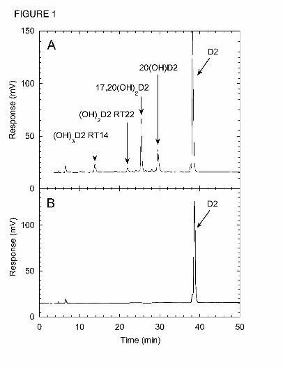
Pathways for the metabolism of vitamin D2 by P450scc. Incubation of cytochrome P450scc
with D2 in 0.45% cyclodextrin and analysis of metabolites by reverse-phase HPLC gave 4
products in sufficient quantities for characterization, which were not present in the zero-time
control incubation (Fig. 1). The two major metabolites were 20(OH)D2 and 17,20(OH)2D2,
which we have previously isolated by TLC and identified by NMR (Slominski et al., 2006). The
additional two products detected by HPLC had retention times of 14 min and 22 min and had UV
spectra similar to D2. The electrospray mass spectrum of the product with RT = 22 min in Fig. 1
showed the most abundant ion at m/z = 451.1 (428.1 + Na+), and ions with m/z = 467.1 ( 428.1 +
K+) and m/z = 879.3 (2M + Na+) from which the sample was identified as dihydroxyvitamin D2.
The electrospray mass spectrum of the RT = 14 min product showed the most abundant ion at
m/z = 467.1 (444.1 + Na+) and ions with m/z = 483.1 (444.1 = K+) and 911.1 (2M + Na+),
corresponding to trihydroxyvitamin D2. This product was subsequently identified as
17,20,24(OH)3D2 by NMR (see below). This name describing its full identification will be used
throughout to avoid confusion.
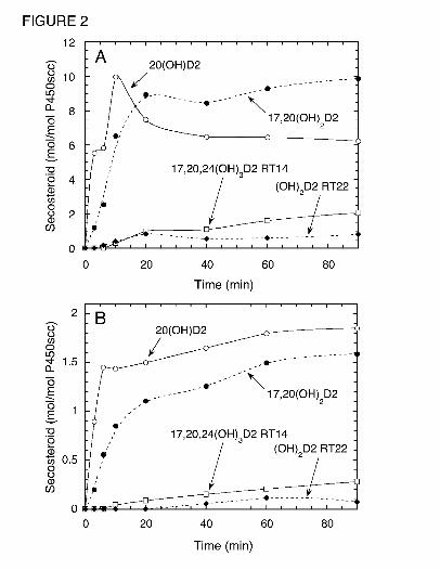
A time course for the metabolism of D2 in 0.45% cyclodextrin is shown in Fig. 2A.
20(OH)D2 was the major product for the first 10 min of incubation but 17,20(OH)2D2 became
the major product after this time. A lag in the time course for the production of 17,20(OH)2D2
was apparent, indicating that some accumulation of the immediate substrate, 20(OH)D2, was
necessary for its synthesis. Similarly, lags were seen in the time courses for production of the
dihydroxyvitamin D2 metabolite with RT = 22 min and 17,20,24(OH)3D2 (RT = 14 min).
This article has not been copyedited and formatted. The final version may differ from this version.DMD Fast Forward. Published on December 30, 2008 as DOI: 10.1124/dmd.108.025619
at ASPE
T Journals on February 26, 2022
dmd.aspetjournals.org
Dow
nloaded from
DMD #25619 10
Cyclodextrin provides a convenient but artificial means of holding D2 in solution for
access by P450scc. Studies on D3 metabolism by P450scc in cyclodextrin have revealed that the
cyclodextrin concentration used has a dramatic effect on both the Km and kcat for D3
consumption (Tuckey et al., 2008b). We therefore examined the metabolism of D2 in
phospholipid vesicles made from phosphatidylcholine and cardiolipin, closely resembling the
normal environment of the cytochrome in the inner mitochondrial membrane (Headlam et al.,
2003; Tuckey, 2005; Tuckey and Stevenson, 1985b). Both D2 and D3 have been shown to
partition quantitatively into the bilayer of phospholipid membranes (Kazanci et al., 2001; Merz
and Sternberg, 1994; Tuckey et al., 2008a). The products of P450scc action on D2 observed in
cyclodextrin, 20(OH)D2, 17,20(OH)2D2, 17,20,24(OH)3D2 and dihydroxyvitamin D2 (RT = 22
min), were also seen in vesicles (Figure 2B). In contrast to cyclodextrin, 20(OH)D2, remained
the major product throughout the 90 min incubation in vesicles.
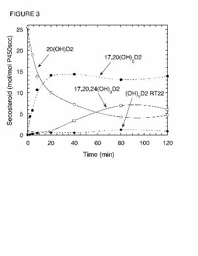
To elucidate the pathways leading to production of trihydroxyvitamin D2 by P450scc, we
examined the products resulting from incubation of P450scc with purified 20(OH)D2,
17,20(OH)2D2 or dihydroxyvitamin D2 (RT = 22 min). Fig. 3 shows the time course for
metabolism of 20(OH)D2 by P450scc. Results show that the 20(OH)D2 serves as a precursor for
production of 17,20(OH)2D2, dihydroxyvitamin D2 (RT = 22 min) and 17,20,24(OH)3D2. A lag
was observed for the production of 17,20,24(OH)3D2 suggesting that accumulation of at least
one of the dihydroxy metabolites was necessary to serve as its immediate substrate. Incubation
of dihydroxyvitamin D2 (RT = 22 min) with P450scc in 0.45% cyclodextrin revealed that it is
not further metabolized by P450scc (not shown), indicating that it is a terminal product of the
This article has not been copyedited and formatted. The final version may differ from this version.DMD Fast Forward. Published on December 30, 2008 as DOI: 10.1124/dmd.108.025619
at ASPE
T Journals on February 26, 2022
dmd.aspetjournals.org
Dow
nloaded from
DMD #25619 11
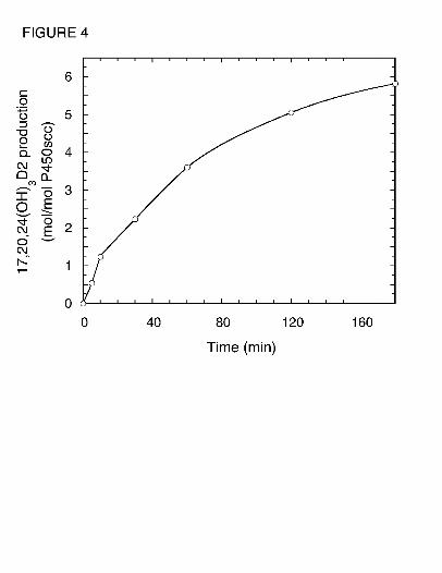
pathway and does not serve as a substrate for 17,20,24(OH)3D2. In contrast, 17,20(OH)2D2 was
converted to 17,20,24(OH)3D2 (Fig. 4). This product was identical to the trihydroxyvitamin D2
product (RT = 14 min) made directly from D2 (Fig. 1) based on their mass spectra, HPLC
retention times and Rf values obtained by TLC. The position of the new hydroxyl groups in this
metabolite, compared to its immediate substrate, was determined by NMR (see below). No other
products were observed in Fig. 4 indicating that 17,20,24(OH)3D2 is not further metabolized.
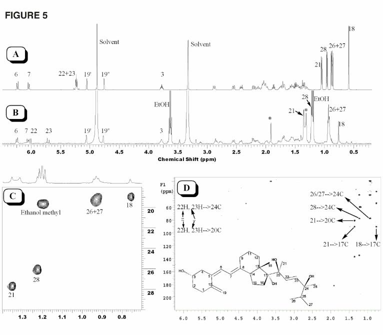
NMR of Trihydroxyvitamin D2. To identify the sites of hydroxylation of trihydroxyvitamin
D2 by P450scc, 50 µg of this product was prepared from 17,20(OH)2D2 and analyzed by NMR
(Fig. 5). Compared with the 1D proton NMR spectrum of vitamin D2 (Fig. 5A), the proton
NMR spectrum of trihydroxyvitamin D2 showed downfield shifts for several protons (Fig. 5B)
due to the hydroxyl groups. While protons in the methyl groups at positions 26 and 27 have a
slight downfield shift of 0.05 ppm, protons in the methyl groups at positions 18, 28 and 21 have
downfield shifts of 0.17 ppm, 0.26 ppm, and 0.29 ppm, respectively. More importantly, the
methyl groups in both positions 28 and 21 became singlet due to the absence of vicinal coupling
in this metabolite (Fig. 5B and 5C). The downfield shift of the methyl group at C18 is due to
hydroxylation at C17 while the change of peak pattern and downfield shift for the methyl at C21
is due to hydroxylation at C20, confirming the hydroxylation pattern of the starting material.
The peak pattern change and downfield shift for the methyl at C28 strongly indicates
hydroxylation at C24. This is consistent with the proton NMR spectrum in which both 22H and
23H show a substantial downfield shift (Fig. 5A). Further analysis of the proton-carbon HMBC
spectrum confirmed hydroxylation at C24 (Fig. 5D). The three quaternary carbons bearing
hydroxyl groups (carbon chemical shifts at 77.5 ppm, 80.6 ppm and 90.4 ppm) clearly display all
This article has not been copyedited and formatted. The final version may differ from this version.DMD Fast Forward. Published on December 30, 2008 as DOI: 10.1124/dmd.108.025619
at ASPE
T Journals on February 26, 2022
dmd.aspetjournals.org
Dow
nloaded from
DMD #25619 12
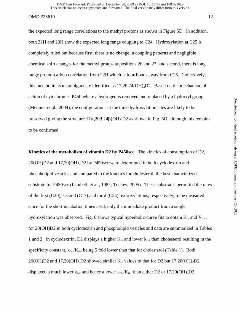
the expected long range correlations to the methyl protons as shown in Figure 5D. In addition,
both 22H and 23H show the expected long range coupling to C24. Hydroxylation at C25 is
completely ruled out because first, there is no change in coupling patterns and negligible
chemical shift changes for the methyl groups at positions 26 and 27, and second, there is long
range proton-carbon correlation from 22H which is four-bonds away from C25. Collectively,
this metabolite is unambiguously identified as 17,20,24(OH)3D2. Based on the mechanism of
action of cytochromes P450 where a hydrogen is removed and replaced by a hydroxyl group
(Meunier et al., 2004), the configurations at the three hydroxylation sites are likely to be
preserved giving the structure 17α,20β,24β(OH)3D2 as shown in Fig. 5D, although this remains
to be confirmed.
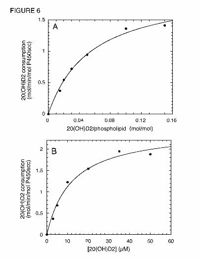
Kinetics of the metabolism of vitamin D2 by P450scc. The kinetics of consumption of D2,
20(OH)D2 and 17,20(OH)2D2 by P450scc were determined in both cyclodextrin and
phospholipid vesicles and compared to the kinetics for cholesterol, the best characterized
substrate for P450scc (Lambeth et al., 1982; Tuckey, 2005). These substrates permitted the rates
of the first (C20), second (C17) and third (C24) hydroxylations, respectively, to be measured
since for the short incubation times used, only the immediate product from a single
hydroxylation was observed. Fig. 6 shows typical hyperbolic-curve fits to obtain Km and Vmax
for 20(OH)D2 in both cyclodextrin and phospholipid vesicles and data are summarized in Tables
1 and 2. In cyclodextrin, D2 displays a higher Km and lower kcat than cholesterol resulting in the
specificity constant, kcat/Km, being 5 fold lower than that for cholesterol (Table 1). Both
20(OH)D2 and 17,20(OH)2D2 showed similar Km values to that for D2 but 17,20(OH)2D2
displayed a much lower kcat and hence a lower kcat/Km, than either D2 or 17,20(OH)2D2.
This article has not been copyedited and formatted. The final version may differ from this version.DMD Fast Forward. Published on December 30, 2008 as DOI: 10.1124/dmd.108.025619
at ASPE
T Journals on February 26, 2022
dmd.aspetjournals.org
Dow
nloaded from
DMD #25619 13
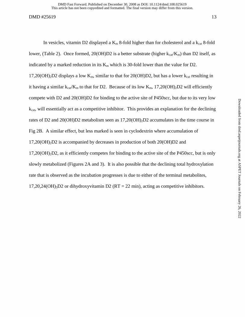
In vesicles, vitamin D2 displayed a Km 8-fold higher than for cholesterol and a kcat 8-fold
lower, (Table 2). Once formed, 20(OH)D2 is a better substrate (higher kcat/Km) than D2 itself, as
indicated by a marked reduction in its Km which is 30-fold lower than the value for D2.
17,20(OH)2D2 displays a low Km, similar to that for 20(OH)D2, but has a lower kcat resulting in
it having a similar kcat/Km to that for D2. Because of its low Km, 17,20(OH)2D2 will efficiently
compete with D2 and 20(OH)D2 for binding to the active site of P450scc, but due to its very low
kcat, will essentially act as a competitive inhibitor. This provides an explanation for the declining
rates of D2 and 20(OH)D2 metabolism seen as 17,20(OH)2D2 accumulates in the time course in
Fig 2B. A similar effect, but less marked is seen in cyclodextrin where accumulation of
17,20(OH)2D2 is accompanied by decreases in production of both 20(OH)D2 and
17,20(OH)2D2, as it efficiently competes for binding to the active site of the P450scc, but is only
slowly metabolized (Figures 2A and 3). It is also possible that the declining total hydroxylation
rate that is observed as the incubation progresses is due to either of the terminal metabolites,
17,20,24(OH)3D2 or dihydroxyvitamin D2 (RT = 22 min), acting as competitive inhibitors.
This article has not been copyedited and formatted. The final version may differ from this version.DMD Fast Forward. Published on December 30, 2008 as DOI: 10.1124/dmd.108.025619
at ASPE
T Journals on February 26, 2022
dmd.aspetjournals.org
Dow
nloaded from
DMD #25619 14
Discussion
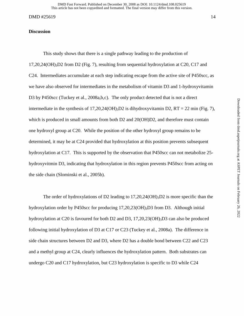
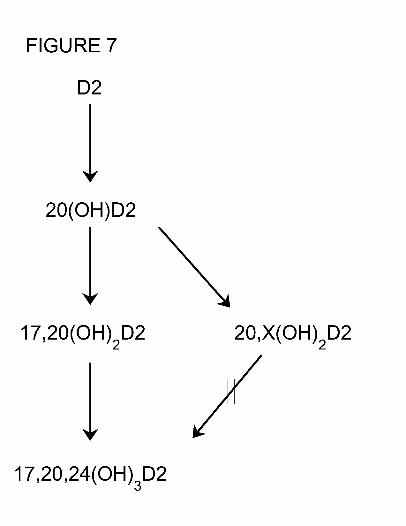
This study shows that there is a single pathway leading to the production of
17,20,24(OH)3D2 from D2 (Fig. 7), resulting from sequential hydroxylation at C20, C17 and
C24. Intermediates accumulate at each step indicating escape from the active site of P450scc, as
we have also observed for intermediates in the metabolism of vitamin D3 and 1-hydroxyvitamin
D3 by P450scc (Tuckey et al., 2008a,b,c). The only product detected that is not a direct
intermediate in the synthesis of 17,20,24(OH)3D2 is dihydroxyvitamin D2, RT = 22 min (Fig. 7),
which is produced in small amounts from both D2 and 20(OH)D2, and therefore must contain
one hydroxyl group at C20. While the position of the other hydroxyl group remains to be
determined, it may be at C24 provided that hydroxylation at this position prevents subsequent
hydroxylation at C17. This is supported by the observation that P450scc can not metabolize 25-
hydroxyvitmin D3, indicating that hydroxylation in this region prevents P450scc from acting on
the side chain (Slominski et al., 2005b).
The order of hydroxylations of D2 leading to 17,20,24(OH)3D2 is more specific than the
hydroxylation order by P450scc for producing 17,20,23(OH)3D3 from D3. Although initial
hydroxylation at C20 is favoured for both D2 and D3, 17,20,23(OH)3D3 can also be produced
following initial hydroxylation of D3 at C17 or C23 (Tuckey et al., 2008a). The difference in
side chain structures between D2 and D3, where D2 has a double bond between C22 and C23
and a methyl group at C24, clearly influences the hydroxylation pattern. Both substrates can
undergo C20 and C17 hydroxylation, but C23 hydroxylation is specific to D3 while C24
This article has not been copyedited and formatted. The final version may differ from this version.DMD Fast Forward. Published on December 30, 2008 as DOI: 10.1124/dmd.108.025619
at ASPE
T Journals on February 26, 2022
dmd.aspetjournals.org
Dow
nloaded from
DMD #25619 15
hydroxylation is specific to D2. Ergosterol, the vitamin D2 precursor is also hydroxylated at
C24 by P450scc, as well as at C17 (Slominski et al., 2005a).
P450scc displays differences in the efficiency of metabolism of D2 and D3. Initial
hydroxylation of D3 at C20 in 0.45% cyclodextrin occurs with a kcat/Km of 666 mM-1min-1
(Tuckey et al., 2008b), which is 5-fold higher than that reported here for D2 measured under
comparable conditions. In vesicles, initial hydroxylation of D3 at C20 occurs with a kcat/Km
approximately 2-fold higher than for C20 hydroxylation of D2 (Tuckey et al., 2008b). Both D2
and D3 display high Km values in vesicles, relative to that for cholesterol, making it difficult to
achieve substrate saturation.
Since our initial studies show that the major products of D2 metabolism by P450scc are
biologically active in inhibiting keratinocyte proliferation and promoting differentiation
(Slominski et al, 2006), we were interested in optimizing their production to facilitate further
biological testing. The relevance of this is highlighted by our detailed testing of some of the
vitamin D3 metabolites produced by P450scc, which display potency on skin cells at least as
good as the hormonally active form of D3, 1,25-dihydroxyvitamin D3, and may be of use
pharmacologically (Janjetovic et al., 2008; Zbytek et al., 2008). Our kinetic study shows that the
cyclodextrin system is superior to vesicles for large-scale production of D2 metabolites. While
the kcat for initial hydroxylation at C20 in 0.45% cyclodextrin is only 40% of that in vesicles,
this is more than compensated for by the ability to solubilize the D2 substrate to a concentration
equivalent to at least three times Km, which cannot be done in vesicles due to the high Km
This article has not been copyedited and formatted. The final version may differ from this version.DMD Fast Forward. Published on December 30, 2008 as DOI: 10.1124/dmd.108.025619
at ASPE
T Journals on February 26, 2022
dmd.aspetjournals.org
Dow
nloaded from
DMD #25619 16
relative to the amount of vitamin D that can be incorporated into the phospholipid bilayer
(Tuckey et al., 2008b).
It remains to be established whether dietary vitamin D2, or D2 administered to vitamin D
deficient patients, can be metabolized by P450scc in vivo. Little is known about the ability of
tissues expressing cytochrome P450scc, such as the adrenal cortex, gonads, placenta, skin, brain
and others, to take up vitamin D from the plasma (Slominski et al, 2005.). We have shown that
vitamin D3 exchanges between membranes more rapidly than cholesterol, plus the StAR protein,
which delivers cholesterol to the inner mitochondrial membrane for steroid synthesis, can also
transport D3 (Tuckey et al., 2008b) and D2 (unpublished). Thus if D2 reaches steroidogenic
tissues, transport into the mitochondria is likely. Effective competition of D2 with cholesterol
would be required for D2 metabolism by P450scc, but given that P450scc is never saturated with
cholesterol in the gonads or adrenals, even following tropic hormone stimulation, competition
would be minimal (Jefcoate et al., 1973; Tuckey et al., 1985).
In conclusion, this study clearly defines the pathway of D2 metabolism by P450scc
through to 17,20,24(OH)3D2, with characterization of the major kinetic constants for the three
hydroxylations involved. The structure of the 17,20,24(OH)3D2 has been solved by NMR
providing an additional product, besides the 20(OH)D2 and 17,20(OH)2D2, for future testing of
biological activity.
This article has not been copyedited and formatted. The final version may differ from this version.DMD Fast Forward. Published on December 30, 2008 as DOI: 10.1124/dmd.108.025619
at ASPE
T Journals on February 26, 2022
dmd.aspetjournals.org
Dow
nloaded from
DMD #25619 17
Acknowledgement
We thank Dr. Tony Reeder for recording mass spectra.
This article has not been copyedited and formatted. The final version may differ from this version.DMD Fast Forward. Published on December 30, 2008 as DOI: 10.1124/dmd.108.025619
at ASPE
T Journals on February 26, 2022
dmd.aspetjournals.org
Dow
nloaded from
DMD #25619 18
References
Armas LAG, Hollis BW, and Heaney RP (2004) Vitamin D2 is much less effective than
vitamin D3 in humans. J Clin Endocrinol Metab 89: 5387-5391.
Bikle DD, Oda Y, and Xie Z (2004) Calcium and 1,25(OH)2D: interacting drivers of epidermal
differentiation. J Steroid Biochem Mol Biol 89-90: 355-360.
Brown AJ, Ritter CS, Holliday LS, Knutson JC, and Strugnell SA (2004) Tissue distribution
and activity studies of 1,24-dihydroxyvitamin D2, a metabolite of vitamin D2 with low
calcemic activity in vivo. Biochem Pharmacol 68: 1289-1296.
Headlam MJ, Wilce MCJ, and Tuckey RC (2003) The F-G loop region of cytochrome P450scc
(CYP11A1) interacts with the phospholipid membrane. BBA Biomembranes 1617: 96-108.
Hiwatashi A, Nishii Y, and Ichikawa Y (1982) Purification of cytochrome P-450D1a (25-
hydroxyvitamin D3-1α-hydroxylase) of bovine kidney mitochondria. Biochem Biophys Res
Commun 105: 320-327.
Holick MF (2003) Vitamin D: A millenium perspective. J Cell Biochem 88: 296-307.
Holick MF (2004) Vitamin D: importance in the prevention of cancers, type 1 diabetes, heart
disease, and osteoporosis. Am J Clin Nutr 79: 362-371.
Holick MF, Biancuzzo RM, Chen TC, Klein EK, Young A, Bibuld D, Reitz R, Salameh W,
Ameri A, and Tannenbaum AD (2008) Vitamin D2 is effective as vitamin D3 in maintaining
circulating concentrations of 25-hydroxyvitamin D. J Clin Endocrinol Metab 93: 677-681.
Hume R, Kelly RW, Taylor PL, and Boyd GS (1984) The catalytic cycle of cytochrome P-
450scc and intermediates in the conversion of cholesterol to pregnenolone. Eur J Biochem
140: 583-591.
This article has not been copyedited and formatted. The final version may differ from this version.DMD Fast Forward. Published on December 30, 2008 as DOI: 10.1124/dmd.108.025619
at ASPE
T Journals on February 26, 2022
dmd.aspetjournals.org
Dow
nloaded from
DMD #25619 19
Janjetovic Z, Zmijewski M, Tuckey RC, Nguyen MN, Slominski A (2008) 20-
Hydroxycholecalciferol and 20, 23-dihydroxycholecalciferol, products of vitamin D3
hydroxylation by P450-scc, decrease NFκB activity by increasing IκBα levels in normal and
HaCaT-keratinocytes. J Invest Dermatol 128: S20, 116.
Jefcoate CR, Simpson ER, Boyd GS, Brownie AC, and Orme-Johnson WH (1973) The
detection of different states of the P-450 cytochromes in adrenal mitochondria; changes
induced by ACTH. Ann NY Acad Sci 212: 243-261.
Kazanci N, Toyran N, Haris PI, and Severcan F (2001) Vitamin D2 at high and low
concentrations exert opposing effects on molecular order and dynamics of dipalmitoyl
phosphatidylcholine membranes. Spectroscopy 15: 47-55.
Lambeth JD, Kitchen SE, Farooqui AA, Tuckey R, and Kamin H (1982) Cytochrome P-450scc-
substrate interactions. Studies of binding and catalytic activity using hydroxycholesterols. J
Biol Chem 257: 1876-1884.
Merz K, and Sternberg B (1994) Incorporation of vitamin D3-derivatives into liposomes of
different lipid types. J Drug Targeting 2: 411-417.
Meunier B, de Visser SP, and Shaik S (2004) Mechanism of oxidation reactions catalyzed by
cytochrome P450 enzymes. Chem Rev 104: 3947-3980.
Mitani H, Naru E, Yamashita M, Arakane K, Suzuki T, and Imanari T (2004) Ergocalciferol
promotes in vivo differentiation of keratinocytes and reduces photodamage caused by
ultraviolet irradiation in hairless mice. Photodermatol Photoimmunol Photomed 20: 215-
223.
Omura T, and Sato R (1964) The carbon monoxide binding pigment of liver microsomes. I.
Evidence for its hemoprotein nature. J Biol Chem 239: 2370-2378.
This article has not been copyedited and formatted. The final version may differ from this version.DMD Fast Forward. Published on December 30, 2008 as DOI: 10.1124/dmd.108.025619
at ASPE
T Journals on February 26, 2022
dmd.aspetjournals.org
Dow
nloaded from
DMD #25619 20
Prosser DE, and Jones G (2004) Enzymes involved in the activation and inactivation of vitamin
D. Trends Biochem Sci 29: 664-673.
Slominski A, Semak I, Wortsman J, Zjawiony J, Li W, Zbytek B, and Tuckey RC (2006) An
alternative pathway of vitamin D2 metabolism. Cytochrome P450scc (CYP11A1)-mediated
conversion to 20-hydroxyvitamin D2 and 17,20-dihydroxyvitamin D2. FEBS J 273: 2891-
2901.
Slominski A, Semak I, Zjawiony J, Wortsman J, Gandy MN, Li J, Zbytek B, Li W, and Tuckey
RC (2005a) Enzymatic metabolism of ergosterol by cytochrome P450scc to biologically
active 17a,24-dihydroxyergosterol. Chem Biol 12: 931-939.
Slominski A, Semak I, Zjawiony J, Wortsman J, Li W, Szczesniewski A, and Tuckey RC
(2005b) The cytochrome P450scc system opens an alternate pathway of vitamin D3
metabolism. FEBS J 272: 4080-4090.
Slominski A, Zjawiony J, Wortsman J, Semak I, Stewart J, Pisarchik A, Sweatman T, Marcos J,
Dunbar C, and Tuckey RC (2004) A novel pathway for sequential transformation of 7-
dehydrocholesterol and expression of the P450scc system in mammalian skin. Eur J Biochem
271: 4178-4188.
Tuckey RC (2005) Progesterone synthesis by the human placenta. Placenta 26: 273-281.
Tuckey RC, and Cameron KJ (1993) Human placental cholesterol side-chain cleavage:
enzymatic synthesis of (22R)-20α,22-dihydroxycholesterol. Steroids 58: 230-233.
Tuckey RC, Janjetovic Z, Li W, Nguyen MN, Zmijewski MA, Zjawiony JK, and Slominski A
(2008c) Metabolism of 1α-hydroxyvitamin D3 by cytochrome P450scc to biologically active
1α,20-dihydroxyvitamin D3. J Steroid Biochem Mol Biol doi:10.1016/j.jsbmb.2008.10.005
This article has not been copyedited and formatted. The final version may differ from this version.DMD Fast Forward. Published on December 30, 2008 as DOI: 10.1124/dmd.108.025619
at ASPE
T Journals on February 26, 2022
dmd.aspetjournals.org
Dow
nloaded from
DMD #25619 21
Tuckey RC, Li W, Zjawiony JK, Zmijewski MA, Nguyen MN, Sweatman T, Miller D, and
Slominski A (2008a) Pathways and products for the metabolism of vitamin D3 by
cytochrome P450scc. FEBS J 275: 2585-2596.
Tuckey RC, Nguyen MN, and Slominski A (2008b) Kinetics of vitamin D3 metabolism by
cytochrome P450scc (CYP11A1) in phospholipid vesicles and cyclodextrin. Int J Biochem
Cell Biol 40: 2619-2626.
Tuckey RC, and Stevenson PM (1984a) Properties of ferredoxin reductase and ferredoxin from
the bovine corpus luteum. Int J Biochem 16: 489-495.
Tuckey RC, and Stevenson PM (1984b) Properties of bovine luteal cytochrome P-450scc
incorporated into artificial phospholipid vesicles. Int J Biochem 16: 497-503.
Tuckey RC, and Stevenson PM (1985) Purification and analysis of phospholipids in the inner
mitochondrial membrane fraction of the bovine corpus luteum, and properties of cytochrome
P-450scc incorporated into vesicles prepared from these phospholipids. Eur J Biochem 148:
379-384.
Zbytek B, Janjetovic Z, Tuckey RC, Zmijewski MA, Sweatman T, Jones E, Nguyen MN, and
Slominski A (2008) 20-Hydroxyvitamin D3, a product of vitamin D3 hydroxylation by
cytochrome P450sc, stimulates keratinocyte differentiation. J Invest Dermatol 128: 2271-
2280.
This article has not been copyedited and formatted. The final version may differ from this version.DMD Fast Forward. Published on December 30, 2008 as DOI: 10.1124/dmd.108.025619
at ASPE
T Journals on February 26, 2022
dmd.aspetjournals.org
Dow
nloaded from
DMD #25619 22
Footnotes
This work was supported by the National Institutes of Health [Grant R01AR052190] to AS and
RCT and by the University of Western Australia.
This article has not been copyedited and formatted. The final version may differ from this version.DMD Fast Forward. Published on December 30, 2008 as DOI: 10.1124/dmd.108.025619
at ASPE
T Journals on February 26, 2022
dmd.aspetjournals.org
Dow
nloaded from
DMD #25619 23
Figure Legends
Fig. 1. Vitamin D2 Metabolites Produced by P450scc. (A) HPLC chromatogram showing
products from the metabolism of vitamin D2 (50 µM) in 0.45% cyclodextrin by P450scc (1.0
µM) from a 1 h incubation in a reconstituted system containing adrenodoxin (15 µM) and
adrenodoxin reductase (0.4 µM). (B) Chromatogram for a control incubation (zero time)
showing the vitamin D2 substrate. RT, retention time in minutes. Abbreviations for products
are: (OH)D2, monohydroxyvitamin D2; (OH)2D2, dihydroxyvitamin D2; (OH)3D2,
trihydroxyvitamin D2.
Fig. 2. Hydroxylation of Vitamin D2 by P450scc in Cyclodextrin and Vesicles. (A) Time-
course for metabolism of vitamin D2 (50 µM) dissolved in 0.45% cyclodextrin and incubated
with 1.0 µM P450scc. (B) Time-course for metabolism of vitamin D2 in phospholipid vesicles
containing 0.2 mol D2/mol phospholipid were incubated with 2.0 µM P450scc. Samples were
taken at the times indicated and products measured by HPLC. Abbreviations are as for Fig. 1.
Fig. 3. Time-course for metabolism of 20(OH)D2 in phospholipid vesicles. Vesicles containing
0.1 mol 20(OH)D2/mol phospholipid were incubated with 2.0 µM P450scc. Samples were taken
at the times indicated and products measured by HPLC. Abbreviations are as for Fig. 1.
Fig. 4. Time-course for conversion of 17,20(OH)2D2 to 17,20,24(OH)3D2 in cyclodextrin.
17,20(OH)2D2 (50 µM) dissolved in 0.45% cyclodextrin was incubated with 1.0 µM P450scc.
Samples were taken at the times indicated and product measured by HPLC.
This article has not been copyedited and formatted. The final version may differ from this version.DMD Fast Forward. Published on December 30, 2008 as DOI: 10.1124/dmd.108.025619
at ASPE
T Journals on February 26, 2022
dmd.aspetjournals.org
Dow
nloaded from
DMD #25619 24
Fig. 5. NMR structure elucidation of trihydroxyvitamin D2. (A) 1D proton NMR of parent D2,
(B) 1D proton NMR of trihydroxyvitamin D2, (C) methyl region of proton-carbon HSQC
spectrum of trihydroxyvitamin D2, and (D) proton-carbon HMBC of trihydroxyvitamin D2. The
residue ethanol is carry over from a previous UV quantification measurement. An unknown
impurity is indicated by the star in the 1D NMR spectrum. The identified structure is shown in
panel D with the likely configurations for each hydroxy group, 17α,20β,24β(OH)3D2.
Fig. 6. Michaelis-Menten plots for metabolism of 20(OH)D2 by P450scc in vesicles and
cyclodextrin. Cytochrome P450scc was incorporated into (A) phospholipid vesicles containing
20(OH)D2, or (B) 0.45% cyclodextrin containing 20(OH)D2, and incubated at 37˚C for 3 min or
2 min, respectively. Products were extracted and analyzed by reverse-phase HPLC. Hyperbolic
curves were fitted by non-linear least squares analysis using Kaleidagraph 3.6. The correlation
coefficients for the curve fits were 0.9965 and 0.9924 for 20(OH)D2 in vesicles and
cyclodextrin, respectively.
Fig. 7. Pathway for the metabolism of vitamin D2 by cytochrome P450scc. X represents a
hydroxyl group in an unknown position. ( ), indicates reaction does not occur.
This article has not been copyedited and formatted. The final version may differ from this version.DMD Fast Forward. Published on December 30, 2008 as DOI: 10.1124/dmd.108.025619
at ASPE
T Journals on February 26, 2022
dmd.aspetjournals.org
Dow
nloaded from
DMD #25619 25
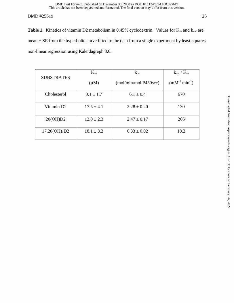
Table 1. Kinetics of vitamin D2 metabolism in 0.45% cyclodextrin. Values for Km and kcat are
mean ± SE from the hyperbolic curve fitted to the data from a single experiment by least-squares
non-linear regression using Kaleidagraph 3.6.
SUBSTRATES Km
(µM)
kcat
(mol/min/mol P450scc)
kcat / Km
(mM-1 min-1)
Cholesterol 9.1 ± 1.7 6.1 ± 0.4 670
Vitamin D2 17.5 ± 4.1 2.28 ± 0.20 130
20(OH)D2 12.0 ± 2.3 2.47 ± 0.17 206
17,20(OH)2D2 18.1 ± 3.2 0.33 ± 0.02 18.2
This article has not been copyedited and formatted. The final version may differ from this version.DMD Fast Forward. Published on December 30, 2008 as DOI: 10.1124/dmd.108.025619
at ASPE
T Journals on February 26, 2022
dmd.aspetjournals.org
Dow
nloaded from
DMD #25619 26
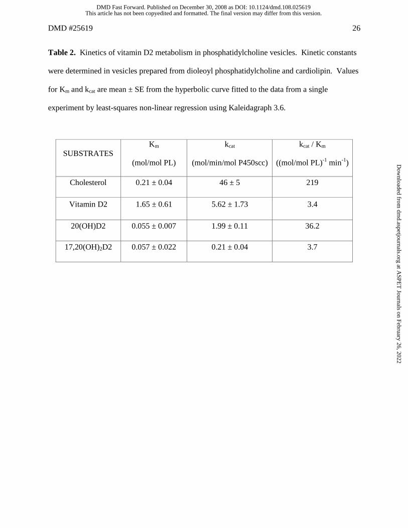
Table 2. Kinetics of vitamin D2 metabolism in phosphatidylcholine vesicles. Kinetic constants
were determined in vesicles prepared from dioleoyl phosphatidylcholine and cardiolipin. Values
for Km and kcat are mean ± SE from the hyperbolic curve fitted to the data from a single
experiment by least-squares non-linear regression using Kaleidagraph 3.6.
SUBSTRATES Km
(mol/mol PL)
kcat
(mol/min/mol P450scc)
kcat / Km
((mol/mol PL)-1 min-1)
Cholesterol 0.21 ± 0.04 46 ± 5 219
Vitamin D2 1.65 ± 0.61 5.62 ± 1.73 3.4
20(OH)D2 0.055 ± 0.007 1.99 ± 0.11 36.2
17,20(OH)2D2 0.057 ± 0.022 0.21 ± 0.04 3.7
This article has not been copyedited and formatted. The final version may differ from this version.DMD Fast Forward. Published on December 30, 2008 as DOI: 10.1124/dmd.108.025619
at ASPE
T Journals on February 26, 2022
dmd.aspetjournals.org
Dow
nloaded from
This article has not been copyedited and formatted. The final version may differ from this version.DMD Fast Forward. Published on December 30, 2008 as DOI: 10.1124/dmd.108.025619
at ASPE
T Journals on February 26, 2022
dmd.aspetjournals.org
Dow
nloaded from
This article has not been copyedited and formatted. The final version may differ from this version.DMD Fast Forward. Published on December 30, 2008 as DOI: 10.1124/dmd.108.025619
at ASPE
T Journals on February 26, 2022
dmd.aspetjournals.org
Dow
nloaded from
This article has not been copyedited and formatted. The final version may differ from this version.DMD Fast Forward. Published on December 30, 2008 as DOI: 10.1124/dmd.108.025619
at ASPE
T Journals on February 26, 2022
dmd.aspetjournals.org
Dow
nloaded from
This article has not been copyedited and formatted. The final version may differ from this version.DMD Fast Forward. Published on December 30, 2008 as DOI: 10.1124/dmd.108.025619
at ASPE
T Journals on February 26, 2022
dmd.aspetjournals.org
Dow
nloaded from
This article has not been copyedited and formatted. The final version may differ from this version.DMD Fast Forward. Published on December 30, 2008 as DOI: 10.1124/dmd.108.025619
at ASPE
T Journals on February 26, 2022
dmd.aspetjournals.org
Dow
nloaded from
This article has not been copyedited and formatted. The final version may differ from this version.DMD Fast Forward. Published on December 30, 2008 as DOI: 10.1124/dmd.108.025619
at ASPE
T Journals on February 26, 2022
dmd.aspetjournals.org
Dow
nloaded from
This article has not been copyedited and formatted. The final version may differ from this version.DMD Fast Forward. Published on December 30, 2008 as DOI: 10.1124/dmd.108.025619
at ASPE
T Journals on February 26, 2022
dmd.aspetjournals.org
Dow
nloaded from