Embed Size (px)
DESCRIPTION
Mimicking biological
Citation preview
Subscriber access provided by HELSINKI UNIV OF TECH
Langmuir is published by the American Chemical Society. 1155 Sixteenth Street N.W.,Washington, DC 20036
Letter
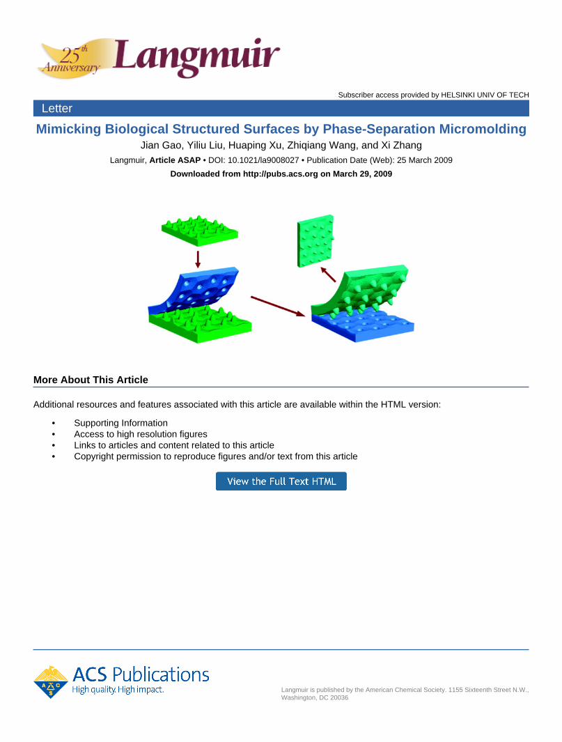
Mimicking Biological Structured Surfaces by Phase-Separation MicromoldingJian Gao, Yiliu Liu, Huaping Xu, Zhiqiang Wang, and Xi Zhang
Langmuir, Article ASAP • DOI: 10.1021/la9008027 • Publication Date (Web): 25 March 2009
Downloaded from http://pubs.acs.org on March 29, 2009
More About This Article
Additional resources and features associated with this article are available within the HTML version:
• Supporting Information• Access to high resolution figures• Links to articles and content related to this article• Copyright permission to reproduce figures and/or text from this article
pubs.acs.org/Langmuir
Mimicking Biological Structured Surfaces by Phase-Separation
Micromolding
Jian Gao, Yiliu Liu, Huaping Xu,* Zhiqiang Wang, and Xi Zhang*
Key Laboratory of Organic Optoelectronics & Molecular Engineering, Department of Chemistry,Tsinghua University, Beijing 100084, PR China
Received March 5, 2009. Revised Manuscript Received March 17, 2009
In this letter, we present a very convenient and efficient technique of direct replication of biological structures via atwo-step phase-separation micromolding process (PSμM). Our study has demonstrated that PSμM can be used toreplicate the surface structure of a lotus leaf. On one hand, the micro/nanostructures of the lotus leaf are well replicatedafter a two-step PSμM.On the other hand, the replicated artificial lotus leaf shows good superhydrophobicity, similar tothat of the natural lotus leaf. In addition, we have also applied the same technique to replicate a rice leaf and haveconfirmed that replicated artificial rice leaves can exhibit not only a very similar structure of the natural rice leaf but alsosurface anisotropic wetting. It is greatly anticipated that this PSμMcan be extended to mimic many other biostructures,therefore opening new avenues for surface molecular engineering.
Introduction
Biological materials found in nature exhibit many fascinatingbiofunctionalities owing to their unique morphologies and phy-sicochemical properties. These special biofunctionalities includethe self-cleaning ability of lotus leaves,1 the anisotropic wettingproperties of rice leaves,2 and the antireflective property of cicadawings,3 which are attributed to the arranged micro/nanostruc-tures on their surfaces. Inspired by these biofunctionalities fromthe nature, many approaches have been developed to fabricateartificial biostructures with specific functionality.1-22 Jiang et al.fabricated a series of surfaces based on aligned carbon tubes thatpossessed superhydrophobility similar to that of lotus or riceleaves.2,4 Fuchs and Chi et al. demonstrated the anisotropicwetting properties of surfaces prepared by Langmuir-Blodgett
lithography.5,6 Recently, Dai and Wang et al. reported carbonnanotube arrays that mimic gecko feet with a shear adhesive forceof close to 100N 3 cm
-2, almost 10 timeshigher than that of the footof a gecko.7 Our group has developed amethod for the fabricationof superhydrophobic surfaces on gold threads by combining thelayer-by-layer assembly of polyelectrolytes with the electrodeposi-tion of gold to mimic the legs of water striders.8-10
Most of the above-mentioned approaches focused on usingfabrication techniques and synthetic nanomaterials to mimic thefunctionality of biological materials. However, there are only afew successful examples of the direct replication of biologicalstructures, also known as “bioreplication”. Wang et al. reportedthe controlled replication of butterfly wings to achieve tunablephotonic properties using a low-temperature atomic layer deposi-tion technique.17 Liu et al. presented the fabrication of cicadawinglike structure based onnanoimprint lithography.18However,these reports often rely on expensive equipment to replicate thebiological structures. Moreover, it will be helpful if the methodallows for the regeneration of the biotemplates. Therefore, it is asignificant challenge to develop a simple approach for the directreplication of natural biological structures and the realization oftheir biofunctionality.
Wessling et al. first introduced phase-separationmicromolding(PSμM) as a microfabrication technique to replicate the patternsfrom their templates.23 It relies on the phase separation of apolymer solution when in contact with a structured surface.22-26
Compared with regular replica molding (REM), which normallyuses poly(dimethylsiloxane) (PDMS) as the replication materi-al,19 PSμM is a convenient and versatile technique that can beused to structure a broad range of polymers, including blockcopolymers and biodegradable and conductive polymers, withoutthe need for expensive facilities. Herein we present a convenient
*Corresponding authors. E-mail: [email protected], [email protected].(1) Barthlott, W.; Neinhuis, C. Planta 1997, 202, 1.(2) Feng, L.; Li, S. H.; Li, Y. S.; Li, H. J.; Zhang, L. J.; Zhai, J.; Song, Y. L.; Liu,
B. Q.; Jiang, L.; Zhu, D. B. Adv. Mater. 2002, 14, 1857.(3) Watson, G. S.; Watson, J. A. Appl. Surf. Sci. 2004, 235, 139.(4) Sun, T. L.; Feng, L.; Gao, X. F.; Jiang, L. Acc. Chem. Res. 2005, 38, 644.(5) Gleiche, M.; Chi, L. F.; Fuchs, H. Nature (London) 2000, 403, 173.(6) Gleiche, M.; Chi, L. F.; Gedig, E.; Fuchs, H. ChemPhysChem 2001, 2, 187.(7) Qu, L. T.; Dai, L. M.; Stone, M.; Xia, Z. H.; Wang, Z. L. Science 2008, 322,
238.(8) Shi, F.; Wang, Z. Q.; Zhang, X. Adv. Mater. 2005, 17, 1005.(9) Shi, F.; Niu, J.; Liu, J. L.; Liu, F.; Wang, Z. Q.; Feng, X. Q.; Zhang, X. Adv.
Mater. 2007, 19, 2257.(10) Zhang, X.; Shi, F.; Yu, X.; Liu, H.; Fu, Y.; Wang, Z. Q.; Jiang, L.; Li, X. Y.
J. Am. Chem. Soc. 2004, 126, 3064.(11) Vukusic, P.; Hooper, I. Science 2005, 310, 1151.(12) Ma, Y.; Sun, J. Q. Chem. Mater. 2009, 21, 898.(13) Sanchez, C.; Arribart, H.; Guille, M. M. G. Nat. Mater. 2005, 4, 277.(14) Zhang, X.; Shi, F.; Niu, J.; Jiang, Y. G.;Wang, Z. Q. J.Mater. Chem. 2008,
18, 621.(15) Mitragotri, S.; Lahann, J. Nat. Mater. 2009, 8, 15.(16) Sotiropoulou, S.; Sierra-Sastre, Y.; Mark, S. S.; Batt, C. A. Chem. Mater.
2008, 20, 821.(17) Huang, J. Y.; Wang, X. D.; Wang, Z. L. Nano Lett. 2006, 6, 2325.(18) Zhang, G. M.; Zhang, J.; Xie, G. Y.; Liu, Z. F.; Shao, H. B. Small 2006, 2,
1440.(19) (a) Sun, M. H.; Luo, C. X.; Xu, L. P.; Ji, H.; Qi, O. Y.; Yu, D. P.; Chen, Y.
Langmuir 2005, 21, 8978. (b) Liu, B.; He, Y. N.; Fan, Y.; Wang, X. G.Macromol.Rapid Commun. 2006, 27, 1859.(20) Xi, J. M.; Jiang, L. Ind. Eng. Chem. Res. 2008, 47, 6354.(21) Saison, T.; Peroz, C.; Chauveau, V.; Berthier, S.; Sondergard, E.; Arribart,
H. Bioinsp. Biomim. 2008, 3, 046004.(22) Bhushan, B.; Jung, Y. C.; Koch, K. Langmuir 2009, 25, 3240.
(23) Vogelaar, L.; Barsema, J. N.; van Rijn, C. J. M.; Nijdam, W.; Wessling, M.Adv. Mater. 2003, 15, 1385.
(24) Vogelaar, L.; Lammertink, R. G. H.; Barsema, J. N.; Nijdam, W.; Bolhuis-Versteeg, L. A. M.; van Rijn, C. J. M.; Wessling, M. Small 2005, 1, 645.
(25) Vogelaar, L.; Lammertink, R. G. H.; Wessling, M. Langmuir 2006, 22,3125.
(26) Xu, H. P.; Ling, X. Y.; van Bennekom, J.; Duan, X. X.; Ludden, M. J. W.;Reinhoudt, D. N.;Wessling, M.; Lammertink, R. G. H.; Huskens, J. J. Am. Chem.Soc. 2009, 131, 797.
© XXXX American Chemical Society
DOI: 10.1021/la9008027Langmuir XXXX, XXX(XX), 000–000 A
technique for the direct replication of biostructures via the PSμMprocess. Our technique features a combination of (i) low fabrica-tion costs, with no specific equipments are needed; (ii) thepossibility to recycle the biotemplates for multiple use; and (iii)an ambient processing environment to replicate biological struc-tures directly from natural materials. The morphological replica-tions of lotus and rice leaves are demonstrated by a two-stepPSμM process. In addition, the biofunctionality of such artificiallotus and rice leaves (i.e., their respective superhydrophobicityand anisotropic wetting properties) is examined.
Experimental Section
Materials. Poly(etherimide) (PEI) (Ultem 1000) was a com-mercial product of General Electrics. Hyflon AD 80X (a copo-lymer of 80 mol % 2,2,4-trifluoromethoxy-1,3-dioxole and 20mol% tetrafluorethylene) was obtained fromSolvay Solexis. SV110, a good solvent for Hyflon AD 80X, was also from SolvaySolexis. NMP and DMF were purchased from SinopharmChemical Reagent Co., Ltd. Ethanol was obtained from BeijingChemical Reagents Company, and n-pentane was purchasedfrom Beijing Modern Eastern Fine Chemical Co., Ltd. Lotusleaves were picked from the lotus pond at Tinghua University.Rice leaves were from Yixing, Jiangsu Province, China.
Method. Scheme 1 shows the two-step PSμM process. Tobegin, a natural biomaterial (e.g., a lotus leaf and/or a rice leaf)was used as a biotemplate. A concentrated PEI (20 wt%, unlessotherwise stated) solution in NMP was cast onto a lotus leaf.Subsequently, the PEI film was solidified by immersing it inwater. After a few minutes of immersion, the PEI film withnegative replica of a lotus leaf was spontaneously released fromthe natural biotemplate. After drying, 17.5 wt % Hyflon AD80X in SV 110 was cast onto the negative replicated PEI film.Subsequently, the precipitation-hardening of Hyflon AD 80Xwas achieved by immersion in n-pentane and ethanol. Afterdetaching the Hyflon AD 80X film from the negative template,the resulting film was dried in air for further study. A positivelyreplicated artificial lotus leaf was obtained.
Characterization. The surface structures were imaged with aJEOL JSM-7401F field-emission scanning electron microscope(SEM) at 1.0 kV. Static, advancing, and receding contact angleswere measured at room temperature by an optical contact anglemeasuring device (OCA20,Dataphysics InstrumentsGmbH).Awater droplet of 2 μL was used.
Results and Discussions
Natural lotus leaves are the first subject of our biologicalreplication study owing to their self-cleaning properties, asrevealed by Barthlott and Neinhuis et al.1 Scheme 1 shows theexperimental strategy of using a two-step PSμM process toreplicate the surface structures of a lotus leaf. To begin, aconcentrated poly(etherimide) (PEI) solution in 1-methyl-2-pyr-rolidinone (NMP)was directly cast onto the surface of a lotus leafattached to a glass substrate. Subsequently, the entire substratewas immediately immersed in water, a nonsolvent of PEI. By thesolvent exchange between NMP and water, the structured PEIfilm was easily detached from the lotus leaf, giving rise to a PEIstructure with a negative replica of a lotus leaf. In the second stepof the PSμM process, a secondary polymer solution;HyflonAD80 (a polymer with low surface energy) in SV 110;was castonto the structured PEI film stuck to a glass substrate.By immersing the substrate in n-pentane (a nonsolvent ofHyflon AD80), the Hyflon AD80 film was detached from thePEI film; therefore, a positive replica structure of a lotus leaf wasobtained.
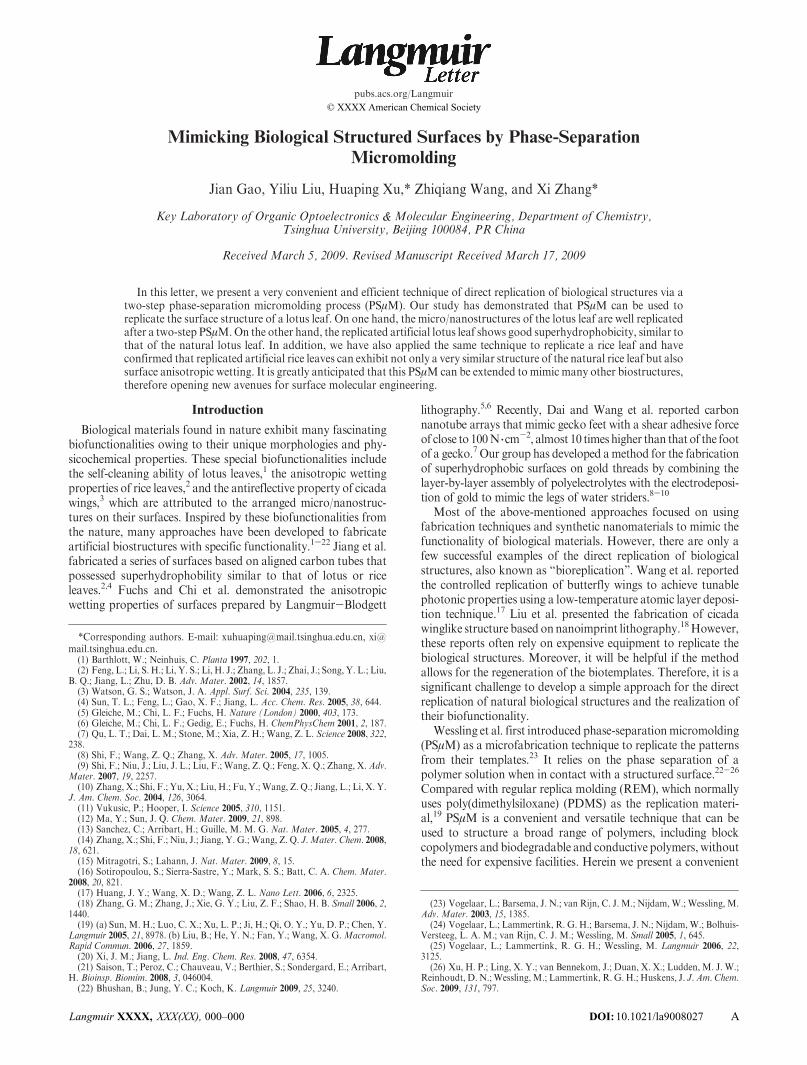
The natural lotus leaves, as shown in Figure 1A,B, consist ofrandomly distributed micropapillae with diameters of several
micrometers. Moreover, there are also some branchlike nanos-tructures on the papillae on the scale of hundreds of nanometers.As shown in Figure 1C,D, a large area of randomly distributedmicropits can be clearly observed on the surface of the replicatedPEI film after the first PSμM step. This showed that during thesolidification process the replicated PEI has maintained thenegative surface structure with respect to that of the lotus leaftemplate. By comparing SEM images of the natural lotus leafand their negative replica by the PSμM process in Figure 1, it isconfirmed that the resulting replicated structures agree well withthe negative replication of the original surface structures of thelotus leaves. Hence, the negative structures of a lotus leaf havebeen successfully obtained by the first step in the PSμM process.It is also noted during the first-step PSμMprocess that the solventexchange between NMP and water was so fast and efficientthat the PEI film can be detached from the original lotus leaf inless than 3 min, which prevented the destruction of the lotus leafby NMP.
For the convenience of fabrication and cost-saving purposes, itis important that the biotemplates for bioreplication can berecycled for multiple replications. Hence, the original lotus leafwas repeatedly used for five replications, and its contact angleafter each replication process was examined. Table 1 shows therespective static, advancing, and receding contact angles (CAs) of
Scheme 1. Schematic Representation of a Two-Step PSμM Processa
a (i) Negative replication of a lotus leaf by PEI, followed by PEI filmdetachment from the lotus leaf inwater. (ii) Positive replication of a lotusleaf by casting a film of Hyflon AD 80X onto the negatively replicatedPEI film as the template and immersion of the entire substrate inn-pentane. (iii) Detachment of the replicated Hyflon AD 80X film fromits template.
Figure 1. SEM images of the original surface structures of anatural lotus leaf (A, B) and a PEI film with a negative replica ofa lotus leaf prepared by a one-step PSμM process (C, D).
DOI: 10.1021/la9008027 Langmuir XXXX, XXX(XX), 000–000
Letter
B
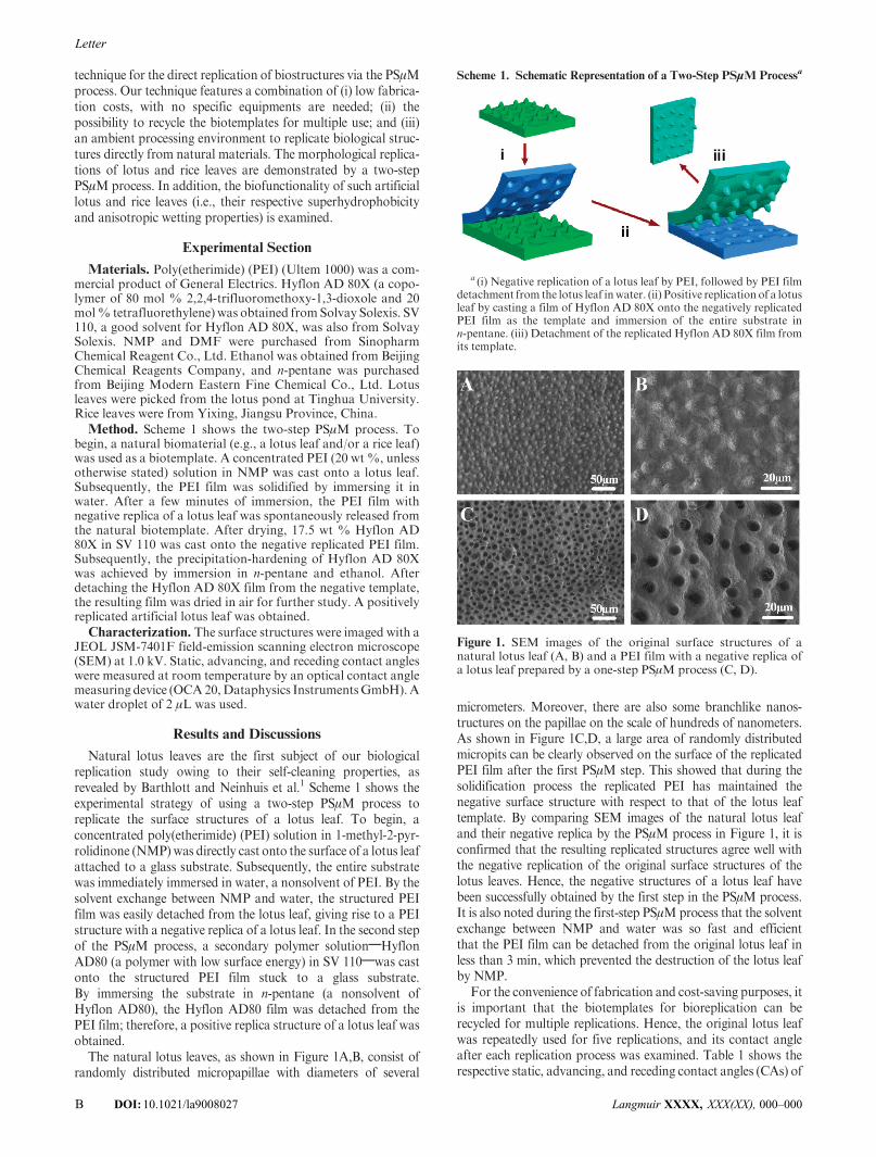
the lotus leaf template after each replication process. The resultsshowed a negligible change in all CAs in all of the replicationprocesses. After the fifth replication by PEI, the original lotus leaftemplate still exhibited good hydrophobicity, a vital featurecontributing to the easy detachment of the PEI film from thelotus leaf during the replication process. In addition, in compar-isons between the replicas made by the same lotus leaf template,there is no obvious change in the morphology, in particular, themicropit structure of the negative replica of the lotus leaf template(Figure 2). It should be noted that all of the SEM images abovewere collected randomly from different areas each time. All of theresults highlight the PSμM technique as a convenient and recycl-able method for replicating the biological structures.
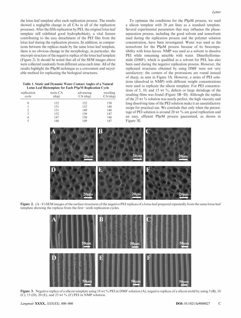
To optimize the conditions for the PSμM process, we useda silicon template with 20 μm lines as a standard template.Several experimental parameters that may influence the phase-separation process, including the good solvent and nonsolventused during the replication process and the polymer solutionconcentration, have been investigated. Water was used as thenonsolvent for the PSμM process because of its biocompa-tibility with lotus leaves. NMP was used as a solvent to dissolvePEI while remaining miscible with water. Dimethylforma-mide (DMF), which is qualified as a solvent for PEI, has alsobeen used during the negative replication process. However, thereplicated structures obtained by using DMF were not verysatisfactory: the corners of the protrusions are round insteadof sharp, as seen in Figure 3A. However, a series of PEI solu-tions (dissolved in NMP) with different weight concentrationswere used to replicate the silicon template. For PEI concentra-tions of 5, 10, and 15 wt %, defects or large shrinkage of theresulting films was found (Figure 3B-D). Although the replicaof the 25 wt % solution was nearly perfect, the high viscosity andlong dissolving time of the PEI solutionmake it an unsatisfactoryrecipe for practical use. We conclude that only when the percen-tage of PEI solution is around 20 wt % are good replication andan easy, efficient PSμM process guaranteed, as shown inFigure 3E.
Figure 2. (A-F)SEM images of the surface structures of the negativePEI replicas of a lotus leaf prepared repeatedly from the same lotus leaftemplate showing the replicas from the first-sixth replication cycles.
Figure 3. Negative replica of a silicon template using 18wt%PEI inDMF solution (A), negative replicas of a siliconmold by using 5 (B), 10(C), 15 (D), 20 (E), and 25 wt % (F) PEI in NMP solution.
Table 1. Static and Dynamic Water Contact Angles of a Natural
Lotus Leaf Biotemplate for Each PSμM Replication Cycle
replicationcycle
static CA(deg)
advancingCA (deg)
recedingCA (deg)
0 152 152 1501 151 152 1482 151 149 1483 152 150 1474 147 150 1485 148 149 147
DOI: 10.1021/la9008027Langmuir XXXX, XXX(XX), 000–000
Letter
C
It would be of more interest to researchers and industry if thenegative structures could be converted into the positive replicatedstructure, resembling that of the natural lotus leaf. To realizepositive replication by using the PSμM process, we performed asecond PSμM step using the formed negative structure preparedby the one-step PSμM process as the subsequent template.Because the low-surface-energy surface is one of the key factorsin the self-cleaning properties of lotus leaves, perfluoropolymerHyflon AD 80X was selected for the second replication step. Agood solvent for Hyflon AD 80X is SV110, a perfluorinatedsolvent that does not dissolve PEI. The replication of the secondstep is similar to that of the first step. Immersing the film ofnegative structures cast with Hyflon AD 80X in n-pentane andethanol resulted in the detachment of the Hyflon AD 80X filmfrom the substrate. As shown in Figure 4A-C, the positivelyreplicated Hyflon AD 80X film consists of randomly distributedmicropapillae with diameters of several micrometers. In addition,nanostructures were also observed on the papillae. Therefore, wehave obtained a Hyflon AD 80X film with lotus-leaf-like surfacestructures.
The superhydrophobicity of such a replicated artificial lotusleaf is examined by contact angle measurements. Shown inFigure 5A is the 151� static CA of the replicated artificial lotusleaf, which is indicative of good superhydrophobicity. Moreimportantly, the hysteresis between the surface advancing CA(152�) and receding CA (150�) is very small, suggesting that auniform structured surface has been successfully fabricated.Considering that the surface wettability of the replicated artificiallotus leaf is almost the same as that of the natural lotus leaf, weconclude that we have successfully mimicked the superhydropho-bicity of the natural lotus leaf by the two-step PSμM process.
The superhydrophobicity of the above replicated artificial lotusleaf is further described by the Cassie-Baxter equation (i.e., cosθCB= f1 cos θ- f2, where θ is the CA on a flat surface, θCB is CAon a rough surface, and f1 and f2 are the fractions of the solidsurface and air in contact with the liquid, respectively).27 Thestatic CA of a flat Hyflon AD 80X film is 112� (Figure 5B),whereas the static CA of the replicated Hyflon AD 80X filmis 151�. Therefore, the values of f1 = 0.20 and f2 = 0.80 areestimated. Our analysis showed that a large fraction of airis trapped within the interspace of the rough replicated arti-ficial lotus leaf, which consequently enhances the apparent con-tact angle.
To demonstrate the versatility and a broader applicability ofthe PSμM technique for the replication of biological structures,we have also employed a rice leaf as a template. Figure 6A,Bshowed the morphology of the rice leaf with orderly arrangedmicropapillae in the direction parallel to the edge of the rice leafand irregularly distributed papillae in the perpendicular direction.After using PEI to replicate the rice leaf in the first-step PSμM,wehave obtained the negative structures of the rice leaf, as shown in
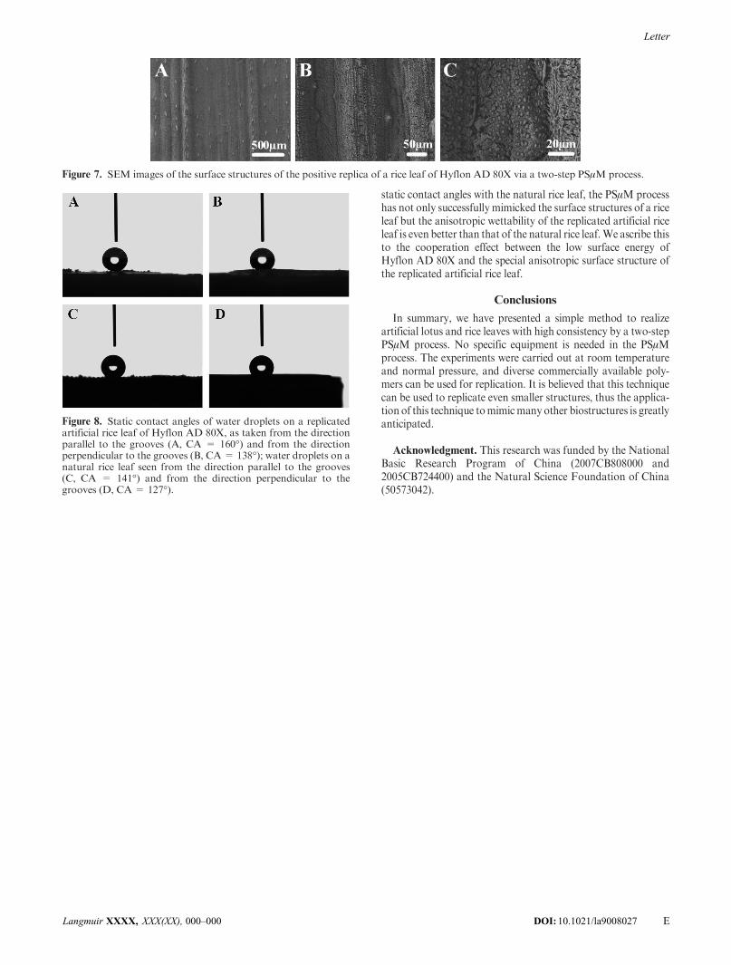
Figure 6C,D. One can see that there are many orderly arrangedmicropits in one dimension on the surface of the PEI film, whichagrees well with the negative structures of the rice leaf. Similarly,we have then employed the replicated PEI film with the negativestructure of the rice leaf as a template for the second step PSμM.Again, the Hyflon AD 80X was used for positive replicationbecause of its low surface energy. As shown in Figure 7A-C,some grooves on the macroscopic scale are arranged on thesurface of the Hyflon AD 80X film. In addition, the higher-resolution SEM image clearly indicates the existence of micro-protrusions arranged in an orderly manner in the directionparallel to the grooves and randomly in the perpendicular direc-tion. Therefore, the PSμM technique has been successfullydemonstrated for the artificial mimicking of rice leaf of aniso-tropic surface structure.
A more interesting finding is that the replicated artificial riceleaf exhibits very good anisotropic wettability, as shown inFigure 8. The static CA measured in the direction parallel tothe grooves is 160�, and that in the perpendicular direction is 138�.It should be noted that for a natural rice leaf its static CAs in thetwo directions are 141 and 127�, respectively. Comparing these
Figure 5. Static contact angles of water droplets on a positivelyreplicated artificial lotus leaf of Hyflon AD 80X (A, CA = 151�)and a flat Hyflon AD 80X surface (B, CA= 112�).
Figure 6. SEM images of the surface structures of a natural riceleaf (A, B) and its negative replica (C, D).
Figure 4. SEM images of the surface structures of the HyflonAD 80X filmwith positive replica of a lotus leaf via a two-step PSμMprocess.
(27) Yu, X.; Wang, Z. Q.; Jiang, Y. G.; Shi, F.; Zhang, X. Adv. Mater. 2005, 17,1289.
DOI: 10.1021/la9008027 Langmuir XXXX, XXX(XX), 000–000
Letter
D
static contact angles with the natural rice leaf, the PSμM processhas not only successfully mimicked the surface structures of a riceleaf but the anisotropic wettability of the replicated artificial riceleaf is even better than that of the natural rice leaf.We ascribe thisto the cooperation effect between the low surface energy ofHyflon AD 80X and the special anisotropic surface structure ofthe replicated artificial rice leaf.
Conclusions
In summary, we have presented a simple method to realizeartificial lotus and rice leaves with high consistency by a two-stepPSμM process. No specific equipment is needed in the PSμMprocess. The experiments were carried out at room temperatureand normal pressure, and diverse commercially available poly-mers can be used for replication. It is believed that this techniquecan be used to replicate even smaller structures, thus the applica-tion of this technique tomimicmany other biostructures is greatlyanticipated.
Acknowledgment. This research was funded by the NationalBasic Research Program of China (2007CB808000 and2005CB724400) and the Natural Science Foundation of China(50573042).
Figure 8. Static contact angles of water droplets on a replicatedartificial rice leaf of Hyflon AD 80X, as taken from the directionparallel to the grooves (A, CA = 160�) and from the directionperpendicular to the grooves (B, CA= 138�); water droplets on anatural rice leaf seen from the direction parallel to the grooves(C, CA = 141�) and from the direction perpendicular to thegrooves (D, CA= 127�).
Figure 7. SEM images of the surface structures of the positive replica of a rice leaf of Hyflon AD 80X via a two-step PSμM process.
DOI: 10.1021/la9008027Langmuir XXXX, XXX(XX), 000–000
Letter
E


![SMASH: Structured Matrix Approximation by Separation and ...yxi26/PDF/smash.pdf · The resulting hierarchical rank structured matrices [3,4,5,6], culminated in H2 matrices[7,6], provide](https://img.pdfslide.us/doc/110x75/5ed837c50fa3e705ec0e0e15/smash-structured-matrix-approximation-by-separation-and-yxi26pdfsmashpdf.jpg)