Embed Size (px)
Citation preview

MidbrainMidbrain
Slide 7.39Copyright © 2003 Pearson Education, Inc. publishing as Benjamin Cummings
Mostly composed of tracts of nerve fibers
Reflex centers for vision and hearing

PonsPons
Slide 7.40Copyright © 2003 Pearson Education, Inc. publishing as Benjamin Cummings
The bulging center part of the brain stem
Mostly composed of fiber tracts
Includes nuclei involved in the control of breathing

Medulla OblongataMedulla Oblongata
Slide 7.41Copyright © 2003 Pearson Education, Inc. publishing as Benjamin Cummings
The lowest part of the brain stem Merges into the spinal cord Includes important fiber tracts Contains important control centers
Heart rate control Blood pressure regulation Breathing Swallowing Vomiting

CerebellumCerebellum
Slide 7.43a
Copyright © 2003 Pearson Education, Inc. publishing as Benjamin Cummings
Two hemispheres with convoluted surfaces
Provides involuntary coordination of body movements

CerebellumCerebellum
Slide 7.43b
Copyright © 2003 Pearson Education, Inc. publishing as Benjamin Cummings
Figure 7.15a
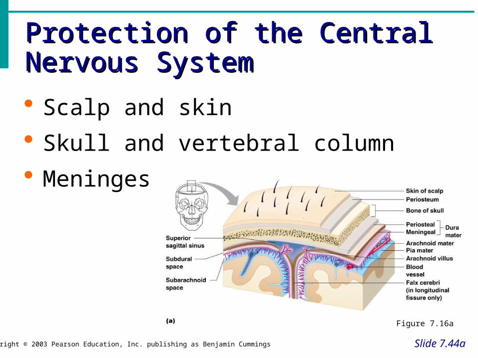
Protection of the Central Nervous Protection of the Central Nervous SystemSystem
Slide 7.44a
Copyright © 2003 Pearson Education, Inc. publishing as Benjamin Cummings
Scalp and skin
Skull and vertebral column
Meninges
Figure 7.16a

Protection of the Central Nervous Protection of the Central Nervous SystemSystem
Slide 7.44b
Copyright © 2003 Pearson Education, Inc. publishing as Benjamin Cummings
Cerebrospinal fluid
Blood brain barrier
Figure 7.16a

MeningesMeninges
Slide 7.45a
Copyright © 2003 Pearson Education, Inc. publishing as Benjamin Cummings
Dura matter
Double-layered external covering
Periosteum – attached to surface of the skull
Meningeal layer – outer covering of the brain
Folds inward in several areas

Cerebrospinal FluidCerebrospinal Fluid
Slide 7.46Copyright © 2003 Pearson Education, Inc. publishing as Benjamin Cummings
Similar to blood plasma composition
Formed by the choroid plexus
Forms a watery cushion to protect the brain

Ventricles and Location of the Ventricles and Location of the Cerebrospinal FluidCerebrospinal Fluid
Slide 7.47a
Copyright © 2003 Pearson Education, Inc. publishing as Benjamin Cummings
Figure 7.17a

Ventricles and Location of the Ventricles and Location of the Cerebrospinal FluidCerebrospinal Fluid
Slide 7.47b
Copyright © 2003 Pearson Education, Inc. publishing as Benjamin Cummings
Figure 7.17b
Blood Brain BarrierBlood Brain Barrier
Slide 7.48Copyright © 2003 Pearson Education, Inc. publishing as Benjamin Cummings
Includes the least permeable capillaries of the body
Excludes many potentially harmful substances
Useless against some substances Fats and fat soluble molecules Respiratory gases Alcohol Nicotine Anesthesia

Traumatic Brain Injuries (TBI)Traumatic Brain Injuries (TBI)
Slide 7.49Copyright © 2003 Pearson Education, Inc. publishing as Benjamin Cummings
Concussion Slight or mild brain injury Bleeding & tearing of nerve fibers happened Recovery likely with some memory loss
Contusion A more severe TBI Nervous tissue destruction occurs Nervous tissue does not regenerate
Cerebral edema Swelling from the inflammatory response May compress and kill brain tissue

Cerebrovascular Accident (CVA)Cerebrovascular Accident (CVA)
Slide 7.50Copyright © 2003 Pearson Education, Inc. publishing as Benjamin Cummings
Commonly called a stroke
The result of a ruptured blood vessel supplying a region of the brain
Brain tissue supplied with oxygen from that blood source dies
Loss of some functions or death may result

Alzheimer’s DiseaseAlzheimer’s Disease
Slide 7.51Copyright © 2003 Pearson Education, Inc. publishing as Benjamin Cummings
Progressive degenerative brain disease
Mostly seen in the elderly, but may begin in middle age
Structural changes in the brain include abnormal protein deposits and twisted fibers within neurons
Victims experience memory loss, irritability, confusion and ultimately, hallucinations and death

Structural Classification of the Structural Classification of the Nervous SystemNervous System
Slide 7.2Copyright © 2003 Pearson Education, Inc. publishing as Benjamin Cummings
Central nervous system (CNS)
Brain
Spinal cord
Peripheral nervous system (PNS)
Nerve outside the brain and spinal cord

Functional Classification of the Functional Classification of the Peripheral Nervous SystemPeripheral Nervous System
Slide 7.3aCopyright © 2003 Pearson Education, Inc. publishing as Benjamin Cummings
Sensory (afferent) division
Nerve fibers that carry information to the central nervous system
Figure 7.1

Functional Classification of the Functional Classification of the Peripheral Nervous SystemPeripheral Nervous System
Slide 7.3bCopyright © 2003 Pearson Education, Inc. publishing as Benjamin Cummings
Motor (efferent) division
Nerve fibers that carry impulses away from the central nervous system
Figure 7.1

Functional Classification of the Functional Classification of the Peripheral Nervous SystemPeripheral Nervous System
Slide 7.3cCopyright © 2003 Pearson Education, Inc. publishing as Benjamin Cummings
Motor (efferent) division Two subdivisions
Somatic nervous system = voluntary Autonomic nervous system = involuntary
Figure 7.1

Organization of the Nervous Organization of the Nervous SystemSystem
Slide 7.4Copyright © 2003 Pearson Education, Inc. publishing as Benjamin Cummings
Figure 7.2

Nervous Tissue: Support Cells Nervous Tissue: Support Cells (Neuroglia or Glia)(Neuroglia or Glia)
Slide 7.5Copyright © 2003 Pearson Education, Inc. publishing as Benjamin Cummings
Astrocytes Abundant, star-shaped cells
Brace neurons
Form barrier between capillaries and neurons
Control the chemical environment of the brain (CNS)
Figure 7.3a

Nervous Tissue: Support CellsNervous Tissue: Support Cells
Slide 7.6Copyright © 2003 Pearson Education, Inc. publishing as Benjamin Cummings
Microglia (CNS) Spider-like phagocytes
Dispose of debris
Ependymal cells (CNS) Line cavities of the
brain and spinal cord
Circulate cerebrospinal fluid
Figure 7.3b, c

Nervous Tissue: Support CellsNervous Tissue: Support Cells
Slide 7.7aCopyright © 2003 Pearson Education, Inc. publishing as Benjamin Cummings
Oligodendrocytes(CNS)
Produce myelin sheath around nerve fibers in the central nervous system Figure 7.3d

Neuroglia vs. Neurons
• Neuroglia divide.– Not really neurons, provide support and
nutrition
• Neurons do not divide.• Most brain tumors are “gliomas.”• Most brain tumors involve the neuroglia
cells, not the neurons.• Consider the role of cell division in cancer!

Support Cells of the PNSSupport Cells of the PNS
Slide 7.7bCopyright © 2003 Pearson Education, Inc. publishing as Benjamin Cummings
Satellite cells Protect neuron cell bodies
Schwann cells Form myelin sheath in the peripheral
nervous system (myelin allows impulses to conduct faster)
Figure 7.3e