Embed Size (px)
Citation preview
Ceramics International 42 (2016) 11562–11567
Contents lists available at ScienceDirect
Ceramics International
http://d0272-88
n Corr(PCSED)
E-m
journal homepage: www.elsevier.com/locate/ceramint
Microwave-assisted synthesis of ZnO doped CeO2 nanoparticles aspotential scaffold for highly sensitive nitroaniline chemical sensor
Naushad Ahmad a, Ahmad Umar b,c,n, Rajesh Kumar d, Manawwer Alam e
a Department of Chemistry, College of Science, King Saud University, Riyadh 11451, Saudi Arabiab Promising Centre for Sensors and Electronic Devices (PCSED), Najran University, P.O. Box-1988, Najran 11001, Saudi Arabiac Department of Chemistry, College of Science and Arts, Najran University, P.O. Box-1988, Najran 11001, Saudi Arabiad PG Department of Chemistry, JCDAV College (Panjab University), Dasuya 144205, Punjab, Indiae Research Centre, College of Science, King Saud University, Riyadh 11451, Saudi Arabia
a r t i c l e i n f o
Article history:Received 16 March 2016Received in revised form1 April 2016Accepted 4 April 2016Available online 6 April 2016
Keywords:ZnO doped CeO2
NanoparticlesChemical sensorsNitroaniline
x.doi.org/10.1016/j.ceramint.2016.04.01342/& 2016 Elsevier Ltd and Techna Group S.r
esponding author at: Promising Centre for Se, Najran University, P.O.Box-1988, Najran 1100ail address: [email protected] (A. Um
a b s t r a c t
Herein, we report the successful fabrication of highly sensitive, reproducible and reliable nitroanilinechemical sensor based on ZnO doped CeO2 nanoparticles. The ZnO doped CeO2 nanoparticles weresynthesized through a simple, facile and rapid microwave-assisted method and characterized by severaltechniques. The detailed characterizations confirmed that the synthesized nanoparticles were mono-disperse and grown in high density and possessing good crystallinity. Further, the synthesized ZnOdoped CeO2 nanoparticles were used as efficient electron mediators for the fabrication of high sensitivenitroaniline chemical sensors. The fabricated nitroaniline chemical sensor exhibited very high sensitivityof 550.42 μA mM�1 cm�2 and experimental detection limit of 0.25 mM. To the best of our knowledge,this is the first report in which CeO2–ZnO nanoparticles were used as efficient electron mediators for thefabrication of nitroaniline chemical sensors. Thus, presented work demonstrates that ZnO doped CeO2
nanoparticles are potential material to fabricate highly efficient and reliable chemical sensors.& 2016 Elsevier Ltd and Techna Group S.r.l. All rights reserved.
1. Introduction
Recently, doped and composite lanthanide oxide materials havebeen explored extensively for a variety of applications. Ceria(CeO2) is one of the most studied oxide among the various lan-thanide oxides owing to its excellent mechanical strength, highoxygen storage capacity (OSC), good optical properties, high con-ductivity, good redox performances, high specific surface areaparticularly in nano regime, high thermal stability, abundance ofactive sites and oxygen vacancies on the surface, and most im-portantly low cost synthesis [1,2]. These properties make thismaterial a promising candidate for a number of applications. Theperformance of these properties, and hence the versatility forpotential applications can be enhanced by either doping or makingits composites with other metals or metal oxides. Transition metallike Ni, Cu, Zn, Co, Sc, Y, Zr, Ti, Fe [3–10] and metal oxides such asZnO, TiO2, CuO, CaO, NiO, Tb2O3, MnO2 [11–21] etc. are some of thereported examples of dopants and additive materials, respectivelyfor CeO2. Doped or composite CeO2 materials are potentially used
.l. All rights reserved.
nsors and Electronic Devices1, Saudi Arabia.ar).
as photocatalysts [22–24], electrochemical sensors [25,26], gassensors [27,28], biosensors [29,30], anodic materials for fuel cellsand lithium ion batteries [31], super-capacitors [19], catalyst for COoxidation [16,32], electrochemical charge storage devices [33,34]and many more. Singh et al. [25] successfully synthesized wellcrystalline CeO2–ZnO nano-ellipsoids via facile hydrothermalprocess at low-temperature conditions. These nano-ellipsoids ex-hibited a high sensitivity of �0.120 μA/μM cm2 with a low de-tection limit of 1.163 μM for p-nitrophenol.
CeO2-ZnO hexagonal nanodisks were synthesized via chemicalroute at low-temperature by Lamba et al. [22]. The preparedhexagonal nanodisks acted as efficient photocatalyst for the de-gradation of Direct Blue-15 dye under solar light irradiation. Ha-medani et al. [27] reported that ZnO nanostructures doped with5 wt% CeO2 showed improved gas sensing response, reduced re-covery time and highly selective sensing of ethanol even in thepresence of CO and CH4. It was proposed that large oxygen storagecapacity and the presence of a large number of oxygen vacancieson the surface of CeO2 with fluorite structure deposited over ZnOsurface resulted in the rapid diffusion of oxygen, easily oxidizingthe analyte molecules [35,36]. Similar results were observed byRajgure et al. [28] for 2 wt% CeO2 doped ZnO nanocomposites. Heet al. [34] fabricated dumbbell-like ZnO nanoparticles deposited
N. Ahmad et al. / Ceramics International 42 (2016) 11562–11567 11563
over the surface of CeO2 nanorods via the hydrothermal methodand reported excellent electrochemical performance as comparedto pure CeO2 nanorods. Li et al. [37] and Wan et al. [38] observedhigh sensitivity and selectivity with fast response for sensorsbased on Ce-doped ZnO hollow nanofibers, synthesized throughfacile single capillary electro-spinning technique, as compared topure ZnO nanomaterials. Similarly, Yan et al. [39] reported ethanolsensing applications of porous CeO2–ZnO hollow fibers synthe-sized on cotton bio-template.
Doping is supposed to influence the active surface area, surfacedefects, oxygen vacancies and the diffusion rates. Due to the pre-sence of surface defects and oxygen vacancies, cerium can undergofast and reversible Ceþ4–Ceþ3 redox interconversions [27]. BothCeO2 (Eg¼3.15 eV) and ZnO (Eg¼3.37) are n-type semiconductorsand thus form n–n type hetero-junction [1,12,39]. The electronictransitions in such heterojunctions result in the formation of anelectron depletion layer at the interface of ZnO and CeO2, therebyimproving the electrocatalytic properties [40]. Thus, CeO2–ZnOmaterials can efficiently act as transducers for converting chemicalenergies produced during redox processes occurring at their sur-face into electrical signals.
In this paper, well crystalline ZnO doped CeO2 composite na-noparticles were synthesized by the facile chemical microwave-assisted process. The prepared nanoparticles were characterized indetail using different characterization techniques. Further, thesynthesized nanoparticles were used as potential electron med-iators for the fabrication of nitroaniline chemical sensors. To thebest of our knowledge, this is the first report in which well-crys-talline ZnO doped CeO2 composite nanoparticles were used asefficient electron mediator to fabricate highly sensitive nitroani-line chemical sensor.
2. Experimental details
2.1. Materials
All chemicals procured for the synthesis ZnO doped CeO2 na-noparticles were used as received without any further purifica-tions. For the synthesis, Zinc Chloride and Cerium Nitrate wereobtained from BDH Chemicals Ltd Poole England, Ammonia So-lution (about 35%) was procured from Avonchem, Cheshire, UK.
2.2. Synthesis of ZnO doped CeO2 nanoparticles
In order to synthesize ZnO doped CeO2nanoparticles, 50 mL of0.25 M Zinc chloride solution was mixed with 50 mL equimolarsolution of cerium nitrate with continuous stirring. The pH of thereaction solution was adjusted to 8 by the careful dropwise addi-tion of 1:1 aqueous NH3 solution. All the solutions were made intriple de-ionized (DI) water. The reaction solution was thentransferred to a Teflon microwave beaker and the contents weresubjected to microwave treatment for 30 min at 200 °C tempera-ture in a Microwave oven (MicroSynth Plus Model, Milestone, US).After the completion of the reaction, the solution was allowed tocool to room temperature followed by the filtration and washingswith distilled water for several times to remove any unreactedcompound. The yellowish white product was dried in an air ovenfor 6 h at 70 °C.
2.3. Characterizations of ZnO doped CeO2 nanoparticles
The detailed analysis for the structural, morphological, micro-structural and compositional properties of ZnO doped CeO2 com-posite nanoparticles was carried out using different techniques.Field emission scanning electron microscopy (FESEM; JEOL-JSM-
7600F) and high-resolution TEM (HR-TEM) were used for mor-phological and structural analysis. The phases, micro-structuralproperties and crystallinity of ZnO doped CeO2 composite nano-particles were studied by X-ray diffraction (XRD; PAN analyticalXpert Pro.) in the scan range of 15–75° (2θ) using Cu–Kα radiation(λ¼1.54178 Å). Fourier transform infrared spectroscopic analysis(FTIR; Perkin Elmer-FTIR Spectrum-100) was used for the com-positional studies of the composites with KBr pelletization in thescan range of 450–4000 cm�1. For FTIR characterization, the ZnOdoped CeO2 powder was mixed with dry solid KBr and ground to afine powder with the help of mortar and pestle. Finally, the groundpowder was pressed to form a transparent pellet. The scatteringproperty of ZnO doped CeO2 composite nanoparticles was studiedat room temperature by using Raman-scattering spectroscopy inthe scan range of 200–800 cm�1 (Perkin Elmer-Raman Station 400series).
2.4. Fabrication of nitroaniline chemical sensor based on ZnO dopedCeO2 nanoparticles
For the fabrication of electrochemical sensor, ZnO doped CeO2
nanoparticles were mixed with conducting agent butyl carbitalacetate (BCA) to form a homogeneous and thin paste which waspasted on the surface of pre-cleaned silver (Ag) electrode. To fixthe sensor material layer on the surface of the Ag electrode, it wasdried in an air oven for 6 h at 70–72 °C. For measuring the elec-trochemical sensing parameters Keithley 6517 A, USA electrometerwas used as a source of voltage and to record the current. ZnOdoped CeO2 nanoparticles modified Ag electrode (AgE) and Pt wirewere used as working and counter electrodes, respectively. All theexperiments were performed at room temperature in the presenceof 0.1 M phosphate buffer solution (PBS) with pH¼7.4.
3. Results and discussion
3.1. Characterization and properties of ZnO doped CeO2
nanoparticles
The morphological properties of the ZnO doped CeO2 nano-particles were characterized by field emission scanning electronmicroscopy (FESEM) and transmission electron microscopy (TEM).
Fig. 1(a) exhibits the typical low-magnification FESEM image ofsynthesized ZnO-doped CeO2 which confirmed that the preparedmaterials are nano-sized particles grown in very high density. Thenanoparticles are monodisperse and spherical in shape. Most of thenanoparticles possess similar sizes while some small nanoparticlescan also be seen in the micrograph (Fig. 1(b)). Fig. 1(c) and (d) depictthe typical TEM images of synthesized ZnO doped CeO2 nano-particles which revealed that the nanoparticles are grown in highdensity. As it can be seen from the observed TEM images that due tohigh-density growth some agglomeration in the nanoparticles oc-curs. The prepared ZnO doped CeO2 nanoparticles possess differentsizes with almost spherical shapes and are well dispersed.
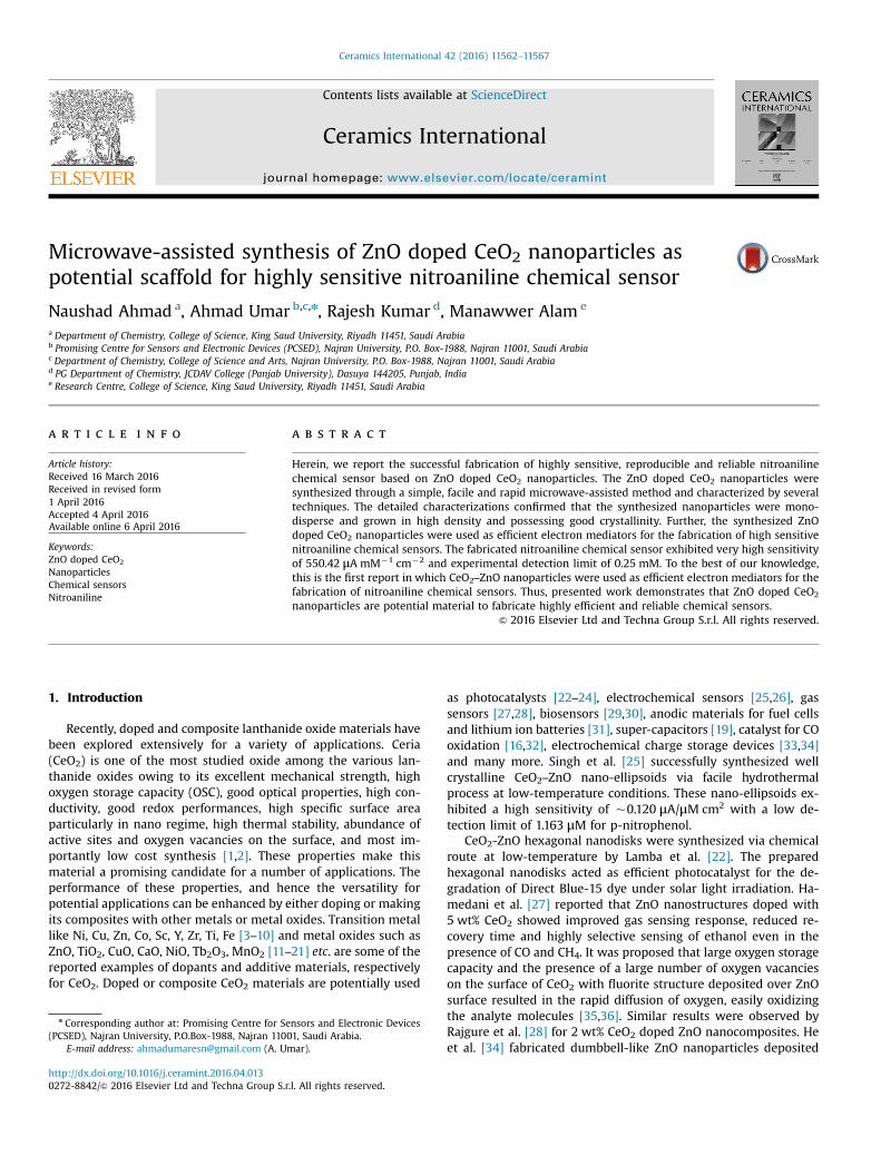
Fig. 2 represents the XRD patterns of the ZnO dopedCeO2nanoparticles. Well defined diffraction peaks at diffractionangles at 2θ¼28.6°, 33.05°, 47.64°, 56.49°, 59.16°, and69.72°corresponds to the (111), (200), (220), (311), (222) and (400)planes of cubic ceria (CeO2) phase with fluorite structure, respec-tively (JCPDS card number 34-394) [1,22,25,41]. On the other hand,the diffraction patterns at 2θ¼36.33°, 62.93° and 68.03° are thecharacteristics peaks corresponding to (101), (103) and (112) dif-fraction planes of wurtzite hexagonal phase of ZnO (JCPDS card no.36-1451) [42–44]. No other diffraction peak in the XRD spectrumexcept for CeO2 and ZnO confirms the fact that composites are ofhigh purity and are composed of only CeO2 and ZnO. Well-defined
Fig. 1. (a) Low and (b) high-resolution FESEM images and (c and d) TEM images of ZnO doped CeO2 nanoparticles.
Fig. 2. Typical XRD diffraction patterns of ZnO doped CeO2 nanoparticles.
Fig. 3. Typical FTIR spectrum of ZnO doped CeO2 nanoparticles.
N. Ahmad et al. / Ceramics International 42 (2016) 11562–1156711564
and sharp reflections of the XRD peaks reveal the good crystallinityof the ZnO doped CeO2nanoparticles. The obtained XRD patternsalso match well with the reported literature.
The crystallite sizes of ZnO and CeO2 were determined fromDebye–Scherrer formula (Eq. 1).
θ= λ
β ( )d
0.89.cos 1
Where λ¼the wavelength of X-rays used (0.1541 nm), θ is theBragg diffraction angle corresponding to ZnO (101) peak situatedat 36.33° and CeO2 (111) peak at 28.60° and β is the peak width athalf maximum (FWHM) [36,45]. The values for β, as derived fromthe XRD patterns were 0.32873 and 0.92124 for ZnO and CeO2,respectively. The crystallite sizes of the ZnO nanoparticles andCeO2were found to be 25.17 and 8.81 nm, respectively.
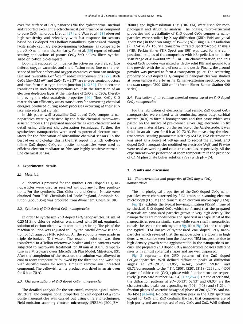
To study the chemical composition of the ZnO doped CeO2
nanoparticles, FTIR studies were performed and the results arerepresented in Fig. 3.Three well-defined absorption peaks at
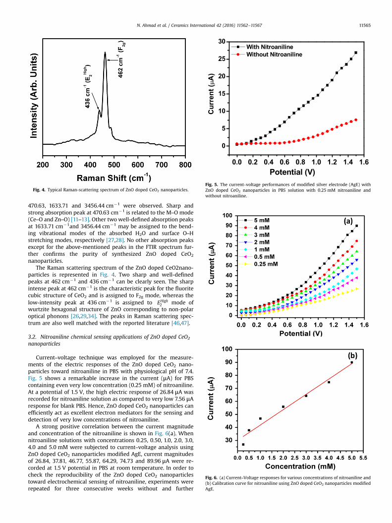
Fig. 4. Typical Raman-scattering spectrum of ZnO doped CeO2 nanoparticles.
0.0 0.2 0.4 0.6 0.8 1.0 1.2 1.4 1.6
0
5
10
15
20
25
30
Potential (V)
Cur
rent
( µA
)
With Nitroaniline Without Nitroaniline
Fig. 5. The current–voltage performances of modified silver electrode (AgE) withZnO doped CeO2 nanoparticles in PBS solution with 0.25 mM nitroaniline andwithout nitroaniline.
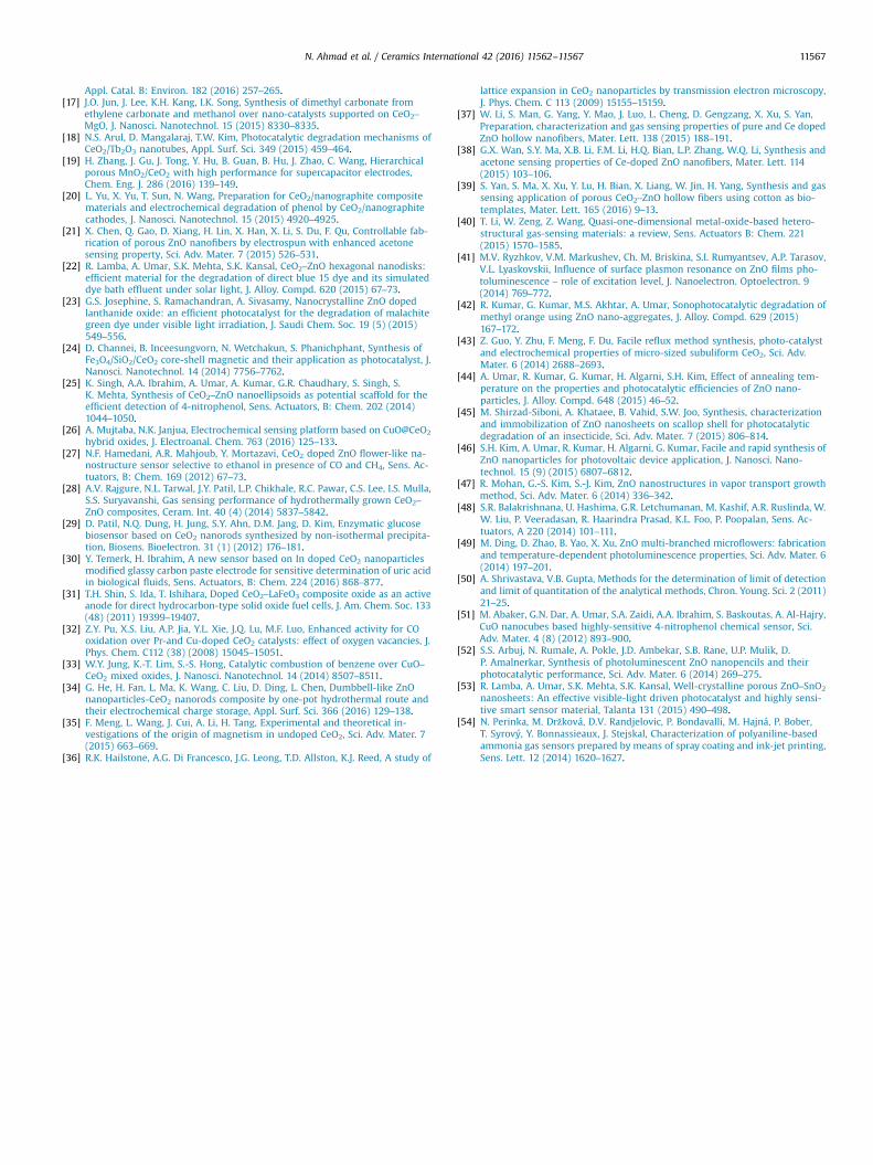
Fig. 6. (a) Current–Voltage responses for various concentrations of nitroaniline and(b) Calibration curve for nitroaniline using ZnO doped CeO2 nanoparticles modifiedAgE.
N. Ahmad et al. / Ceramics International 42 (2016) 11562–11567 11565
470.63, 1633.71 and 3456.44 cm�1 were observed. Sharp andstrong absorption peak at 470.63 cm�1 is related to the M–O mode(Ce–O and Zn–O) [11–13]. Other two well-defined absorption peaksat 1633.71 cm�1and 3456.44 cm�1 may be assigned to the bend-ing vibrational modes of the absorbed H2O and surface O–Hstretching modes, respectively [27,28]. No other absorption peaksexcept for the above-mentioned peaks in the FTIR spectrum fur-ther confirms the purity of synthesized ZnO doped CeO2
nanoparticles.The Raman scattering spectrum of the ZnO doped CeO2nano-
particles is represented in Fig. 4. Two sharp and well-definedpeaks at 462 cm�1 and 436 cm�1 can be clearly seen. The sharpintense peak at 462 cm�1 is the characteristic peak for the fluoritecubic structure of CeO2 and is assigned to F2g mode, whereas thelow-intensity peak at 436 cm�1 is assigned to EHigh
2 mode ofwurtzite hexagonal structure of ZnO corresponding to non-polaroptical phonons [26,29,34]. The peaks in Raman scattering spec-trum are also well matched with the reported literature [46,47].
3.2. Nitroaniline chemical sensing applications of ZnO doped CeO2
nanoparticles
Current–voltage technique was employed for the measure-ments of the electric responses of the ZnO doped CeO2 nano-particles toward nitroaniline in PBS with physiological pH of 7.4.Fig. 5 shows a remarkable increase in the current (μA) for PBScontaining even very low concentration (0.25 mM) of nitroaniline.At a potential of 1.5 V, the high electric response of 26.84 μA wasrecorded for nitroaniline solution as compared to very low 7.56 μAresponse for blank PBS. Hence, ZnO doped CeO2 nanoparticles canefficiently act as excellent electron mediators for the sensing anddetection of very low concentrations of nitroaniline.
A strong positive correlation between the current magnitudeand concentration of the nitroaniline is shown in Fig. 6(a). Whennitroaniline solutions with concentrations 0.25, 0.50, 1.0, 2.0, 3.0,4.0 and 5.0 mM were subjected to current–voltage analysis usingZnO doped CeO2 nanoparticles modified AgE, current magnitudesof 26.84, 37.81, 46.77, 55.87, 64.29, 74.73 and 89.96 μA were re-corded at 1.5 V potential in PBS at room temperature. In order tocheck the reproducibility of the ZnO doped CeO2 nanoparticlestoward electrochemical sensing of nitroaniline, experiments wererepeated for three consecutive weeks without and further
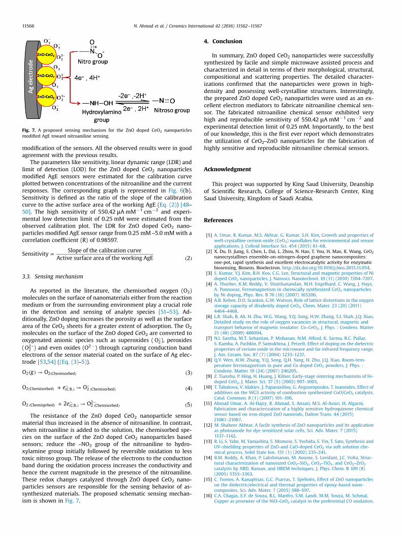
Fig. 7. A proposed sensing mechanism for the ZnO doped CeO2 nanoparticlesmodified AgE toward nitroaniline sensing.
N. Ahmad et al. / Ceramics International 42 (2016) 11562–1156711566
modification of the sensors. All the observed results were in goodagreement with the previous results.
The parameters like sensitivity, linear dynamic range (LDR) andlimit of detection (LOD) for the ZnO doped CeO2 nanoparticlesmodified AgE sensors were estimated for the calibration curveplotted between concentrations of the nitroaniline and the currentresponses. The corresponding graph is represented in Fig. 6(b).Sensitivity is defined as the ratio of the slope of the calibrationcurve to the active surface area of the working AgE (Eq. (2)) [48–50]. The high sensitivity of 550.42 μA mM�1 cm�2 and experi-mental low detection limit of 0.25 mM were estimated from theobserved calibration plot. The LDR for ZnO doped CeO2 nano-particles modified AgE sensor range from 0.25 mM –5.0 mMwith acorrelation coefficient (R) of 0.98597.
=( )
SensitivitySlope of the calibration curve
Active surface area of the working AgE 2
3.3. Sensing mechanism
As reported in the literature, the chemisorbed oxygen (O2)molecules on the surface of nanomaterials either from the reactionmedium or from the surrounding environment play a crucial rolein the detection and sensing of analyte species [51–53]. Ad-ditionally, ZnO doping increases the porosity as well as the surfacearea of the CeO2 sheets for a greater extent of adsorption. The O2
molecules on the surface of the ZnO doped CeO2 are converted tooxygenated anionic species such as superoxides ( −O2), peroxides( −O2
2 ) and even oxides (O2�) through capturing conduction bandelectrons of the sensor material coated on the surface of Ag elec-trode [53,54] ((Eq. (3)–5)).
( ) → ( )( )gO O 32 2 Chemisorbed
+ → ( )( ) ( )−
( )−eO O 42 Chemisorbed C.B. 2 Chemisorbed
+ → ( )( ) ( )−
( )−eO 2 O 52 Chemisprbed C.B. 2 Chemisorbed
2
The resistance of the ZnO doped CeO2 nanoparticle sensormaterial thus increased in the absence of nitroaniline. In contrast,when nitroaniline is added to the solution, the chemisorbed spe-cies on the surface of the ZnO doped CeO2 nanoparticles basedsensors; reduce the –NO2 group of the nitroaniline to hydro-xylamine group initially followed by reversible oxidation to lesstoxic nitroso group. The release of the electrons to the conductionband during the oxidation process increases the conductivity andhence the current magnitude in the presence of the nitroaniline.These redox changes catalyzed through ZnO doped CeO2 nano-particles sensors are responsible for the sensing behavior of as-synthesized materials. The proposed schematic sensing mechan-ism is shown in Fig. 7.
4. Conclusion
In summary, ZnO doped CeO2 nanoparticles were successfullysynthesized by facile and simple microwave assisted process andcharacterized in detail in terms of their morphological, structural,compositional and scattering properties. The detailed character-izations confirmed that the nanoparticles were grown in high-density and possessing well-crystalline structures. Interestingly,the prepared ZnO doped CeO2 nanoparticles were used as an ex-cellent electron mediators to fabricate nitroaniline chemical sen-sor. The fabricated nitroaniline chemical sensor exhibited veryhigh and reproducible sensitivity of 550.42 μA mM�1 cm�2 andexperimental detection limit of 0.25 mM. Importantly, to the bestof our knowledge, this is the first ever report which demonstratesthe utilization of CeO2–ZnO nanoparticles for the fabrication ofhighly sensitive and reproducible nitroaniline chemical sensors.
Acknowledgment
This project was supported by King Saud University, Deanshipof Scientific Research, College of Science-Research Center, KingSaud University, Kingdom of Saudi Arabia.
References
[1] A. Umar, R. Kumar, M.S. Akhtar, G. Kumar, S.H. Kim, Growth and properties ofwell-crystalline cerium oxide (CeO2) nanoflakes for environmental and sensorapplications, J. Colloid Interface Sci. 454 (2015) 61–68.
[2] X. Du, D. Jiang, S. Chen, L. Dai, L. Zhou, N. Hao, T. You, H. Mao, K. Wang, CeO2
nanocrystallines ensemble-on-nitrogen-doped graphene nanocomposites:one-pot, rapid synthesis and excellent electrocatalytic activity for enzymaticbiosensing, Biosens. Bioelectron. http://dx.doi.org/10.1016/j.bios.2015.11.054.
[3] S. Kumar, Y.J. Kim, B.H. Koo, C.G. Lee, Structural and magnetic properties of Nidoped CeO2 nanoparticles, J. Nanosci. Nanotechnol. 10 (11) (2010) 7204–7207.
[4] A. Thurber, K.M. Reddy, V. Shutthanandan, M.H. Engelhard, C. Wang, J. Hays,A. Punnoose, Ferromagnetism in chemically synthesized CeO2 nanoparticlesby Ni doping, Phys. Rev. B 76 (16) (2007) 165206.
[5] A.B. Kehoe, D.O. Scanlon, G.W. Watson, Role of lattice distortions in the oxygenstorage capacity of divalently doped CeO2, Chem. Mater. 23 (20) (2011)4464–4468.
[6] L.R. Shah, B. Ali, H. Zhu, W.G. Wang, Y.Q. Song, H.W. Zhang, S.I. Shah, J.Q. Xiao,Detailed study on the role of oxygen vacancies in structural, magnetic andtransport behavior of magnetic insulator: Co–CeO2, J. Phys. : Condens. Matter21 (48) (2009) 486004.
[7] N.I. Santha, M.T. Sebastian, P. Mohanan, N.M. Alford, K. Sarma, R.C. Pullar,S. Kamba, A. Pashkin, P. Samukhina, J. Petzelt, Effect of doping on the dielectricproperties of cerium oxide in the microwave and far-infrared frequency range,J. Am. Ceram. Soc. 87 (7) (2004) 1233–1237.
[8] Q.Y. Wen, H.W. Zhang, Y.Q. Song, Q.H. Yang, H. Zhu, J.Q. Xiao, Room-tem-perature ferromagnetism in pure and Co doped CeO2 powders, J. Phys. :Condens. Matter 19 (24) (2007) 246205.
[9] Z. Tianshu, P. Hing, H. Huang, J. Kilner, Early-stage sintering mechanisms of Fe-doped CeO2, J. Mater. Sci. 37 (5) (2002) 997–1003.
[10] T. Tabakova, V. Idakiev, J. Papavasiliou, G. Avgouropoulos, T. Ioannides, Effect ofadditives on the WGS activity of combustion synthesized CuO/CeO2 catalysts,Catal. Commun. 8 (1) (2007) 101–106.
[11] Ahmad Umar, A. Al-Hajry, R. Ahmad, S. Ansari, M.S. Al-Assiri, H. Algarni,Fabrication and characterization of a highly sensitive hydroquinone chemicalsensor based on iron-doped ZnO nanorods, Dalton Trans. 44 (2015)21081–21087.
[12] M. Shaheer Akhtar, A facile synthesis of ZnO nanoparticles and its applicationas photoanode for dye sensitized solar cells, Sci. Adv. Mater. 7 (2015)1137–1142.
[13] R. Li, S. Yabe, M. Yamashita, S. Momose, S. Yoshida, S. Yin, T. Sato, Synthesis andUV-shielding properties of ZnO-and CaO-doped CeO2 via soft solution che-mical process, Solid State Ion. 151 (1) (2002) 235–241.
[14] B.M. Reddy, A. Khan, P. Lakshmanan, M. Aouine, S. Loridant, J.C. Volta, Struc-tural characterization of nanosized CeO2–SiO2, CeO2–TiO2, and CeO2–ZrO2
catalysts by XRD, Raman, and HREM techniques, J. Phys. Chem. B 109 (8)(2005) 3355–3363.
[15] C. Tsonos, A. Kanapitsas, G.C. Psarras, T. Speliotis, Effect of ZnO nanoparticleson the dielectric/electrical and thermal properties of epoxy-based nano-composites, Sci. Adv. Mater. 7 (2015) 588–597.
[16] C.A. Chagas, E.F. de Souza, R.L. Manfro, S.M. Landi, M.M. Souza, M. Schmal,Copper as promoter of the NiO–CeO2 catalyst in the preferential CO oxidation,
N. Ahmad et al. / Ceramics International 42 (2016) 11562–11567 11567
Appl. Catal. B: Environ. 182 (2016) 257–265.[17] J.O. Jun, J. Lee, K.H. Kang, I.K. Song, Synthesis of dimethyl carbonate from
ethylene carbonate and methanol over nano-catalysts supported on CeO2–
MgO, J. Nanosci. Nanotechnol. 15 (2015) 8330–8335.[18] N.S. Arul, D. Mangalaraj, T.W. Kim, Photocatalytic degradation mechanisms of
CeO2/Tb2O3 nanotubes, Appl. Surf. Sci. 349 (2015) 459–464.[19] H. Zhang, J. Gu, J. Tong, Y. Hu, B. Guan, B. Hu, J. Zhao, C. Wang, Hierarchical
porous MnO2/CeO2 with high performance for supercapacitor electrodes,Chem. Eng. J. 286 (2016) 139–149.
[20] L. Yu, X. Yu, T. Sun, N. Wang, Preparation for CeO2/nanographite compositematerials and electrochemical degradation of phenol by CeO2/nanographitecathodes, J. Nanosci. Nanotechnol. 15 (2015) 4920–4925.
[21] X. Chen, Q. Gao, D. Xiang, H. Lin, X. Han, X. Li, S. Du, F. Qu, Controllable fab-rication of porous ZnO nanofibers by electrospun with enhanced acetonesensing property, Sci. Adv. Mater. 7 (2015) 526–531.
[22] R. Lamba, A. Umar, S.K. Mehta, S.K. Kansal, CeO2–ZnO hexagonal nanodisks:efficient material for the degradation of direct blue 15 dye and its simulateddye bath effluent under solar light, J. Alloy. Compd. 620 (2015) 67–73.
[23] G.S. Josephine, S. Ramachandran, A. Sivasamy, Nanocrystalline ZnO dopedlanthanide oxide: an efficient photocatalyst for the degradation of malachitegreen dye under visible light irradiation, J. Saudi Chem. Soc. 19 (5) (2015)549–556.
[24] D. Channei, B. Inceesungvorn, N. Wetchakun, S. Phanichphant, Synthesis ofFe3O4/SiO2/CeO2 core-shell magnetic and their application as photocatalyst, J.Nanosci. Nanotechnol. 14 (2014) 7756–7762.
[25] K. Singh, A.A. Ibrahim, A. Umar, A. Kumar, G.R. Chaudhary, S. Singh, S.K. Mehta, Synthesis of CeO2–ZnO nanoellipsoids as potential scaffold for theefficient detection of 4-nitrophenol, Sens. Actuators, B: Chem. 202 (2014)1044–1050.
[26] A. Mujtaba, N.K. Janjua, Electrochemical sensing platform based on CuO@CeO2
hybrid oxides, J. Electroanal. Chem. 763 (2016) 125–133.[27] N.F. Hamedani, A.R. Mahjoub, Y. Mortazavi, CeO2 doped ZnO flower-like na-
nostructure sensor selective to ethanol in presence of CO and CH4, Sens. Ac-tuators, B: Chem. 169 (2012) 67–73.
[28] A.V. Rajgure, N.L. Tarwal, J.Y. Patil, L.P. Chikhale, R.C. Pawar, C.S. Lee, I.S. Mulla,S.S. Suryavanshi, Gas sensing performance of hydrothermally grown CeO2–
ZnO composites, Ceram. Int. 40 (4) (2014) 5837–5842.[29] D. Patil, N.Q. Dung, H. Jung, S.Y. Ahn, D.M. Jang, D. Kim, Enzymatic glucose
biosensor based on CeO2 nanorods synthesized by non-isothermal precipita-tion, Biosens. Bioelectron. 31 (1) (2012) 176–181.
[30] Y. Temerk, H. Ibrahim, A new sensor based on In doped CeO2 nanoparticlesmodified glassy carbon paste electrode for sensitive determination of uric acidin biological fluids, Sens. Actuators, B: Chem. 224 (2016) 868–877.
[31] T.H. Shin, S. Ida, T. Ishihara, Doped CeO2–LaFeO3 composite oxide as an activeanode for direct hydrocarbon-type solid oxide fuel cells, J. Am. Chem. Soc. 133(48) (2011) 19399–19407.
[32] Z.Y. Pu, X.S. Liu, A.P. Jia, Y.L. Xie, J.Q. Lu, M.F. Luo, Enhanced activity for COoxidation over Pr-and Cu-doped CeO2 catalysts: effect of oxygen vacancies, J.Phys. Chem. C112 (38) (2008) 15045–15051.
[33] W.Y. Jung, K.-T. Lim, S.-S. Hong, Catalytic combustion of benzene over CuO–CeO2 mixed oxides, J. Nanosci. Nanotechnol. 14 (2014) 8507–8511.
[34] G. He, H. Fan, L. Ma, K. Wang, C. Liu, D. Ding, L. Chen, Dumbbell-like ZnOnanoparticles-CeO2 nanorods composite by one-pot hydrothermal route andtheir electrochemical charge storage, Appl. Surf. Sci. 366 (2016) 129–138.
[35] F. Meng, L. Wang, J. Cui, A. Li, H. Tang, Experimental and theoretical in-vestigations of the origin of magnetism in undoped CeO2, Sci. Adv. Mater. 7(2015) 663–669.
[36] R.K. Hailstone, A.G. Di Francesco, J.G. Leong, T.D. Allston, K.J. Reed, A study of
lattice expansion in CeO2 nanoparticles by transmission electron microscopy,J. Phys. Chem. C 113 (2009) 15155–15159.
[37] W. Li, S. Man, G. Yang, Y. Mao, J. Luo, L. Cheng, D. Gengzang, X. Xu, S. Yan,Preparation, characterization and gas sensing properties of pure and Ce dopedZnO hollow nanofibers, Mater. Lett. 138 (2015) 188–191.
[38] G.X. Wan, S.Y. Ma, X.B. Li, F.M. Li, H.Q. Bian, L.P. Zhang, W.Q. Li, Synthesis andacetone sensing properties of Ce-doped ZnO nanofibers, Mater. Lett. 114(2015) 103–106.
[39] S. Yan, S. Ma, X. Xu, Y. Lu, H. Bian, X. Liang, W. Jin, H. Yang, Synthesis and gassensing application of porous CeO2–ZnO hollow fibers using cotton as bio-templates, Mater. Lett. 165 (2016) 9–13.
[40] T. Li, W. Zeng, Z. Wang, Quasi-one-dimensional metal-oxide-based hetero-structural gas-sensing materials: a review, Sens. Actuators B: Chem. 221(2015) 1570–1585.
[41] M.V. Ryzhkov, V.M. Markushev, Ch. M. Briskina, S.I. Rumyantsev, A.P. Tarasov,V.L. Lyaskovskii, Influence of surface plasmon resonance on ZnO films pho-toluminescence – role of excitation level, J. Nanoelectron. Optoelectron. 9(2014) 769–772.
[42] R. Kumar, G. Kumar, M.S. Akhtar, A. Umar, Sonophotocatalytic degradation ofmethyl orange using ZnO nano-aggregates, J. Alloy. Compd. 629 (2015)167–172.
[43] Z. Guo, Y. Zhu, F. Meng, F. Du, Facile reflux method synthesis, photo-catalystand electrochemical properties of micro-sized subuliform CeO2, Sci. Adv.Mater. 6 (2014) 2688–2693.
[44] A. Umar, R. Kumar, G. Kumar, H. Algarni, S.H. Kim, Effect of annealing tem-perature on the properties and photocatalytic efficiencies of ZnO nano-particles, J. Alloy. Compd. 648 (2015) 46–52.
[45] M. Shirzad-Siboni, A. Khataee, B. Vahid, S.W. Joo, Synthesis, characterizationand immobilization of ZnO nanosheets on scallop shell for photocatalyticdegradation of an insecticide, Sci. Adv. Mater. 7 (2015) 806–814.
[46] S.H. Kim, A. Umar, R. Kumar, H. Algarni, G. Kumar, Facile and rapid synthesis ofZnO nanoparticles for photovoltaic device application, J. Nanosci. Nano-technol. 15 (9) (2015) 6807–6812.
[47] R. Mohan, G.-S. Kim, S.-J. Kim, ZnO nanostructures in vapor transport growthmethod, Sci. Adv. Mater. 6 (2014) 336–342.
[48] S.R. Balakrishnana, U. Hashima, G.R. Letchumanan, M. Kashif, A.R. Ruslinda, W.W. Liu, P. Veeradasan, R. Haarindra Prasad, K.L. Foo, P. Poopalan, Sens. Ac-tuators, A 220 (2014) 101–111.
[49] M. Ding, D. Zhao, B. Yao, X. Xu, ZnO multi-branched microflowers: fabricationand temperature-dependent photoluminescence properties, Sci. Adv. Mater. 6(2014) 197–201.
[50] A. Shrivastava, V.B. Gupta, Methods for the determination of limit of detectionand limit of quantitation of the analytical methods, Chron. Young. Sci. 2 (2011)21–25.
[51] M. Abaker, G.N. Dar, A. Umar, S.A. Zaidi, A.A. Ibrahim, S. Baskoutas, A. Al-Hajry,CuO nanocubes based highly-sensitive 4-nitrophenol chemical sensor, Sci.Adv. Mater. 4 (8) (2012) 893–900.
[52] S.S. Arbuj, N. Rumale, A. Pokle, J.D. Ambekar, S.B. Rane, U.P. Mulik, D.P. Amalnerkar, Synthesis of photoluminescent ZnO nanopencils and theirphotocatalytic performance, Sci. Adv. Mater. 6 (2014) 269–275.
[53] R. Lamba, A. Umar, S.K. Mehta, S.K. Kansal, Well-crystalline porous ZnO–SnO2
nanosheets: An effective visible-light driven photocatalyst and highly sensi-tive smart sensor material, Talanta 131 (2015) 490–498.
[54] N. Perinka, M. Držková, D.V. Randjelovic, P. Bondavalli, M. Hajná, P. Bober,T. Syrový, Y. Bonnassieaux, J. Stejskal, Characterization of polyaniline-basedammonia gas sensors prepared by means of spray coating and ink-jet printing,Sens. Lett. 12 (2014) 1620–1627.