Embed Size (px)
Citation preview
CHEMISTRY & CHEMICAL TECHNOLOGY
Vol. 8, No. 1, 2014 Chemical Technology
Anna Korneva1, 2, Izabela Orlicka2, Krzysztof Sztwiertnia1,2 and Gennady Zaikov3
MICROSTRUCTURE OF DENTAL CASTING ALLOY Ni-Cr-Mo (RODENT)
1 Institute of Metallurgy and Materials Science, Polish Academy of Sciences, Krakow, Poland 2 Department of Anatomy and Physiology, The Alfred Meissner Graduate School
of Dental Engineering and Humanities, Ustron, Poland 3 N. M. Emanuel Institute of Biochemical Physics, Russian Academy of Sciences, Moscow, Russia
Received: November 01, 2012 / Revised: November 26, 2012 / Accepted: March 20, 2013
Korneva A., Orlicka I., Sztwiertnia K., Zaikov G., 2014
Abstract. Microstructure of the dental alloy Ni-Cr-Mo (Rodent) after casting in the vacuum-pressure furnace has been examined. Chemical analysis by using energy dispersive spectroscopy as well as crystallographic orientations topography by using backscattered electron diffraction in the scanning electron microscope was carried out. It was found that after casting the microstructure consists of dendrites of solid-solution strengthened nickel–chromium matrix (γ phase). Inside the dendrites the precipitations of molybdenum silicides, eutectics γ–P and few particles of aluminum oxide were observed. Keywords: microstructure, dental alloy, casting.
1. Introduction
We observed the rising interest in cobalt-chrome and chrome-nickel alloys since they became available for dentists which used them for casting removable partial dentures. At present these alloys replaced the gold type IV, and almost all of the frame dentures are performed at them.
Furthermore these materials are used also as substitutes for gold alloy type III [1]. Chrome-nickel and chromium-cobalt alloys are also used in metal-ceramic prosthetic restorations.
New dental casting alloy Ni-Cr-Mo (Rodent) has been developed for permanent, porcelain faced dentures at the Meissner Higher School of Dental Engineering in Ustron.
Its chemical composition fulfills the requirements of the European standard ADA. Nevertheless both the chemical composition, the microstructure and hardness slightly differ from other dental alloys of the same type [2].
2. Experimental
The Rodent for testing was obtained by melting the original alloys. At first a prototype was made of wax then the mold was prepared. Casting and annealing of the mold were performed in a vacuum/pressure furnace.
Examinations of the microstructure were carried out using optical microscope Leica QWin and the scanning electron microscope SEM XL30, Philips. In order to illustrate microstructures (the phase contrast and crystallographic orientation maps) backscattered electrons (BSE) and diffractions of them (EBSD) in SEM were used. For observations of surface morphology the secondary electrons (SE) were used.
Microsections of samples were prepared by grinding with abrasive paper and then polished using diamond pastes of decreasing grit (up to 0.25 µm). Electrolytic polishing and etching were carried out in the reagent composed of: 100 ml of HCrO4 perchloric acid and 900 ml of CH3COOH acetic acid, at the voltage of 20 and 13 V, respectively.
The chemical composition of the Rodent alloy was tested in XL30 SEM, Philips by EDS Link ISIS X-ray. The share of alumina was calculated using appropriate software QWin Leica for optical microscope.
X-ray phase analysis was carried out with the help of PW1710 diffractometer (Philips), CoKα.
3. Results and Discussion
The chemical composition of the Rodent alloy is shown in Table 1.
Microstructure after casting of Rodent consists of grains/dendrites sizes up to a few hundred micrometers. Second phase precipitations that originated during alloy
Anna Korneva et al.
104
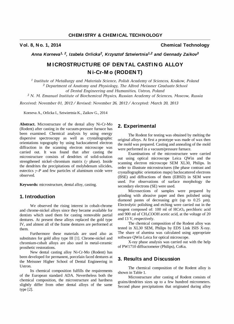
solidification as a result of a superfusion on growing dendrites boundaries are uniformly distributed inside the grains (Fig. 1a).
Table 1
The chemical composition of the Rodent alloy The content of elements, wt %
Ni Cr Mo Si Fe 62.7 ± 0.2 24.3 ± 0.1 10.2 ± 0.1 1.7 ± 0.1 1.1 ± 0.1
The matrix of the alloy is the solution of chromium
and molybdenum in nickel, crystallizes as FCC austenite (γ phase, Fig. 1b). EDS/SEM investigations have revealed that mainly precipitations of three phases are dispersed in the matrix. These are eutectics γ-P (shown in Fig. 1b as A), precipitations of molybdenum silicides (marked as B) and
precipitations of alumina (marked as C). Very few particles of alumina are evenly distributed in the matrix, and their global share was estimated at approximately of 0.1 %. The chemical composition of the matrix and the main precipitates of the Rodent alloy are shown in Table 2. In comparison with matrix the phase P contains 2.5 times more Mo, while the precipitations of molybdenum silicide contain 5 times more Mo, 4.4 times more Si and 1.9 times less Ni, 1.5 times less Cr as well as 4 times less Fe.
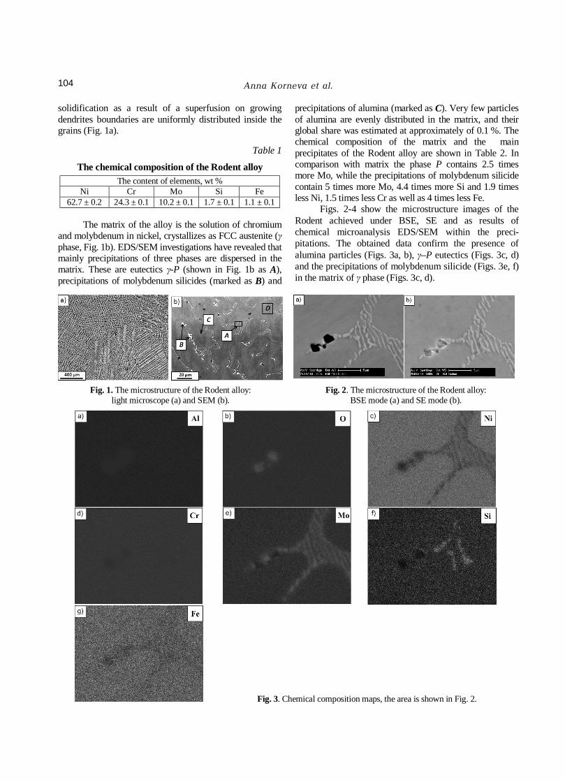
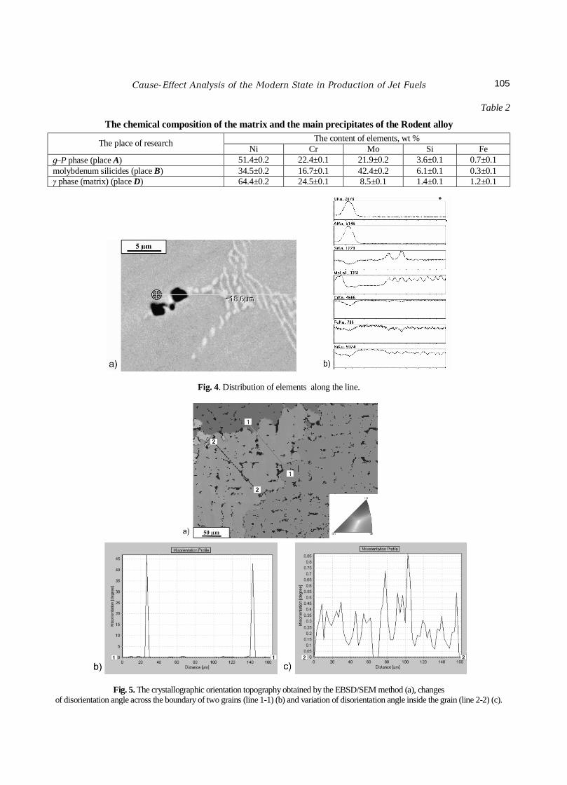
Figs. 2-4 show the microstructure images of the Rodent achieved under BSE, SE and as results of chemical microanalysis EDS/SEM within the preci-pitations. The obtained data confirm the presence of alumina particles (Figs. 3a, b), γ–P eutectics (Figs. 3c, d) and the precipitations of molybdenum silicide (Figs. 3e, f) in the matrix of γ phase (Figs. 3c, d).
Fig. 1. The microstructure of the Rodent alloy:
light microscope (a) and SEM (b). Fig. 2. The microstructure of the Rodent alloy:
BSE mode (a) and SE mode (b).
Fig. 3. Chemical composition maps, the area is shown in Fig. 2.
Cause-Effect Analysis of the Modern State in Production of Jet Fuels
105
Table 2
The chemical composition of the matrix and the main precipitates of the Rodent alloy The content of elements, wt % The place of research Ni Cr Mo Si Fe
γ–P phase (place A) 51.4±0.2 22.4±0.1 21.9±0.2 3.6±0.1 0.7±0.1 molybdenum silicides (place B) 34.5±0.2 16.7±0.1 42.4±0.2 6.1±0.1 0.3±0.1 γ phase (matrix) (place D) 64.4±0.2 24.5±0.1 8.5±0.1 1.4±0.1 1.2±0.1
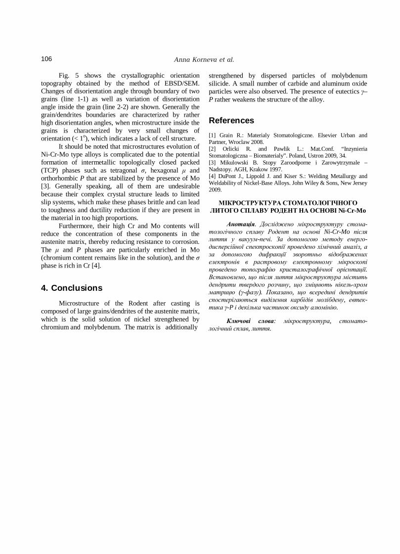
Fig. 4. Distribution of elements along the line.
Fig. 5. The crystallographic orientation topography obtained by the EBSD/SEM method (a), changes of disorientation angle across the boundary of two grains (line 1-1) (b) and variation of disorientation angle inside the grain (line 2-2) (c).
Anna Korneva et al.
106
Fig. 5 shows the crystallographic orientation topography obtained by the method of EBSD/SEM. Changes of disorientation angle through boundary of two grains (line 1-1) as well as variation of disorientation angle inside the grain (line 2-2) are shown. Generally the grain/dendrites boundaries are characterized by rather high disorientation angles, when microstructure inside the grains is characterized by very small changes of orientation (< 1o), which indicates a lack of cell structure.
It should be noted that microstructures evolution of Ni-Cr-Mo type alloys is complicated due to the potential formation of intermetallic topologically closed packed (TCP) phases such as tetragonal σ, hexagonal μ and orthorhombic P that are stabilized by the presence of Mo [3]. Generally speaking, all of them are undesirable because their complex crystal structure leads to limited slip systems, which make these phases brittle and can lead to toughness and ductility reduction if they are present in the material in too high proportions.
Furthermore, their high Cr and Mo contents will reduce the concentration of these components in the austenite matrix, thereby reducing resistance to corrosion. The μ and P phases are particularly enriched in Mo (chromium content remains like in the solution), and the σ phase is rich in Cr [4].
4. Conclusions
Microstructure of the Rodent after casting is composed of large grains/dendrites of the austenite matrix, which is the solid solution of nickel strengthened by chromium and molybdenum. The matrix is additionally
strengthened by dispersed particles of molybdenum silicide. A small number of carbide and aluminum oxide particles were also observed. The presence of eutectics γ–P rather weakens the structure of the alloy.
References
[1] Grain R.: Materialy Stomatologiczne. Elsevier Urban and Partner, Wroclaw 2008. [2] Orlicki R. and Pawlik L.: Mat.Conf. “Inzynieria Stomatologiczna – Biomaterialy”. Poland, Ustron 2009, 34. [3] Mikulowski B. Stopy Zaroodporne i Zarowytrzymale – Nadstopy. AGH, Krakow 1997. [4] DuPont J., Lippold J. and Kiser S.: Welding Metallurgy and Weldability of Nickel-Base Alloys. John Wiley & Sons, New Jersey 2009.
МІКРОСТРУКТУРА СТОМАТОЛОГІЧНОГО
ЛИТОГО СПЛАВУ РОДЕНТ НА ОСНОВІ Ni-Cr-Mo
Анотація. Досліджено мікроструктуру стома-тологічного сплаву Родент на основі Ni-Cr-Mo після лиття у вакуум-печі. За допомогою методу енерго-дисперсійної спектроскопії проведено хімічний аналіз, а за допомогою дифракції зворотньо відображених електронів в растровому електронному мікроскопі проведено топографію кристалографічної орієнтації. Встановлено, що після лиття мікроструктура містить дендрити твердого розчину, що зміцнють нікель-хром матрицю (γ-фазу). Показано, що всередині дендритів спостерігаються виділення карбідів молібдену, евтек-тика γ-P і декілька частинок оксиду алюмінію.
Ключові слова: мікроструктура, стомато-
логічний сплав, лиття.