Embed Size (px)
Citation preview
5/11/2015
1
UNIVERSITY OF PÉCSMEDICAL SCHOOL
www.medschool.pte.hu
BIOPHYSICS 2.2015 18th MarchDr. Beáta BugyiDepartment of Biophysics
MICROSCOPIC TECHNIQUES 1� LIGHT MICROSCOPY� FLUORESCENCE
MICROSCOPY
courtesy: see last slide
human lung tissue (histology) microsurgery
cell migration
(phase contrast microscopy)
mitosis
actin, microtubule
(confocal microscopy)
mitosis, starfish oocyte
actin, microtubule, chromosome
(3D confocal microscopy)
blood flow in living mouse
dextran, hepatocyte
(intravital microscopy)
individual molecules - formin, actin
(TIRFM)
� IMAGING TECHNIQUES
� MICROSCOPIC TECHNIQUES
• LIGHTLIGHTLIGHTLIGHT MICROSCOPYMICROSCOPYMICROSCOPYMICROSCOPY
» principles of image formation in the light microscope
� light-matter interaction: REFRACTION, DIFFRACTIONREFRACTION, DIFFRACTIONREFRACTION, DIFFRACTIONREFRACTION, DIFFRACTION
� MAGNIFICATION, RESOLUTION, CONTRASTMAGNIFICATION, RESOLUTION, CONTRASTMAGNIFICATION, RESOLUTION, CONTRASTMAGNIFICATION, RESOLUTION, CONTRAST
– FLUORESENCE MICROSCOPYFLUORESENCE MICROSCOPYFLUORESENCE MICROSCOPYFLUORESENCE MICROSCOPY
» special components in the fluorescence microscope
Overview Microscopy, light microscopy
� MICROSCOPYMICROSCOPYMICROSCOPYMICROSCOPY = MIKROS (small) + SZKOPEIN (to see)
- vizualize small objects that are „invisible” for the human eyes: magnifying device
- observe biological objects at different levels: from organs (cm 10-2m) to single molecules (nm 10-9m)
� SCANNINGSCANNINGSCANNINGSCANNING PROBEPROBEPROBEPROBE MICROSCOPYMICROSCOPYMICROSCOPYMICROSCOPY
� ELECTRONELECTRONELECTRONELECTRON MICROSCOPYMICROSCOPYMICROSCOPYMICROSCOPY
� LIGHT MICROSCOPYLIGHT MICROSCOPYLIGHT MICROSCOPYLIGHT MICROSCOPY
� Image formation is based on visible light (400 – 800 nm) and the use of glass lenses.
Principles of image formation in the light microscope
� MAGNIFICATION
� RESOLUTION
� CONTRAST
� …do all of the above while introducing as little
distortion as possible
Principles of image formation in the light microscope
� MAGNIFICATION
� small objects have to be large enough to see them by eyes
� RESOLUTION
� CONTRAST
� …do all of the above while introducing as little
distortion as possible
5/11/2015
2
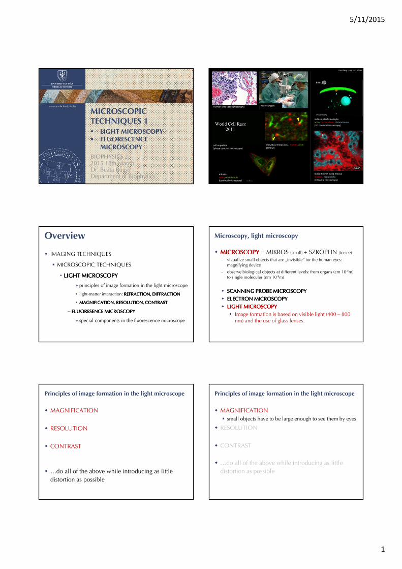
The simple magnifying glass, loupe1x magnification
von Leeuwenhoek
(1632-1723 Dutch
zoologist, microbiologist)
reading stone (~ 810-887
B.C. Abbas Ibn Firnas)
CONVERGINGCONVERGINGCONVERGINGCONVERGING LENSLENSLENSLENS
O I1
observer
MAGNIFIEDMAGNIFIEDMAGNIFIEDMAGNIFIED
real
inverted
Magnification in the „compound” microscope2x magnification
Hans & Zacharias Jansen
(~ 1590 Dutch spectacle-
maker)
OBJECTIVEOBJECTIVEOBJECTIVEOBJECTIVEconverging lens
close to the object
O I1I2
OCULAR, EYEPIECEOCULAR, EYEPIECEOCULAR, EYEPIECEOCULAR, EYEPIECEconverging lens
close to the observer
observer
MAGNIFIEDMAGNIFIEDMAGNIFIEDMAGNIFIED
real
inverted
MAGNIFIEDMAGNIFIEDMAGNIFIEDMAGNIFIED
virtual
inverted
Lens systems in a modern light microscope
OBJECTIVEOBJECTIVEOBJECTIVEOBJECTIVE
OCULAROCULAROCULAROCULAR
CONDENSORCONDENSORCONDENSORCONDENSORuniform illumination
Köhler
Magnification of the light microscope
OBJECTIVE lens: ���������~2.5 � 150
OCULAR lens: �������~10 � 25
�����������~20 � 1000
��������� �!�"�# $����%&�'%
!()%� &�'%��������� �!�"�# $
����%&�'%
!()%� &�'%
����������� $ ��������� ∗ ������������������ $ ��������� ∗ �������
http://www.olympusmicro.com/primer/virtual/magnifying/index.html
Lenses, lens systemsnumerical aperture
http://zeiss-campus.magnet.fsu.edu/tutorials/basics/oilimmersionrefractiveindex/indexflash.html
http://www.microscopyu.com/articles/formulas/formulasna.html
NUMERICALNUMERICALNUMERICALNUMERICAL APERTUREAPERTUREAPERTUREAPERTURE (NA(NA(NA(NA))))� light collecting ability of a lens (system)
� NA ↑ more light is captured
+, $ � ∗ &��-+, $ � ∗ &��- nnnn: refactive index of the medium between the lens
and the object
αααα: aperture angle, half-angle of the light cone
captured by the objective
If we had a lens with infinitely high magnification
could we see infinitely small things?
NO!
The wave nature of light has to be considered!
DIFFRACTION, INTERFERENCE(previously: EM waves, X-ray diffraction)
5/11/2015
3
Principles of image formation in the light microscope
� MAGNIFICATION
� RESOLUTION
� small details have to be distinguishable from each other
� CONTRAST
� …do all of the above while introducing as little
distortion as possible
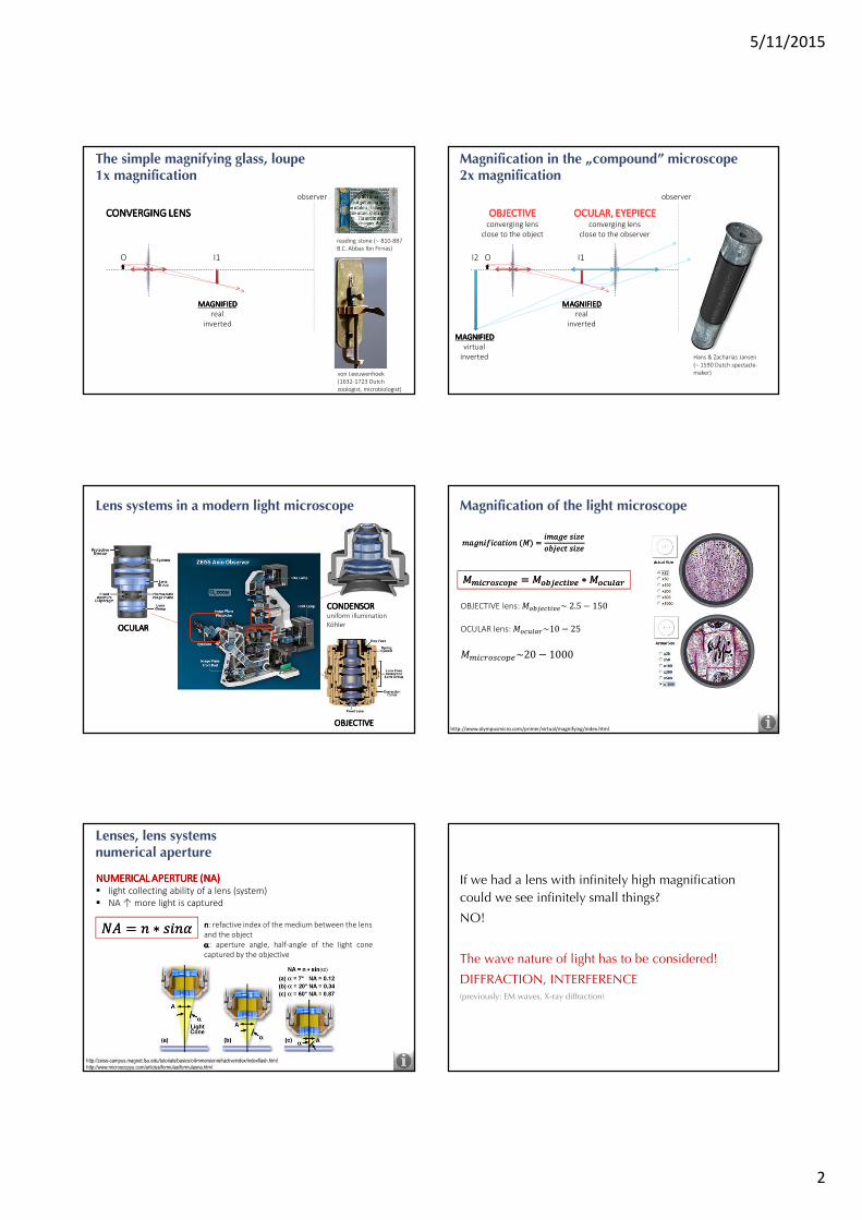
Resolwing power of the light microscope
RESOLUTIONRESOLUTIONRESOLUTIONRESOLUTION LIMITLIMITLIMITLIMIT
the shortest distance between two points of the object
that can be distinguished as separate entities on the image
(d)
Diffraction in the light microscope
SPREAD IN SPACE
lightlightlightlight sourcesourcesourcesource
OBJECTOBJECTOBJECTOBJECT
opticalopticalopticaloptical gratinggratinggratinggratingdrating constant: d
periodic optical properties
CONSTRUCTIVEmaximum - bright
DESTRUCTIVEminimum - dark
INFORMATION
http://zeiss-campus.magnet.fsu.edu/articles/basics/imageformation.html
INTERFERENCEINTERFERENCEINTERFERENCEINTERFERENCE
DIFFRACTIONDIFFRACTIONDIFFRACTIONDIFFRACTION
IMAGEIMAGEIMAGEIMAGE
intensityintensityintensityintensity distributiondistributiondistributiondistribution////diffractiondiffractiondiffractiondiffraction patternpatternpatternpattern
diffraction orders: . $ 0, 01, 02
-
1
∆& $ 1&��- $ .3
objective
back focal plane
Image as a diffraction patternJohn Herschel (1792-1871, English astronomer), George Biddell Airy (1801-1892, English astronomer)
AIRYAIRYAIRYAIRY PATTERNPATTERNPATTERNPATTERN: : : : diffraction limited image of a single point-like object (concentric dark/bright
fringes)
in 3D: PSF (POINT in 3D: PSF (POINT in 3D: PSF (POINT in 3D: PSF (POINT SPREAD SPREAD SPREAD SPREAD FUNCTION)FUNCTION)FUNCTION)FUNCTION)
Richard W Cole et al Measuring and interpreting point spread functions to determine confocal microscope resolution and ensure quality control Nature Protocols (2011)
XY direction – lateral
Z direction – axial
Abbe’s limit of resolutionErnst Abbe (1840-1905)
Image formation: besides the 0th order maximum at least the 1th order maximum
have to be captured.
. $ 0
. $ �1 . $ 41
Maxima are observed for angles (-):
∆& $ 1&��- $ .3. $ 0,01, 02
Participate in image formation:
for . $ 1:
1 $3
&��-5
Have to be captured:
1 $3
267869$
3
2:;
objective
-5 -5
Resolwing power of a light microscopediffraction limit
1<,= $ 0.613
+,~200��1<,= $ 0.61
3
+,~200��
1? $ 2�3
+, @~800��1? $ 2�
3
+, @~800��
λ: wavelenght of the illuminating light
NA: numerical aperture of the lens system
XY direction – lateral
Z direction – axial
Rayleigh’s criterion:
the central maximum of the diffraction
pattern of one point-source has to be
centered on the first minimum of the
diffraction pattern of the other point source
5/11/2015
4
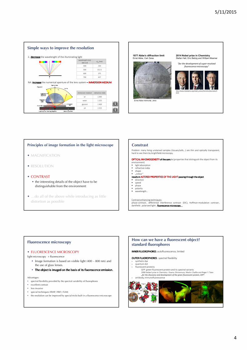
Simple ways to improve the resolution
λ: decreasedecreasedecreasedecrease the wavelenght of the illuminating light
NA: increaseincreaseincreaseincrease the numerical aperture of the lens system –––– IMMERSION MEDIUMIMMERSION MEDIUMIMMERSION MEDIUMIMMERSION MEDIUM
immersion medium refractive index
air 1.000
water 1.333
glycerol 1.469
oil 1.515
wavelength (nm)
NA= 0.8dx,y (nm)
400 250
500 312.5
600 375
700 437
2014 Nobel prize in ChemistryStefan Hell, Eric Betzig and William Moerner
"for the development of super-resolved
fluorescence microscopy"
1877 Abbe’s diffraction limitErnst Abbe, Carl Zeiss
Ernst Abbe memorial, Jena
http://www.nobelprize.org/nobel_prizes/chemistry/laureates/2
014/
Principles of image formation in the light microscope
� MAGNIFICATION
� RESOLUTION
� CONTRAST
� the interesting details of the object have to be
distinguishable from the environment
� …do all of the above while introducing as little
distortion as possible
ConstrastProblem: many living unstained samples (tissues/cells…) are thin and optically transparent,
hard to see them by brightfield microscopy.
OPTICAL INHOMOGENEITYOPTICAL INHOMOGENEITYOPTICAL INHOMOGENEITYOPTICAL INHOMOGENEITY of of of of the samthe samthe samthe sample (properties that distinguish the object from its
environment)
� light absorption
� refractive index
� shape
� „colour”
results in results in results in results in ALTERED PROPERTIES OF THE LIGHT ALTERED PROPERTIES OF THE LIGHT ALTERED PROPERTIES OF THE LIGHT ALTERED PROPERTIES OF THE LIGHT passing through the object passing through the object passing through the object passing through the object
� direction
� speed
� phase
� polarity
� wavelength…
Contrast enhancing techniques:
phase-contrast-, differential interference contrast- (DIC), Hoffman-modulation contrast-,
darkfield-, polarized light-, fluorescencefluorescencefluorescencefluorescence microscopymicroscopymicroscopymicroscopy,…
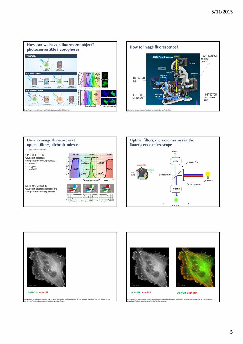
Fluorescence microscopy
� FLUORESCENCE MICROSCOPYlight microscopy + fluorescence
� Image formation is based on visible light (400 – 800 nm) and
the use of glass lenses.
� The The The The objectobjectobjectobject is is is is imagedimagedimagedimaged onononon thethethethe basisbasisbasisbasis of of of of itsitsitsits fluorescencefluorescencefluorescencefluorescence emissionemissionemissionemission....
Advantages:
� spectral flexibility provided by the spectral variability of fluorophores
� excellent contrast
� less invasive
� special techniques (FRAP, FRET, FLIM)
� the resolution can be improved by special tricks built in a fluorescence microscope
How can we have a fluorescent object?standard fluorophores
INNERINNERINNERINNER FLUOROPHORESFLUOROPHORESFLUOROPHORESFLUOROPHORES: autofluorescence, limited
OUTEROUTEROUTEROUTER FLUOROPHORESFLUOROPHORESFLUOROPHORESFLUOROPHORES : spectral flexibility� synthetic dye
� quantum dot
� fluorescent proteins
GFP: green fluorescent protein and its spectral variants2008 Noberl prize in Chemistry: Osamu Shimomura, Martin Chalfie and Roger Y. Tsien
„for the discovery and development of the green fluorescent protein, GFP".
� antibody, immunofluorescence
5/11/2015
5
How can we have a fluorescent object?photoconvertible fluorophores
http://www.olympusfluoview.com/applications/opticalhighlighters.html
STANDARDSTANDARDSTANDARDSTANDARD
PHOTOACTIVABLEPHOTOACTIVABLEPHOTOACTIVABLEPHOTOACTIVABLE
PHOTOSWITCHABLEPHOTOSWITCHABLEPHOTOSWITCHABLEPHOTOSWITCHABLE
How to image fluorescence?
http://zeiss-campus.magnet.fsu.edu/tutorials/axioobserver/index.html
LIGHT SOURCEarc lamp
LASER
DETECTORCCD camera
PMT
DETECTOReye
FILTERS
MIRRORS
sample
trans
epi
How to image fluorescence?optical filters, dichroic mirrors
OPTICAL FILTERSwavelength dependent
absorption/transmission properties
� shortpass
� longpass
� bandpass
DICHROIC MIRRORSwavelength dependent reflection and
absorption/transmission properties
(see: Flow cytometry)
Optical filters, dichroic mirrors in thefluorescence microscope
emission filter
excitation
filterdichroic
mirror
VASP-GFP actin-RFP
Shows again the localization of VASP at protruding lamellipodia and filopodia tips in a fish fibroblast expressing VASP-GFP and Actin-RFP.
forrás: http://cellix.imba.oeaw.ac.at/motility/nucleationfactors
VASP-GFP actin-RFP
Shows again the localization of VASP at protruding lamellipodia and filopodia tips in a fish fibroblast expressing VASP-GFP and Actin-RFP.
forrás: http://cellix.imba.oeaw.ac.at/motility/nucleationfactors
VASP-GFP actin-RFP
5/11/2015
6
www.aok.pte.hu
Light-, fluorescence microscopekeywords
� PrinciplesPrinciplesPrinciplesPrinciples of image of image of image of image formationformationformationformation in a in a in a in a lightlightlightlight microscopemicroscopemicroscopemicroscope::::
� refractionrefractionrefractionrefraction and and and and diffractiondiffractiondiffractiondiffraction
� magnificationmagnificationmagnificationmagnification, , , , resolutionresolutionresolutionresolution, , , , contrastcontrastcontrastcontrast
� Image Image Image Image formationformationformationformation bybybyby convergingconvergingconvergingconverging lenseslenseslenseslenses
� NumericalNumericalNumericalNumerical apertureapertureapertureaperture
� AiryAiryAiryAiry patternpatternpatternpattern, , , , diffractiondiffractiondiffractiondiffraction limit, limit, limit, limit, PSFPSFPSFPSF
� SpecialSpecialSpecialSpecial elementselementselementselements in a in a in a in a fluorescencefluorescencefluorescencefluorescence microscopemicroscopemicroscopemicroscope: : : : fluorophoresfluorophoresfluorophoresfluorophores, , , , lightlightlightlight
sourcessourcessourcessources, , , , detectorsdetectorsdetectorsdetectors, , , , opticalopticalopticaloptical filtersfiltersfiltersfilters, , , , mirrorsmirrorsmirrorsmirrors
www.aok.pte.hu
Recomended web resources
http://www.olympusmicro.com/index.html
http://www.microscopyu.com
/
http://zeiss-campus.magnet.fsu.edu/index.html
http://www.ibiology.org/

![Blind Deconvolution of Widefield Fluorescence Microscopic ... · eral deconvolution methods in widefield microscopy. In [3] several nonlinear deconvolution methods as the Lucy-Richardson](https://img.pdfslide.us/doc/110x75/5f6dfa53e2931769252d0293/blind-deconvolution-of-widefield-fluorescence-microscopic-eral-deconvolution.jpg)