Embed Size (px)
Citation preview
Microscopic Identification of Turfgrass Diseases
Alan Windham Frank Wong Brandon Horvath
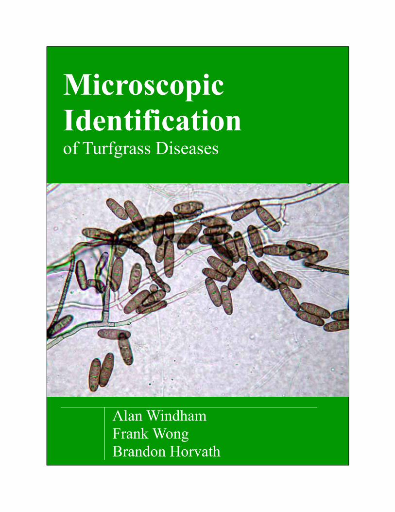
Microscopy 101 The use of a microscope can be fascinating or in some cases frustrating if you have lim-ited experience with microscopy. Ideally, if you wish to become proficient at identifying turf diseases, it’s best to have a dissecting microscope (6-40X) and a compound mi-croscope (40-400X). Each microscope is valuable and has particular strengths. Dissecting Microscope A dissecting microscope is quite useful in disease diagnosis. It’s a good place to start with a sample. In many cases you may put all or a portion of a plug from a green on the stage of the microscope and examine the foli-age, stolons and roots for the presence of fungal hyphae, fruiting bodies and, in some instances, masses of spores. A dissecting microscope is very useful to pinpoint foliage or fungal structures that can be transferred to a glass slide for examination with a com-pound microscope.
A dissecting microscope may also be used to scan plant material for mites and small insects. You can also observe galled roots infected with root knot nematodes. Other nematodes are difficult to observe without special prepara-tion of a soil or plant sample. Leave most nematode assays to the experts.
A Meiji trinocular dissecting microscope. The photo tube accommodates a camera for photo- graphy.
Mycelium of Sclerotium rolfsii as viewed with a dis-secting microscope (40X). Mycelium may be re-trieved with forceps or a probe and placed on a glass slide for examination with a compound microscope.
Runner hyphae(RH) and hyphopodia (H) of Gaeu-mannomyces graminis var graminis on a ultradwarf bermudagrass stolon (40X).
H
RH
2
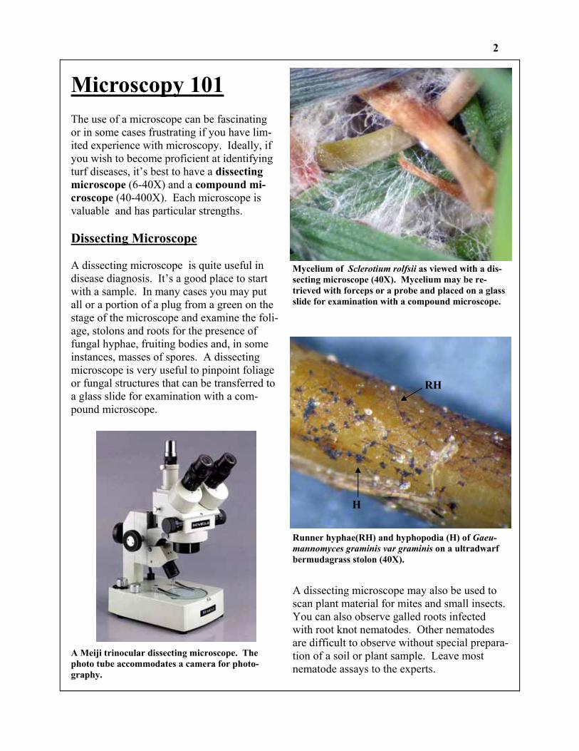
Dissecting Microscope
Photo tube (camera mount)
Eyepieces (oculars)
Zoom Control Knob
Objective Focusing Knob
Incident Illumina-tor Lamp Cover
Base with Transformer and Transmitted Illuminator
Illuminator Selector Switch
Rheostat
Clamp
Lever for switch-ing the image from one binocu-lar eyepiece to phototube for photography
Stage Clips
Stage Plate
Diopter Adjust-ment Ring
3
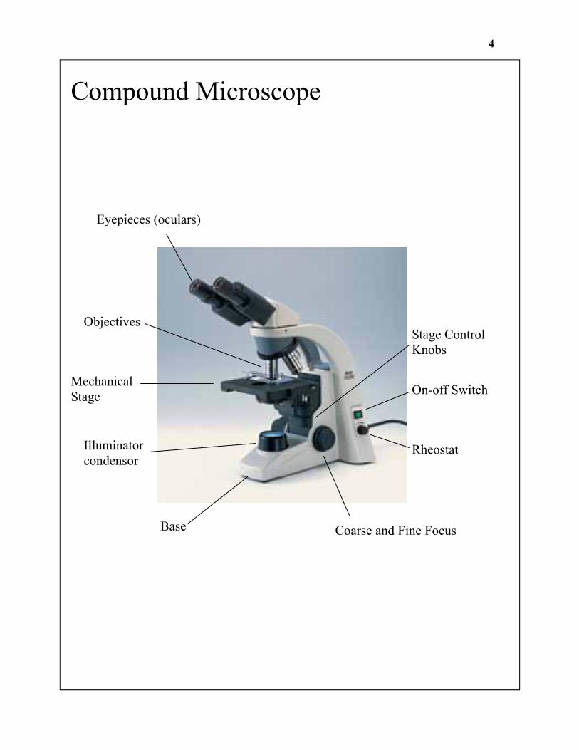
Eyepieces (oculars)
Mechanical Stage
Illuminator condensor
Coarse and Fine Focus
On-off Switch
Rheostat
Base
Objectives
Compound Microscope
Stage Control Knobs
4
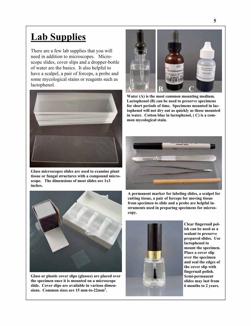
Lab Supplies There are a few lab supplies that you will need in addition to microscopes. Micro-scope slides, cover slips and a dropper-bottle of water are the basics. It also helpful to have a scalpel, a pair of forceps, a probe and some mycological stains or reagents such as lactophenol.
Glass microscopes slides are used to examine plant tissue or fungal structures with a compound micro-scope. The dimensions of most slides are 1x3 inches.
Glass or plastic cover slips (glasses) are placed over the specimen once it is mounted on a microscope slide. Cover slips are available in various dimen-sions. Common sizes are 15 mm-to-22mm2.
A B C Water (A) is the most common mounting medium. Lactophenol (B) can be used to preserve specimens for short periods of time. Specimens mounted in lac-tophenol will not dry out as quickly as those mounted in water. Cotton blue in lactophenol, ( C) is a com-mon mycological stain.
A permanent marker for labeling slides, a scalpel for cutting tissue, a pair of forceps for moving tissue from specimen to slide and a probe are helpful in-struments used in preparing specimens for micros-copy.
Clear fingernail pol-ish can be used as a sealant to preserve prepared slides. Use lactophenol to mount the specimen. Place a cover slip over the specimen and seal the edges of the cover slip with fingernail polish. Semi-permanent slides may last from 6 months to 2 years.
5
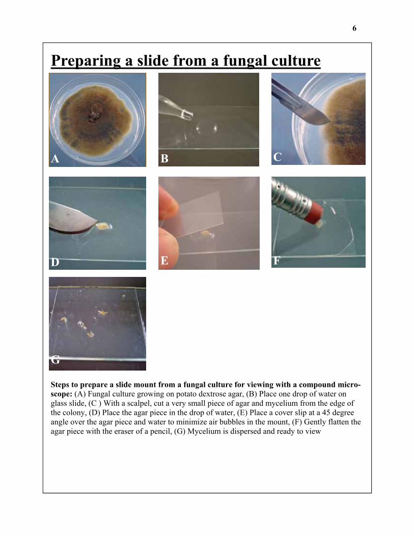
Preparing a slide from a fungal culture
A B C
D E F
G
Steps to prepare a slide mount from a fungal culture for viewing with a compound micro-scope: (A) Fungal culture growing on potato dextrose agar, (B) Place one drop of water on glass slide, (C ) With a scalpel, cut a very small piece of agar and mycelium from the edge of the colony, (D) Place the agar piece in the drop of water, (E) Place a cover slip at a 45 degree angle over the agar piece and water to minimize air bubbles in the mount, (F) Gently flatten the agar piece with the eraser of a pencil, (G) Mycelium is dispersed and ready to view
6

Preparing a slide from a grass plug
Steps to prepare a bentgrass specimen and view with a compound microscope: (A) bent-grass plug taken from the margin of a patch disease, (B) a portion of the plug from the sympto-matic area was collected and washed to remove sand (you may also prepare an unwashed speci-men), (C ) bentgrass plants are placed on a glass slide, (D) the slide is flooded with water, (E) cover slip is gently placed on plants, (F) slide is viewed first with the 4X and 10X objective, hyphae is observed along the edge of the leaf, (G) once fungal structures are observed, increase magnification to the 20X or 40X objective so that you may clearly see morphological character-istics which aid in identification of the fungus. In this case, the pathogen in Rhizoctonia which has right angle branching, septations and hypha that is slightly constricted where branching oc-curs.
A B C
D E
F G
7

Rhizoctonia Diseases: brown patch, large patch, yellow patch, leaf and sheath spot
Diagnostic tips: • Hyphae generally large diameter, usually
consistent diameter • Septate hyphae • Right angle branching of hyphae • Hyphae may be constricted near point of
branching • Identification by hyphal characteristics
only; no spores associated with Rhizocto-nia with only rare exceptions
• Start with a dissecting microscope and scan foliage of hyphae
• Remove foliage with hyphae and mount in water on a slide.
• Examine with 4 or 10X objective to find hyphae
• View at 20 or 40X to clearly see hyphal morphology
• Mycelium may be easier to see if leaf tis-sue is mounted in a stain such as cotton blue in lactophenol
Common species: Rhizoctonia solani Rhizoctonia cerealis Rhizoctonia zeae Binucleate Rhizoctonia-like fungi
Figures Top: Rhizoctonia hyphae in a water mount (400X) Middle: Rhizoctonia solani growing among bentgrass leaves (60X) Bottom: Brown patch on a creeping bentgrass green; smoke ring is visible at edge of patch
8

Sclerotinia Diseases: dollar spot
Diagnostic tips: • Septate hyphae • Hyphae vary in diameter • Mycelium white • Cytoplasm in hyphal cells may be grainy • Septations may be difficult to spot in water
mounts, but easily spotted after staining with cotton blue or aniline blue
• Mycelium is often observed on grass when dew is present
• If mycelium is not present incubate plug in a moist chamber for 12-48 hrs
• Observe hyphae in a water mount at 40, 100 and 400X
Common species: Sclerotinia homeocarpa
Figures Top: Sclerotinia hyphae mounted in water (400X) Middle: Sclerotinia hyphae mounted in aniline blue (400X) Bottom: Sclerotinia hyphae visible in a dollar spot patch on a creeping bentgrass green.
9
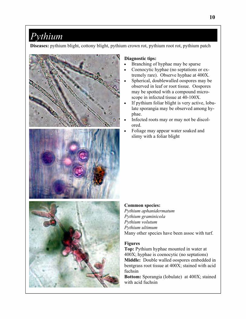
Pythium Diseases: pythium blight, cottony blight, pythium crown rot, pythium root rot, pythium patch
Diagnostic tips: • Branching of hyphae may be sparse • Coenocytic hyphae (no septations or ex-
tremely rare). Observe hyphae at 400X. • Spherical, doublewalled oospores may be
observed in leaf or root tissue. Oospores may be spotted with a compound micro-scope in infected tissue at 40-100X.
• If pythium foliar blight is very active, lobu-late sporangia may be observed among hy-phae.
• Infected roots may or may not be discol-ored.
• Foliage may appear water soaked and slimy with a foliar blight
Common species: Pythium aphanidermatum Pythium graminicola Pythium volutum Pythium ultimum Many other species have been assoc with turf.
Figures Top: Pythium hyphae mounted in water at 400X; hyphae is coenocytic (no septations) Middle: Double walled oospores embedded in bentgrass root tissue at 400X; stained with acid fuchsin Bottom: Sporangia (lobulate) at 400X; stained with acid fuchsin
10

Gaeumannomyces graminis var graminis Diseases: bermudagrass decline, take-all patch (St. Augustine), zoysia decline, root decline
Diagnostic tips: • Runner hyphae is darkly pigmented, sep-
tate and well branched • Hyphopodia are darkly pigmented, deeply
lobed • With a dissecting microscope look for run-
ner hyphae on stolons, rhizomes and roots (10-60X)
• With a scalpel remove a thin slice of the epidermis of the stolon containing runner hyphae and hyphopodia and mount in wa-ter; observe with a compound microscope at 40, 100 and 400X.
Common species: Gaeumannomyces graminis var graminis Gaeumannomyces graminis var avenae (take-all patch on bentgrass) Gaeumannomyces incrustans
Figures Top: Runner hyphae and hyphopodia of Ggg on a bermudagrass stolon (60X) Upper middle: Deeply lobed hyphopodia and runner hyphae of Ggg (400X) Lower middle: Lobed hyphopodia (1000X) Bottom: Root decline of bermudagrass
11

Colletotrichum cereale Disease: anthracnose (foliar blight, basal rot)
Diagnostic tips: • Observe diseased plants with a dissecting
scope at 10-60X to observe fruiting bodies (acervulii) with black, hair-like structures (setae)
• Infection mats (dark brown to black) may also be visible on the leaf sheath at 10-60X
• Wash specimen and observe leaf sheath, crown of plant and upper roots for appres-soria with a dissecting microscope 40-60X(dark gray, charcoal); appressoria anchor the fungus on plant tissue
• Mount leaf sheath tissue and/or whole plants in water and observe at 40-100X with a compound microscope
• Setae, appressora, infection mats and spores may be observed on some samples
• Spores (one celled, crescent shaped) are best viewed at 400X
Common species: Colletotrichum cereale Colletotrichum graminicola
Figures Top: Spores (conidia) 400X Upper middle: Fruiting bodies (acervulii) with black, hair-like setae (60X) Middle: Appresoria visible on bentgrass crown (40X) Lower middle: Appresoria 400X Lower: Infection mats on bentgrass leaf sheath tissue (60X)
12
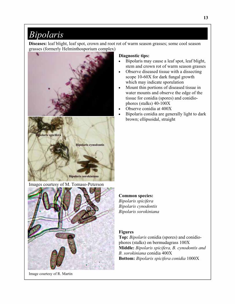
Bipolaris Diseases: leaf blight, leaf spot, crown and root rot of warm season grasses; some cool season grasses (formerly Helminthosporium complex) Diagnostic tips:
• Bipolaris may cause a leaf spot, leaf blight, stem and crown rot of warm season grasses
• Observe diseased tissue with a dissecting scope 10-60X for dark fungal growth which may indicate sporulation
• Mount thin portions of diseased tissue in water mounts and observe the edge of the tissue for conidia (spores) and conidio-phores (stalks) 40-100X
• Observe conidia at 400X • Bipolaris conidia are generally light to dark
brown; ellipsoidal, straight
Common species: Bipolaris spicifera Bipolaris cynodontis Bipolaris sorokiniana
Figures Top: Bipolaris conidia (spores) and conidio-phores (stalks) on bermudagrass 100X Middle: Bipolaris spicifera, B. cynodontis and B. sorokiniana conidia 400X Bottom: Bipolaris spicifera conidia 1000X
Images courtesy of M. Tomaso-Peterson
13
Image courtesy of R. Martin

Drechslera Diseases: leaf spot, leaf blight, crown rot; generally on cool season grasses
Diagnostic tips: • Usually found on cool season grasses • Sporulation may be difficult to observe on
lesions with a dissecting scope, but attempt at 40-60X
• Cut and remove lesions from leaf blades and mount in water
• Conidia (spores) may be observed adjacent to plant tissue at 40-100X
• Conidia are mostly tapered and have 4-9 septations
Common species: Drechslera nobleae Drechslera catenaria Drechslera dictyoides
Figures Top: Drechslera conidia Middle: Drechslera conidia attached to a co-nidiophore Bottom: Drechslera leaf spot on ryegrass
Images courtesy of Maria Tomaso-Peterson
14
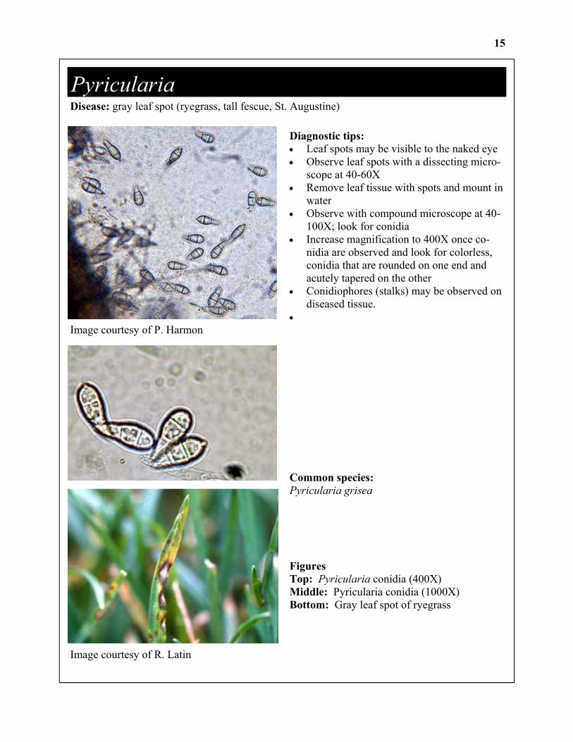
Pyricularia Disease: gray leaf spot (ryegrass, tall fescue, St. Augustine)
Image courtesy of P. Harmon
Diagnostic tips: • Leaf spots may be visible to the naked eye • Observe leaf spots with a dissecting micro-
scope at 40-60X • Remove leaf tissue with spots and mount in
water • Observe with compound microscope at 40-
100X; look for conidia • Increase magnification to 400X once co-
nidia are observed and look for colorless, conidia that are rounded on one end and acutely tapered on the other
• Conidiophores (stalks) may be observed on diseased tissue.
•
Common species: Pyricularia grisea
Figures Top: Pyricularia conidia (400X) Middle: Pyricularia conidia (1000X) Bottom: Gray leaf spot of ryegrass
Image courtesy of R. Latin
15
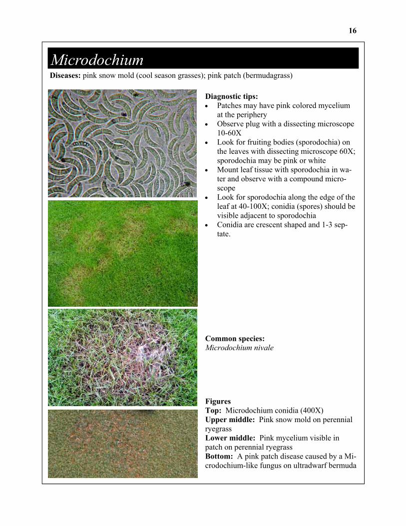
Microdochium Diseases: pink snow mold (cool season grasses); pink patch (bermudagrass)
Diagnostic tips: • Patches may have pink colored mycelium
at the periphery • Observe plug with a dissecting microscope
10-60X • Look for fruiting bodies (sporodochia) on
the leaves with dissecting microscope 60X; sporodochia may be pink or white
• Mount leaf tissue with sporodochia in wa-ter and observe with a compound micro-scope
• Look for sporodochia along the edge of the leaf at 40-100X; conidia (spores) should be visible adjacent to sporodochia
• Conidia are crescent shaped and 1-3 sep-tate.
Common species: Microdochium nivale
Figures Top: Microdochium conidia (400X) Upper middle: Pink snow mold on perennial ryegrass Lower middle: Pink mycelium visible in patch on perennial ryegrass Bottom: A pink patch disease caused by a Mi-crodochium-like fungus on ultradwarf bermuda
16
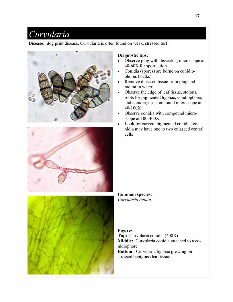
Curvularia Disease: dog print disease, Curvularia is often found on weak, stressed turf
Diagnostic tips: • Observe plug with dissecting microscope at
40-60X for sporulation • Conidia (spores) are borne on conidio-
phores (stalks) • Remove diseased tissue from plug and
mount in water • Observe the edge of leaf tissue, stolons,
roots for pigmented hyphae, condiophores and conidia; use compound microscope at 40-100X
• Observe conidia with compound micro-scope at 100-400X
• Look for curved, pigmented conidia; co-nidia may have one to two enlarged central cells
Common species: Curvularia lunata
Figures Top: Curvularia conidia (400X) Middle: Curvularia conidia attached to a co-nidiophore Bottom: Curvularia hyphae growing on stressed bentgrass leaf tissue
17