Embed Size (px)
Citation preview
MICROSCOPIC ANALYSIS OF OIL PALM (ELAEIS GUINEENSIS)
INFECTION BY GANODERMA BONINENSE
Emmanuel Albert Gorea
BSc. Biology
The University of Papua New Guinea
A thesis submitted for the degree of Master of Philosophy at
The University of Queensland in 2016
School of Agriculture and Food Sciences

i
Abstract
Oil Palm (Elaeis guineensis Jacq.) is an important agricultural commodity for many developing
countries in South East Asia including Papua New Guinea and the Solomon Islands. Currently basal
stem rot (BSR) disease caused by Ganoderma boninense is the major disease of economic
importance causing severe crop losses. BSR disease has been known for over 100 years however,
progress in disease control and management has been hampered by lack of understanding of
Ganoderma-oil palm interaction which is essential in developing better strategies to control BSR
incidence. To date, there are no effective control methods to manage BSR disease. The viable long
term alternative would be to breed for resistant germplasm. However, this strategy is dependent on
improved understanding of the factors associated with resistance/tolerance and susceptibility. These
include genetic, molecular, physiological and microscopic factors. Development of suitable
screening trials will also facilitate Ganoderma-oil palm interaction studies and to better screen
germplasm in breeding studies.
A reliable pathological test depends on well-characterised pathogen strains. For this reason a
Ganoderma isolate (3039) used routinely in nursery pathological testing by OPRA (Oil Palm
Research Association) was imported from PNG. However, this meant that under quarantine
conditions experimental design would be restricted. Thus additional Australian isolates were sought
for this study. Brackets of Ganoderma-like fungi were collected from around Brisbane and Cairns,
Queensland. All new Australian isolates were described based on bracket morphology, basidiospore
characteristics (where these were available), vegetative growth rates and sequencing of the ITS
regions. The two isolates RD1 and RD2 which were sampled from golden cane (Dypsis lutescens)
palms in Cairns, were found to have identical ITS sequences to the PNG isolate 3039 and a G.
boninense reference sequence, thus these three isolates were considered to be G. boninense. Both
RD1 and RD2 were tested for pathogenicity on oil palm. G. boninense was re-isolated from roots of
oil palm seedlings inoculated with RD1, thus indicating that it was pathogenic on oil palm. At this
stage G. boninense was unable to be re-isolated from RD2 inoculated oil palm seedlings.
PNG isolate 3039 was used for pathogenicity assays under controlled environmental conditions in
growth cabinets on oil palm seedlings. It was found to infect roots as early as two months post
inoculation, indicating that it followed a similar pattern of infection in a growth cabinet as in a
ii
nursery environment. G. boninense does not readily produce spores in artificial media, thus it is first
grown on a substrate which is then used as inoculum. Traditionally rubber wood blocks are used,
but since these are not readily available in Brisbane, various other substrates were tested. Of the
substrates tested the two best and most reliable proved to be potato dextrose agar and sorghum
grains. Thus inoculum delivery tests were carried out on oil palm seedlings under growth cabinet
conditions using agar plugs and sorghum grains colonised with 3039. BSR symptoms usually
appear 4 mpi in nursery inoculated plants. Microscopic examination under fluorescence microscopy
revealed that both inoculum delivery methods caused colonisation, however earlier infection by
3039 using sorghum grain inoculum as compared to using PDA plugs was observed. Sorghum grain
inoculations were found have better infection potential than agar plugs, possibly due to higher
Ganoderma biomass in the grains.
Bright field and fluorescence microscopy was used to follow Ganoderma infection of oil palm
seedlings. Microscopy revealed that Ganoderma appears to have a preference for colonising root
tissue as opposed to bole tissue, even though the bole was the sight of inoculation. Therefore
Ganoderma’s survival and spread in soil was tested, whereby PDA and sorghum grains colonised
with 3039 were placed in soil for two weeks and re-isolation from soil up to 2cm away from
original site was attempted. Live Ganoderma was not found away from inoculation site indicating
that this fungus does not survive away from a food source, whether that be a live host or artificial
medium. Hyphae were observed in the sub epidermal highly lignified corky cell layer found in
primary and lateral roots. Other plant tissues colonised by Ganoderma included lateral roots
emerging from primary roots, and the root-bole interface region and dead tissue areas associated
with roots and bole. The tissue regions where Ganoderma has been observed did not fluoresce
under fluorescence microscopy when stained with calcofluor white indicating that these were either
dead or so highly lignified that these could not be stained. Furthermore, this may also suggest that
initial establishment of Ganoderma in oil palm is more saprophytic than pathogenic. No specialized
hyphal structures were observed during this early infection study. This study describes for the first
time an account of early processes of Ganoderma colonisation and infection on oil palm seedlings.
iii
Declaration by author
This thesis is composed of my original work, and contains no material previously published or
written by another person except where due reference has been made in the text. I have clearly
stated the contribution by others to jointly-authored works that I have included in my thesis.
I have clearly stated the contribution of others to my thesis as a whole, including statistical
assistance, survey design, data analysis, significant technical procedures, professional editorial
advice, and any other original research work used or reported in my thesis. The content of my thesis
is the result of work I have carried out since the commencement of my research higher degree
candidature and does not include a substantial part of work that has been submitted to qualify for
the award of any other degree or diploma in any university or other tertiary institution. I have
clearly stated which parts of my thesis, if any, have been submitted to qualify for another award.
I acknowledge that an electronic copy of my thesis must be lodged with the University Library and,
subject to the policy and procedures of The University of Queensland, the thesis be made available
for research and study in accordance with the Copyright Act 1968 unless a period of embargo has
been approved by the Dean of the Graduate School.
I acknowledge that copyright of all material contained in my thesis resides with the copyright
holder(s) of that material. Where appropriate I have obtained copyright permission from the
copyright holder to reproduce material in this thesis.

iv
Publications during candidature
None.
Publications included in the thesis
None.
Contributions by others to the thesis
None.
Statement of parts of the thesis submitted to qualify for the
award of another degree
None.

v
Acknowledgements
I am extremely grateful for the excellent support, guidance and advice from my principal
supervisorDr Agnieszka Mudge and associate supervisor Professor Ian Godwin throughout my
journey. You both have been absolutely wonderful! I would also like to extend my gratitude as well
to Dr Elizabeth Aitken for her many helpful comments and suggestions during my candidature
milestones and Dr Harshi Gamage for her kind assistance, especially on the microscopy work.
The staff and students of the Godwin lab, I am grateful for all your assistance and our many
interesting and fun conversations. My gratitude to Mr Liu for the sorghum grains for my inoculation
work and Mr Kyle Lamont for your assistance with the vibratome and sectioning of my samples.
To the staff of the glass house, especially Ken and Daniel, I am very grateful for all your kind
assistance.
To my employer, Papua New Guinea Oil Palm Research Association, my gratitude for allowing me
to undertake this study full time. I would like to extend my gratitude to my superior Dr. Carmel
Pilotti for all your support and mentoring over the years.
A huge thank you to my mum, dad and siblings for your love and support throughout all these
years. Finally, I am grateful to my wife, Maria and our children – thank you for all your support and
most of all thank you for sharing this experience with me.
vi
Key words
Ganoderma boninense, Ganoderma-oil palm interaction, basal stem rot, light microscopy, oil palm
(Elaeis guineensis), pathogenicity
Australian and New Zealand Standard Research
Classifications (ANZSRC)
ANZSRC code: 060704 (Plant Pathology) 50%
ANZSRC code: 070308 (Crop and Pasture Protection) 50%
Fields of Research (FoR) Classification
FoR code: 0704 (Plant Pathology) 50%
FoR code: 0308 (Crop and Pasture Protection) 50%
vii
Table of Contents
Abstract ................................................................................................................................................ i
Declaration by author ....................................................................................................................... iii
Publications during candidature ..................................................................................................... iv
Publications included in the thesis ................................................................................................... iv
Contributions by others to the thesis............................................................................................... iv
Statement of parts of the thesis submitted to qualify for the award of another degree ............. iv
Acknowledgements............................................................................................................................. v
Key words .......................................................................................................................................... vi
Australian and New Zealand Standard Research Classifications (ANZSRC) ............................ vi
Fields of Research (FoR) Classification .......................................................................................... vi
Table of Contents ............................................................................................................................. vii
List of Figures ..................................................................................................................................... x
List of Tables ................................................................................................................................... xiii
Abbreviations .................................................................................................................................. xiv
Chapter 1 GENERAL INTRODUCTION ................................................................................... 1
1.1 OIL PALM INDUSTRY IN PAPUA NEW GUINEA ......................................................... 1
1.2 BASAL STEM ROT A MAJOR DISEASE OF OIL PALM ............................................... 1
1.3 AIMS OF STUDY................................................................................................................. 2
Chapter 2 LITERATURE REVIEW............................................................................................ 3
2.1 THE OIL PALM INDUSTRY .............................................................................................. 3
2.1.1 Origin and distribution ............................................................................................................................. 3
2.1.2 Cultivation and use ................................................................................................................................... 5
2.1.2.1 Seed preparation and germination ............................................................................................................ 5
2.1.2.2 Nursery and field planting ........................................................................................................................ 5
2.1.2.3 Palm oil products ...................................................................................................................................... 6
2.1.3 Development of oil palm trade ................................................................................................................. 6
2.1.4 Oil Palm industry in Papua New Guinea .................................................................................................. 7
2.1.5 Diseases of oil palm .................................................................................................................................. 7
2.1.5.1 Stem and root disease ............................................................................................................................... 7
2.1.5.2 Stem diseases ............................................................................................................................................ 9
2.2 BASAL STEM ROT OF OIL PALM ................................................................................. 10
2.2.1 Basal stem rot a major disease of oil palm ............................................................................................. 10
2.2.2 The causal pathogen ............................................................................................................................... 10
viii
2.2.3 Biology ................................................................................................................................................... 11
2.2.4 Infection and symptoms of BSR ............................................................................................................. 12
2.2.4.1 Field symptoms ...................................................................................................................................... 12
2.2.4.2 Internal symptoms .................................................................................................................................. 13
2.2.5 Mode of infection ................................................................................................................................... 14
2.2.6 Wood decay and rot diseases .................................................................................................................. 15
2.2.7 Control of basal stem rot ........................................................................................................................ 16
2.2.7.1 Control at new establishments and at replant ......................................................................................... 16
2.2.7.2 Control in established plantations ........................................................................................................... 17
2.2.7.3 Short term control strategies ................................................................................................................... 17
2.2.7.4 Long term control ................................................................................................................................... 18
2.2.8 Early detection ........................................................................................................................................ 19
2.2.9 Conclusion .............................................................................................................................................. 19
Chapter 3 CHARACTERISATION OF NEW ISOLATES OF GANODERMA SPP ........... 20
3.1 INTRODUCTION ............................................................................................................... 20
3.2 MATERIALS AND METHODS ........................................................................................ 21
3.2.1 Collection of Ganoderma samples ......................................................................................................... 21
3.2.2 Ganoderma selective media (GSM) ....................................................................................................... 22
3.2.3 Molecular identification using Internal Transcribed Spacer ITS1 .......................................................... 22
3.2.3.1 Fungal genomic DNA miniprep for PCR ............................................................................................... 22
3.2.3.2 Polymerase chain reaction (PCR) of internal transcribed spacers .......................................................... 23
3.2.3.3 Cleaning PCR product ............................................................................................................................ 23
3.2.3.4 Sequencing reactions .............................................................................................................................. 23
3.2.4 Morphological identification .................................................................................................................. 23
3.2.5 Bracket and basidiospore production ...................................................................................................... 24
3.2.6 Koch’s postulates.................................................................................................................................... 24
3.2.7 Ganoderma soil media test ..................................................................................................................... 24
3.3 RESULTS ............................................................................................................................ 26
3.3.1 Ganoderma morphology......................................................................................................................... 26
3.3.1.1 Basidiocarp and basidiospore morphology ............................................................................................. 26
3.3.1.2 Cultural characteristics ........................................................................................................................... 29
3.3.2 Bracket and spore production ................................................................................................................. 30
3.3.3 Sequencing ............................................................................................................................................. 31
3.3.4 Testing for Koch’s postulates ................................................................................................................. 33
3.3.5 Ganoderma soil media test ..................................................................................................................... 33
3.4 DISCUSSION ..................................................................................................................... 33
Chapter 4 NOVEL INOCULATION METHODS .................................................................... 37
4.1 INTRODUCTION ............................................................................................................... 37
4.2 MATERIALS AND METHODS ........................................................................................ 40
ix
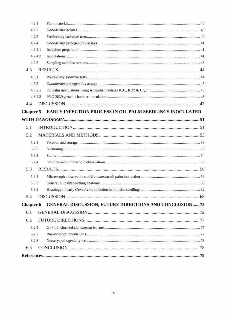
4.2.1 Plant material .......................................................................................................................................... 40
4.2.2 Ganoderma isolates ................................................................................................................................ 40
4.2.3 Preliminary substrate tests ...................................................................................................................... 40
4.2.4 Ganoderma pathogenicity assays ........................................................................................................... 41
4.2.4.1 Inoculum preparation .............................................................................................................................. 41
4.2.4.2 Inoculations ............................................................................................................................................ 41
4.2.5 Sampling and observations ..................................................................................................................... 43
4.3 RESULTS ............................................................................................................................ 44
4.3.1 Preliminary substrate tests ...................................................................................................................... 44
4.3.2 Ganoderma pathogenicity assays ........................................................................................................... 45
4.3.2.1 Oil palm inoculations using Australian isolates RD1, RD2 & UQ3 ....................................................... 45
4.3.2.2 PNG 3039 growth chamber inoculation ................................................................................................. 45
4.4 DISCUSSION ..................................................................................................................... 47
Chapter 5 EARLY INFECTION PROCESS IN OIL PALM SEEDLINGS INOCULATED
WITH GANODERMA...................................................................................................................... 51
5.1 INTRODUCTION ............................................................................................................... 51
5.2 MATERIALS AND METHODS ........................................................................................ 53
5.2.1 Fixation and storage ............................................................................................................................... 53
5.2.2 Sectioning ............................................................................................................................................... 53
5.2.3 Stains ...................................................................................................................................................... 54
5.2.4 Staining and microscopic observations .................................................................................................. 55
5.3 RESULTS ............................................................................................................................ 56
5.3.1 Microscopic observations of Ganoderma-oil palm interaction .............................................................. 56
5.3.2 General oil palm seedling anatomy ........................................................................................................ 58
5.3.3 Histology of early Ganoderma infection in oil palm seedlings .............................................................. 62
5.4 DISCUSSION ..................................................................................................................... 68
Chapter 6 GENERAL DISCUSSION, FUTURE DIRECTIONS AND CONCLUSION ...... 72
6.1 GENERAL DISCUSSION .................................................................................................. 72
6.2 FUTURE DIRECTIONS ..................................................................................................... 77
6.2.1 GFP transformed Ganoderma isolates .................................................................................................... 77
6.2.2 Basidiospore inoculations ....................................................................................................................... 77
6.2.3 Nursery pathogenicity tests .................................................................................................................... 78
6.3 CONCLUSION ................................................................................................................... 78
References ......................................................................................................................................... 79
x
List of Figures
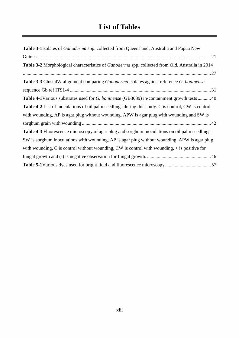
Figure 2-1Cross section of intraspecific fruit forms of oil palm (Elaeis guineensis) (Photo with
permission from Dr. A.Mudge)............................................................................................................ 4
Figure 2-2Upper stem rot (USR) showing fracture at point of infection (Photo by Dr. Carmel
Pilotti)................................................................................................................................................... 9
Figure 3-1Internal composition of Ganoderma basidiocarp showing (A) pileus, (B) context and (C)
tubes. .................................................................................................................................................. 22
Figure 3-2Ganoderma growth test in sterilised sand media. (A) sand inoculation with agar plug
colonised with GB3039, (B) sand inoculation with 1 g of sorghum grain colonised with GB3039
and (C) arrows showing sampling points one week after inoculations.............................................. 25
Figure 3-3Basidiospores of Ganoderma spp. collected from Qld, Australia showing spore shape
and size under bright field.(A) RD1 and (B) RD2 showing elongated and narrowly ellipsoid spores.
(C) UQ3 and (D) UQ4 showing truncated and ovoid basidiospores. ................................................ 27
Figure 3-4Basidiocarp collection from Queensland, Australia. (A) and (B) laccate species collected
from Cairns, Australia from golden cane host, (C) UQ3 laccate species sampled from ex-
litter/mulch, (D) UQ4 non-laccate species collected from ex-litter/mulch, (E) EG1 non-laccate
species sampled from hard wood spp. ............................................................................................... 28
Figure 3-5Ganoderma mycelia showing numerous clamp junctions (arrows) under bright field. (A)
RD1 and (B) RD2. ............................................................................................................................. 29
Figure 3-6Ganoderma growth rates after one week incubation at 28oC and room temperature. ..... 29
Figure 3-7Basidiocarp formation of Ganoderma isolate, RD1, growing on golden cane logs. ....... 30
Figure 3-8Neighbour joining tree using Jukes-Cantor genetic distance model. ............................... 32
Figure 4-1Wounded area (indicated by arrow) of a three month old seedling ready for inoculation
(A).(B) Three leaf stage seedling inoculated with Ganoderma infested sorghum grains. (C) Three
leaf stage seedling inoculated with agar plug colonised by Ganoderma. .......................................... 43
Figure 4-2Sorghum grains colonised by Ganoderma isolate RD1 (A), (B) PDA colonised with RD2
and (C) agar plugs colonised with PNG isolate GB3039 (1 cm dia.) ................................................ 44
Figure 5-1 Oil palm seedling showing root and bole tissue that were sampled for microscopy. (A)
Bole showing area of artificial injury with primary root below it. Areas which were sampled are
indicated by arrows. (B) Internal bole-root interface indicated by red circle showing regions of
interest for microscopic evaluation .................................................................................................... 53
xi
Figure 5-2 General anatomy of oil palm root and bole tissues. (A-C) Bright field micrographs of oil
palm bole (A) showing numerous vascular bundles (vb) embedded in ground parenchyma (p) tissue,
root-bole interface (B) showing vascular cylinder (vc) of root in bole tissue and later root (lr) and
(C) root tissue showing whole primary root section with emerging lateral root (lr), vascular cylinder
(vc), lacunae (l) also known as air spaces, (c) cortex and epidermis (ep). All sections were stained
with toluidine and observed at x5 magnification. (D-F) Macroscopic images of (D) sites of tissue
sampling showing bole (b), root-bole interface (rbi) and primary root (pr). ..................................... 59
Figure 5-3Cross-section of control OP primary root. Root cross-section of stained with calcofluor
white under UV fluorescence showing non-fluorescence of sclerenchyma (sc) ring of the outer
cortex (oc) (A) and under bright field stained with toluidine blue (B) showing unstained
sclerenchyma ring with tannins. (C) root tissue stained with safranin o with dark red regions
showing highly lignified sclerenchyma tissue. (D) Low magnification of whole root section stained
with calcofluor white ......................................................................................................................... 60
Figure 5-4 Cross-section of oil palm bole tissue. (A) x 5 magnification showing vascular bundles
embedded in ground parenchyma tissue. Staining with safranin showing highly lignified vascular
bundles and cells in red and ground parenchyma in pink. (B) Close up image of bole showing
lignified vascular fibres (vf) in red against ground parenchyma (p) in pink with abundant starch
granules (s). (C) Calcium oxalate Raphide crystals (r) were also found to be associated with the bole
tissue................................................................................................................................................... 61
Figure 5-5 Oil palm root epidermis showing controls and Ganoderma inoculated plants. All root
samples were stained with calcofluor white to visualise fungal hyphae in roots. (A) Oil palm root
epidermis and sclerenchyma ring tissue with multiple fungal hyphae (arrows). (B) High definition
image showing fungal hyphae fluorescing in root epidermis of Ganoderma inoculated oil palm. (C)
Control root showing no fungal hyphae in epidermis or non-fluorescent sclerenchyma ring. (D)
Root tissue showing fungal hyphae in epidermis/sclerenchyma ring region. .................................... 65
Figure 5-6 Ganoderma inoculated oil palm seedlings showing hyphal colonisation of lateral root
emerging from primary root. (A) Bright field image of whole root section stained with toluidine
blue showing lateral roots emerging from primary root with square indicating region were hyphae
were observed. (B) Same root tissue stained with calcofluor white showing non-fluorescent region
of lateral root (square) where hyphae were observed. (C) High magnification image of (B) showing
hyphae (arrows) in lateral root tissue. (D) Bright field image of root cross-section indicating lateral
root (square) region that showed hyphal structures with clamped connections under UV
fluorescence in image (D). Bright field image of primary root with lateral root (square) indicating
region of hyphal association (arrows) under UV fluorescence as shown for images (G) and (H). ... 65
xii
Figure 5-7 Bole and root-bole interface tissue of Ganoderma inoculated oil palm seedling showing
brown pigmented tissue under bright field and non-fluorescent under UV light. (A) Bright field and
(B) UV fluorescence image showing bole tissue region of hyphal association (square). (C) Root-
bole interface region under bright field showing brown pigmented tissue of lateral root that showed
hyphae (arrows) under fluorescence after staining with calcofluor white as shown in image (D). (E)
High magnification of image (A) showing fungal hyphae (arrow heads) within cells of the bole
tissue................................................................................................................................................... 66
Figure 5-8 Ganoderma inoculated oil palm seedlings showing root and root-bole interface region
and root tissue cell wall degradation. (A) Root-bole interface region showing degraded cell walls
with multiple pockets (arrows) in cell walls. (B) Cross-section of root from inoculated oil palm
showing non-fluorescent region (rectangle) of lesion. (C) Image (B) at x100 magnification showing
interface between decayed tissues of non-fluorescence (arrows) adjacent to thick fluorescent cells
that appear to be loosely arranged. (D) x100 magnification of (B) showing hyphae (arrows) inside
of brightly fluorescing cell. ................................................................................................................ 67
xiii
List of Tables
Table 3-1Isolates of Ganoderma spp. collected from Queensland, Australia and Papua New
Guinea. ............................................................................................................................................... 21
Table 3-2 Morphological characteristics of Ganoderma spp. collected from Qld, Australia in 2014
............................................................................................................................................................ 27
Table 3-3 ClustalW alignment comparing Ganoderma isolates against reference G. boninense
sequence Gb ref ITS1-4 ..................................................................................................................... 31
Table 4-1Various substrates used for G. boninense (GB3039) in-containment growth tests ........... 40
Table 4-2 List of inoculations of oil palm seedlings during this study. C is control, CW is control
with wounding, AP is agar plug without wounding, APW is agar plug with wounding and SW is
sorghum grain with wounding ........................................................................................................... 42
Table 4-3 Fluorescence microscopy of agar plug and sorghum inoculations on oil palm seedlings.
SW is sorghum inoculations with wounding, AP is agar plug without wounding, APW is agar plug
with wounding, C is control without wounding, CW is control with wounding, + is positive for
fungal growth and (-) is negative observation for fungal growth. ..................................................... 46
Table 5-1Various dyes used for bright field and fluorescence microscopy ...................................... 57
xiv
Abbreviations
BF Bright Field
BR Bud rot
BSR Basal Stem Rot
CS Cross section
CW Calcofluor White
DNA Deoxyribose Nucleic Acid
en endodermis
ex exodermis
GB Ganoderma boninense
GSM Ganoderma selective media
ITS Internal Transcribed Spacer
LM Light Microscopy
LSCM Laser scanning confocal microscope
mtDNA Mitochondrial Deoxyribose Nucleic Acid
PCR Polymerase Chain Reaction
PDA Potato Dextrose Agar
PDB Potato Dextrose Broth
PNG Papua New Guinea
PNG OPRA Papua New Guinea Oil Palm Research Association
RAMS Random Amplified Microsatellites
RAPD Random Amplified Polymorphic DNA
rDNA Ribosomal DNA
RFLPS Restriction Fragment Length Polymorphism
RSPO Roundtable for Sustainable Palm Oil
SE Asia South East Asia
SEM Scanning Electron Microscopy
SI Solomon Islands
TBO Toluidine Blue O
TEM Transmission Electron Microscopy
TS Transverse section
USR Upper Stem Rot
YEP Yeast Extract Peptone
1
Chapter 1 GENERAL INTRODUCTION
1.1 OIL PALM INDUSTRY IN PAPUA NEW GUINEA
Palm oil is an important export crop in Papua New Guinea and the industry is the second largest
employer after the public service (ITS Global, 2011). Palm oil exports have increased steadily in the
last decade and contribute almost 60% of all agricultural exports (ITS Global, 2011). The total area
currently under production is 144, 405 ha of which 40.6 % are small holder producers (PNG, Palm
Oil Council, 2013) who rely heavily on revenue generated from palm oil production
1.2 BASAL STEM ROT A MAJOR DISEASE OF OIL PALM
Basal Stem Rot (BSR) caused by Ganoderma boninense is a major threat to the oil palm industry in
South East Asia, including Papua New Guinea and the Solomon Islands (Pilotti et al. 2002; Rees et
al. 2007).Ganoderma species also affect other crops such as coconut, betel nut, tea and forest
species such as acacia grown in PNG and the Solomon Is (Lee et al. 2000; Rolph et al.
2000).Studies have shown (Turner, 1981; Corley & Tinker, 2003) that stem rot caused by
G.boninense cause considerable crop damage and subsequent yield loss in oil palm and is therefore
considered a disease of economic importance in Malaysia and Indonesia (Corley & Tinker, 2003).
However, while disease incidences remain relatively low at this point in PNG and the Solomon Is. it
is more likely to follow the same trend as predicted by Turner (1981) for Malaysia and Indonesia
where BSR incidences as high as 80% have been recorded. Factors that contributed to greater
disease severity in Indonesia and Malaysia are; (a) Initial plantings in Indonesia and Malaysia were
mostly deli dura which were found to be more susceptible to BSR infections than the hybrid tenera
(deli dura x pisifera) plantings which are widely planted in PNG, (b) as predicted by Turner (1981),
disease severity increases with each replanting cycle. Plantations in Indonesia and Malaysia have
gone through several re-planting cycles (>7) and as such have contributed to a build-up of inoculum
over time. In addition, the lack of appropriate control measures (rogueing) throughout each
cropping cycle has aggravated the situation. In PNG, the situation is less severe as most of the
plantations are in their second replant
The current control strategies of basal stem rot are focused on long term and short term control by
keeping disease incidence below the economical thresh hold of 20% of infected palms (Turner,
1981). Short term control strategies include the routine roguing of infected stands, fungicide
intervention and use of bio control agents whilst long term strategies include breeding (screening)

2
for resistance. Both have been hampered by a lack of understanding of disease mechanisms and the
host-pathogen interactions, which are associated with factors of pathogenicity, resistance, tolerance
and susceptibility.
An understanding of the Ganoderma-oil palm interaction is essential in developing strategies to
control BSR infections. As there is very little knowledge in this area, progress could be made on a
number of fronts. The use of histological studies will help with better understanding of disease
mechanisms and elucidating the key routes of infection and host responses. Combining microscopic
analysis with other methods, e.g.: transcriptome analysis of the infection processes will shed light
on the various mechanisms involved in this particular host-pathogen interaction.
Control and management of BSR infections have been hampered by lack of understanding in
Ganoderma-oil palm interactions and as a result current control methods based on our limited
understanding are inadequate in the successful management of BSR. Currently there are still a
number of gaps in our knowledge regarding Ganoderma-oil palm interactions. For example, what
are the various stages in the establishment and development of BSR in oil palm? Is wounding of
roots or stem tissue a pre-requisite for infection? Does Ganoderma produce special structures (e.g.
appressoria)? Which tissues are preferentially colonised in the early stages of infection? Are there
any discernible differences between susceptible, resistant or tolerant oil palm seedlings? It has been
reported by Chan et al. (2011) in nursery pathogenicity tests that only dikaryons are pathogenic in
contrast to monokaryons. However, it is unknown if monokaryons are able to elicit a defense
response from the oil palm host.
1.3 AIMS OF STUDY
The objectives of the present study are to:
1. Determine the conditions necessary to initiate infection of oil palm seedlings by Ganoderma
boninense.
2. Assess novel inoculation methods for disease screening.
3. Increase our understanding of the mechanism(s) of infection by Ganoderma boninense in oil
palm seedlings and study host response at a microscopic level.

3
Chapter 2 LITERATURE REVIEW
2.1 THE OIL PALM INDUSTRY
The oil palm (Elaeis guineensis Jacq) is one of the world’s most important sources of vegetable oils
among other primary oil crops such as soya bean, rape seed and sun flower. Worldwide production
of oil palm constitutes 33% of all vegetable oils yet only covers 5% of the total world area planted
(Singh et al., 2013), thus making oil palm the highest yielding oil crop per ha (Corley & Tinker,
2003). Oil palm cultivation is estimated to cover 18 million ha in total world area planted with an
estimated value of US $ 33 billion (FAOSTAT, 2015).
2.1.1 Origin and distribution
The oil palm is native to Africa were historical evidence of its origins is supported by oil palm
pollen retrieved from two deep core samples in Congo dating to the period since 24, 000 years BP
(Elenga et al. 1994: as cited in Corley & Tinker, 2003). Its botanical name, Elaeis guineensis, was
given by Jacquin in 1763 as an attribute to its high oil content and origin - elaion, meaning oil in
Greek and guineensis, from Guinea coast of West Africa, (Corley and Tinker, 2003). The oil palm
is a monocotyledon which belongs to the family Aracaceae (Palmae) and subfamily cocosideae that
also includes coconut. Three species are recognized, Elaeis guineensis, E. oleifera, and E. odora
(Hartley, 1988) of which E. oleifera and E. odora are of South American origin (Corley & Tinker,
2003). The African oil palm, E. guineensis, is most widely exploited commercially in contrast to
E.odora that exists only in wild groves in Latin America. E.oleifera is a parent of the O x E hybrid
which is widely planted in South America. E. oleifera is also known to be resistant to sudden
wither, fatal yellowing diseases (Corley & Tinker, 2003) in South America and is thought to
possibly hold sources of genetic resistance to basal stem rot (BSR) caused by Ganoderma spp. and
is therefore deemed important in breeding trials (Durand-Gasselin et al. 2005).
E. guineensis has three distinct fruit forms (Fig.2.1) that are characterized by the thickness of the
shell surrounding the seed which is controlled by a single locus.
a) dura: thick-shelled
b) tenera: medium or thin-shelled, hybrid of dura x pisifera
c) pisifera: shell-less
In commercial plantations yield (bunch numbers and bunch weight) and palm oil (CPO & KPO)
extraction rates are important in oil palm production. Initial cultivation of oil palm used dura fruit
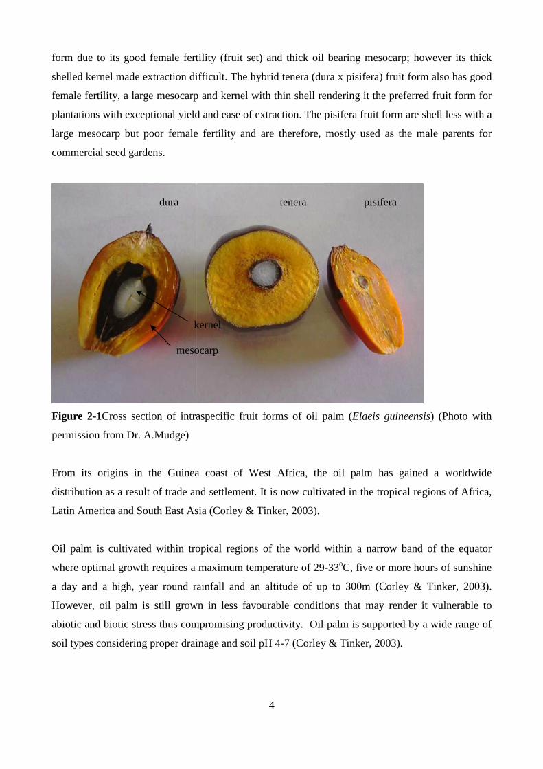
form due to its good female fertility (fruit set) and thick oil bearing mesocarp; however its thick
shelled kernel made extraction difficult. The h
female fertility, a large mesocarp and kernel with thin shell rendering it the preferred fruit form for
plantations with exceptional yield and ease of extraction. The pisifera fruit form are shell les
large mesocarp but poor female fertility and are therefore, mostly used as the male parents for
commercial seed gardens.
Figure 2-1Cross section of intraspecific fruit forms of oil palm (
permission from Dr. A.Mudge)
From its origins in the Guinea coast of West Africa, the oil palm has gained a worldwide
distribution as a result of trade and settlement. It is
Latin America and South East Asia (Corley & Tinker, 2003).
Oil palm is cultivated within tropical regions of the world within a narrow band of the equator
where optimal growth requires a maximum temperatur
a day and a high, year round rainfall and an altitude of up to 300m (Corley &
However, oil palm is still grown in less
abiotic and biotic stress thus compromising productivity. Oil palm is supported by a wide range of
soil types considering proper drainage and soil pH 4
dura
mesocarp
kernel
4
form due to its good female fertility (fruit set) and thick oil bearing mesocarp; however its thick
shelled kernel made extraction difficult. The hybrid tenera (dura x pisifera) fruit form also has good
female fertility, a large mesocarp and kernel with thin shell rendering it the preferred fruit form for
plantations with exceptional yield and ease of extraction. The pisifera fruit form are shell les
large mesocarp but poor female fertility and are therefore, mostly used as the male parents for
section of intraspecific fruit forms of oil palm (Elaeis guineensis
From its origins in the Guinea coast of West Africa, the oil palm has gained a worldwide
distribution as a result of trade and settlement. It is now cultivated in the tropical regions of Africa,
Latin America and South East Asia (Corley & Tinker, 2003).
Oil palm is cultivated within tropical regions of the world within a narrow band of the equator
where optimal growth requires a maximum temperature of 29-33oC, five or more hours of sunshine
a day and a high, year round rainfall and an altitude of up to 300m (Corley &
However, oil palm is still grown in less favourable conditions that may render it vulnerable to
ess thus compromising productivity. Oil palm is supported by a wide range of
soil types considering proper drainage and soil pH 4-7 (Corley & Tinker, 2003).
tenera pisifera
mesocarp
kernel
form due to its good female fertility (fruit set) and thick oil bearing mesocarp; however its thick
ybrid tenera (dura x pisifera) fruit form also has good
female fertility, a large mesocarp and kernel with thin shell rendering it the preferred fruit form for
plantations with exceptional yield and ease of extraction. The pisifera fruit form are shell less with a
large mesocarp but poor female fertility and are therefore, mostly used as the male parents for
Elaeis guineensis) (Photo with
From its origins in the Guinea coast of West Africa, the oil palm has gained a worldwide
now cultivated in the tropical regions of Africa,
Oil palm is cultivated within tropical regions of the world within a narrow band of the equator
C, five or more hours of sunshine
a day and a high, year round rainfall and an altitude of up to 300m (Corley & Tinker, 2003).
conditions that may render it vulnerable to
ess thus compromising productivity. Oil palm is supported by a wide range of
7 (Corley & Tinker, 2003).
pisifera
5
2.1.2 Cultivation and use
The initial establishment of plantations throughout South East Asia used extensively the Deli (dura)
oil palm line derived from four palms in Bogor Botanical gardens, Indonesia, in 1848. Similarities
within progenies assume that these four palms may have originated from a single parent palm
(Corley & Tinker, 2003) and as such create a genetic bottle neck effect within the Deli palms.
Gurand-Gasselin et al. (2005) observed that Deli lines were more susceptible to BSR infections than
material from Africa. Current planting material within plantations is F1 tenera hybrids and breeding
programs have incorporated new material from Africa to encourage genetic diversity.
2.1.2.1 Seed preparation and germination
Seeds are prepared from commercial seed gardens by firstly removing the fruits by placing the
harvested bunches into gunny sacks for several days to naturally loosen the fruits (Corley & Tinker,
2003). The mesocarp is mechanically de-pulped in a revolving cage fitted with scarifying screens
for 30-40 minutes. Finally, workers sort for poorly cleaned and damaged seeds and only batches
that have been properly de-pulped are used for germination (Turner, 1981).
The dry heat method is widely used in commercial seed production where germination is achieved
in approximately 80 days with a success rate of around 90 percent. The success of germination rates
are attributed to the use of dry heat that reduce early germination and disease. Firstly, moisture
content of the seeds is reduced from 22% to an average 17% on dry matter by drying in an oven at
40oC for two days. The seeds are then placed in heavy-gauged polythene bags and stored in an
insulated chamber at 38oC to 40oC for an additional 40 days. After the heat treatment, the moisture
content is adjusted to 22% on dry matter by soaking the seeds for two days in water followed by
storage at ambient temperature in polythene bags. Germination commences after 7-10 days and
seeds can be planted 10-14 days after germination (Turner, 1981).
2.1.2.2 Nursery and field planting
The establishment of new oil palm plantings require a constant supply of healthy and uniform seeds.
The most common plantation set up requires a pre-nursery and main nursery with a standard final
seedling out-turn of ≥ 70 % (Rankie & Fairhurst, 1999). Pre-germinated seeds are maintained in a
pre-nursery for 10-14 weeks and transplanted to the main nursery to develop for an additional 10-14
months prior to field planting. Palms are planted in densities ranging from 128 palms/ha to 145
palms /ha depending on climatic conditions (Rankie & Fairhurst, 1999).
Harvesting of fresh fruit bunches (FFB) is carried out manually by removal of lower fronds to
access the bunch peduncle which is severed with a sharp sequel attached to a harvesting pole up to
6
20 m in height depending on the age of the palms. Harvest begins round two years after planting
and is typically carried out every ten to six days for peak fruiting production during rainy seasons.
The size and weight of the fresh fruit bunches increase with maturity of the palms up to 60kg per
bunch (PNGOPRA). In a well maintained block palms begin yielding fruit bunches two years after
planting with an average fresh bunch (FFB) weight of 5 kg and peak crop production of 35 kg at 6
years after planting and slowly tapering off nearing replant (Dr. M.Barnabas, personal
communication,). The normal cropping cycle of oil palm is 20-25 years before the next replant and
extended periods are not usually encouraged because palms become too tall (≥ 20m) and thus
difficult to manage. Furthermore, high BSR incidences associated with senescing palms or earlier
infections have resulted in both plantations and small holders going into early replants as it is often
economically necessary.
2.1.2.3 Palm oil products
Oil palm is cultivated for two primary products. These are derived from the mesocarp of the fruit,
yielding crude palm oil (CPO) and the seed or kernel, yielding kernel palm oil (KPO). These are the
main products of international trade. A spent product, palm kernel cake, high in protein is used as
feed for livestock such as cattle. In addition, bi-product uses of oil palm include spent bunches as
mulch for immature plantings and electricity production utilizing methane bio-gas from mill refuse
(Corley & Tinker, 2003; NBPOL, 2013).
2.1.3 Development of oil palm trade
European discovery of oil palm was made by Portuguese sailors along the Guinea coast of West
Africa during explorations and trade in 1434. Early trade of palm oil was associated with the West
African slave trade in 1562 where oil palm was traded as food for slaves until abolishment of the
slave trade in the early 1800s. During this period palm oil trade remained domestic due to large
local consumption where palm oil was used for cooking and palm wine production and a small
surplus was exported to Europe. It was not until the 1800s that international trade of palm oil
become established due to two main factors; firstly, due to economic development and industrial
revolution in Europe where new inventions for palm oil use such as the manufacture of margarines,
soaps, candles, lubricants for machinery and industrial processes saw increased demand; and
secondly, the abolition of the slave trade that prompted an alternative trade in timber, ivory and
palm oil (Corley & Tinker, 2003). The increasing demand in palm oil saw a shift from the
exploitation of semi-wild groves to the establishment of plantations and several mills in Congo that
also took fruits from small holders and local farmers. However, production in Africa declined after

7
World War II and new establishments were made in the Far East with the first plantations in
Indonesia in 1911 and Malaysia in 1917 (Corley & Tinker, 2003). Since then the oil palm industry
has grown rapidly in the twentieth century and is now established world-wide as a crop of economic
importance for many developing countries in the major regions of Africa, South East Asia and Latin
America where South East Asia is the largest palm oil producer accounting for 90% total world
production (FAOSTAT, 2015).
2.1.4 Oil Palm industry in Papua New Guinea
The development of oil palm cultivation in Papua New Guinea began as early as the 1920s (Grieve,
1986). However, it was not until the late 1960s that the first commercial plantations were
established in Hoskins, West New Britain, at the advice of the World Bank to diversify the
agricultural economy of Papua New Guinea (Grieve, 1986). The success of this scheme saw further
developments in Northern Province in the early 1970s followed by Milne Bay and New Ireland
Provinces in 1985 and 1988 respectively.
The oil palm industry in PNG is the second largest employer after the public service and contributes
significantly both socially and economically. Palm oil exports have grown steadily in the last
decade exceeding copra, coco, tea, coffee and rubber comprising nearly 60% of all agricultural
exports (ITS Global, 2011). The oil palm industry is comprised of commercial plantations owned
and run by milling companies and small- holder blocks owned by individual growers who supply
the mills with fresh fruit bunches. The total area under oil palm cultivation currently is 144, 405 ha
of which small holders constitute 58, 408 Ha (Palm Oil Council, 2013).
2.1.5 Diseases of oil palm
Oil palm is cultivated in various countries along the equatorial regions of Africa, South and Central
America and South East Asia. The exposure to a wide range of soil types and environmental
conditions predisposes oil palm to a range of pest, diseases and disorders that can affect seeds,
seedlings and matured palms. However, only diseases of economic importance within major oil
palm cultivation regions will be discussed here and further details of pests and disorders can be
found in Turner, (1981); and Corley and Tinker (2003).
2.1.5.1 Stem and root disease
Several countries in West Africa have been devastated by vascular wilt disease caused by Fusarium
oxysporum f.sp. elaeidisis (Turner, 1981; Corley & Tinker, 2003; Flood, 2006). The soil borne
pathogen produces microspores, macrospores and chlamydospores in soil which, upon germination
enter through the roots and develop in xylem vessels causing the production of tyloses, gums and
8
gels that restrict water uptake leading to acute or chronic wilting. In addition, vascular wilt is
characterized internally by browning of xylem vessels (Flood, 2006; Cooper et al. 2011). Disease
incidences of 25% have been recorded in Zaire (Turner, 1981) and as high as 50% of 10 year old
palms after replant have been affected (Corley & Tinker, 2003) making vascular wilt disease an
important disease of oil palm in Africa. Furthermore, localized outbreaks have been recorded in
Ecuador and Brazil however; there have been no reports of its occurrences in South East Asia
(Flood, 2006).
Root and stem rot caused by the fungus Armillaria mellea have been reported in Colombia and
Ghana however, severe outbreaks of economic importance have been observed in Zaire only.
Disease symptoms exhibit premature falling away of stem butts close to the base of the palm
followed by subsequent foliar wilting associated with generalized rapid decay of internal tissue of
base stem (Turner, 1981).
Dry basal rot caused by Ceratocystis paradoxa (Anamorph: Thielaviopsis paradoxa) is a disease
which in the 1960s became epidemic in several localities within West Africa (Corley & Tinker,
2003) causing severe crop loss with incidences as high as 70% affecting palms 4-8 years of age.
Palms that recovered from the disease had abnormalities in trunk and foliage growth and yield
production were compromised for several years (Turner, 1981). Furthermore, deaths were common
in the first epidemic, however, recovery became more common and further serious out breaks have
not been reported (Corley & Tinker, 2003). Earlier reports have suggested that the imperfect stage
of C.paradoxa, T.paradoxa, is associated with bud rot disease in northern Colombia (Corley &
Tinker, 2003; Alvarez et al. 2012). However, recent reports (Torres et al. 2016) indicate the
Phytophthora palmivora is the causal agent of bud rot disease.
Severe losses due to sudden wither disease had been recorded in South America between 1963 and
1976 were mortality of 15 – 20 % were recorded in young (2-3 years old) palms with progressive
losses of 90% in 12 year old palms (Turner, 1981). The disease is possibly caused by a protozoan
flagellate, Phytomonas staheli (Corley & Tinker, 2003). It is known that the native E.oleifera
species is resistant to sudden wither disease (Turner, 1981).

9
2.1.5.2 Stem diseases
Stem rot occurring at 2 m above ground level and further up are regarded as upper stem rot (USR)
disease and the causal agent was earlier established as Phellinus noxious which had been reported in
Peninsular Malaysia, Sabah, Indonesia and Papua New Guinea (Turner, 1981). However, later
investigations have identified Ganoderma boninense as the causal agent associated with upper stem
rots in plantations (Pilotti, 2005). The symptoms of USR infection is often not noticeable until
collapse of the palm at the point of infection is seen leaving behind a stump as seen in Figure 2.2
The incidences of USR in plantations is low and therefore, regarded as of no economic significance
except in commercial seed production where loss of a single palm can be significant (Turner, 1981).
However, anecdotal evidence suggests that USR incidence is on the rise in parts of South East Asia.
Figure 2-2Upper stem rot (USR) showing fracture at point of infection (Photo by Dr. Carmel Pilotti)

10
2.2 BASAL STEM ROT OF OIL PALM
2.2.1 Basal stem rot a major disease of oil palm
Basal stem rot was first described in the Republic of Congo, West Africa in 1915 as a disease of
senescing palms (Wakefield, 1920; cited in Ariffin et al. 2000) with similar occurrences in Malaysia
(Thompson, 1931). BSR infections became of economic significance in the 1960s occurring on
younger palms (10-15 years) after replants from oil palm and coconut (Turner, 1981). Later, earlier
infections were seen on palms as early as 12-24 months especially after replant (Singh, 1990) or
when under planted with coconuts with increased incidences in 4-6 year old plantings (Ariffin et al.
1996). Since then BSR of oil palm has emerged as the most devastating disease of oil palm in South
East Asia. Disease incidences have been observed to increase with each planting cycle with earlier
infections of young oil palms. However, this may be associated with other factors such as type of
genetic material planted, type of soils and cultural practices rather than direct links to previous
crops (Turner, 1981; Durand-Gasselin et al. 2005).
Basal stem rot (BSR) caused by G. boninense is a major disease of economic importance to oil palm
production in South East Asia including Papua New Guinea (PNG) and Solomon Islands (Hushirian
et al. 2013; Pilotti et al. 2002; Rees et al. 2007). G. boninense and related species are white rot fungi
that decompose lignin as well as cellulose and other related polysaccharides (Rees, 2006). BSR
infections manifest as decay of the lower stem base commonly referred to as basal stem rot (BSR)
or upper stem rot (USR) when rot occurs two meters above soil. Effects of stem rots lead to
economic losses due to decline in yield and subsequent loss of productive stands (Ariffin et al.
2000). Turner (1981) recorded 50% infection in several plantations in Indonesia and Lim (1992)
recorded 80% infections of 13 year old palms in Malaysia with 50% loss of yield.
2.2.2 The causal pathogen
Ganoderma spp. have a wide distribution in both temperate and tropical regions of the world (Pilotti
et al. 2004) and occur predominantly as saprophytes on dead wood in the natural environment.
Ganoderma spp. are white rotting fungi that simultaneously degrade lignin and polysaccharides in
wood (Adaskaveg et al. 1990; Rees et al. 2006). The genus Ganoderma belongs to the family
Ganodermataceae in the order of polyporales that have distinctive double-walled basidiospores
(Smith & Sivasithamparam, 2003). Ganoderma spp. have been known to cause diseases on several
tropical crops including Acacia mangium (Lee et al. 2000; Coetzee et al. 2011), Eucalyptus spp.
(Coetzee et al. 2011), areca nut (Areca catechu), Cocos nucifera (Rolph et al. 2000) and ornamental
11
palms such as Crytostachys renda, Chrysalidocarpus lutescens and Ptychosperma macarthurri
(Lim & Fong, 2005).
Ganoderma spp. has a world-wide distribution of over 200 species and at least 15 species have been
recorded to affect oil palm (Turner, 1981). However, according to Monoclavo and Ryvarden (1997)
some of these names may be synonyms and a revision in their nomenclature is required due to the
chaotic nature of Ganoderma taxonomy. G. lucidum had earlier been associated as the causal
pathogen (Wakefield, 1920; Thompson, 1931) however; due to uncertainties about Ganoderma
taxonomy is was most commonly referred to as Ganoderma rot of palms. G. boninense was later
identified by Ho and Nawawi, 1985 as the causal pathogen using the system of Steyaert (1967,
1972) for the identification of Ganoderma species based on morphological characterisation of the
basidiome and basidiospores. Confirmation of pathogenicity satisfying Koch’s postulates was
carried out by inoculating oil palm seedlings with Ganoderma infected rubber wood block Sariah et
al. 1994; Miller, 1995).
2.2.3 Biology
G.boninense is characterized based on the morphology of the basidiocarp and more importantly
basidiospores which are more reliable characteristics (Steryaert, 1972; Pilotti et al. 2004). Much
confusion has arisen in the taxonomy of the genus due to morphological plasticity of basidiocarps at
a species level (Monclavo, 2000; Pilotti et al. 2004). The main features of Ganoderma spp. are the
formation of shell shaped brackets with or without a stipe and the surface colour may range from
light orange to dark reddish brown (Fig.2.3B). The surface of the brackets may appear to have a
varnished or unvarnished appearance with concentric zonation and the underside is creamy white in
colour to greyish white (Pilotti et al. 2004).
G. boninense has a heterothallic, tetrapolar mating system with multiple alleles at both mating type
loci (Pilotti et al. 2002) and favours out-crossing leading to a large genetic variability within its
population. Matured basidiocarps give rise to monokaryotic spores that are thought to colonize
through wounds and injuries in oil palm. Germination of monokaryotic spores is followed by
anastomosis of compatible mating types forming dikaryotic mycelia that further develop into
basidiocarps thus continuing the life cycle (Pilotti et al. 2002).
12
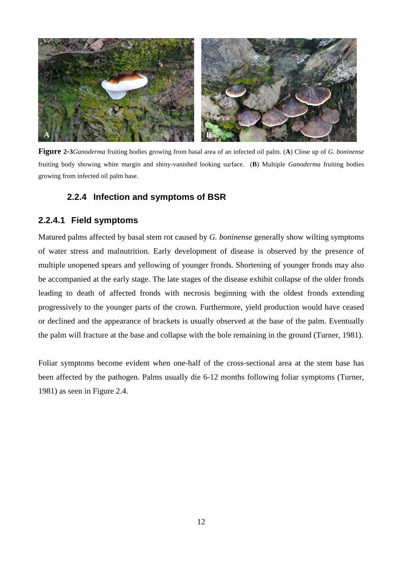
Figure 2-3Ganoderma fruiting bodies growing from basal area of an infected oil palm. (A) Close up of G. boninense
fruiting body showing white margin and shiny-vanished looking surface. (B) Multiple Ganoderma fruiting bodies
growing from infected oil palm base.
2.2.4 Infection and symptoms of BSR
2.2.4.1 Field symptoms
Matured palms affected by basal stem rot caused by G. boninense generally show wilting symptoms
of water stress and malnutrition. Early development of disease is observed by the presence of
multiple unopened spears and yellowing of younger fronds. Shortening of younger fronds may also
be accompanied at the early stage. The late stages of the disease exhibit collapse of the older fronds
leading to death of affected fronds with necrosis beginning with the oldest fronds extending
progressively to the younger parts of the crown. Furthermore, yield production would have ceased
or declined and the appearance of brackets is usually observed at the base of the palm. Eventually
the palm will fracture at the base and collapse with the bole remaining in the ground (Turner, 1981).
Foliar symptoms become evident when one-half of the cross-sectional area at the stem base has
been affected by the pathogen. Palms usually die 6-12 months following foliar symptoms (Turner,
1981) as seen in Figure 2.4.
A B

2.2.4.2 Internal symptoms
Internal symptoms of the disease are characterized by a dry, light brown rot with yellow margins
between the lesion and healthy tissues (Fig.2.
with defense mechanism of host (Turner, 1981). Darmono (1998) suggested that the yellow zones
could be a result of enzyme production by fungi. However, ultrastructural studies of infected oil
palm seedlings by Rees (2006) showed no evidence of fu
Instead an increased activity of host cells within the yellow zone was observed associated with large
amounts of vesicular budding onto the outer membrane suggesting possible cell wall alteration such
as suberisation and lignification in response to cell wall degrading enzymes (Merrill, 1992; Pearce,
1996; Rees, 2006). Narrow dark zones termed ‘black lines’ are embedded with masses of swollen
hyphal cells which may be resting structures (Ariffin et al
A
Figure 2-4 Field symptoms of oil palm affected by basal stem rot caused by
symptoms of water stress showing collapsed canopy and narrowing of crown. (
infection showing rot of infected bole (Photographs with permission from Dr. A. Mudge)
13
Internal symptoms
Internal symptoms of the disease are characterized by a dry, light brown rot with yellow margins
between the lesion and healthy tissues (Fig.2.5). The yellow zones are speculated to be
with defense mechanism of host (Turner, 1981). Darmono (1998) suggested that the yellow zones
could be a result of enzyme production by fungi. However, ultrastructural studies of infected oil
palm seedlings by Rees (2006) showed no evidence of fungal activity or cell wall degradation.
Instead an increased activity of host cells within the yellow zone was observed associated with large
amounts of vesicular budding onto the outer membrane suggesting possible cell wall alteration such
and lignification in response to cell wall degrading enzymes (Merrill, 1992; Pearce,
1996; Rees, 2006). Narrow dark zones termed ‘black lines’ are embedded with masses of swollen
hyphal cells which may be resting structures (Ariffin et al. 1989).
B
Field symptoms of oil palm affected by basal stem rot caused by G. boninense
symptoms of water stress showing collapsed canopy and narrowing of crown. (B) Collapsed palm due to BSR
le (Photographs with permission from Dr. A. Mudge)
Internal symptoms of the disease are characterized by a dry, light brown rot with yellow margins
). The yellow zones are speculated to be involved
with defense mechanism of host (Turner, 1981). Darmono (1998) suggested that the yellow zones
could be a result of enzyme production by fungi. However, ultrastructural studies of infected oil
ngal activity or cell wall degradation.
Instead an increased activity of host cells within the yellow zone was observed associated with large
amounts of vesicular budding onto the outer membrane suggesting possible cell wall alteration such
and lignification in response to cell wall degrading enzymes (Merrill, 1992; Pearce,
1996; Rees, 2006). Narrow dark zones termed ‘black lines’ are embedded with masses of swollen
(A) Typical foliar
) Collapsed palm due to BSR
14
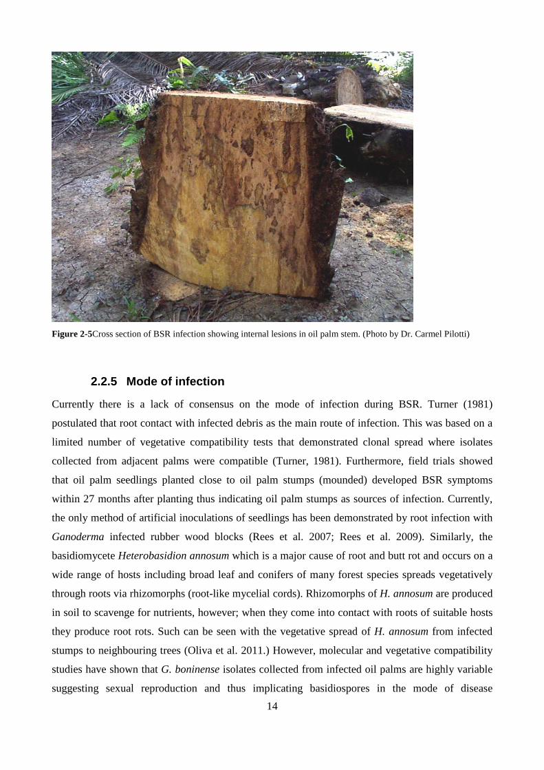
Figure 2-5Cross section of BSR infection showing internal lesions in oil palm stem. (Photo by Dr. Carmel Pilotti)
2.2.5 Mode of infection
Currently there is a lack of consensus on the mode of infection during BSR. Turner (1981)
postulated that root contact with infected debris as the main route of infection. This was based on a
limited number of vegetative compatibility tests that demonstrated clonal spread where isolates
collected from adjacent palms were compatible (Turner, 1981). Furthermore, field trials showed
that oil palm seedlings planted close to oil palm stumps (mounded) developed BSR symptoms
within 27 months after planting thus indicating oil palm stumps as sources of infection. Currently,
the only method of artificial inoculations of seedlings has been demonstrated by root infection with
Ganoderma infected rubber wood blocks (Rees et al. 2007; Rees et al. 2009). Similarly, the
basidiomycete Heterobasidion annosum which is a major cause of root and butt rot and occurs on a
wide range of hosts including broad leaf and conifers of many forest species spreads vegetatively
through roots via rhizomorphs (root-like mycelial cords). Rhizomorphs of H. annosum are produced
in soil to scavenge for nutrients, however; when they come into contact with roots of suitable hosts
they produce root rots. Such can be seen with the vegetative spread of H. annosum from infected
stumps to neighbouring trees (Oliva et al. 2011.) However, molecular and vegetative compatibility
studies have shown that G. boninense isolates collected from infected oil palms are highly variable
suggesting sexual reproduction and thus implicating basidiospores in the mode of disease

15
transmission (Lim & Fong. 2005; Miller et al. 1999; Rees et al. 2012; Pilotti, 2005; Pilotti et al.
2004; Rees et al. 2012). It is postulated that basidiospores infect through wounds and injury on
fronds (Pilotti, personal communication). In support of this view Panchal and Bridge (2005) carried
out sampling of fronds over a two year period of 2 and 7 years palms and were able to detect
Ganoderma via PCR using Ganoderma specific primers. Furthermore, occurrences of upper stem
rot (USR) caused by G. boninense have been sighted to be independent of any basal rot further
indicating the importance of basidiospores in the spread of BSR of oil palm (Pilotti, 2005). A
similar case of pathogenic fungi infecting via basidiospores are Witches Broom of cacao caused by
Crinipellis perniciosus and BSR of Onicidium orchid which is caused by Marasmiellus inoderma (
Lim et al. 2005). In addition, it is estimated that Ganoderma can produce approximately 140, 000
spores per minute and these can be sucked into freshly cut fronds by capillary action where they
readily germinate (Rees et al. 2012).
Knowledge and understanding on the modes of BSR infection are essential in order to develop best
management practices for disease control (Cooper et al. 2011). Past and previous methods of BSR
control and management have been based solely on root – root as the mode of infection and in
effect these methods have been futile in limiting the incidences of BSR infections (Soepena et al.
2000; Naher et al. 2012; Hushiarian et al. 2013). Therefore, a more integrated approach considering
both root contact and basidiospores is needed in the control and management of BSR of oil palm.
Currently, control strategies developed with this approach are used in Papua New Guinea and the
Solomon Is (Sanderson et al. 2000).
2.2.6 Wood decay and rot diseases
Wood decay caused by fungi can be categorized as soft rots, brown rots and white rots which play
an important role in the recycling of wood plant material in natural ecosystems. Soft rot fungi attack
the polysaccharide component of wood, mainly in water saturated areas such as bogs and swamps.
Examples of soft rot include the Ascomycetes Trichoderma, Thielvia and Penicillium species (Rees,
2006).
Brown rot fungi which include a small minority of Basidiomycetes (Rees, 2006) are able to attack
cellulose and hemicellulose with only minor modifications to lignin (Geib et al. 2008).
Transmission electron microscopy of brown rot decay of wood tissue reveals intact cell structure
and darkly stained middle lamella with intact lignin (Rees, 2006).
16
White rot fungi include Basidiomycetes that degrade cellulose, hemicellulose and lignin component
of wood tissue. White rot fungi are known to simultaneously remove all components of wood tissue
or be selective in removing only lignin (Adaskaveg et al. 1990). Species of Ganoderma cover a
wide host range and as such a number of important crops have been affected by rots caused by
Ganoderma. Tropical Acacia species are grown throughout South East Asia and are economically
important cash crops for many small holders. For example, the Indonesian forest industry
contributes an estimated 3.5% of the country’s GDP and provides employment in rural areas. Both
Acacia and Eucalyptus species are grown for pulpwood (Hidayati et al. 2014). Red root rot disease
caused by G.philippii is a serious forest pathogen affecting rubber as well as Acacia mangium and
Eucalyptus pellita. Infections causing root rot can be as high as 28% in second crop rotation. In a
recent study, G. steyaertanum was proved using pathogenicity testing to be pathogenic to
A.mangium where disease spread is supposedly through root contact with infected debris (Hidayati
et al. 2014).
2.2.7 Control of basal stem rot
The control and management of BSR infections is approached at three stages during the cropping
cycle in plantations: new establishments, during the productive phase and more importantly at
replants where younger palms are more susceptible to BSR infections (Sanderson et al. 2000).
Several control measures, both past and present, have been used to control BSR infections with
varying effectiveness.
2.2.7.1 Control at new establishments and at replan t
Newly established plantations after successive oil palm or coconut generations are more susceptible
to early BSR infections. Studies have shown that first generation oil palm planted after previous
crops of coconut or oil palms, where stumps were left to rot in-situ, were more susceptible to early
BSR infections affecting palms as early as 4-6 years after planting (Ariffin et al. 1996), in contrast
to oil palm stands planted after primary forest. In addition, it has been also established that
incidences of BSR infections increase with each successive replant (Turner, 1981), therefore
rendering the control of BSR at planting crucial for the productivity of new oil palm stands.
Several methods of control in the past and present have been developed to remove potentially
infective material that may be carried over from the previous stands thus minimising early
infections at establishment and more importantly at replant. For instance, to reduce the risk of early
infection from previous stands, all stumps are removed and boles are dug out and windrowed.
Furthermore, larger plantations may employ two rounds of mechanical ploughing to a depth of 60

17
cm to bring to the surface root and other woody material and one round of harrowing to chop up the
remaining diseased roots (Flood et al. 2000). Where incidences of BSR have exceeded 20% a
fallow period of two years is recommended however, economic losses due to a fallow of two years
and an additional two years before newly planted palms begin yielding render this approach
unviable and is seldom employed by plantations and smallholders. In addition, smallholders have an
average block size of 2-3 ha which is not big enough for partial fallowing. Virdiana (2010) has
shown significant effects on slow development of BSR after fallowing and studies are continuing to
determine the optimum fallow period and the potential for other crops to balance associated
economic losses.
2.2.7.2 Control in established plantations
The most economical control of BSR infections commonly used by commercial and small holder
plantations in PNG and the Solomon Is. is based on timely identification and removal of infected
palms. Surveys are carried within plantations to identify symptomatic palms (foliar symptoms,
brackets, dead palms, stumps) which are recorded (physical location: block number, harvest road,
palm row and palm number) and marked with red paint for sanitation (Sanderson et al. 2000).
Surveys and sanitation are carried out twice a year beginning when palms have reached six years
and are routinely done up until replant (25 years). Sanitation involves the manual felling of BSR
infected palms exposing decay in the bole that is chiselled into smaller chippings. The root ring is
removed by digging to a depth of 10-15 cm and all infected tissue spread along the windrow.
Finally, the vacant bole area is covered with soil to ground level (Sanderson et al. 2000).
2.2.7.3 Short term control strategies
The control and management of BSR of oil palm is difficult due to difficulty in detecting the
disease at its early stages. Therefore, integrating both short and long term approaches are needed to
minimise damage in current stands and more importantly new replants were oil palm are more
susceptible to early infections (Ariffin et al. 2000). Short term control strategies such as fungicides
and biocontrol agents have been investigated for early intervention into the control of BSR. Earlier
studies (Jollands, 1983) using in-vitro and in-vivo investigations have shown great potential for use
of fungicide in controlling BSR. Field studies (George et al. 1996; Teh, 1996) carried out showed
that systemic fungicides such as triadimenol (triazole group) and hexaconazole (Muhammed et al.
2014) were effective in retarding disease progress in palms with BSR symptoms. However, the use
of fungicides on a commercial scale is not practical particularly on smallholder farms as the effects
of fungicide use diminish rapidly (Darmono, 2000) leading to repeated applications which are too
18
costly as a control option. Furthermore, fungicide use is not environmentally friendly and may
inhibit beneficial microbes (Naher et al. 2013) that may be natural antagonists towards Ganoderma.
Soil saprophytes such as Trichoderma spp., Actinomycete spp. and Bacillus spp. are natural
antagonists towards G.boninense (Soepena et al. 2000; Susanto et al. 2005). In vitro studies
(Susanto et al. 2005) carried out using dual culture method have identified T.harzianum, T.viride
and Gliocladium viride as potential biocontrol agents with 84% - 97% inhibition of Ganoderma
while Bacillus spp. was least inhibitory with 50% inhibition. In a similar study a combination of
Psuedomonas spp. and Arbuscular mycorrhizal fungi inhibited Ganoderma growth by 86%
(Sundram et al. 2011). There is potential to use Trichoderma spp. formulations as prophylactic
against BSR within the first five years after replant (Soepena et al. 2000). Trichoderma
formulations may be applied into plating holes prior to replant, on old logs and stumps that are
potential sources of Ganoderma inoculum and on pruned frond wounds of young palms that may
also provide an entry point for Ganoderma basidiospores. In light of the potential use of biocontrol
agents in control of BSR more work is still needed to develop suitable Trichoderma field
formulations at reasonable cost that can encourage their use by small holders and plantations
considering that repeat treatments may be costly. To date, no field trials have been successful in
supporting results from in-vitro tests.
2.2.7.4 Long term control
The greatest hope for the oil palm industry is in the breeding of oil palm cultivars that are either
resistant or tolerant to BSR as this approach is environmentally compatible and inexpensive for
plantations and smallholders (Idris et al. 2004; Cooper et al. 2011). Durand-Gasselin et al. 2005
suggest genetic sources of resistance do exist in oil palm using hybrids between E.oleifera and
E.guineensis. Nursery trials using rubber wood blocks infected with Ganoderma to artificially
induce BSR infections have also indicated differences in susceptibly of progenies (Idris et al. 2004).
This indication would allow for the identification and subsequent removal of the most susceptible
oil palm germ plasm.

19
2.2.8 Early detection
The effectiveness of short term control strategies for BSR control and management are limited by a
lack of rapid, inexpensive and accurate diagnostic techniques for the early detection of BSR
infections which are specific for large scale applications within plantations (Utomo & Niepold,
2000). Previously G. lucidum causing Thanjavur wilt on coconut was detected with
ethylenediaminetetraacetic acid (EDTA) via colorimetric analysis (Natarajan, 1986) and
Ganoderma was detected in oil palm by plating drill shavings taken from infected tissue 10 cm
above the soil and culturing on Ganoderma selective medium (Ariffin et al. 1993). Both methods
were time consuming and inaccurate. Studies carried out using immunosorbent enzyme-linked
assay (ELISA) using both polyclonal and monoclonal antibodies showed good results but cross
reacted with saprophytic fungi and therefore were not species-specific (Utomo and Niepold, 2000;
Shamala et al. 2006). The use of polymerase chain reaction using ITS region showed more accuracy
to the detection of Ganoderma. Although PCR methods of disease detection have been developed
with great sensitivity it is impractical for a wider application in field screening. More recently the
development of a mobile gamma ray scanner has been prototyped to capture internal basal rot of
asymptomatic palms with greater accuracy however; more work is required to improve faster
scanning and usability under field conditions (Abdullah et al. 2013).
2.2.9 Conclusion
There is still a lot to learn about the Ganoderma-oil palm interaction and what mechanism(s) are
responsible for pathogenicity and aggressiveness in the pathogen and what responses take place
with regard to host defence mechanisms. Filling the gaps in our knowledge may lead to better
understanding of Ganoderma-oil palm pathogen system which is essential in developing strategies
to control BSR infections. A microscopic study of Ganoderma-oil palm interactions may shed light
to the disease mechanism(s) involved and possibly causes of susceptibility and resistance in oil
palm.
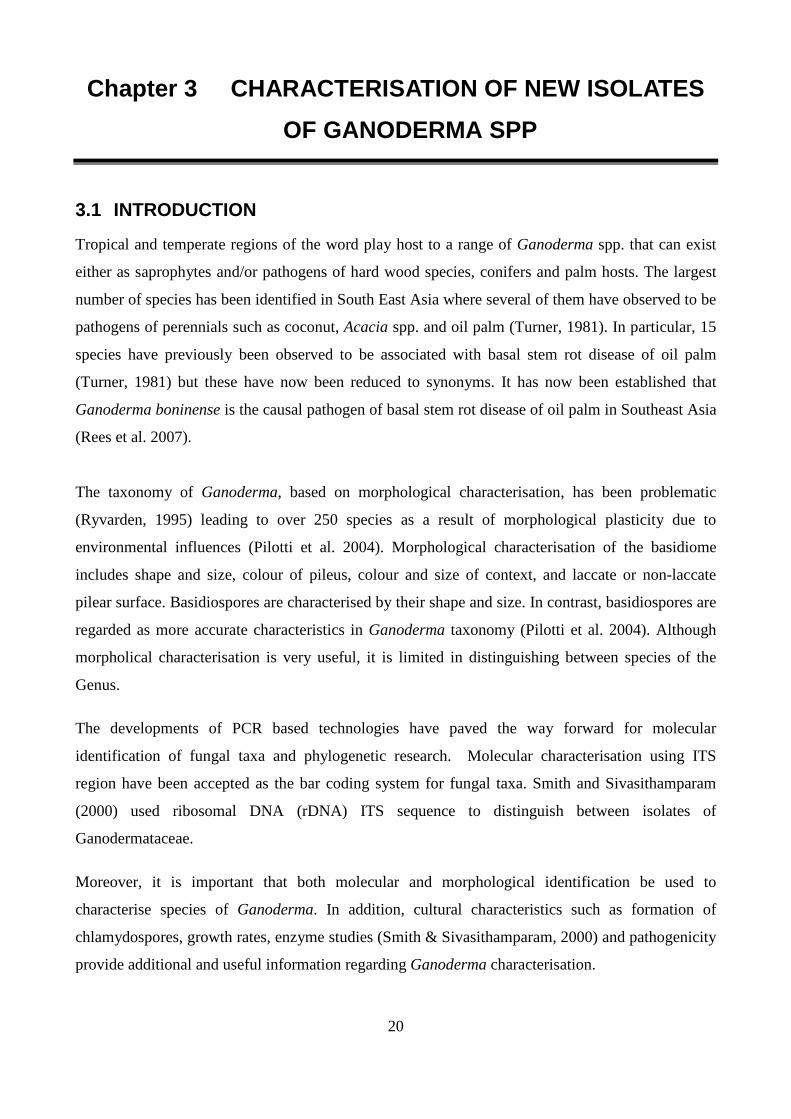
20
Chapter 3 CHARACTERISATION OF NEW ISOLATES
OF GANODERMA SPP
3.1 INTRODUCTION
Tropical and temperate regions of the word play host to a range of Ganoderma spp. that can exist
either as saprophytes and/or pathogens of hard wood species, conifers and palm hosts. The largest
number of species has been identified in South East Asia where several of them have observed to be
pathogens of perennials such as coconut, Acacia spp. and oil palm (Turner, 1981). In particular, 15
species have previously been observed to be associated with basal stem rot disease of oil palm
(Turner, 1981) but these have now been reduced to synonyms. It has now been established that
Ganoderma boninense is the causal pathogen of basal stem rot disease of oil palm in Southeast Asia
(Rees et al. 2007).
The taxonomy of Ganoderma, based on morphological characterisation, has been problematic
(Ryvarden, 1995) leading to over 250 species as a result of morphological plasticity due to
environmental influences (Pilotti et al. 2004). Morphological characterisation of the basidiome
includes shape and size, colour of pileus, colour and size of context, and laccate or non-laccate
pilear surface. Basidiospores are characterised by their shape and size. In contrast, basidiospores are
regarded as more accurate characteristics in Ganoderma taxonomy (Pilotti et al. 2004). Although
morpholical characterisation is very useful, it is limited in distinguishing between species of the
Genus.
The developments of PCR based technologies have paved the way forward for molecular
identification of fungal taxa and phylogenetic research. Molecular characterisation using ITS
region have been accepted as the bar coding system for fungal taxa. Smith and Sivasithamparam
(2000) used ribosomal DNA (rDNA) ITS sequence to distinguish between isolates of
Ganodermataceae.
Moreover, it is important that both molecular and morphological identification be used to
characterise species of Ganoderma. In addition, cultural characteristics such as formation of
chlamydospores, growth rates, enzyme studies (Smith & Sivasithamparam, 2000) and pathogenicity
provide additional and useful information regarding Ganoderma characterisation.

21
The current study was initiated to identify possible G. boninense isolates from palm hosts that could
be used for pathogenicity assays of Ganoderma on oil palm. The need for this approach was to
identify and work with an Australian G. boninense isolate without the stringent quarantine
restrictions that are associated with working with imported fungal isolates. Therefore, the aim of
this study was to identify Ganoderma isolates to their species level using morphological
characteristics of basidiome and basidiospores parallel to molecular methods using PCR and
Ganoderma spcific primers, to confirm PNG isolate GB3039 as G.boninense for lab pathogenicity
trials, and to determine pathogenicity of Australian Ganoderma isolates.
3.2 MATERIALS AND METHODS
3.2.1 Collection of Ganoderma samples
Ganoderma isolates used in this study came from PNG and Australia. All isolates are listed in Table
3.1. Isolate GB3039 was isolated from a basidiocarp of a BSR infected oil palm in Numundo, West
New Britain Province, PNG. Australian isolates RD1 and RD2 were isolated from basidiocarps
growing on golden cane palms (Dypsis lutescens) in Cairns and were provided by Dr. Richard
Davis, while isolates UQ3 and UQ4 were collected from garden mulch at the University of
Queensland, St Lucia campus. Isolate EG1 was found growing on the trunk of a deciduous tree in St
Lucia, Brisbane.
Table 3-1Isolates of Ganoderma spp. collected from Queensland, Australia and Papua New Guinea.
Ganoderma isolates Location Host GB3039 West New Britain, PNG Oil palm RD1 Cairns, Australia Golden cane RD2 Cairns, Australia Golden cane UQ3 University of Queensland, Australia ex litter / mulch UQ4 University of Queensland, Australia ex litter / mulch EG1 Brisbane, Australia Hard wood spp.
All basidiocarps were cleaned off any superficial debris and surface sterilised with 70% ethanol. A
rectangular piece (approx. 1cm2) was cut out from the basidiocarp using a sterile scalpel and blade.
Dikaryotic mycelia was isolated from the context tissue (Fig. 3.1 B) and placed directly on to
Ganoderma selective media (GSM) for one to two weeks. Isolates confirmed for clamp connections
under bright field microscope were sub cultured to potato dextrose agar (PDA, Difco) and incubated
at room temperature. Long term storage of isolates were made possible by removing agar plugs
from the growing edges of each isolate and placed into sterile 1.5 ml micro centrifuge tubes
containing either 1 ml of sterile distilled water or 10% glycerol respectively.
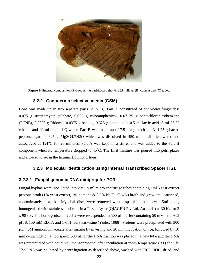
22
Figure 3-1Internal composition of Ganoderma basidiocarp showing (A) pileus, (B) context and (C) tubes.
3.2.2 Ganoderma selective media (GSM)
GSM was made up in two separate parts (A & B). Part A constituted of antibiotics/fungicides:
0.075 g streptomycin sulphate, 0.025 g chloramphenicol, 0.07125 g pentachloronitrobenzene
(PCNB), 0.0325 g Ridomil, 0.0375 g benlate, 0.625 g tannic acid, 0.5 ml lactic acid, 5 ml 95 %
ethanol and 40 ml of milli Q water. Part B was made up of 7.5 g agar tech no. 3, 1.25 g bacto-
peptone agar, 0.0625 g MgSO4.7H2O which was dissolved in 450 ml of distilled water and
autoclaved at 121oC for 20 minutes. Part A was kept on a stirrer and was added to the Part B
component when its temperature dropped to 45oC. The final mixture was poured into petri plates
and allowed to set in the laminar flow for 1 hour.
3.2.3 Molecular identification using Internal Trans cribed Spacer ITS1
3.2.3.1 Fungal genomic DNA miniprep for PCR
Fungal hyphae were inoculated into 2 x 1.5 ml micro centrifuge tubes containing 1ml Yeast extract
peptone broth (1% yeast extract, 1% peptone & 0.5% NaCl, all w/v) broth and grew until saturated,
approximately 1 week. Mycelial discs were removed with a spatula into a new 1.5mL tube,
homogenised with stainless steel rods in a Tissue Lyser (QIAGEN Pty Ltd, Australia) at 30 Hz for 2
x 90 sec. The homogenised mycelia were resuspended in 500 µL buffer containing 50 mM Tris-HCl
pH 8, 150 mM EDTA and 1% N-laurylsarkosine (Yoder, 1988). Proteins were precipitated with 300
µL 7.5M ammonium acetate after mixing by inverting and 20 min incubation on ice, followed by 10
min centrifugation at top speed. 500 µL of the DNA fraction was placed in a new tube and the DNA
was precipitated with equal volume isopropanol after incubation at room temperature (RT) for 1 h.
The DNA was collected by centrifugation as described above, washed with 70% EtOH, dried, and
A
B
C
23
resuspended in 50 µL water for injection BP (Pfizer, Australia). For PCR applications DNA was
diluted 1:10 and 1/10th of reaction volume was used.
3.2.3.2 Polymerase chain reaction (PCR) of internal transcribed spacers
PCR was done using primers ITS1 and ITS4 (Latiffah et al. 2002) which amplify a 655bp fragment
spanning from 3’ end of 18S rRNA gene, both ITS intergenic regions and the 5.8S rRNA gene to
5’end of the 28S rRNA gene. Amplification reactions were performed in 10 µl volumes containing
Multiplex PCR Master Mix (QIAGEN Pty Ltd, Australia), 0.2 µM of each primer and 1µl of 1:10
diluted template DNA. Cycling conditions included a 15 min denaturation step at 95oC, followed by
30 cycles of 95oC for 30 sec, 53oC for 90 sec and 72oC for 90 sec with a final extension of 72oC for
10 min. Amplification (4ul) products were separated on a 2% agarose gel in 1x TAE buffer (40 mM
Tris pH 8, 20 mM acetic acid and 1 mM EDTA) and DNA was visualised on a GelDoc system
(BioRad Laboratories, USA) after staining with GelRed (Biotium Inc, US) as per manufacturer’s
instructions.
3.2.3.3 Cleaning PCR product
Six µl of PCR reaction were cleaned in a total volume of 20 µl consisting of 1x Antarctic
phosphatase buffer (New England Biolabs), 20U ExoI, 5U Antarctic phosphatase, for 15 min at
37oC, followed by 15 min at 80oC to deactivate the enzymes.
3.2.3.4 Sequencing reactions
Cleaned PCR products were mixed with either 1µl of 10µM ITS1 or 1 µl of 10µM ITS4 primer and
sent to the Australian Genome Research Facility (Brisbane) for Sanger sequencing and fragment
separation on an ABI 3730 capillary sequencer (Applied Biosystems).
ITS sequences were aligned using the ClusterW algorithm with 1000 bootstrap replications, and a
Neighbour Joining tree was drawn using the Jukes–Cantor genetic distance model using the
software Geneious 7.1. A previously published G. boninense sequence (Gbref) was used as a
reference (Bridge et al. 2000).
3.2.4 Morphological identification
Macroscopic features such as size and shape of basidiocarp and colour and texture of pilear surface
were recorded. Basidiospores were harvested using a moist cotton bud to collect spores from the
underside of the bracket. The cotton buds containing the spores were dispensed in sterile distilled
water and pipetted on a glass slide for light microscopy of basidiospore size and shape.
24
All Ganoderma isolates were viewed under light microscope and recorded for clamp connections.
Growth rates of mycelia on PDA were determined for 28oC and bench temperatures. All isolates
were tested for vegetative self and non-self- compatibility on PDA.
3.2.5 Bracket and basidiospore production
Autoclaved golden cane logs approximately 15cm high x 8cm diameter were inoculated with
isolates RD1 and RD2 under sterile conditions. The logs were incubated in the dark at 28oC for five
months, and then placed in 12h day/night cycle for five months.
3.2.6 Koch’s postulates
Australian isolates RD1, RD2 and UQ3 were trialled for their pathogenicity on oil palm seedlings
under glass house conditions. Oil palm seedlings were grown for 2-3 months till they reached 2-3
leaf stage. Artificial inoculations were carried out by placing Ganoderma infested sorghum grains
or agar plugs on the bole region of the seedlings, just above soil level. Pots were covered with foil
to seal in moisture and encourage Ganoderma to grow. Three experiments were carried out to
determine the pathogenicity of the above isolates.
Palms were examined at four months post inoculation and root and bole tissue were excised,
washed, surface sterilised, air dried and put onto GSM. Putative Ganoderma were subcultured from
GSM onto PDA and viewed under LM for clamp connections. Isolates with clamp connections
were subjected to molecular identification for G. boninense as described above in section 3.2.3.
3.2.7 Ganoderma soil media test
PNG Ganoderma isolate GB3039 was allowed to colonise 10 g of sterile sorghum grains for one
month and PDA plates one week before inoculation tests. A total of 15 jars containing 180 g of sand
media were sterilised by autoclaving at 121oC for 1 hr. Prior to inoculation of sand media with
Ganoderma, 80 ml of sterile distilled water was added to each jar or until saturated to allow
Ganoderma to grow. Using a sterile spatula, 1 g of sorghum grain was removed from the stock
culture and buried 2 cm below the sand in the treated jars. Similarly, agar plugs were made by using
a sterile core bore, 1 cm dia., and transferred to the sand media and buried to a depth of 2 cm below
(Fig.3.2). A total of five jars each were treated with sorghum grains and agar plugs respectively.
Uninoculated sand media served as controls for the experiment. All treatments were covered in foil
to seal in moisture and reduce illumination to allow for Ganoderma growth. The experiment was set
up in a growth chamber (model: Percival) with the following conditions: temperature 28oC, 80%
humidity and 12 hr photo period. The experiments were allowed to run for one week before
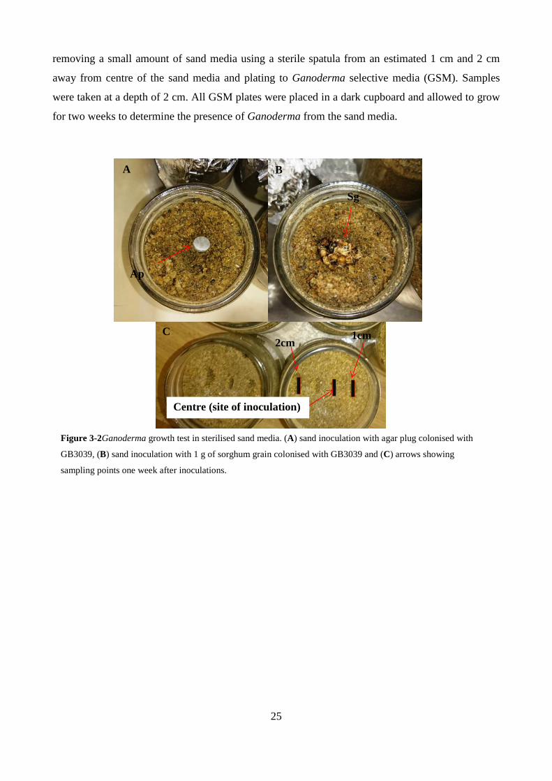
25
removing a small amount of sand media using a sterile spatula from an estimated 1 cm and 2 cm
away from centre of the sand media and plating to Ganoderma selective media (GSM). Samples
were taken at a depth of 2 cm. All GSM plates were placed in a dark cupboard and allowed to grow
for two weeks to determine the presence of Ganoderma from the sand media.
A B
Ap
Sg
C Figure 3-2Ganoderma growth test in sterilised sand media. (A) sand inoculation with agar plug colonised with
GB3039, (B) sand inoculation with 1 g of sorghum grain colonised with GB3039 and (C) arrows showing
sampling points one week after inoculations.
C 2cm
1cm
Centre (site of inoculation)

26
3.3 RESULTS
3.3.1 Ganoderma morphology
3.3.1.1 Basidiocarp and basidiospore morphology
Morphological characterisation of both basidiocarp and basidiospores were not sufficient in
separating the isolates by species as no holo types were available for comparisons. At best,
Ganoderma specimens were separated according to their basidiocarps as laccate or non-laccate
species. The Ganoderma specimens were further grouped or separated according to their
basidiospores as either narrowly ellipsoid or ovoid.
Specimens RD1, RD2 and UQ3 were all grouped as laccate species (Fig3.4 A-C) based on the
texture of their basidiocarps while UQ4 and EG1 were identified as non-laccate (Fig.3.4 D-E) based
on their respective morphology.
Basidiospore morphology for the Cairns isolates (RD1 and RD2) were found to be similar with both
having elongated and narrowly ellipsoid spores (Fig.3.3 A and B) while UQ3 and UQ4 had
basidiospores that were observed to be similar, having truncated and ovoid spores (Fig3.3 C and D).
Spore dimensions for length and width could not be accurately confirmed as approximately 20-100
basidiospores are necessary for determining average length and width. Basidiospores were able to
be salvaged from the pores of the basidiocarps and rough estimates of their dimensions were able to
be determined from an average of four basidiospores per slide per specimen. Isolates RD1 and RD2
had a spore length of 8.2-9.4 µm, width of 3.82-4.38 µm and an index of 2.1 (Table 3.2). In
contrast, UQ3 and UQ had spore dimensions that had a length of 10.04-7.49 µm, width 7.50-5.64
µm and an index of 1.4 and 1.3 respectively
Isolates RD1 and RD2 from a palm host (golden cane) were found to be similar in morphology and
had similar basidiospores and were tentatively grouped together as a single species. UQ3 was also
found to be laccate as well but had basidiospores that were more ovoid and truncated thus rendering
it dissimilar and was identified as being a separate species from the Cairns isolates RD1 and RD2.
UQ4 was easily distinct in this case as it was the only isolate that was non-laccate and having
basidiospores that were different to RD1, RD2 but similar to UQ3. Therefore, these isolates were
separated into three tentative species, A (RD1, RD2), B (UQ3) and C (UQ4) based on their
morphology.
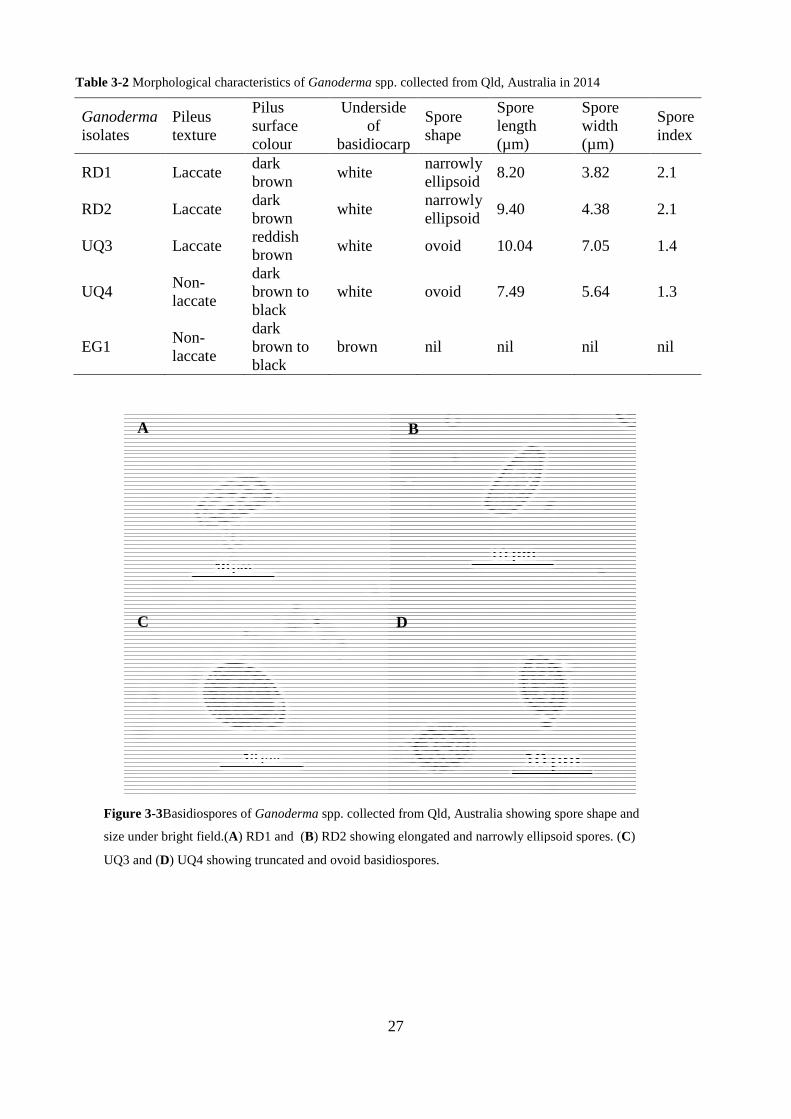
Table 3-2 Morphological characteristics of
Ganoderma isolates
Pileus texture
Pilus surface colour
RD1 Laccate dark brown
RD2 Laccate dark brown
UQ3 Laccate reddish brown
UQ4 Non-laccate
dark brown to black
EG1 Non-laccate
dark brown to black
A
C
Figure 3-3Basidiospores of Ganoderma
size under bright field.(A) RD1 and (
UQ3 and (D) UQ4 showing truncated and ovoid basidiospores.
27
cal characteristics of Ganoderma spp. collected from Qld, Australia in 2014
Pilus surface colour
Underside of
basidiocarp
Spore shape
Spore length (µm)
brown white
narrowly ellipsoid
8.20
brown white
narrowly ellipsoid
9.40
reddish brown
white ovoid 10.04
brown to black
white ovoid 7.49
brown to black
brown nil nil
D
B
Ganoderma spp. collected from Qld, Australia showing spore shape and
) RD1 and (B) RD2 showing elongated and narrowly ellipsoid spores. (
) UQ4 showing truncated and ovoid basidiospores.
spp. collected from Qld, Australia in 2014
Spore width (µm)
Spore index
3.82 2.1
4.38 2.1
7.05 1.4
5.64 1.3
nil nil
spp. collected from Qld, Australia showing spore shape and
narrowly ellipsoid spores. (C)

C
A
B
D
Figure 3-4Basidiocarp collection f
from Cairns, Australia from golden cane host
(D) UQ4 non-laccate species collected from ex
from hard wood spp.
28
C
E
Basidiocarp collection from Queensland, Australia. (A) and (B) laccate species collected
Cairns, Australia from golden cane host, (C) UQ3 laccate species sampled from ex
laccate species collected from ex-litter/mulch, (E) EG1 non-laccate species sampled
) laccate species collected
UQ3 laccate species sampled from ex-litter/mulch,
laccate species sampled
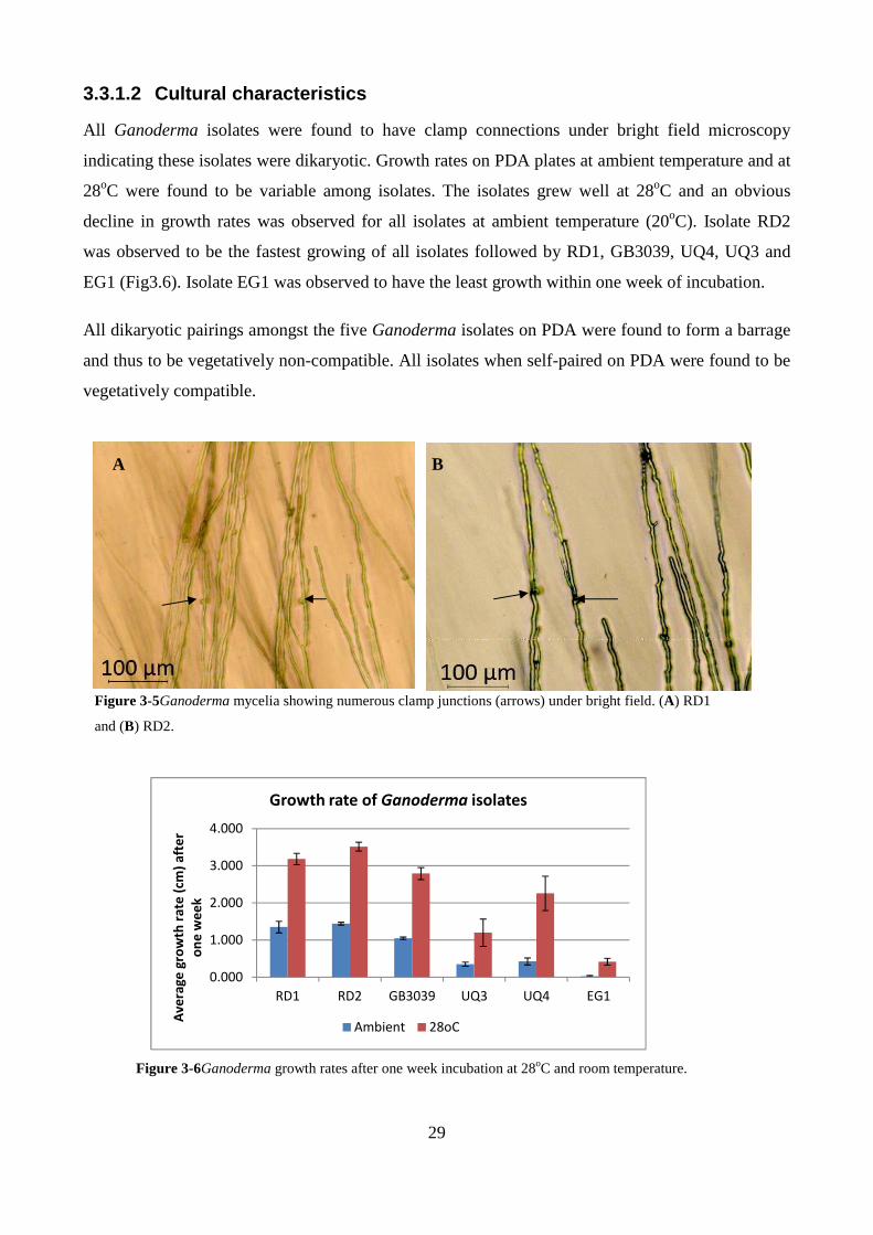
29
0.000
1.000
2.000
3.000
4.000
RD1 RD2 GB3039 UQ3 UQ4 EG1
Av
era
ge
gro
wth
ra
te (
cm)
aft
er
on
e w
ee
k
Growth rate of Ganoderma isolates
Ambient 28oC
3.3.1.2 Cultural characteristics
All Ganoderma isolates were found to have clamp connections under bright field microscopy
indicating these isolates were dikaryotic. Growth rates on PDA plates at ambient temperature and at
28oC were found to be variable among isolates. The isolates grew well at 28oC and an obvious
decline in growth rates was observed for all isolates at ambient temperature (20oC). Isolate RD2
was observed to be the fastest growing of all isolates followed by RD1, GB3039, UQ4, UQ3 and
EG1 (Fig3.6). Isolate EG1 was observed to have the least growth within one week of incubation.
All dikaryotic pairings amongst the five Ganoderma isolates on PDA were found to form a barrage
and thus to be vegetatively non-compatible. All isolates when self-paired on PDA were found to be
vegetatively compatible.
A B
Figure 3-5Ganoderma mycelia showing numerous clamp junctions (arrows) under bright field. (A) RD1
and (B) RD2.
Figure 3-6Ganoderma growth rates after one week incubation at 28oC and room temperature.

30
3.3.2 Bracket and spore production
The aim of inducing bracket formation was to obtain basidiospores for further morphological
characterisation of isolates RD1 and RD2, and perhaps to attempt inoculation if enough spores were
able to be collected. Despite two attempts being made at bracket production, mature basidiocarps
did not develop. Both RD1 and RD2 grew very well on the autoclaved golden cane logs, rapidly
colonising the substrate. Within 6 months bracket buds started to form, at which time the samples
were placed into a 12h photoperiod incubator as Ganoderma likes some light for bracket formation
(Seo & Kirk, 2000). However, these bracket buds (Fig. 3.7) did not develop any further, and thus
basidiospores were not produced either.
Figure 3-7Basidiocarp formation of Ganoderma isolate, RD1, growing on golden cane logs.

31
3.3.3 Sequencing
Sequencing confirmed the identity of both the PNG (GB3039) and Australia isolates (RD1 & RD2)
as G. boninense after BLASTn analysis. Furthermore, ClustalW alignment of GB3039, RD1, and
RD2 were found to be identical with Ganoderma reference sequence from Bridge et al. (2000). ITS
sequencing and BLAST results identify UQ3 as G. tropicum and UQ4 as G. australe.
Neighbour joining tree using Jukes-Cantor genetic distance model group RD1, RD2, PNG Gb 3039
isolate and the Ganoderma reference sequence from Bridge et al. (2000) in one group (Fig.3.8).
Table 3-3 ClustalW alignment comparing Ganoderma isolates against reference G. boninense sequence Gb ref ITS1-4

32
Figure 3-8Neighbour joining tree using Jukes-Cantor genetic distance model.

33
3.3.4 Testing for Koch’s postulates
Four months after inoculations no macroscopic symptoms by G. boninense were observed in the
root and bole sections of seedlings. Root and bole tissues from seedlings inoculated with RD1, RD2
and UQ3 were plated onto GSM. A high level of contamination was present, making re-isolation of
these isolates difficult. However, a sample with cultural characteristics of Ganoderma including
clamped connections on hyphae (as seen under light microscope) was recovered from roots of palm
seedlings inoculated with RD1. This isolate was vegetatively self-compatible, as expected of
Ganoderma. Sequencing of the ITS1-4 region confirmed that the re-isolated fungus was indeed G.
boninense.
3.3.5 Ganoderma soil media test
Sand media inoculated with Ganoderma colonised sorghum grains and agar plugs showed no
visible signs of any hyphal mass growing on the surface after one week. Isolation of sand samples
both 1 cm and 2 cm away from the initial site of inoculation showed nil growth on all GSM plates
after two weeks.
3.4 DISCUSSION
Cultural and morphological characterisation was able to determine that the Australian isolates RD1,
RD2, UQ3 and UQ4 were indeed Ganoderma. Although these methods provided useful information
it was difficult to separate these isolates to their species level. In contrast, PNG isolate GB3039 was
tentatively identified as G. boninense based on its morphology and its association to oil palm.
Molecular identification using ITS sequencing confirmed PNG isolate GB3039 as G. boninense.
Interestingly, isolates RD1 and RD2 from golden cane palm host in Cairns, Australia were also
identified as G. boninense after ITS sequencing. ITS sequencing of UQ3 and UQ4 identified these
as G. tropicum and G. australe based on highest blast scores respectively. Australian G.boninense
isolate RD1 was re-isolated from root tissue five months after inoculations and was confirmed as G.
boninense using ITS sequencing.
Morphological characterisation alone using bracket size, shape, colour and texture of pilear surface
has not been very reliable and has led to over 250 species being named in the genus Ganoderma,
most of which have been misnamed or are synonyms. Differences in morphological characteristics
alone are not a distinctive diagnostic feature as phenotypic differences are influenced by the
environment (Pilotti et al. 2004). The most stable and distinctive feature of characterisation is by
using basidiospores and this method has been used by Steyaert (1980) in Ganoderma taxonomy as
well as other workers in separating between species (Pilotti et al. 2004). In the present studies
34
basidiocarp morphology could not be reliably used to accurately separate the small collection of
Ganoderma isolates into their species level. Furthermore, proper identification using spores were
not able to be achieved due to low number of spores that were able to be salvaged from the
brackets. However, molecular methods, such as sequencing of rDNA genes and ITS regions, is
more reliable.
The confirmation of PNG isolate GB3039 as G.boninense further supports the view by Pilotti et al.
(2004) that G. boninense is the major pathogen of oil palm in PNG. G. boninense is also known to
occur as a saprophyte on coconuts and exist on other palm hosts as well. Isolates RD1 and RD2
from golden cane palm were also identified as G.boninense. These findings have a significant
implication in field control as this suggests that G.boninense is not only able to colonise oil palms
and dead coconuts but also able to establish in other live palm hosts which may act as potential
reservoirs for the Ganoderma life cycle.
The identification of RD1 and RD2 as G. boninense was an important find allowing a possibility of
setting up alternate experiments to study host – pathogen interactions in glass house settings without
the quarantine restrictions that are imposed when working with imported isolates. G. tropicum was
also used for inoculations along with isolates RD1 and RD2. G.australe was not used in any of the
pathogenicity tests; however, both species are useful as outliers for future genetic studies. Both
G.tropicum and G.australe are widely distributed throughout the tropics and occur on either dead or
live tree species. G.tropicum found in tropical China is used locally as a health supplement or herbal
medicine for liver disease and coronary heart disease (Ma et al. 2013). G.lucidum has been used for
centuries for its medicinal properties. Smith and Savasithamparam (2000) were the first to report an
Australian laccate species that was closely related to medicinal strains that are found in Taiwan. As
such, there is a vested interest here in Australia for research into pharmalogical and commercial
production of Ganoderma (Smith & Savasithamparam, 2000).
Attempts using three month old oil palm seedlings inoculated with Ganoderma colonised sorghum
grains and agar plugs did not induce BSR symptoms at four months post inoculation (described in
chapter 4). However, because the hosts used were F1 hybrid seedlings, as used in commercial
plantations, the susceptibility and resistance of individuals is bound to be variable from individual
to individual, and this pathogen–host system is known for the non-synchronous development of
BSR symptoms. Re-isolation from inoculated plants to satisfy Koch’s postulates was conducted on
root and bole tissues and G. boninense like fungus was recovered from RD1 inoculated host.
Further confirmation using ITS sequencing showed that the re-isolation from root was G.boninense.

35
This proves the infective potential of RD1 from these trials but as this was an early stage infection
process it is still unknown as to whether this would have progressed further to the typical BSR
symptoms of internal lesions.
Basidiospores have been implicated in their role of BSR spread and as such the non-quarantine
isolates RD1 and RD2 were grown on golden cane stem trunks in an attempt to initiate bracket
production and consequently spores that could be potentially used for spore inoculations of oil palm
seedlings. Despite best efforts only poorly shaped brackets were formed with no spore production at
all. In the natural field setting bracket production on BSR infected palms does not always occur and
information regarding the mechanism of basidiocarp formation is vague for Ganoderma.
The effect of temperature on mycelial growth showed that growths of all isolates were variable and
were affected noticeably by temperature, with faster growth at 28oC, as expected of
tropical/subtropical species. Although this data may not be suitable for diagnostic characterisation
of Ganoderma isolates, it provides practical and useful information on Ganoderma growth for each
isolate with regard to temperature. For instance, in-vitro growth rate tests reported by Rees et al.
(2007) showed Ganoderma growth was severely impaired when temperatures exceeded 40oC.
Optimal temperatures for Ganoderma growth ranged from 25-30oC. The effects of temperature and
shading were demonstrated by Rees et al. (2007) where shaded oil palm seedling inoculated with
Ganoderma showed early BSR symptoms and higher infection rates compared to inoculated
seedlings that were unshaded. The effects of temperature had a great influence on the growth rates
of Ganoderma, however it is undetermined if the differences in growth rates of individual
Ganoderma isolates may be related to pathogenicity. As such, future pathogenicity seedling trials
should determine the growth rates and optimal temperature of each individual Ganoderma isolate
prior to experimentation, the purpose of which could be to potentially identify whether there is true
expression of differences in susceptibility of oil palm to Ganoderma or as a result of the differences
in Ganoderma growth rates. A better strategy to achieve this would be to use clonal seedlings
inoculated with several G.boninense isolates of variable but significantly different growth rates as a
potential test to determine if growth rates could possibly be associated with
pathogenicity/aggressiveness.
Ganoderma soil tests were carried out using agar plugs and sorghum grains colonised with
G.boninense to inoculate sand media in order to determine if Ganoderma would grow through the
sand. The results from this study showed that no Ganoderma hyphae were able to be re-isolated
from the sand media. These results further support the view by Rees et al. (2007) that Ganoderma is
36
a poor soil competitor and cannot survive in soils that are void of organic matter. The fact that
Ganoderma cannot directly infect through soil could suggest that Ganoderma needs to establish and
build up inoculum as a saprophyte on dead logs, stumps and frond butts before becoming infective
on live palms.
To conclude, the current study reports the confirmation of PNG isolate GB3039 and the Australian
isolates (RD1 & RD2) as G.boninense after ITS sequencing. Isolate GB3039 has been routinely
used for nursery inoculation of oil palm seedlings at PNGOPRA, Milne Bay Research Station,
Alotau, Papua New Guinea and its pathogenicity on oil palm has already been determined. It has
been used for pathogenicity assay under growth chamber conditions for inoculum testing (Chapter
4). Australian isolates (RD1 & RD2) of an unknown pathogenicity on oil palm were used for
nursery pathogenicity tests to satisfy Koch’s postulate. Only RD1 was able to be re-isolated from
the root of an inoculated seedling. Identification of G.boninense isolates from within Australia will
now facilitate future trials without stringent quarantine restrictions which are imposed when
working with imported fungal samples. The Australian isolate, G. tropicum, has been used as part
of the Koch’s postulate tests, however its pathogenicity/infectiveness on oil palm remains
undetermined. Isolates G. tropicum and G.australe may be useful for future pathogenicity work to
determine possible responses to saprophytes. Furthermore, these isolates will be used as outliers for
future phylogeny studies.

37
Chapter 4 NOVEL INOCULATION METHODS
4.1 INTRODUCTION
Although basal stem rot (BSR) disease of oil palm caused by Ganoderma has been known for over
100 years, it continues to be a limiting factor for oil palm production in South East Asia. It is has
been observed that BSR disease increases at each successive replant with infections as early as two
years after planting due to inoculum build up from previous stands (Miller et al. 2000). Both
Malaysia and Indonesia have gone through several replants and severe BSR incidences have been
recorded with more than 50% damage to plantations in some areas (Pilotti, 2005; Su’ud et al. 2007).
Although low levels of BSR infections have been observed in PNG and the Solomon Islands during
first generation plantings, it is envisaged to follow a similar trend to that found in Malaysia and
Indonesia as they move into successive replants. The effects of BSR disease have led to economic
losses due to yield decline and loss of productive stands. Earlier observations by Turner (1981)
suggest that the economic threshold of BSR disease is 20 percent of trees before financial losses
come into effect. It should however, be considered that other abiotic and biotic factors such as type
of genetic material being planted, planting densities, soil types and environment may also have an
influence on yield production. The overall approach and recommendation by plantations affected by
BSR disease is to minimise disease incidences to remain profitable and viable.
Various control and management measures have been used over the years in an attempt to control
the spread of BSR with varying results. Often the high costs and logistics associated with these are a
limiting factor for the oil palm industry in curbing the issue, especially among the smallholder
farmers. The current best management practice is to maintain BSR incidences below the economic
threshold of 20 percent (Turner, 1981) which is carried out by routine disease surveys, and removal
of BSR infected palms’. Total eradication of BSR is highly unlikely and a sustainable long term
approach such as breeding for resistance and/ or tolerance against BSR disease is favourable (Rees
et al. 2007). Progress in this area has been slow and could be addressed by developing suitable
pathogenicity assay that can be used in Ganoderma-oil palm interaction studies to determine factors
that are associated with resistance and susceptibility.
38
Initial pathogenicity work by Navaratnam and Chee (1965) demonstrated that BSR infection in oil
palm seedlings could be induced with a large inoculum (750 cm3) sourced from a naturally infected
BSR palm. Although a non-axenic (wild) Ganoderma culture was used in the trial, an infection rate
of 68 % was achieved. In a separate study carried out by Lim et al. (1992), colonised oat grains
were able to cause 100% infection rate in seedlings within ten weeks however, no further success of
this method has been reported (Rees et al. 2007). Idris et al., (2004) inoculated seedlings by placing
single roots in Ganoderma infested substrate which included oil palm saw dust. Infection via roots
was estimated to be < 2 cm month-1 and mean death of various progenies was calculated at 25.6 %
after 12 months. Sariah et al. (1994) used 5 x 5 x 15 cm rubber wood blocks (Hevea brasiliensis)
infested with Ganoderma and achieved 100% infection rate in oil palm seedlings by placing roots in
close proximity to the wood blocks. Furthermore, pathogenicity trials carried out by Rees et al.
(2007) using different inoculum sized rubber wood and oil palm substrate colonised with a single
Ganoderma isolate showed that a large inoculum size (12 x 6 x 6 cm) was required to cause
infection where 90% of seedlings became infected within 6 – 8 months. In a similar trial Rees et al.
(2007) used smaller rubber wood blocks (3 cm3) colonised with G. boninense. The infected wood
blocks were directly attached to the roots and rapid infection of roots was able to be achieved in
three months suggesting that a larger inoculum size may not be necessary and that intimate contact
with infectious source increases the potential for infection by Ganoderma (Lim et al. 1992; Rees et
al. 2007).
Current pathogenicity testing in nurseries use large rubber wood blocks and take several months to
complete which does not facilitate testing for a large number of oil palm progenies. Therefore, a
more suitable method of pathogenicity testing is needed where a large number of progenies can be
screened and assessed for factors associated with both aggressiveness of pathogen and susceptibility
or resistance of the host plant. There is only limited amount of information regarding natural
virulence/aggressiveness levels found in Ganoderma strains. Information regarding genetics and
nature of resistance or tolerance of oil palm to G. boninense is still unknown (Cooper et al. 2011;
Hushiarain et al. 2013), nor is there much knowledge regarding virulence factors in Ganoderma.
The aim of this chapter was to determine a suitable inoculation method for Ganoderma-oil palm
interaction study of young seedlings suitable for microscopy analysis of early infection processes.
Ganoderma colonised rubber wood blocks have been the preferred choice at inducing BSR
infections in oil palm seedlings in glass house trials (Rees et al. 2007) and are routinely used in
pathogenicity testing of oil palm seedlings for BSR. However, due to the difficulty of acquiring

39
rubber wood in Australia, and because site of pathogen entry into the host was desirable, a range of
inoculation techniques were investigated using agar plugs, pine wood blocks, and sorghum grains
colonised with Ganoderma and artificial wounding of plants to induce BSR infection
40
4.2 MATERIALS AND METHODS
4.2.1 Plant material
Oil palm seedlings used for this study were hybrid F1 tenera (Deli dura x AVROS pisifera) that are
commercially available from Dami Research station, Kimbe, West New Britain, PNG. All seedlings
were potted into sterile sand/sand-compost (50:50) substrate and placed under 1-2 layers of shade
cloth to reduce light intensity. Seedlings were routinely watered with a weekly dose of 5 ml / pot of
N – P – K + trace elements (20-3.5-16.6 + TE) and maintained under glass house conditions at the
University of Queensland, St Lucia campus, Brisbane, Australia.
4.2.2 Ganoderma isolates
Ganoderma isolates used in this study have been previously described in Chapter 3 and listed in
Table 3.1. G. boninense PNG isolate 3039 was used exclusively for lab in containment
pathogenicity assays under quarantine conditions. Australian G. boninense isolates (RD1 and RD2)
and G. tropicum (UQ3) were used for oil palm inoculations under glass house conditions. All
isolates were grown and maintained as dikaryotic mycelium.
4.2.3 Preliminary substrate tests
Rubber wood blocks colonised with Ganoderma are routinely used to artificially induce BSR
symptoms in oil palm seedling pathogenicity assays. However, due to the unavailability of rubber
wood blocks in Australia, a number of substrates were explored to determine which could
adequately support Ganoderma growth. Isolate GB3039 was grown on PDA for a week and
mycelia from the leading edge was isolated using a sterile scalpel and blade and transferred onto
various substrates as listed in Table 4.1 and incubated at room (25oC ± 2oC) temperature for 2-4
weeks.
Table 4-1Various substrates used for G. boninense (GB3039) in-containment growth tests
Substrate Unit quantity per inoculation Potato dextrose broth (PDB) 100 ml Yeast extract peptone broth (YEPB) 100 ml Pine wood blocks 1 cm3 Sorghum grain 10 g Potato dextrose agar (PDA) petri dish (90 mm)
5 x 90 mm petri dish
Brown rice 10 g
41
4.2.4 Ganoderma pathogenicity assays
4.2.4.1 Inoculum preparation
Fifteen grams of sorghum (Sorghum bicolor) grains of Tx430 were placed in a glass jar and
hydrated with an equivalent amount (1 g : 1 ml) of sterile distilled water (SDW) and autoclaved at
121oC for 30 min. The sterile sorghum grains were then inoculated with mycelia from the leading
edge of a PDA plate colonised with Ganoderma. The inoculated grains were placed in a dark
cupboard at room temperature to allow for colonization of substrate for approx. 1 month, or until
fully grown over (Fig 4.2A). Similarly, plate cultures of Ganoderma were grown on PDA and
incubated with the same conditions as the sorghum grains and allowed to colonize the media for
two weeks in preparation for inoculation trials. Pine blocks were cut to 1cm3, autoclaved, then
inoculated with PDA plugs containing Ganoderma and kept at room temperature. 10g samples of
brown rice, hydrated and non-hydrated, were autoclaved and inoculated with PDA plugs containing
Ganoderma and incubated at room temperature.
4.2.4.2 Inoculations
Oil palm seedlings were grown as described in section 4.2.1., for approximately one to two months
or until three leaf stage. The seedlings were subject to wounding (Fig. 4.1A) using a clean scalpel
blade to make a small cut on the bole near the root-bole interface area. This inoculation site is based
on the notion that BSR infection of lower frond would ultimately lead into infection in the bole.
Wounding is representative of the field scenario where continuous wounds are made during
harvesting of fruits where fronds have to be pruned off to access the fruits. Some test and control
plants were left non-wounded. Test plants were inoculated either with sorghum grain (1g/plant) or
agar (1 cm dia. plug /plant) colonized with G. boninense (GB3039, RD1or RD2) or G. tropicum
(UQ3). Mock inoculated plants (wounded and non-wounded) served as controls for the assay.
Finally, all inoculation sites (real and mock) were covered in aluminium foil to facilitate
Ganoderma growth as it thrives better in low light conditions. Plants for inoculations with GB3039
(and controls) were kept in a quarantine facility in a growth chamber (model: Percival) with the
following parameters: temperature 28oC, humidity 80 % and a photo period of 12 hrs. Plants were
watered every two days. All other plants were kept under glasshouse conditions, and watered every
two days.
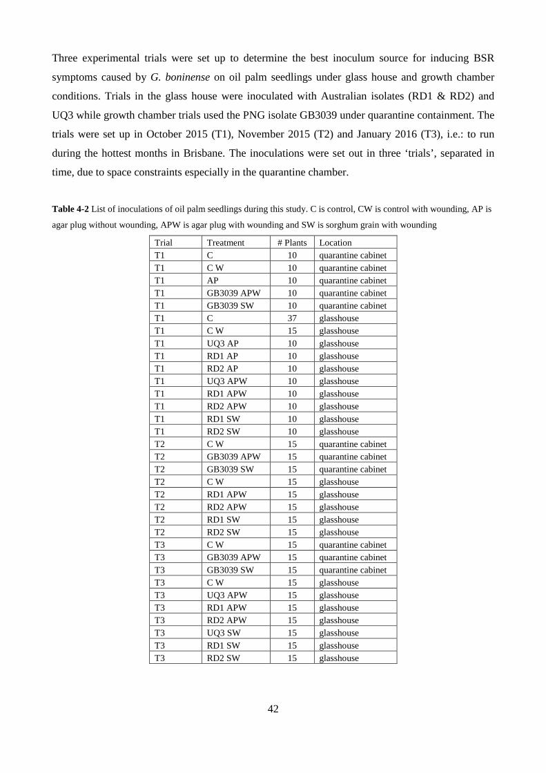
42
Three experimental trials were set up to determine the best inoculum source for inducing BSR
symptoms caused by G. boninense on oil palm seedlings under glass house and growth chamber
conditions. Trials in the glass house were inoculated with Australian isolates (RD1 & RD2) and
UQ3 while growth chamber trials used the PNG isolate GB3039 under quarantine containment. The
trials were set up in October 2015 (T1), November 2015 (T2) and January 2016 (T3), i.e.: to run
during the hottest months in Brisbane. The inoculations were set out in three ‘trials’, separated in
time, due to space constraints especially in the quarantine chamber.
Table 4-2 List of inoculations of oil palm seedlings during this study. C is control, CW is control with wounding, AP is
agar plug without wounding, APW is agar plug with wounding and SW is sorghum grain with wounding
Trial Treatment # Plants Location T1 C 10 quarantine cabinet T1 C W 10 quarantine cabinet T1 AP 10 quarantine cabinet T1 GB3039 APW 10 quarantine cabinet T1 GB3039 SW 10 quarantine cabinet T1 C 37 glasshouse T1 C W 15 glasshouse T1 UQ3 AP 10 glasshouse T1 RD1 AP 10 glasshouse T1 RD2 AP 10 glasshouse T1 UQ3 APW 10 glasshouse T1 RD1 APW 10 glasshouse T1 RD2 APW 10 glasshouse T1 RD1 SW 10 glasshouse T1 RD2 SW 10 glasshouse T2 C W 15 quarantine cabinet T2 GB3039 APW 15 quarantine cabinet T2 GB3039 SW 15 quarantine cabinet T2 C W 15 glasshouse T2 RD1 APW 15 glasshouse T2 RD2 APW 15 glasshouse T2 RD1 SW 15 glasshouse T2 RD2 SW 15 glasshouse T3 C W 15 quarantine cabinet T3 GB3039 APW 15 quarantine cabinet T3 GB3039 SW 15 quarantine cabinet T3 C W 15 glasshouse T3 UQ3 APW 15 glasshouse T3 RD1 APW 15 glasshouse T3 RD2 APW 15 glasshouse T3 UQ3 SW 15 glasshouse T3 RD1 SW 15 glasshouse T3 RD2 SW 15 glasshouse
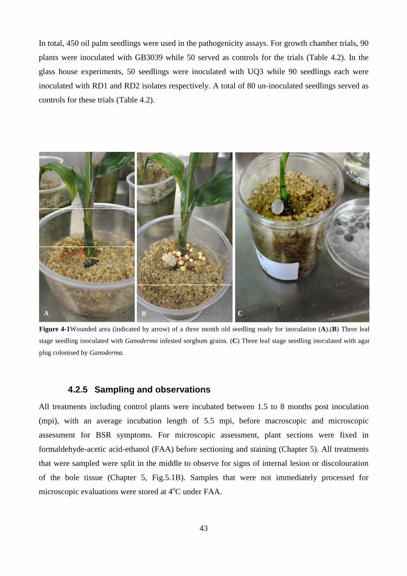
43
In total, 450 oil palm seedlings were used in the pathogenicity assays. For growth chamber trials, 90
plants were inoculated with GB3039 while 50 served as controls for the trials (Table 4.2). In the
glass house experiments, 50 seedlings were inoculated with UQ3 while 90 seedlings each were
inoculated with RD1 and RD2 isolates respectively. A total of 80 un-inoculated seedlings served as
controls for these trials (Table 4.2).
4.2.5 Sampling and observations
All treatments including control plants were incubated between 1.5 to 8 months post inoculation
(mpi), with an average incubation length of 5.5 mpi, before macroscopic and microscopic
assessment for BSR symptoms. For microscopic assessment, plant sections were fixed in
formaldehyde-acetic acid-ethanol (FAA) before sectioning and staining (Chapter 5). All treatments
that were sampled were split in the middle to observe for signs of internal lesion or discolouration
of the bole tissue (Chapter 5, Fig.5.1B). Samples that were not immediately processed for
microscopic evaluations were stored at 4oC under FAA.
A B C
Figure 4-1Wounded area (indicated by arrow) of a three month old seedling ready for inoculation (A).(B) Three leaf
stage seedling inoculated with Ganoderma infested sorghum grains. (C) Three leaf stage seedling inoculated with agar
plug colonised by Ganoderma.
44
4.3 RESULTS
4.3.1 Preliminary substrate tests
Of the substrates listed in Table 4.1, pine wood blocks did not support Ganoderma growth at all. It
was assumed that some component of pine wood was toxic/antagonistic to hyphal growth, so the
blocks were soaked in water in an attempt to leach out any growth inhibitors and re-autoclaved, but
with no success. Brown rice was also tested as it is fairly readily available in both Australia and
PNG. However, it was found to be an excellent source of food for many micro-organisms, not just
Ganoderma, as contamination was a problem. Growth rates in liquid culture, both potato dextrose
(PDB) and yeast peptone extract peptone (YEPB) broths were trialled. Although Ganoderma grew
in both PDB and YEPB, growth rates were slow taking more than two weeks to saturate the culture.
Furthermore, growth rates were too variable, making these broths unreliable.
Fresh sorghum grain was readily available in this laboratory; therefore this substrate was also
trialled. With adequate hydration, Ganoderma grew very well and reliably in autoclaved sorghum
grains. A jar containing 15g grain with equal amount of water was readily colonised within a month
by all Ganoderma isolates tested (Fig.4.2A), and provided enough inoculum for 15 plants.
Furthermore, this substrate was found to be easy to handle during plant inoculations, as it can be
delivered by a spatula or spoon. As described in Chapter 3, Ganoderma grows readily and reliably
on PDA (Fig.4.2B). Using a cork borer (1 cm dia.) to cut out agar plugs of a culture provided a
standardised inoculum source (Fig4.2C).
A B C
Figure 4-2Sorghum grains colonised by Ganoderma isolate RD1 (A), (B) PDA colonised with RD2 and (C) agar
plugs colonised with PNG isolate GB3039 (1 cm dia.)
45
4.3.2 Ganoderma pathogenicity assays
4.3.2.1 Oil palm inoculations using Australian isol ates RD1, RD2 & UQ3
After six to eight months post inoculations, no macroscopic symptoms of BSR disease were
visually observed for all treatments. Foliage of palms showed no major differences between
controls and tests plants. No lesions were observed when boles were split from the crown down to
the bole of asymptomatic seedlings. A total of 108 seedlings were sampled and root and bole tissue
were plated onto GSM to determine if Ganoderma could be grown from asymptomatic palms.
These composed 27 control plants and 81 plants inoculated with RD1 and RD2 using either
sorghum grain inoculation or agar plug inoculations. All bole samples from all treatments were
found to be negative for Ganoderma on GSM plates after almost a month in incubation. Only one
root sample from the 81 plants inoculated with sorghum grain using isolate RD1 was positive for
Ganoderma on GSM. This was confirmed with microscopic observations of clamp connections and
ITS sequencing of the isolate (see Chapter 3) confirmed to be G. boninense. Of the 310 seedlings
used for this trial, 18.7 % of the seedlings including controls died during the trial. Although some of
these plants showed internal lesions, re-isolation attempts on GSM were unsuccessful due to a large
number of contaminants on the plates. Due to the unknown pathogenicity status of the Australian
isolates on oil palm, no microscopy work was carried out as priority was given to the growth
chamber inoculations using the PNG isolate GB3039.
4.3.2.2 PNG 3039 growth chamber inoculation
General symptoms of test and control seedlings showed necrosis of leaf tips and presence of
necrotic lesions on the leaves. This feature was seen randomly for all treatments including control
plants particularly for trials inoculated in October and November, 2015. Of the 140 seedlings that
were used for growth chamber inoculations, 48 seedlings died during the course of this study with
64.6% of these seedlings originating from trials initiated in November, 2015. Plating of root and
bole tissue of the dead seedlings to GSM did not support Ganoderma growth after one month,
indicating the seedlings died from other causes. Macroscopic features of foliar symptoms and status
of internal bole structure showed no discernible differences between controls and inoculated plants.

46
Table 4-3 Fluorescence microscopy of agar plug and sorghum inoculations on oil palm seedlings. SW is sorghum
inoculations with wounding, AP is agar plug without wounding, APW is agar plug with wounding, C is control without
wounding, CW is control with wounding, + is positive for fungal growth and (-) is negative observation for fungal
growth.
Trial Code Date sampled Root Bole
Degree of fungal colonisation of tissue
T1 SW6 5 mpi +++ - 1
T2 SW2 2 mpi +++ ++ 2
T2 SW5 2 mpi +++ ++ 2
T2 SW11 2 mpi +++ ++ 2
T3 SW10 4 mpi +++ ++ 2
T3 SW12 4 mpi +++ ++ 2
T3 SW13 4 mpi +++ - 1
T3 SW14 4 mpi - + 1
T1 AP9 5 mpi - + 1
T1 AP7 5 mpi - - 0
T1 APW3 5 mpi - + 1
T1 APW7 7 mpi - - 0
T2 APW2 2 mpi + - 1
T2 APW3 2 mpi - + 1
T3 APW1 4 mpi + - 1
T3 APW6 4 mpi + + 2
T3 APW7 4 mpi - + 1
T3 APW9 4 mpi - + 1
T1 CW10 5 mpi - - 0
T2 CW5 2 mpi - + 1
T2 CW10 2 mpi - + 1
T3 CW2 4 mpi - + 1
T3 CW3 4 mpi - + 1
T3 CW12 4 mpi - + 1
T3 CW13 4 mpi - + 1 Scale for hyphal occurrence in tissue type: (-) = no fungal hyphae for both bole and roots, (+) = fungal hyphae in either root or bole tissue only, 2 = fungal hyphae in both root and bole tissues and (+++) = high degree of fungal colonisation Representative samples were removed and processed for microscopic evaluation of Ganoderma
infections by agar plug and sorghum grain inoculations. Rather than looking at how many plants
were infected here the focus was based on the type of tissue that were colonised by Ganoderma.
From table 4.3 there is a higher frequency of infection events occurring for both root and bole
tissues using the sorghum inoculations in contrast to agar plug inoculations that has slightly fewer
infections for both root and bole tissue. Control plants also showed presence of fungal hyphae in the
47
dead region of the bole tissue and it is anticipated that this may have arisen initially from rearing of
the plants in the glass house where non sterile conditions may have allowed for contaminants and
opportunists to colonise the site of inoculation. Although not included in this chapter but in the next
chapter, microscopic observations of the inoculated plants show a far greater number of hyphae
than in the controls.
4.4 DISCUSSION
The present study reports the use of sorghum grains and agar plugs in inoculations of oil palm
seedlings to establish BSR. Preliminary substrate tests have identified sorghum grains and agar
plugs as suitable inoculum for early infection study based on rapid colonisation of the substrate and
relative ease of use. Early infection studies using sorghum grain and agar plugs showed no
discernible differences between controls and treatments. Although early deaths of seedlings were
recorded and had internal discolouration and lesion, plating of these bole and root tissues onto GSM
failed to confirm the presence of Ganoderma. Although macroscopic observations showed no
symptoms of early BSR infection, microscopic observations using fluorescence microscopy
revealed early establishment using both methods of inoculations were achieved and the results
suggest that sorghum grains have a better infection potential than agar plugs, possibly due to higher
mycelial mass.
Rubber wood is the preferred substrate that is used widely for artificially inducing BSR infections
in nursery oil palm seedlings (Rees et al. 2007). The work by Rees et al. (2007) reported that
inoculum potential is influenced by shading, soil temperature, inoculum load, aggressiveness of
Ganoderma isolate, intimate contact with Ganoderma inoculum and oil palm via root inoculations.
Furthermore, although inoculum size may have an influence in faster disease development, intimate
contact of the fungus using a small inoculum is more important than the size of the inoculum as
reported by Rees et al (2007). As such, the current pathogenicity trials carried out using agar plugs
and sorghum grains were inoculated within close proximity to the injured root-bole interface to
mimic colonisation of the lower fronds by Ganoderma and possible movement into the bole region.
The results from table 4.4 indicate more establishments of fungal hyphae in root and bole tissue of
sorghum inoculation as opposed to agar plug inoculations most probably due to the effect of
inoculum. What seems intriguing at this point is that there seems to be slightly more fungal
establishment in root tissue than the bole tissue suggesting a possible preference for root tissue in
contrast to bole tissue.

48
The artificial method of inducing BSR symptoms in juvenile oil palm seedlings had been earlier
demonstrated by Navaratnam and Chee (1965) and this was achieved by placing fairly large wood
blocks (750 cm3) colonised with Ganoderma under growing oil palm seedlings and caused
infection via roots. This method has been widely adapted and routinely used by plantations for BSR
screening against Ganoderma. Although G. boninense is not a pathogen on rubber, it has been
found to support Ganoderma growth very well and has been the preferred choice of inoculum
substrate for pathogenicity studies. Previous pathogenicity studies (Rees et al. 2007) using both
rubber and oil palm wood blocks as inoculum sources demonstrated that rubber wood blocks had
higher disease incidences than oil palm wood inoculations. The effect of inoculum on BSR
infections had been previously demonstrated by Rees et al. (2007) where larger rubber wood blocks
(12 x 12 x 6 cm) colonised with Ganoderma induced faster infection with higher disease rates
compared to smaller rubber blocks ( ≤ 3 x 3 x 3 cm) within a 15 month period. The limitations in
using large rubber wood blocks is that they take up a lot of space to prepare and so a limited number
of breeding lines can be tested at any one time. Thus there is a need for a smaller inoculum size
with a more rapid infection rate for screening of a large number of progenies against BSR disease.
Oil palm seedlings inoculated using sorghum and agar plugs colonised with Australian isolates
RD1, RD2 and UQ3 showed no macroscopic differences. Assays using PNG isolate GB3039
showed a similar trend, most probably because plants were sampled at early stages in the infection
process and therefore, no macroscopic symptoms were observed. No internal lesions were observed
for all asymptomatic plants except for seedlings that had either died of possible wet rot. In addition,
none of the plants that had died were from a direct result of basal stem rot. The lack of any
symptoms is a typical feature of BSR disease as foliar symptoms in matured stands only appear
where more than half of the bole has been destroyed by Ganoderma (Turner, 1981). Furthermore,
only one sample dissected from 108 seedlings roots and boles on GSM were able to successfully re-
isolate Australian Ganoderma isolate RD1 from the roots of an asymptomatic palm. Confirmation
that it was Ganoderma was done after ITS sequencing. Due to the lack of discernible differences
associated with both inoculations methods, it remains undetermined which is better as early
infection process of Ganoderma is difficult to determine visually.
Inoculations using both sorghum grains and agar plugs showed no macroscopic differences between
each other and in contrast to controls. All inoculated and test plants were split in the middle with a
sharp knife to observe for possible internal lesions. Although a few plants exhibited leaf lesions
49
these did not relate to BSR infections as control plants also exhibited the same leaf lesions and this
may be possible to growing conditions such a prolonged exposure to high temperatures.
Basal stem rot disease caused by G. boninense is a slow but progressive disease taking several years
for symptoms to develop. It has been estimated that symptoms appear when more than 50 % of the
bole has been affected by BSR (Turner, 1981). In brief, matured palms affected by BSR show
general wilting and nutritional deficiency. Immature plantings (2-5 yrs) affected by BSR are often
difficult to identify and remain healthy until they suddenly collapse. Occasionally young palms
show signs of early yellowing and stunting but these may be related to nutrition or genetic factors as
well. Nursery pathogenicity tests usually take between 6-18 months where BSR symptoms can
appear as early as four months after inoculations and can cause 80-90% BSR incidences within
eight months post inoculations (Rees et al. 2007). In these instances, fairly large inoculum sizes (12
x 12x 6cm) were used to induce a quicker response. In has also been demonstrated that smaller
inoculum (3 x 3 x 3 cm) was able to induce BSR symptoms after firm attachment of inoculum to
roots (Rees et al. 2009) with in a duration of five months.
Ganoderma has been suggested to enter though wounds and injuries and has been postulated that
wounds may play an important role in the infection process by inducing a faster rate of infection.
Due to the limitation in space requirements, a comparison between wounding and non-wounding of
oil palm seedlings was not thoroughly addressed in this present study. However, it has been
reported by Rees et al. (2007) that wounding is not a prerequisite to infection and that pathogen
aggressiveness and intimate contact of Ganoderma with oil palm plays a larger role. In addition,
injury was found to facilitate entry of weaker pathogens and/or opportunists (Rees et al. 2007).
Spores have been implicated to play a vital role in the spread of BSR disease and this notion has
been supported by genetic studies that show that Ganoderma populations in the field are genetically
variable thus indicating sexual recombination between compatible monokaryons (Pilotti et al.
2003). Implications for spores to be used for pathogenicity testing would be more representative of
the natural process in the plantations-but getting enough spores from a single isolate is problematic
and so far no one has managed to show infection with spores. In contrast, other patho-systems such
as Heterobasidion annosum initially infect by basidiospores on a suitable host and spread through
secondary means by root contact (Woodward et al. 1998).

50
The overall results suggest that more work is needed to determine the effective inoculum level for
disease expression in oil palm seedlings. However, for the purpose of finding a suitable inoculation
substrate for inducing early BSR infections for microscopic studies, sorghum grains show great
potential for use in host-pathogen interaction studies.
51
Chapter 5 EARLY INFECTION PROCESS IN OIL PALM
SEEDLINGS INOCULATED WITH GANODERMA
5.1 INTRODUCTION
Bright field microscopy has been used for decades in studying fungal disease progress in planta and
more specifically fungal-plant interactions. The development of electron microscopy has greatly
improved the resolution and detailing of both plant and fungal structures with regard to fungal
disease progression in plants. However, the use of electron microscopy often involves complicated
processing (fixation, resin embedding, and ultra-thin sectioning of samples) and that can be time
consuming, plus such expensive equipment is not often readily available. The study of fungal –
plant interactions has improved in particular with the use of counter staining or differential staining
to view fungi in plants but visualisation of fungal structures inside plants is still often difficult. The
visualisation of fungi in plants has been further enhanced with the use of fluorescence microscopy
in combination with appropriate fluorescent staining techniques (Knight & Sutherland, 2011). More
advanced fluorescent microscopy involves confocal laser scanning microscopy where both fungal
and/or plant tissue can be genetically transformed with green fluorescent protein (GFP) or similar
derivatives. Although confocal microscopy may be very accurate in contrasting between fungal
elements and plant tissues, it is often limited by the lengthy period of time required to produce GFP
transformants of either plant or fungus (Knight & Sutherland, 2011). Therefore, other conventional
methods have been developed using dual staining techniques and florescence microscopy where
non-fluorescent stains are used to suppress auto-fluorescence of plant tissue while fluorescent stains
are employed to visualise fungal elements. This differential staining technique has been
successfully used for the study of Fusarium pseudograminearum in cereal tissues during crown rot
infections (Knight & Sutherland, 2011).
Various wood decay fungi have been reported to affect palms with many of them responsible for
causing major diseases. However, there has been very little progress in determining the decay
process of palm wood (Adaskaveg, 1991). Palms, including date and oil palm, which are of
commercial value, have long been reported to be affected by several Ganoderma species. To date,
only a limited number of histological studies have been carried out to investigate the decay process
caused by Ganoderma spp. on date (Adaskaveg, 1991) and oil palm. The work carried out by Rees
et al. (2009) provides the only recent histological study of Ganoderma infection process in oil palm
seedlings. This work has shown Ganoderma to exhibit three development stages during infection:
52
initial biotrophic phase, a necrotrophic phase associated with advance decay and finally, the
formation of melanised mycelia as a final resting stage. Reports from this study were centred on
moderate to advanced stages of infection and do not clearly indicate on how the establishment
phase of Ganoderma in oil palm occurs and the early infection process (es). Therefore, there is still
a gap in knowledge regarding establishment and early infection process of Ganoderma in oil palm
that requires further investigation.
Fungal plant pathogens have the ability to manipulate their hyphal morphology in response to
physiological and structural features of plant hosts. Following establishment in a host, fungi can
exist as a biotroph where it acquires nutrition from the host plant without causing any detrimental
effects; or as a necrotroph where it kills its host plant in order to acquire nutrition. Some fungi
however can initially exist as a biotroph and later switch over to a necrotroph; these are termed
hemi-biotrophs such as several species of Colletotrichum (Perfect et al. 1999). Colletotrichum has
been used as a model genus for pathology and fungal-plant interaction studies (Perfect et al. 1999)
and has revealed the formation of specialised fungal structures involved during infection such as
germ tubes, appressoria, haustoria, intracellular hyphae and secondary necrotic hyphae (Perfect et
al. 1999; Mendgen & Hahn, 2002). In contrast, the plant hosts are able to use an array of defence
mechanisms to fungal pathogens such as lignin deposition in sites of fungal invasion, automatic cell
death, production of gums and tyloses as well as many defence related proteins.
The current study sets out to determine what occurs in oil palm and Ganoderma during early stages
of BSR establishment. Light microscopies with dual staining techniques were employed to visualise
physical changes that may be associated with defence or pathogenicity of oil palm and Ganoderma
respectively during the early stages of establishment and development. The Ganoderma-oil palm
pathosystem has not been studied in detail and information regarding colonisation, infection process
and symptom expression (both macro and micro) is poorly understood. Any information gathered
from this work will contribute to the field of knowledge regarding the Ganoderma-oil palm
pathosystem.
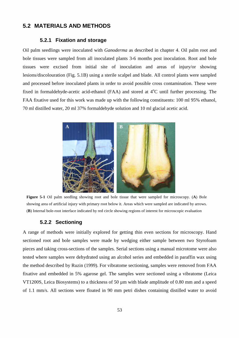
53
5.2 MATERIALS AND METHODS
5.2.1 Fixation and storage
Oil palm seedlings were inoculated with Ganoderma as described in chapter 4. Oil palm root and
bole tissues were sampled from all inoculated plants 3-6 months post inoculation. Root and bole
tissues were excised from initial site of inoculation and areas of injury/or showing
lesions/discolouration (Fig. 5.1B) using a sterile scalpel and blade. All control plants were sampled
and processed before inoculated plants in order to avoid possible cross contamination. These were
fixed in formaldehyde-acetic acid-ethanol (FAA) and stored at 4oC until further processing. The
FAA fixative used for this work was made up with the following constituents: 100 ml 95% ethanol,
70 ml distilled water, 20 ml 37% formaldehyde solution and 10 ml glacial acetic acid.
5.2.2 Sectioning
A range of methods were initially explored for getting thin even sections for microscopy. Hand
sectioned root and bole samples were made by wedging either sample between two Styrofoam
pieces and taking cross-sections of the samples. Serial sections using a manual microtome were also
tested where samples were dehydrated using an alcohol series and embedded in paraffin wax using
the method described by Ruzin (1999). For vibratome sectioning, samples were removed from FAA
fixative and embedded in 5% agarose gel. The samples were sectioned using a vibratome (Leica
VT1200S, Leica Biosystems) to a thickness of 50 µm with blade amplitude of 0.80 mm and a speed
of 1.1 mm/s. All sections were floated in 90 mm petri dishes containing distilled water to avoid
A B
Figure 5-1 Oil palm seedling showing root and bole tissue that were sampled for microscopy. (A) Bole
showing area of artificial injury with primary root below it. Areas which were sampled are indicated by arrows.
(B) Internal bole-root interface indicated by red circle showing regions of interest for microscopic evaluation

54
dehydration of sections while pending staining and microscopy. Blades were replaced after every
four samples sectioned.
5.2.3 Stains
Calcofluor White M2R stain was obtained as a 100 ml solution containing 1g/l calcofluor white and
0.5 g/l of Evans blue (Fluka, Sigma-Aldrich). Calcofluor white is a specific stain for β-glucans and
chitin, staining cellulose in plant tissue, callose (β 1, 3-glucans) which is an important part of post-
infection cell wall reinforcements and fungal cell walls (Rees et al. 2009; Bhadauria et al. 2010).
Calcofluor white has an excitation range of 365-395 nm and emission of 420 nm. Fungal structures
and plant tissue appear fluorescent bright blue under UV. As per the manufacturer’s instruction,
plant samples were placed on a clean slide followed by the addition of one drop of calcofluor white
and one drop of 10% potassium hydroxide. In instances involving large sections, two drops each of
calcofluor white and 10% potassium hydroxide were applied to the slides. Finally, cover slips were
placed on the specimens and allowed to stand for one minute before viewing under UV
fluorescence at x40 and x100 magnification.
Toluidine blue working solution of 0.1% (w/v) was made by dissolving 0.1 g of toluidine blue
(Sigma-Aldrich) in 100 ml sterile distilled water. The polychromatic dye toluidine blue was used to
visualise general root and bole anatomy of oil palm. Tracheary elements (lignified walls) stained
green or bluish-green but occasionally green, lignified sclerenchyma occasionally stained green but
also appeared blue-green, collenchyma and parenchyma cells stained reddish purple, sieve tubes
and companion cells stained red, un-lignified middle lamellae stained reddish purple or red, callose
and starch remained unstained (Haskins, 1982). Sectioned specimens were placed in a petri-dish
and several drops of toluidine blue (0.1% w/v) added to the specimens and allowed to stain for three
minutes. Specimens were finally rinsed with several exchanges of distilled water. The specimens
were mounted with 30-50% glycerol on a clean slide, cover slip placed onto the specimen and
viewed under bright field.
Safranin O (Sigma-Aldrich) was used to visualise general plant anatomy. A 0.2% (w/v) working
solution was made up by dissolving 0.2g in 100 ml 10% ethanol v/v. Specimens were stained for
less than a minute followed by several changes with distilled water. Stained specimens were
mounted with 50% glycerol on a clean glass slide with a cover slip and viewed under bright field.

55
To make up aniline blue, (BDH Chemical Ltd) 0.05g was added to 20 ml of distilled water and
allowed to stand for over for 24 hours. 20g of phenol crystals were dissolved in 20 ml of lactic acid
with stirring. To this solution 40 ml of glycerol was added followed by the aniline blue solution that
was previously prepared. Specimens were stained for two to five minutes, rinsed with several
changes of distilled water, mounted with 50% glycerol on a clean slide with a cover slip and viewed
under bright field. Aniline blue stains callose depositions formed after pathogen attack.
Phloroglucinol stain was used for visualising lignin in root and bole tissues. The working solution
was prepared by dissolving 0.2g in 80 ml 20% ethanol solution followed by the addition of 20 ml of
concentrated HCl (12N). Samples were stained for five minutes and rinsed several times with
distilled water before mounting on a clean glass slide with 50% glycerol with a cover slip. Slides
were observed under bright field and lignified cells appeared red in colour.
5.2.4 Staining and microscopic observations
Various dyes for both bright field and fluorescence microscopy were initially trialled for their
ability to contrast between oil palm and fungal cell walls and other structures. In addition, several
dye permutations were also investigated to determine the best dye combinations to simultaneously
study changes occurring in both plant and fungus during Ganoderma-oil palm interactions. For
plant structure, non-fluorescent dyes such as Phloroglucinol (PHCl), toluidine blue (0.1% w/v) and
safranin (0.2% w/v) were used to stain both root and bole tissue. Fungal hyphae was stained using
non-fluorescent dye aniline blue and fluorescent dye Calcofluor white (CW). It was eventually
resolved that toluidine blue (TBO) gave better visualisation and detail of plant structures under
bright field microscopy while fungal hyphae was better visualised using calcofluor white under
fluorescence microscopy. As a result, a dual staining method was developed where tissues were first
stained for 3 minutes in toluidine blue (0.1% w/v) for bright field microscopy, rinsed with several
changes of distilled water and stained with calcofluor white for visualisation of fungal hyphae under
fluorescence microscopy. Stained tissues were observed using a ZEISS AXIO Fluorescence
Microscope. Dual stained tissues were first observed under bright field followed by fluorescence
microscopy using the DAPI filter and UV lamp with an excitation wave length of 365 nm and
emission wave length of 420-470 nm. Images were taken using the mounted camera on the
microscope (ZEISS AXIO Cam MRc).
56
5.3 RESULTS
5.3.1 Microscopic observations of Ganoderma-oil palm interaction
Hand sectioning was found to be quick and easy; however it was difficult to obtain thin and uniform
sections due to the difficulties of working with hard woody tissues of both oil palm root and bole.
Bright field images of hand sectioned samples were found to be uneven and resulted in low
resolution images due to samples being too thick (> 50 µm). Microtome sectioning provided thin
and even serial sections of 10-15µm with good resolution under bright field microscopy. However,
the steps involved with sample dehydration, wax infiltration and wax embedding of samples was
found to be too time consuming for the number of samples processed in this study and thus the
microtome methods were not further explored. Finally, vibratome sectioning was identified as the
most rapid and convenient method of choice in obtaining uniform thin sections between 40-50 µm.
Various dyes showed a degree of effectiveness in their visualisation of both plant and fungal
elements under bright field and fluorescence microscopy (Table 5.1). However, the most effective
dye for visualisation of oil palm anatomy was toluidine blue while fungal hyphae in oil palm roots
and bole tissue specimens were visualised by staining with calcofluor white under UV light. The
dual staining technique using toluidine blue and calcofluor white provided simultaneous
visualisation of plant cells and fungal hyphae where fungal hyphae could only be viewed under UV
light while plant cell walls could be visualised under bright field and UV light.
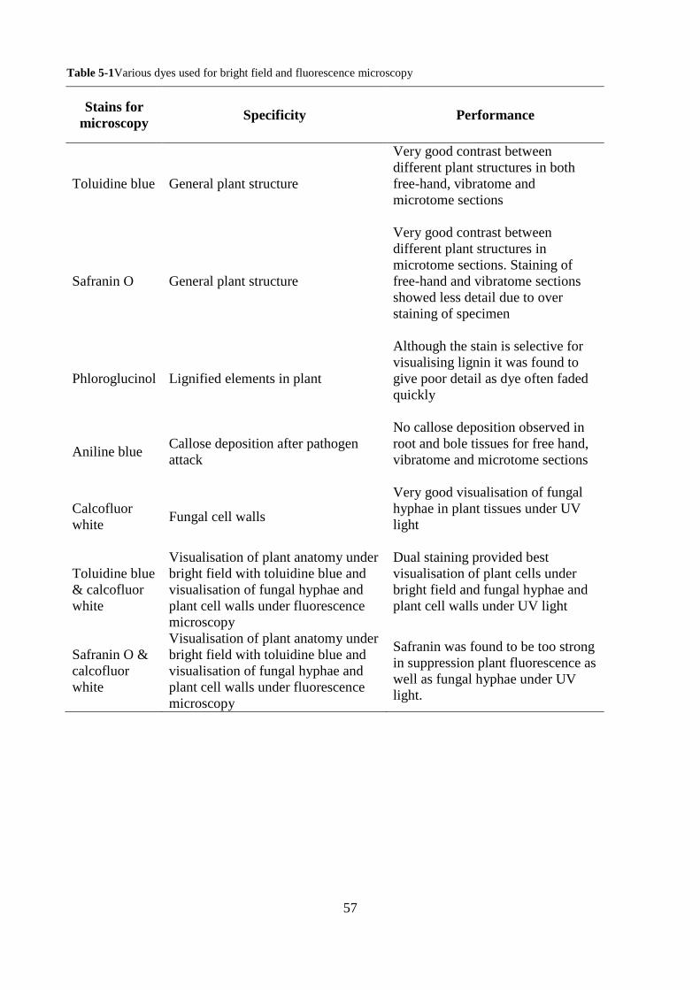
57
Table 5-1Various dyes used for bright field and fluorescence microscopy
Stains for microscopy Specificity Performance
Toluidine blue General plant structure
Very good contrast between different plant structures in both free-hand, vibratome and microtome sections
Safranin O General plant structure
Very good contrast between different plant structures in microtome sections. Staining of free-hand and vibratome sections showed less detail due to over staining of specimen
Phloroglucinol Lignified elements in plant
Although the stain is selective for visualising lignin it was found to give poor detail as dye often faded quickly
Aniline blue Callose deposition after pathogen attack
No callose deposition observed in root and bole tissues for free hand, vibratome and microtome sections
Calcofluor white
Fungal cell walls
Very good visualisation of fungal hyphae in plant tissues under UV light
Toluidine blue & calcofluor white
Visualisation of plant anatomy under bright field with toluidine blue and visualisation of fungal hyphae and plant cell walls under fluorescence microscopy
Dual staining provided best visualisation of plant cells under bright field and fungal hyphae and plant cell walls under UV light
Safranin O & calcofluor white
Visualisation of plant anatomy under bright field with toluidine blue and visualisation of fungal hyphae and plant cell walls under fluorescence microscopy
Safranin was found to be too strong in suppression plant fluorescence as well as fungal hyphae under UV light.
58
5.3.2 General oil palm seedling anatomy
Information regarding the internal anatomy is similar as to most other monocotyledons. Oil palm
stems including lower stem (bole) are set in a matrix of ground parenchyma cells embedded with
numerous vascular bundles and fibres (Fig. 5.2A). The bole tissue was also found to contain
randomly dispersed calcium oxalate crystals known as Raphides. Raphides are needle shaped
crystals (Fig. 5.4C) typically found in the parenchyma cells and are speculated to play a role in
defence against herbivorous pests particularly causing stinging, irritation and inflammation in
mouth parts (Prychid & Rudall, 1999).
The root tissue is made up of several cells layer beginning with the epidermis (outer most layer) of
rectangular shaped cells and usually one cell thick. The outer cortex is composed of two zones, the
first zone consisting of small and thick-walled cells that look like sclerenchyma. This layer is 3-4
cells thick that look dark brown and are hard to visualise. The brown pigmentation probably
contains polyphenols and/or tannins and forms a ring just under the epidermis (Fig. 5.3A & B). This
brown staining of polyphenols/tannins in a sclerenchyma layer has been also observed by Seubert
(1996) in his work on root anatomy of palms under the sub family Calamoideae. These cells also
show very thick secondary cell walls suggesting highly lignified tissue. This is also supported by
staining of the sclerenchyma ring deep red with safranin o indicating high lignification (Fig. 5.3C).
This region however did not stain well with calcofluor white and toluidine blue (Fig. 5.3A & B) and
remained unstained with brown colouration under bright field and non-fluorescent under UV light.
The second zone of the outer cortex contains larger cells with thick walls as shown with by staining
blue-greenish with toluidine blue (Fig 5.3B). This second layer of the outer cortex has gradually
decreasing polyphenol/tannin content. The work by Rees et al. (2009) showed a slightly different
root anatomy where in between the first and second zone of the outer cortex lies a region of thin-
walled transversely stretched cells which was also termed the outer cortex in that work. This
difference may be due to differences in age as Rees et al. (2009) used older seedlings (12 months)
in their study. Genetic variability may also have played a role as that work involved material from
Malaysia as opposed the PNG material used in this study. The inner cortex is composed of several
layers of thin-walled parenchyma cells that also contains lacunae (large, numerous air spaces) that
run longitudinally along the root. (Rees et al. 2009). The endodermis surrounds the vascular
cylinder where phloem and xylem are arranged in a cylindrical pattern supported by lignified
sclerenchyma cells.
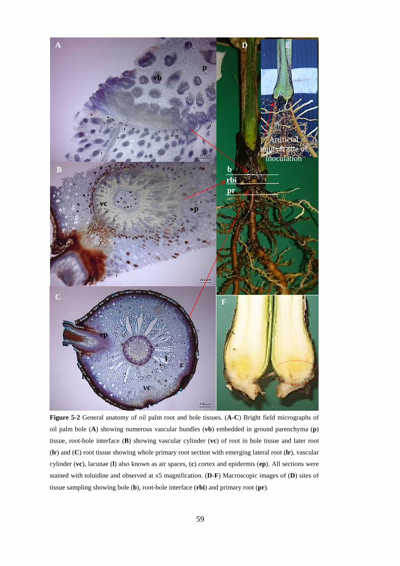
59
A
B
C
D E
Artificial injury& site of
inoculation
pr rbi b
F
vb p
p vc
lr
lr vc
c l
ep
F
Figure 5-2 General anatomy of oil palm root and bole tissues. (A-C) Bright field micrographs of
oil palm bole (A) showing numerous vascular bundles (vb) embedded in ground parenchyma (p)
tissue, root-bole interface (B) showing vascular cylinder (vc) of root in bole tissue and later root
(lr) and (C) root tissue showing whole primary root section with emerging lateral root (lr), vascular
cylinder (vc), lacunae (l) also known as air spaces, (c) cortex and epidermis (ep). All sections were
stained with toluidine and observed at x5 magnification. (D-F) Macroscopic images of (D) sites of
tissue sampling showing bole (b), root-bole interface (rbi) and primary root (pr).

60
.
ep
oc
sc
p
A B
C D
ep sc
oc
p
ep
sc
oc
ic
Figure 5-3Cross-section of control OP primary root. Root cross-section of stained with calcofluor white
under UV fluorescence showing non-fluorescence of sclerenchyma (sc) ring of the outer cortex (oc) (A)
and under bright field stained with toluidine blue (B) showing unstained sclerenchyma ring with tannins.
(C) root tissue stained with safranin o with dark red regions showing highly lignified sclerenchyma tissue.
(D) Low magnification of whole root section stained with calcofluor white

61
A
B
s
vf
C
Figure 5-4 Cross-section of oil palm bole tissue. (A) x 5 magnification showing vascular bundles
embedded in ground parenchyma tissue. Staining with safranin showing highly lignified vascular bundles
and cells in red and ground parenchyma in pink. (B) Close up image of bole showing lignified vascular
fibres (vf) in red against ground parenchyma (p) in pink with abundant starch granules (s). (C) Calcium
oxalate Raphide crystals (r) were also found to be associated with the bole tissue.
62
5.3.3 Histology of early Ganoderma infection in oil palm seedlings
Since this study examined the early stages of BSR infections in oil palm seedlings, only roots and
bole tissue in direct contact with Ganoderma inoculum (Ganoderma colonised agar plugs or
Ganoderma colonised sorghum grains) were excised and processed for examination via bright field
and fluorescence microscopy. Primary roots and bole tissues were excised from plants and further
processed as root, lateral root, root-bole interface preceding the bole, and the bole.
It should be noted that although sampling was carried out between two to six months, all the
specimens used for microscopy were regarded as early infection due to the non-synchronous nature
of BSR development (Rees et al. 2009). The term ‘early infection’ used throughout this thesis refers
to early disease progress rather than processes seen with other fungal pathogens such as
Colletotrichum spp. where the formation of penetration pegs and appressoria are considered as
‘early infection events’.
Although initial site of inoculation was at the lower bole (Fig. 5.2 E) fungal hyphae were generally
observed to favour primary and lateral root as opposed the entry site of inoculation at the injured
bole. Initial penetration was observed in the epidermal layer and sclerenchyma ring comprising the
outer cortex (Fig. 5.6 A, B, D). In lateral roots fungal hyphae were observed to be associated with
non-fluorescent region of the lateral root when stained with calcofluor white and viewed under UV
light. This non-fluorescent region of sclerenchyma was also observed not to absorb toluidine blue
and often maintained a brown pigmentation under bright field microscopy (Fig. 5.5 A, F).
At the bole and the root-bole interface tissues hyphae were seen in lateral roots (Fig. 5.7 C, D) as
well as the brown pigmented parenchyma tissue of the bole region (Fig. 5.7 A, B, E). These brown
pigmented tissues where hyphae were observed appear non-fluorescent when viewed under
fluorescence after staining with calcofluor white.
This study involved early infection progress by Ganoderma in oil palm seedlings and as such it was
expected that macroscopic symptoms, such as internal lesion of both root and bole, would not be
obvious. However, at the microscopic level, evidence of early internal rot was observed on a few
root and root-bole interface sections as demonstrated in Figure 5.8 A-C, where decayed tissue or
lesions appeared to be non-fluorescent under bright field after staining with calcofluor white. As
depicted in Figure 5.8 A and C, the non-fluorescent cells showed thin-walled cells that are observed
to be degraded, adjacent to healthy cell that fluoresce brightly with thicker cell walls (Fig. 5.8 C).
63
Furthermore, the root-bole interface region as shown in Figure 5.8 A shows the presence of hyphae
inside of a cell surrounded by several cells that show partial degradation of cell walls by appearing
to having pockets in their cell walls or simply having discontinuous cell walls.
Comparison of controls to inoculated test plants showed no phenotypic host defence response such
as thickening of cell walls in regions associated with fungal invasion. The formation of specialised
hyphal structures such as appressoria or haustoria were not observed during this work however,
hyphal clamp connections were observed in one instance of a root sample with hyphae in the lateral
root region (Fig. 5.5 E).
Although diligent efforts were made to sterilise all growing substrates (sand & compost) in the glass
house, some but not all controls had fungal contamination and this may possibly be true for the test
plants as well. However, from table 4.4 in chapter 4 it can be seen that fungal establishment in the
tests plants was more extensive than in the controls indicating that what has been observed to a
greater extent was GB3039. Isolation of GB3039 from oil palm tissue was hampered by high
contamination rate. Both controls and test plants from inoculations using GB3039 were put to
Ganoderma selective media (GSM) and turned out negative as well. In the case of contaminations,
other saprophytic and fast growing fungi outgrew Ganoderma making isolation difficult. The only
successful re-isolation was of the Australian isolate RD1on GSM from inoculated roots. The re-
isolated G. boninense was confirmed using ITS sequencing. However, what remains intriguing with
the results from table 4.3 is that there were more establishments of fungal hyphae in root tissue as
compared to bole tissue.
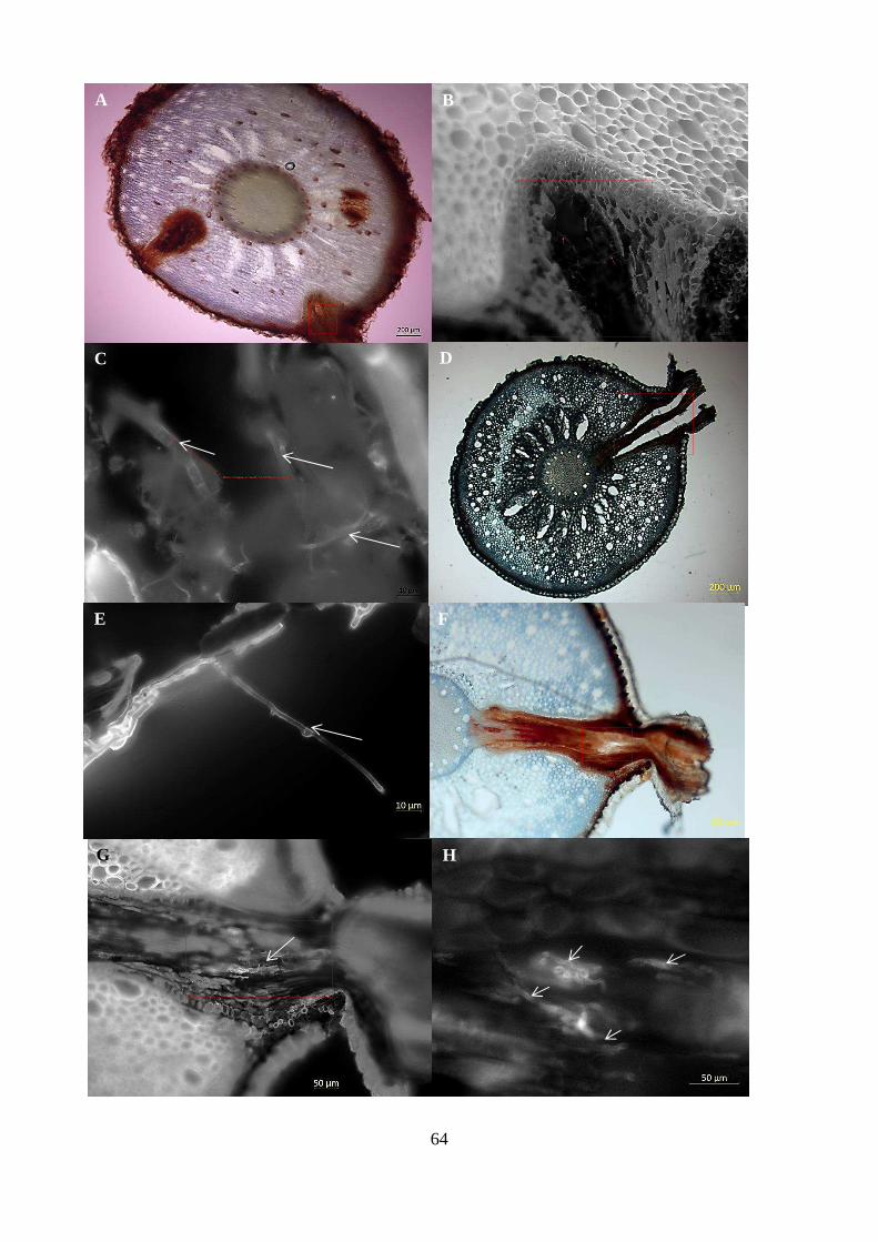
64
A B
C D
E F
G H
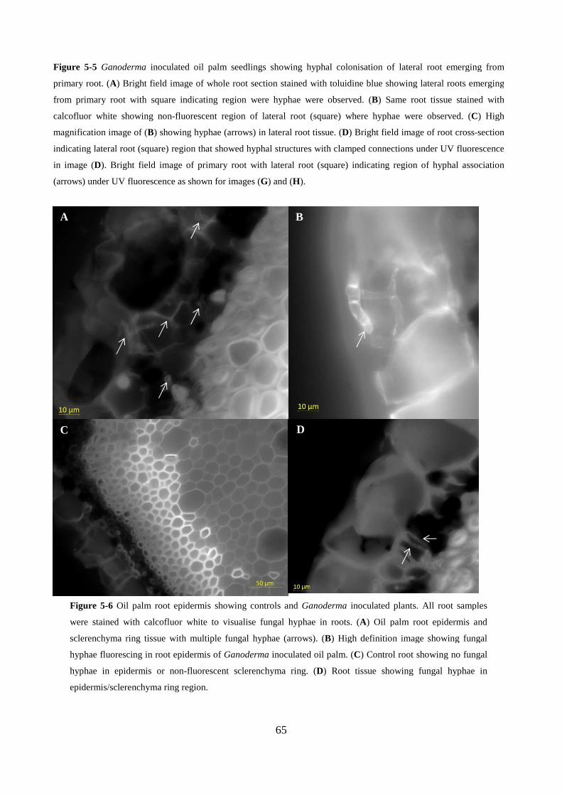
65
A B
C D
Figure 5-5 Ganoderma inoculated oil palm seedlings showing hyphal colonisation of lateral root emerging from
primary root. (A) Bright field image of whole root section stained with toluidine blue showing lateral roots emerging
from primary root with square indicating region were hyphae were observed. (B) Same root tissue stained with
calcofluor white showing non-fluorescent region of lateral root (square) where hyphae were observed. (C) High
magnification image of (B) showing hyphae (arrows) in lateral root tissue. (D) Bright field image of root cross-section
indicating lateral root (square) region that showed hyphal structures with clamped connections under UV fluorescence
in image (D). Bright field image of primary root with lateral root (square) indicating region of hyphal association
(arrows) under UV fluorescence as shown for images (G) and (H).
Figure 5-6 Oil palm root epidermis showing controls and Ganoderma inoculated plants. All root samples
were stained with calcofluor white to visualise fungal hyphae in roots. (A) Oil palm root epidermis and
sclerenchyma ring tissue with multiple fungal hyphae (arrows). (B) High definition image showing fungal
hyphae fluorescing in root epidermis of Ganoderma inoculated oil palm. (C) Control root showing no fungal
hyphae in epidermis or non-fluorescent sclerenchyma ring. (D) Root tissue showing fungal hyphae in
epidermis/sclerenchyma ring region.
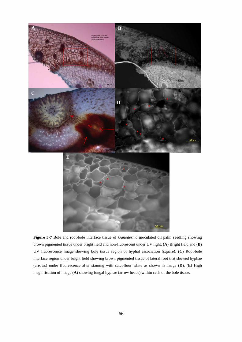
66
A
S
D
C
B
E
Figure 5-7 Bole and root-bole interface tissue of Ganoderma inoculated oil palm seedling showing
brown pigmented tissue under bright field and non-fluorescent under UV light. (A) Bright field and (B)
UV fluorescence image showing bole tissue region of hyphal association (square). (C) Root-bole
interface region under bright field showing brown pigmented tissue of lateral root that showed hyphae
(arrows) under fluorescence after staining with calcofluor white as shown in image (D). (E) High
magnification of image (A) showing fungal hyphae (arrow heads) within cells of the bole tissue.
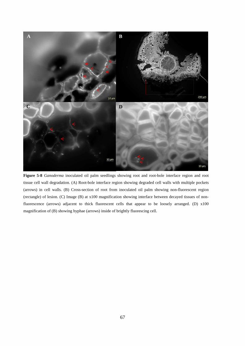
67
A B
C D
Figure 5-8 Ganoderma inoculated oil palm seedlings showing root and root-bole interface region and root
tissue cell wall degradation. (A) Root-bole interface region showing degraded cell walls with multiple pockets
(arrows) in cell walls. (B) Cross-section of root from inoculated oil palm showing non-fluorescent region
(rectangle) of lesion. (C) Image (B) at x100 magnification showing interface between decayed tissues of non-
fluorescence (arrows) adjacent to thick fluorescent cells that appear to be loosely arranged. (D) x100
magnification of (B) showing hyphae (arrows) inside of brightly fluorescing cell.

68
5.4 DISCUSSION
The results from this study give a first account of early infection progress using in-containment
pathogenicity assay and microscopy. Fluorescence microscopy revealed Ganoderma to establish
and colonise epidermis, sclerenchyma ring of the outer cortex, lateral root and the root-bole
interface. It should be noted that the presence of hyphae in all these tissue regions was found to be
associated with brown pigmented tissue assumed to contain polyphenols/tannins and tissue difficult
to stain with the dyes used in this study. General sequence of infection progress showed that as
early as two months post inoculation (mpi) Ganoderma was observed in the root tissue, and was
visible in the root-bole interface approximately three mpi, and then observed in the bole later.
Further microscopic evaluation after five months showed increase of hyphae in the root and root-
bole interface regions. Colonisation of oil palm by Ganoderma appeared to be confined to the
sclerenchyma rings of the outer cortex of the root and other brown pigmented tissues in the bole.
Furthermore, signs of cell wall degradation have been observed in both the root-bole interphase and
the root tissue in a few samples. No discernible host defence responses, such as callose depositions,
were observed in inoculated plants as compared to controls. Rees et al. (2009) also reported similar
results. Specialised hyphal structures such as appressoria and haustoria were not observed in this
study.
For this study, the primary point of inoculation was made at the bole of the seedlings aided by a
small wound to hasten the infection process. This approach was taken to mimic field conditions
where palms are subject to multiple wounds created from frond pruning and bunch harvesting that
may provide a possible entry point for BSR infection. Histology of root and bole tissue suggested
that Ganoderma had a strong affinity for growth in the root. This was evident with frequent
observations of hyphae in the sub epidermal sclerenchyma layer of the root including the primary
root and the lateral root leading into the root-bole interface region. In some instances, hyphae were
only seen in the lateral root and were observed to be moving along this tissue towards the vascular
cylinder centre (Fig.5.7 C) In contrast, for root-bole interface tissue, Ganoderma hyphae were
confined mostly to the non-fluorescent region only with a few instances of advancement leading
into the live parenchyma tissue. Ganoderma is not a soil borne fungus and can only infect through
direct contact between infected roots or other inoculum and healthy roots (Rees et al. 2009).
Therefore, the most probable route of infection in this instance was from the original source of
inoculum (at the bole) down towards the primary roots and lateral roots via the highly lignified
sclerenchyma tissue of the outer cortex. It is possible that Ganoderma grows sparsely in other cell
types, thus making observation more sporadic in those tissues.
69
In root tissue, Ganoderma was associated with the sub epidermal tissue composed of a ‘corky’ layer
of sclerenchyma cells with thick suberinized secondary cell walls (Abdul-Khalil et al. 2010).
Ganoderma was also observed in the brown pigmented tissue in the root-bole interface region. Both
of these cell types in the root and the bole appeared brown under bright field microscopy regardless
of dyes used, and did not seem to absorb stains of any sort. The brown tissue pigment is possibly
due to deposition of polyphenols/tannins which is a common feature of palm root tissue (Seubert,
1996). Furthermore, these cell types did not fluoresce under UV possibly indicating that either these
cells are dead, or that the stains are masked by polyphenols/tannins. The assumption that the non-
fluorescent region/brown pigmented tissue as being dead is highly possible as lesions caused by
injury or fungal invasion are commonly observed in plant tissues as brown discolorations.
Polyphenols/tannins in angiosperms are toxic to fungi; however, these can be broken down by wood
decay fungi (Schwarze, 2007).
Since Ganoderma is a white rot fungus and thus preferentially utilises lignin as primary carbon
source, it is not surprising to find it in the highly lignified sub epidermal sclerenchyma cell layer of
the roots, both primary and lateral. It is possible that as the bole lacks cells with very thick cell
walls at the entry point; Ganoderma grows sparsely, slowly growing down to the roots (perhaps
aided by geotropism). Once in the root, Ganoderma finds the thick walled sclerenchyma cells and
so is able to proliferate at a faster rate, making visualisation more probable. From primary roots the
pathogen can spread into lateral roots. In the bole, the pathogen continues to grow, finally finding
the brown pigmented cells which were hard to visualise and so there is uncertainty regarding the
thickness of their walls. An alternative scenario would be that Ganoderma grows down to the roots
externally (not via the wounded bole), perhaps colonising via lateral roots, than growing up and into
the bole. However, the epidermis of the roots did not appear to be degraded or otherwise breached
and as shown in chapter 3, Ganoderma does not grow in soil. Thus the later theory of infection is
less likely.
The lack of advanced host cell wall degradation and movement of Ganoderma into the cortex is
suggestive of early or slow infection progress given an incubation period of more than five months.
Although no physical-wall related responses were visualised by the host, it is still possible that the
host may be eliciting a chemical defence response (Rees et al. 2009). This however, remains
unknown at this point and may be resolved with a multi-disciplinary approach either employing
molecular and or biochemical studies.

70
The probable action of cell wall degrading enzymes appeared to be involved in the early stages of
infection. Figure 5.8A and C clearly show loosely arranged cells. The observation of pockets in the
cell walls causing discontinuity further suggest lignin degrading enzymes produced by Ganoderma
to play an important role in the infection process (Rees et al. 2009).
Lignin is an important polymer for structure and support in woody tissue and is made of three
monomers, p-hydroxyphenyl (H) units, guaicyl (G) units and syringyl (S) units. Lignin is generally
resistant to fungal degradation and is also reported to play a role in defence mechanism by
reinforcing cell walls during fungal attack (Paterson et al. 2009; Schwarze, 2007). Hard wood trees
contain guaicyl-rich lignin and are more resistant to decay than syringyl-rich lignin. Oil palm is
high in syringyl lignin followed by a small proportion of guaicyl lignin and very little p-
hydroxyphenyl and the proportions of monolignols (S: G: H) are similar to those of grass (Paterson
et al. 2009). Since oil palm is high in syringyl lignin it has been reported to be more susceptible to
white rot decay (Paterson et al. 2009). The occurrence of Ganoderma hyphae in the sub epidermal
area raises some interesting questions with respect to Ganoderma infection of roots. Is Ganoderma
attracted to the lignin content of this tissue area? If so, then what is the lignin composition of root
tissue, bole tissue and stem tissue and how does this influence establishment and infection progress?
The composition of lignin can differ between cell types. It may be that some lignin types are more
attractive to Ganoderma then others, and not just the quantity of lignin. Knowledge of the lignin
composition of tissue types directly associated with BSR infection may further provide information
on the mechanisms involved with Ganoderma during early infection.
Fungal pathogens can have various life styles. Biotrophic fungi acquire nutrition form the plant host
without killing the host, necrotrophic fungi acquire nutrition after killing its host, whilst hemi-
biotrophs start off as biotrophs and then switch to a necrotrophic phase. Wood rots on the other
hand degrade woody material. Wood rots are classed as brown rots, white rots and soft rots. All
wood rot fungi employ cell wall degrading enzymes suited to degrading different wood
constituents. Brown rot fungi such as some ascomycetes and a few basidiomycetes are able to
degrade the hemicellulose content of wood leaving behind lignin, which in some instances can only
partially modify the lignin structure. Soft rot fungi are only able to break down cellulose in wood
leaving behind a soft cavity. White rot is commonly caused by basidiomycete fungi were lignin is
degraded leaving behind the white cellulose thus the term ‘white rot’. White rot fungi can either be
lignin selective where only lignin is degraded, or it can degrade lignin as well as other
71
polysaccharides. Ganoderma is a white rotting fungus which has also been reported to be a hemi-
biotroph (Rees et al. 2009). The association with matured sclerenchyma (possibly dead tissue) of
the root and the brown pigmented (possibly dead) tissue from the root-bole interface could facilitate
nutritional requirements and possible longitudinal growth, potentially without elicitation of a
defence response by the oil palm host. It may be that Ganoderma establishes in these dead regions
as saprophyte and proliferates and possibly grows longitudinally until it reaches the bole or laterally
into the cortex once lignin in the sclerenchyma has been exhausted and then it switches over to a
more classical necrotrophic phase. At later stages of infection it is known that Ganoderma can
colonise the middle of the trunk/bole leading to general degradation of all cell types/tissues with no
apparent preference (Rees et al. 2009)
Overall this study presents a first account of early infection progress in oil palm seedlings by G.
boninense. Bole inoculations revealed that Ganoderma grew faster in the roots, both primary and
lateral roots and the presumably dead region of root-bole interface region, colonising the bole at a
later stage. Moreover, the information gathered from this study has set a foundation for future
Ganoderma-oil palm interaction studies and has contributed new knowledge with respect to the
Ganoderma infection process.
72
Chapter 6 GENERAL DISCUSSION, FUTURE
DIRECTIONS AND CONCLUSION
6.1 GENERAL DISCUSSION
Oil palm is an important agricultural commodity that is cultivated globally throughout the tropical
regions including many developing countries. Oil palm is cultivated both by commercial milling
companies and small holder farmers who depend heavily on this crop as their source of livelihood.
South East Asia accounts for over 80% of total palm oil production of which Malaysia and
Indonesia are the largest producers (Rees et al. 2007). Basal stem rot (BSR) caused by Ganoderma
spp. is the major constraint to oil palm production in SE Asia (Chan et al. 2011; Naher et al. 2013).
Although BSR disease has been known for over 100 years (Turner, 1981), progress into its control
and management has been slow due to our lack of understanding regarding Ganoderma-oil palm
interactions. Currently, there are no effective control measures. At best, BSR management is aimed
at maintaining disease levels below the economic threshold of 20 % (Turner, 1981) by removal of
infected palms throughout the lifespan of a stand (Sanderson, 2005; Pilotti et al. 2004). The long
term sustainable approach to BSR disease would be to breed for resistant and/or tolerant varieties
(Idris et al. 2004). This in turn is dependent on the development of a reliable screening assay to
identify potential candidates to breeders. Once this is achieved it would be possible to identify
factors that are associated with resistance and or tolerance in oil palm and use these traits as
markers to select premium breeding material with enhanced tolerance/resistance to Ganoderma
infection.
Commercially important palms such as coconuts (Kandan et al. 2010), dates (Adaskaveg et al.
1991; Elliott & Broschat, 2001) and oil palm (Turner, 1981) have been known to be affected by a
number of Ganoderma spp., however, there is very limited knowledge regarding mechanism of
infection. Previous studies have looked at wood decay mechanism of white rot fungi for
biotechnology purposes such as wood pulping for paper manufacturing and biofuel production
(Fackler et al. 2007). Only a limited number of histological and chemical studies have been carried
out on the BSR disease mechanism of hard wood species (Adaskaveg et al. 1990), dates
73
(Adaskaveg et al. 1991) and coconuts (Kandan et al. 2009). The only recent histological study on
Ganoderma -oil palm interaction has been carried out by Rees et al. (2009) where Ganoderma was
reported to have a hemi-biotrophic relationship with oil palm. However, information regarding the
early stages of establishment and infection preceding the biotrophic phase was unknown until this
study was conducted. Employing the use of microscopy and specific staining techniques has shed
new light on the Ganoderma-oil palm interactions during the early stages of disease establishment
and progress. In addition, this study has set a foundation and direction for further studies of
Ganoderma-oil palm interactions.
Information regarding the histology of oil palm tissues was very scarce and if present in literature
was often very limited in detail and description. Knowledge and understanding of the oil palm
anatomy was deemed essential as it facilitated better understanding of the different tissues and cells
that may have played a possible role in the interaction with Ganoderma during basal stem rot
infections. The works of Seubert (1996) and Rees et al. (2009) have been useful in providing
relevant information regarding internal oil palm anatomy.
Initial delays in importing Ganoderma isolate 3039 from PNG prompted an investigation to look for
possible G. boninense or other closely related pathogens of BSR disease such as G. zonatum that
could be used for pathogenicity assays. Therefore, a total of five Australian Ganoderma isolates
were collected and identified using morphology and ITS sequencing. Of the five Ganoderma
isolates, two of the isolates (RD1 and RD2) were identified as G. boninense after ITS sequencing
while the other three isolates were putatively identified as G. tropicum (UQ3), G. australe (UQ4)
and Phellinus spp. Isolate 3039 from PNG was also confirmed as G. boninense after ITS
sequencing supporting the view by Pilotti et al. (2004) that G. boninense is the major pathogen of
BSR disease of oil palm in PNG.
The significance of obtaining G. boninense isolates here in Australia means that future work can be
carried without the restrictions and hassles of working with quarantine material. Furthermore,
basidiospores have been implicated in their role of BSR spread; however, to date no one has been
able to successfully demonstrate the use of basidiospore inoculation to induce BSR disease in oil
palm. Therefore, the potential to rear brackets from Australian G. boninense isolates and ultimately
basidiospores for inoculations of oil palm seedlings is very much anticipated as it is thought that
spore inoculations closely follow the natural infection process in the plantations.
74
Other Ganoderma species that have also been previously found to be associated with BSR disease
include G. zonatum, and G. miniatocinctum (Wong et al. 2012). Information regarding Ganoderma
species identification and host associated within an oil palm cropping system is vital for control and
management. For example, from this study is has been found that golden cane plays host to G.
boninense. This implies that other palm hosts such as coconuts may act as reservoirs for the
continual spread of BSR disease (Naher et al. 2013). Indeed, there appears to be an increase in BSR
incidence among ornamental palms in far north of Australia (Hennessy & Daly, 2007). In South
India it has been reported that coconuts are affected by G. lucidum, G. boninense and G.
applanatum (Kandan et al. 2010; Mandal et al. 2014). BSR incidences have been observed in oil
palm planted in ex-BSR infected coconut gardens (Mandal et al. 2014) suggesting possible carry
over from the same pathogens affecting coconuts. In addition, BSR incidences in oil palm planted
after coconut (Turner, 1981) palms is higher than after virgin forest or other crops, suggesting that
coconut palms may also be a reservoir for pathogenic Ganoderma. Therefore, precautionary efforts
should be considered to avoid potential inoculum build up from other alternative palm hosts
including dead palms and stumps. These can be addressed by determining the relationships between
saprophytic Ganoderma isolates growing on dead logs and stumps and their relationships to
pathogenic Ganoderma isolates growing on live palms. An existing ACIAR funded project with
PNG-OPRA in collaboration with the University of Queensland has set out to address this issue for
long term control of BSR disease of oil palm. It is anticipated that knowledge gained from this work
may be beneficial for plantations as informed decisions can be made to improve field sanitation
procedures. For instance, if saprophytic Ganoderma found on windrowed logs is found to be
associated or related to Ganoderma pathogens on live palms then control and field management
procedures should be extended to take into consideration windrowed logs and stumps, including of
other nearby palms. Ideally, the removal of all potential sources of inoculum would be of great
benefit; however it is not economical and is logistically difficult. Alternatively, other methods
should be explored to look at rapid break down of felled oil palm logs, especially at replant, in an
attempt to reduce the inoculum build up and narrow the window of opportunity for BSR infection.
It takes two years for felled oil palm logs (often stacked between inter-rows) to break down and
finding an efficient wood degrader that is compatible with the oil palm cropping system may be of
great benefit.
Preliminary investigations discussed in Chapter 4 have identified sorghum grain and agar plugs as
suitable substrates for Ganoderma growth as inoculum for in-vitro Ganoderma pathogenicity assay.
The choice of these substrates was based on rapid and reliable colonisation of substrates and
75
relative ease of use during preparation and inoculations. The present inoculation procedure used
smaller inoculum loads (1 g sorghum and agar plug 1 cm dia.) as opposed to using large rubber
wood blocks (18-750 cm3) which are routinely used in nursery pathogenicity assays. The use of a
small inoculum load coupled with inoculations to boles with wounding is less artificial and
probably more closely mimics field infections involving basidiospores infections of pruned fronds
leading to stem rots. The use of a small inoculum load is proportional with the smaller sized palm
seedlings used in this study. Basidiospores have been suggested to play an important role in the
spread of BSR disease and it is postulated that air borne basidiospores infect through wounds made
by continuous pruning during harvesting of fruit bunches or natural injury. Therefore, the use of
smaller inoculum load aided by artificial wounding was deemed more reflective of the theory of
infection by basidiospores and thus, this approach was used in the present study. Furthermore,
wounding of plants gave an indication where initial site of inoculation and possible BSR infection
may have occurred allowing for selective sampling and sectioning of regions of interest with higher
chances with higher chances of visualising the pathogen.
Foliar symptoms of wilting and nutrient deficiency have been reported to occur when 50% of the
bole has been damaged as a result of BSR infections (Turner, 1981). There are however, several
variations to field disease symptoms. For example, there are instances in plantations in Milne Bay,
PNG where palms with extensive BSR infections show; no foliar symptoms until they suddenly
fracture or remain highly productive until they facture. These palms may possibly be partially
resistant or at least tolerant to BSR infection. These observations may suggest that the type of
genetic material planted may have an influence on the rate and progress of BSR infection or can just
simply be due to infections with a less aggressive isolate. Similarly, it has also been report by
Durand-Gasselin et al. (2005) that planting material of Deli origin was highly susceptible to BSR
infection that material from Africa. Infections studies from the current work have indicated no
macroscopic symptoms and this could be possible due to slow infection by Ganoderma. Therefore,
a histological approach was taken to assess the status of each inoculation method. Histological
assessment of both sorghum and agar plugs suggest that both sources of inoculum were able to
facilitate Ganoderma establishment and development in the treated seedlings. However, the
sorghum grain inoculum was generally found to demonstrate a better infection potential in contrast
to agar plugs where establishment of Ganoderma on treated seedlings occurred more readily than
during agar plug inoculations. It should be noted here that although sorghum was found to have a
better infection potential, it is most likely that this may be due to the effect of inoculum load (Rees
et al. 2007) rather than the use of the substrate itself. Therefore, further work is still required to

76
determine the appropriate and effective inoculum load as well as to validate whether there are any
significant differences between the two methods.
Ideally, future trials involving Ganoderma-oil palm interaction studies should strongly consider
basidiospore inoculation as a complementary to this study. However, obtaining large quantities of
basidiospores from a pure isolate (as opposed to a mix from various brackets) is not easy, and is
possibly why no one has been able to demonstrate infection using basidiospores (Hasan et al. 2005).
The insufficient quantities of spores are compounded by their short lifespan of only a few days.
Interestingly, nursery seedlings inoculated using rubber wood blocks colonised with monokaryotic
G. boninense mycelia were not able to induce BSR symptoms (Lim & Fong, 2005; Chan et al.
2011) and were suggested to be saprophytic in contrast to dikaryons which were highly pathogenic
on oil palm (Chan et al. 2011). Both monokaryotic and dikaryotic mycelia are able to colonise oil
palm tissue (Rees et al. 2012) however, it still remains unknown if monokaryons play an important
role in BSR establishment and disease progress preceding anastomosis.
Histological assessment using bright field and fluorescence microscopy with appropriate staining
techniques has been beneficial in visualising the establishment and progress of Ganoderma during
early BSR infections. However, more detailed work is still required to fully understand the disease
progress from its early stages through to advanced stages of BSR infection and host responses.
Much of this work would require Ganoderma specific staining and a longer incubation period.
Fluorescence microscopy coupled with calcofluor white staining revealed fungal hyphae associated
with primary root, lateral root emerging from primary root and root-bole interface tissue of the bole.
It should be noted that the initial point of inoculations was made at the bole or lower stem region;
however, fungal hyphae were more often seen in root tissues as opposed to the bole, the site of
inoculation. It could be possible that Ganoderma has an affinity to certain root tissue types.
Furthermore, the cell types in which fungal hyphae were found to be associated with were the
sclerenchyma ring of the outer root cortex and the non-fluorescent/brown pigmented tissue of the
root-bole interface region. The sclerenchyma ring zone is made up of thick matured sclerenchyma
cells that are dead when matured and form a hard corky ring layer around the root. The non-
fluorescent tissue of the bole region is made up of single-cell layer of parenchyma cells. These
tissue regions are presumed to be dead regions. On the contrary, it could be possible that

77
Ganoderma has a liking for the brown pigmentation and not necessarily association to what is
assumed to be dead tissue.
One theory may be that Ganoderma initially establishes as a saprophyte in dead regions of both the
root and bole tissue. It is possible that it may remain in these tissues without triggering a host
defence response as well as attaining a food source. Then, once all food (lignin) is exhausted in this
area and a build-up of biomass has occurred, Ganoderma switches over to become pathogenic
and/or more aggressive and invade live tissue. It still remains unknown what triggers aggressiveness
in Ganoderma. Analysis of Ganoderma transcriptome during infection would show which genes
are involved in BSR and so provide an insight into this pathogen’s lifestyle, and possible clues on
how to control it.
6.2 FUTURE DIRECTIONS
6.2.1 GFP transformed Ganoderma isolates
The visualisation of fungi in planta is often difficult using conventional staining methods and is not
specific to the fungi of interest. Fungi can be visualised either with strong antigens for
immunofluorescence microscopy (Christopher & Nicholas, 2006) or green fluorescent protein
(GFP) for specific viewing of a fungal isolate of specific interest. The gfp gene has been widely
used to transform both plants (Qi et al. 2016) and fungi (Lorang et al. 2001; Oren et al. 2003) and
such GFP expressing transformants have been invaluable in histological work. Generation of GFP-
expressing transgenic Ganoderma could potentially be achieved by particle bombardment and/or
agrobacterium mediated transformation. The advantage of obtaining GFP-tagged Ganoderma
isolates would mean that any contamination by other fungi could be dismissed and that the confocal
microscope could also be used to follow the infection process of Ganoderma in oil palm seedlings.
6.2.2 Basidiospore inoculations
The current study of BSR establishment and development shows that it is possible using a smaller
inoculum load such as agar plugs and sorghum grains colonised with Ganoderma. Basidiospores
have been implicated to play an important role in the spread of BSR disease (Sanderson et al. 2000;
Pilotti et al. 2003). However, no one has been able to artificially induce BSR disease in oil palm
using basidiospores. Therefore, future work should consider in-vitro rearing of basidiospores from
well characterised isolates that can be used for oil palm pathogenicity tests where evaluation of
BSR infections should be assessed at a microscopic level. It is also important to determine whether
78
BSR disease arises from one single infection event or multiple infection events of basidiospores.
The use of basidiospores for inoculations is highly regarded as it is thought to be more reminiscent
of the natural infection process that occurs in plantations. Therefore, its application in Ganoderma-
oil palm interaction studies will be very beneficial as it is a more realistic approach.
6.2.3 Nursery pathogenicity tests
Current nursery testing involves the use of breeding lines or F1 hybrids which are genetically
variable and results in non-synchronous appearance of symptoms. Therefore, the use of clonally
derived plants should be considered in order to standardise trials. For example, in order to properly
assess and evaluate an inoculation technique, all seedling test plants should be clonally derived to
reduce host genetic variables and tested against one Ganoderma isolate. These tests should be
repeated and amended accordingly until a repeatable and reliable result can be achieved. There and
then, can we begin to introduce clonal seedlings inoculated with several Ganoderma isolates and
finally, different breeding lines maybe introduced against one or several Ganoderma isolates. The
consolidated data collected from these sets of experiments would give a more reliable and accurate
representation of the Ganoderma-oil palm interactions. Furthermore, it is imperative that nursery
tests be correlated with field conditions, something which is still lacking for this disease.
6.3 CONCLUSION
The aims of this study have been achieved to a beneficial degree with novel insights into
Ganoderma-oil palm interaction, particularly the early establishment and progress of G. boninense
in oil palm seedlings. Inoculation of seedlings at the bole confirmed Ganoderma’s preference for
both primary and later roots, specifically the highly lignified sclerenchyma cells of the outer cortex.
The tissue regions where Ganoderma has been observed the most did not fluoresce under
fluorescence microscopy when stained with calcofluor white indicating that these were either dead
or so highly lignified that they could not be stained. Furthermore, this may also suggest that initial
establishment of Ganoderma in oil palm is more saprophytic than pathogenic. No specialized
hyphal structures were observed during this early infection study.
Although the work is preliminary, it sets a foundation for future studies involving host – pathogen
interactions towards the long term control of basal stem rot of oil palm caused by Ganoderma.
Future work on Ganoderma-oil palm interactions should consider a longer time frame for
experiments as the infection process and progress by Ganoderma is slow but also progressive.
79
References
Abdullah, J., Hassan, H., Shari, M. R., Mohd, S., Mustapha, M., Mahmood, A. A., Hamid, N. H. (2013). Gamma scorpion: Mobile gamma-ray tomography system for early detection of basal stem rot in oil palm plantations. Optical Engineering, 52(3). doi:10.1117/1.oe.52.3.036502
Adaskaveg, J. E., Gilbertson, R. L., & Blanchette, R. A. (1990). Comparative studies on delignification caused by Ganoderma species. Applied and Environmental Microbiology, 56(6), 1932-1943. Retrieved from <Go to ISI>://CABI:19911368909 http://aem.asm.org/content/56/6/1932.full.pdf
Adaskaveg, J. E., Blanchette, R. A., & Gilbertson, R. L. (1991). Decay of date palm wood by white-rot and brown-rot fungi. Canadian Journal of Botany, 69(3), 615-629. doi:10.1139/b91-083
Alvarez, E., Llano, G. A., Loke, J. B., & Chacon, M. I. (2012). Characterization of Thielaviopsis paradoxa isolates from oil palms in Colombia, Ecuador and Brazil. Journal of Phytopathology, 160(11-12), 690-700. doi:10.1111/jph.12012
Ariffin, D., Idris, A., & Hassan, A. (1989). Significance of the black line within oil palm tissue decayed by Ganoderma boninense. Elaeis, 1(1), 11 - 16.
Ariffin, D., Idris, A. S., & Azahari, M. (1996). Spread of Ganoderma boninense and vegetative compatibility studies of single field palm isolates. Paper presented at the PORIM International Palm Oil Congress, Kuala Lumpur.
Ariffin, D., Idris, A., & Singh, G. (2000). Status of Ganoderma in Oil Palm. In J. Flood, P. Bridge, & M. Holderness, Ganoderma Diseases of Perennial Crops (pp. 49-79). Wallingford, UK: Cabi Publishing.
Bridge, P. D., O'Grady, E., Pilotti, C. A., & Sanderson, F. R. (2000). Development of Molecular Diagnostics for the Detection of Ganoderma Isolates Pathogenic to Oil Palm. In J. Flood, P. D. Bridge, & M. Holderness (Eds.), Ganoderma Diseases of Perennial Crops (pp. 225 - 247). UK: CABI Publishing.
Bhadauria, V., Miraz, P., Kennedy, R., Banniza, S., & Wei, Y. (2010). Dual trypan-aniline blue fluorescence staining methods for studying fungus-plant interactions. Biotechnic & Histochemistry, 85(2), 99-105. doi:10.3109/10520290903132196
Chan, J., Latiffah, Z., Liew, K., & Idris, A. (2011). Pathogenicity of monokaryotic and dikaryotic mycelia of Ganoderma boninense on oil palm seedlings and germinated seeds in Malaysia. Australasian Plant Pathology, 40(3), 222-227. doi:10.1007/s13313-011-0031-4
Christopher, R. T., & Nicholas, J. T. (2006). Immunofluorescence microscopy and immunogold EM for investigating fungal infection of plants. Nature Protocols, 1(5), 2506. doi:10.1038/nprot.2006.347
Coetzee, M. P. A., Wingfield, B. D., Golani, G. D., Tjahjono, B., Gafur, A., & Wingfield, M. J. (2011). A single dominant Ganoderma species is responsible for root rot of Acacia mangium and Eucalyptus in Sumatra. Southern Forests, 73(3-4), 175-180. doi:10.2989/20702620.2011.639488

80
Cooper, R., Flood, J., & Rees, R. (2011). Ganoderma boninense in oil palm plantations: current thinking on epidemiology, resistance and pathology. Planter, 87(1024), 515 - 526.
Corley, R. H. V., & Tinker, P. B. (2003). The Oil Palm. Blackwell Science Ltd.
Darmono, T. W. (1998). Molecular approaches to the elucidation of basal stem rot disease of oil palm. Proceedings of BTIG Workshop on Oil Palm Improvement through Biotechnology, Bogor, Indonesia, April 1997, pp.83-93.
Darmono, T. W. (2000). Ganoderma in oil palm in Indonesia: current status and prospective use of antibodies for the detection of infection. In J. Flood, P. Bridge, & M. Holderness, Ganoderma Diseases of Perennial Crops (pp. 249-266). Wallingford, UK: Cabi Publishing.
Durand-Gasselin, T., Asmady, H., Flori, A., Jacquemard, J. C., Hayun, Z., Breton, F., & de Franqueville, H. (2005). Possible sources of genetic resistance in oil palm (Elaeis guineensis Jacq.) to basal stem rot caused by Ganoderma boninense - prospects for future breeding. Mycopathologia, 159(1), 93-100. doi:10.1007/s11046-004-4429-1
Fackler, K., Schwanninger, M., Gradinger, C., Srebotnik, E., Hinterstoisser, B., & Messner, K. (2007). Fungal decay of spruce and beech wood assessed by near-infrared spectroscopy in combination with uni- and multivariate data analysis. Holzforschung, 61(6), 680-687. doi:10.1515/HF.2007.098
FAOSTAT 2015. Retrieved September 05, 2016, from http://faostat3.fao.org/home/E
Flood, J. (2006). A review of Fusarium wilt of oil palm caused by Fusarium oxysporum f. sp elaeidis. Phytopathology, 96(6), 660-662. doi:10.1094/phyto-96-0660
Flood, J., Hasan, Y., Turner, P., & O'Grady, E. (2000). The spread of Ganoderma from infective sources in the field and its implications for management of the disease in oil palm. Ganoderma Diseases of Perennial Crops, 101 - 112.
Geib, S. M., Filley, T. R., Hatcher, P. G., Hoover, K., Carlson, J. E., Jimenez-Gasco Mdel, M., Tien, M. (2008). Lignin degradation in wood-feeding insects. Proceedings of the National Academy of Sciences USA, 105(35), 12932-12937. doi:10.1073/pnas.0805257105
George, S. T., Chung, G. F., & Zakaria, K. (1996). Updated results (1990–1995) on trunk injection of fungicides for the control of Ganoderma basal stem rot. Paper presented at the 1996 PORIM International Palm Oil Congress (Agriculture), Bangi, Selangor, Malaysia.
Grieve, R. B. (1986). The oil palm industry of Papua New Guinea. Australian Geographer, 17, 72-76.
Hartley, C. W. S. (1988). The Oil Palm (Elaeis guineensis Jacq.). New York: Longman Scientific and Technical Publication.
Haskins, E. F. (1982). Toluidine Blue: A simple, effective stain for plant tissues. The American Biology Teacher, 44(8), 487-489. doi:10.2307/4447575
Hidayati, N., Glen, M., Nurrohmah, S. H., Rimbawanto, A., Mohammed, C. L., & Woodward, S. (2014). Ganoderma steyaertanumas a root-rot pathogen of forest trees. Forest Pathology, 44(6), 460-471. doi:10.1111/efp.12142
Ho, Y. W., & Nawawi, A. (1985). Ganoderma boninense Pat. from basal stem rot of oil palm (Elaeis guineensis) in Peninsular Malaysia. Pertanika, 8(3), 425-428.

81
Hushiarian, R., Yusof, N., & Dutse, S. (2013). Detection and control of Ganoderma boninense: strategies and perspectives. SpringerPlus, 2(1), 555. Retrieved from http://www.springerplus.com/content/2/1/555
Idris, A., Kushairi, A., Ismail, S., & Ariffin, D. (2004). Selection for partial resistance in oil palm progenies to Ganoderma basal stem rot. Journal of Oil Palm Research, 16(2), 12 - 18.
Idris, A., Yamaoka, M., Hayakawa, S., Basri, M., Noorhasimah, I., & Ariffin, D. (2003). PCR technique for detection of Ganoderma. MPOB Inf Ser MPOB TT No.188:MPOB TT No.188, MPOB TT No.188.
ITS Global, (2011). The economic benefits of oil palm in Papua New Guinea. Melbourne: ITS Global.
Jollands, P. (1983). Laboratory investigations on fungicides and biological agents to control three diseases of rubber and oil palm and their potential applications. Tropical Pest Management, 29, 33-38.
Kandan, A., Radjacommare, R., Ramanathan, A., Raguchander, T., Balasubramanian, P., & Samiyappan, R. (2009). Molecular biology of Ganoderma pathogenicity and diagnosis in coconut seedlings. Folia Microbiologica, 54(2), 147-152. doi:10.1007/s12223-009-0022-9
Knight, N. L., & Sutherland, M. W. (2011). A rapid differential staining technique for Fusarium pseudograminearum in cereal tissues during crown rot infections. Plant Pathology, 60(6), 1140-1143. doi:10.1111/j.1365-3059.2011.02462.
Latiffah, Z., Harikrishna, K., Tan, S. G., Tan, S. H., Abdullah, F., & Ho, Y. W. (2002). Restriction analysis and sequencing of the ITS regions and 5.8S gene of rDNA of Ganoderma isolates from infected oil palm and coconut stumps in Malaysia. Annals of Applied Biology, 141(2), 133-142. doi:10.1111/j.1744-7348.2002.tb00205.x
Lee, S. S. (2000). The Current Status of Root Diseases of Acacia mangium Willd. In J. Flood, P. Bridge, & M. Holderness, Ganoderma Diseases of Perennial Crops (pp. 71-79). Wallingford, UK: Cabi Publishing.
Lim, H. P., & Fong, Y. K. (2005). Research on basal stem rot (BSR) of ornamental palms caused by basidiospores from Ganoderma boninense. Mycopathologia, 159(1), 171-179. doi:10.1007/s11046-004-4440-6
Lim, T. K., Chung, G. F., & Ko, W. H. (1992). Basal stem rot of oil palm caused by Ganoderma boninense. Plant Pathology Bulletin, 1(3), 147-152. Retrieved from <Go to ISI>://CABI:19942306566
Lorang, J. M., Tuori, R. P., Martinez, J. P., Sawyer, T. L., Redman, R. S., Rollins, J. A., . . . Ciuffetti, L. M. (2001). Green fluorescent protein is lighting up fungal biology. Applied and Environmental Microbiology, 67(5), 1987.
Ma, Q., Hu, L., Kong, L., Huang, S., Dai, H., LI, Y., & Zhao, Y. (2013). Chemical constituents from the fungus Ganoderma tropicum (Jungh.) Bres. and their cytotoxic activities. African Journal of Microbiology Research, 7(16), 1543-1547. doi:10.5897/AJMR12.1649
Mendgen, K., & Hahn, M. (2002). Plant infection and the establishment of fungal biotrophy. Trends in Plant Science, 7(8), 352-356. doi:http://dx.doi.org/10.1016/S1360-1385(02)02297-5
82
Merrill, W. (1992). Mechanisms to resistance of fungi in woody plants: a historical perspective. In R. A. Blanchette, & A. R. Biggs, Defence Mechanisms of Woody Plants Against Fungi (pp. 1-12). Berlin: Spriner-Verlag.
Miller, R. (1995). The Characterization of Ganoderma Populations in Oil palm cropping systems. PhD thesis, University of Reading, UK.
Miller, R.N.G. Holderness, M., Bridge, P.D., Chung, G.F., & Zakaria, M.H. (1999). Genetic diversity of Ganoderma in oil palm plantings. Plant Pathology, 48(5), 595-603. doi:10.1046/j.1365-3059.1999.00390.x
Moncalvo, J.-M. (2000). Systematics of Ganoderma. In J. Flood, P. Bridge, & M. Holderness, Ganoderma Diseases of Perennial Crops (pp. 23-45). Wallingford, UK: Cabi Publishing.
Moncalvo, J.-M., & Ryvarden, L. (1997). A nomenclatural study of the Ganodermataceae Donk. Synopsis Fungorum II.
Muhammed, C. L., Rimbawanto, A., & Page, D. E. (2014). Management of basidiomycete root- and stem-rot diseases in oil palm, rubber and tropical hardwood plantation crops. Forest Pathology, 44, 428-446.
Naher, L., Tan, S. G., Yusuf, U. K., Ho, C. L., & Siddiquee, S. (2012). Activities of chitinase enzymes in the oil palm (Elaeis guineensis Jacq.) in interactions with pathogenic and non-pathogenic fungi. Plant Omics, 5(4), 333-336. Retrieved from <Go to ISI>://WOS:000310985700003
Naher, L., Yusuf, U. K., Tan, S. G., Siddiquee, S., & Islam, M. R. (2014). In-vitro and in-vivo biocontrol performance of Trichoderma harzianum Rifai on Ganoderma boninense Pat. Related to pathogenicity on oil palm (Elaeis guineensis Jacq.). Journal of Pure and Applied Microbiology, 8(2), 973-978.
Natarajan, S., Bhaskaran, R., & Shanmugan, N. (1986). Preliminary studies to develop techniques for early detection of Thanjavur wilt in coconut. Indian Coconut Journal, 17(3), 3 - 6.
Navaratnam, S., & Chee, K. (1965). Root inoculation of oil palm seedlings with Ganoderma spp. Plant Disease, 49, 1011 - 1012.
NBPOL. (2013). Annual Report 2013. Retrieved from http://www.nbpol.com.pg/wp-content/uploads/downloads/2014/04/New-Britain-Palm-Oil-Limited-NBPOL-Annual-Report-2013.pdf:
Oßwald, W., Fleischmann, F., Rigling, D., Coelho, A. C., Cravador, A., Diez, J., . . . Werres, S. (2014). Strategies of attack and defence in woody plant–Phytophthora interactions. Forest Pathology, 44(3), 169-190. doi:10.1111/efp.12096
Oliva, J., Bendz-Hellgren, M., & Stenlid, J. (2011). Spread of Heterobasidion annosum s.s. and Heterobasidion parviporum in Picea abies 15 years after stump inoculation. FEMS MicrobiologyEcology (Vol. 75).
Oren, L., Ezrati, S., Cohen, D., & Sharon, A. (2003). Early Events in the Fusarium verticillioides-Maize interaction characterized by using a green fluorescent protein-expressing transgenic isolate. Applied and Environmental Microbiology, 69(3), 1695.
83
Panchal, G., & Bridge, P. D. (2005). Following basal stem rot in young oil palm plantings. Mycopathologia, 159(1), 123-127. doi:10.1007/s11046-004-4434-4
Paterson, R. R. M., Moen, S., & Lima, N. (2009). The feasibility of producing oil palm with altered lignin content to control Ganoderma disease. Journal of Phytopathology, 157(11-12), 649-656. doi:10.1111/j.1439-0434.2009.01553.x
Pearce, R. B. (1996). Antimicrobial defences in the wood of living trees. New Phytologist, 132(2), 203-233. doi:10.1111/j.1469-8137.1996.tb01842.x
Perfect, S. E., Hughes, H. B., & O'Connell, R. J. (1999). Colletotrichum: A model genus for studies on pathology and fungal-plant interactions. Fungal Genetics and Biology, 27, 186-198.
Pilotti, C. A. (2005). Stem rots of oil palm caused by Ganoderma boninense: Pathogen biology and epidemiology. Mycopathologia, 159(1), 129-137. doi:10.1007/s11046-004-4435-3
Pilotti, C. A., Sanderson, F. R., & Aitken, E. A. B. (2002). Sexuality and interactions of monokaryotic and dikaryotic mycelia of Ganoderma boninense. Mycological Research, 106, 1315-1322. doi:10.1017/s0953756202006755
Pilotti, C. A., Sanderson, F. R., Aitken, E. A. B., & Armstrong, W. (2004). Morphological variation and host range of two Ganoderma species from Papua New Guinea. Mycopathologia, 158(2), 251-265. doi:10.1023/B:MYCO.0000041833.41085.6f
Prychid, C. J., & Rudall, P. J. (1999). Calcium oxalate crystals in monocotyledons: A Review of their structure and systematics. Annals of Botany, 84(6), 725-739. doi:10.1006/anbo.1999.0975
Qi, J., Li, G.-Q., Dong, Z., & Zhou, W. (2016). Transformation of tobacco plants by Yali PPO-GFP fusion gene and observation of subcellular localization. American journal of translational research, 8(2), 698.
Rankine, I., & Fairhurst, T. (1999). Field Handbook (2 ed. Vol. 1). Singapore: Oxford Graphic Printers.
Rees, R. W. (2006). Ganoderma Stem Rot of Oil Palm (Elaeis guineensis): Mode of infection, Epidemiology and Biological Control. (PHD), University of Bath.
Rees, R. W., Flood, J., Hasan, Y., & Cooper, R. M. (2007). Effects of inoculum potential, shading and soil temperature on root infection of oil palm seedlings by the basal stem rot pathogen Ganoderma boninense. Plant Pathology, 56(5), 862-870. doi:10.1111/j.1365-3059.2007.01621.x
Rees, R. W., Flood, J., Hasan, Y., Potter, U., & Cooper, R. M. (2009). Basal stem rot of oil palm (Elaeis guineensis); mode of root infection and lower stem invasion by Ganoderma boninense. Plant Pathology, 58(5), 982-989. doi:10.1111/j.1365-3059.2009.02100.x
Rees, R. W., Flood, J., Hasan, Y., Wills, M. A., & Cooper, R. M. (2012). Ganoderma boninense basidiospores in oil palm plantations: evaluation of their possible role in stem rots of Elaeis guineensis. Plant Pathology, 61(3), 567-578. doi:10.1111/j.1365-3059.2011.02533.x
Rolph, H., Wijesekara, R., Lardner, R., Abdullah, F., Kirk, P. M., Holderness, M., . . . Flood, J. (2000). Molecular Variation in Ganoderma Isolates from Oil Palm, Coconut and Betelnut. In J. Flood, P. Bridge, & M. Holderness, Ganoderma Diseases of Perennial Crops (pp. 205-221). Wallingford, UK: Cabi Publishing.
84
Ruzin, S. E. (1999). Plant Microtechnique and Microscopy / Steven E. Ruzin. New York: New York : Oxford University Press.
Ryvarden, L. (1995). Can we trust morphology in Ganoderma? Paper presented at the Contributed Symposia 5A,B, 5th International Mycological Congress, Vancouver, 19 - 24.
Sanderson, F. R., Pilotti, C. A., & Bridge, P. D. (Eds.). (2000). Basidiospores: Their Influence on Our Thinking Regarding a Control Strategy for Basal Stem Rot of Oil Palm. In J. Flood, P. Bridge, & M. Holderness, Ganoderma Diseases of Perennial Crops (pp. 113-119). Wallingford, UK: CABI Publishing.
Sariah, M., Hussin, M. Z., Miller, R. N. G., & Holderness, M. (1994). Pathogenicity of Ganoderma. boninense tested by inoculation of oil palm seedlings Plant Pathology, 43(3), 507-510. doi:10.1111/j.1365-3059.1994.tb01584.x
Schwarze, F. W. M. R. (2007). Wood decay under the microscope. Fungal Biology Reviews, 21(4), 133-170. doi:http://dx.doi.org/10.1016/j.fbr.2007.09.001
Seubert, E. (1996). Root anatomy of palms II. Calamoideae. Feddes Repertorium, 107(1‐2), 43-59. doi:10.1002/fedr.19961070108
Shamala, S., Chris, D., Sioban, O., & Idris, A. (2006). Preliminary studies on the development of monoclonal antibodies against mycelia of Ganoderma boninense, the causal pathogen of basal stem rot of oil palm. Malaysian Journal of Microbiology, 2(1), 30 - 34.
Singh, G. (1990). Ganoderma - the scourge of oil palm in the coastal area. Paper presented at the Ganoderma workshop, Bangi, Selangor, Malaysia, 11 September 1990., Bangi, Selangor, Malaysia.
Singh, R., Ong-Abdullah, M., Low, E.-T. L., Manaf, M. A. A., Rosli, R., Nookiah, R., . . . Sambanthamurthi, R. (2013). Oil palm genome sequence reveals divergence of interfertile species in Old and New worlds. Nature, 500(7462), 335-339. doi:10.1038/nature12309
http://www.nature.com/nature/journal/v500/n7462/abs/nature12309.html#supplementary-information
Singh, S., Harms, H., & Schlosser, D. (2014). Screening of ecologically diverse fungi for their potential to pretreat lignocellulosic bioenergy feedstock. Applied Microbiology and Biotechnology, 98(7), 3355-3370. doi:10.1007/s00253-014-5563-4
Smith, B. J., & Sivasithamparam, K. (2000). Internal transcribed spacer ribosomal DNA sequence of five species of Ganoderma from Australia. Mycological Research, 104(8), 943-951. doi:http://dx.doi.org/10.1017/S0953756200002458
Smith, B. J., & Sivasithamparam, K. (2003). Morphological studies of Ganoderma (Ganodermataceae) from the Australasian and Pacific regions. Australian Systematic Botany, 16(4), 487-503. doi:10.1071/sb02001
Soepena, H., Purba, R., & Pawirosukarto, S. (2000). A control strategy for basal stem rot (Ganoderma) on oil palm. Ganoderma Diseases of Perennial Crops, 83 - 88.
85
Steyaert, R. L. (1967). Les Ganoderma palmicoles. Bulletin Jardin Botanique Belgique, 37, 465-492.
Steyaert, R. L. (1972). Species of Ganoderma and related genera mainly of the Bogor and Leiden herbaria. Persoonia, 7, 55-118.
Steyaert, R. L. (1980). Basidiospores of two Ganoderma species and others of two related genera under the scanning electron microscope. Kew Bulletin, 31, 437-422.
Sundram, S., Meon, S., Abu Seman, I., & Othman, R. (2011). Symbiotic Interaction of Endophytic Bacteria with Arbuscular Mycorrhizal Fungi and Its Antagonistic Effect on Ganoderma boninense. Journal of Microbiology, 49(4), 551-557. doi:10.1007/s12275-011-0489-3
Susanto, A., Sudharto, P. S., & Purba, R. Y. (2005). Enhancing biological control of basal stem rot disease (Ganoderma boninense) in oil palm plantations. Mycopathologia, 159(1), 153-157. doi:10.1007/s11046-004-4438-0
Su'ud, M., Loonis, P., & Seman, I. (2007). Towards automatic recognition and grading of Ganoderma infection pattern using fuzzy systems. T Eng Comput Technol, 19.
Teh, K. S. (1996). Curative activity of fungicides against basal stem rot of oil palm. (Master of Agricultural Science), Universiti Putra Malaysia, Malaysia.
Torres, G. A., Sarria, G. A., Martinez, G., Varon, F., Drenth, A., & Guest, D. I. (2015). Bud rot
caused by Phytophthora palmivora: A destructive emerging disease of oil palm. Phytopathology, 106(4), 320-329. doi:10.1094/PHYTO-09-15-0243-RVW
Thompson, A. (1931). Stem-rot of the oil palm in Malaysia: Bulletine Department of Agriculture,
Straits Settlements and F.M.S, Science Series 6.
Turner, P. D. (1981). Oil Palm disease and disorders. United Kingdom: Oxford University Press.
Utomo, C., & Niepold, F. (2000). Development of diagnostic methods for detecting Ganoderma-
infected oil palms. Journal of Phytopathology-Phytopathologische Zeitschrift, 148(9-10), 507-514. doi:10.1046/j.1439-0434.2000.00478.x
Virdiana, I., Hasan, Y., Aditya, R., & Flood, J. (2010). Testing the effects of oil palm replanting practices (windrowing, fallowing and poisoning) on incidence of Ganoderma. Proc IOPC.
Wakefield, E. M. (1920). Diseases of the Oil Palm in West Africa. Kew Bulletin, 306-308.
Wong, L.-C., Bong, C.-F. J., & Idris, A. S. (2012). Ganoderma species associated with basal stem rot disease of oil palm. American Journal of Applied Sciences, 9(6), 879-885.
Yoder, O. C. (1988). Genetics of Cochliobolus heterosphus. Advances in plant pathology, 6, 93-122.

86

87