Embed Size (px)
Citation preview
MicroRNAs and epigenetic regulation in the mammalian innerear: implications for deafness
Lilach M. Friedman • Karen B. Avraham
Received: 17 June 2009 / Accepted: 30 September 2009 / Published online: 30 October 2009
� Springer Science+Business Media, LLC 2009
Abstract Sensorineural hearing loss is the most common
sensory disorder in humans and derives, in most cases,
from inner-ear defects or degeneration of the cochlear
sensory neuroepithelial hair cells. Genetic factors make a
significant contribution to hearing impairment. While
mutations in 51 genes have been associated with hereditary
sensorineural nonsyndromic hearing loss (NSHL) in
humans, the responsible mutations in many other chro-
mosomal loci linked with NSHL have not been identified
yet. Recently, mutations in a noncoding microRNA
(miRNA) gene, MIR96, which is expressed specifically in
the inner-ear hair cells, were linked with progressive
hearing loss in humans and mice. Furthermore, additional
miRNAs were found to have essential roles in the devel-
opment and survival of inner-ear hair cells. Epigenetic
mechanisms, in particular, DNA methylation and histone
modifications, have also been implicated in human deaf-
ness, suggesting that several layers of noncoding genes that
have never been studied systematically in the inner-ear
sensory epithelia are required for normal hearing. This
review aims to summarize the current knowledge about the
roles of miRNAs and epigenetic regulatory mechanisms in
the development, survival, and function of the inner ear,
specifically in the sensory epithelia, tectorial membrane,
and innervation, and their contribution to hearing.
Sensorineural hearing loss and the mammalian
inner ear
Sensorineural hearing loss, or deafness in its most severe
form, is the most common disability in humans, affecting at
least 1 in 500 newborns and over half of individuals older
than 80 years. In regions of high consanguinity or poor
health care, the incidence of hearing loss is even higher.
It is estimated that about 278 million people worldwide
have moderate to profound hearing loss in both ears, and
over 50 million are living in the US and European coun-
tries, with dire consequences on public health and quality
of life of the individual [statistics from the World Health
Organization (WHO), http://www.who.int/mediacentre/
factsheets/fs300/en/index.html, and the US National Insti-
tute on Deafness and Other Communication Disorders
(NIDCD), http://www.nidcd.nih.gov/health/statistics/hearing.
asp].
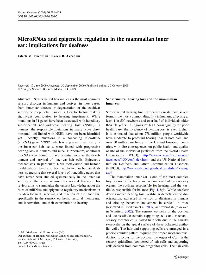
The mammalian inner ear is one of the most complex
tiny organs in the body and is composed of two sensory
organs: the cochlea, responsible for hearing, and the ves-
tibule, responsible for balance (Fig. 1, left). While cochlear
defects induce hearing loss, vestibular defects lead to dis-
orientation, expressed as vertigo or dizziness in humans
and circling behavior (movement in circles) in mice
(reviewed in Friedman et al. 2007) and zebrafish (reviewed
in Whitfield 2002). The sensory epithelia of the cochlea
and the vestibule contain supporting cells and mechano-
sensory receptor cells, called hair cells due to the hairlike
stereocilia on the apical surface of these polarized epithe-
lial cells. The hair and supporting cells are arranged in a
precise cellular pattern required for proper mechanotrans-
duction to occur. In the cochlea, the organ of Corti is the
sensory epithelium, composed of hair cells and supporting
cells derived from common progenitor cells. The hair cells
L. M. Friedman � K. B. Avraham (&)
Department of Human Molecular Genetics and Biochemistry,
Sackler School of Medicine, Tel Aviv University,
Tel Aviv 69978, Israel
e-mail: [email protected]
123
Mamm Genome (2009) 20:581–603
DOI 10.1007/s00335-009-9230-5
are arranged in four rows along the snail-shaped cochlea:
three rows of outer hair cells and a single row of inner hair
cells, surrounded by supporting cells that include pillar
cells, Hensen cells, and Deiter cells (reviewed in Raphael
and Altschuler 2003) (Fig. 1, right). The stereocilia of the
outer hair cells are embedded in the overlying tectorial
membrane, which is composed of collagen fibrils and
glycoproteins (reviewed in Richardson et al. 2008). The
tympanic membrane between the outer ear and the middle
ear and the tiny bones of the middle ear convert auditory
signals to cochlear fluid waves. The specialized stereocilia
of the cochlear hair cells move in response to the fluid
movement in the inner-ear canals, which causes ion
channels on the stereocilia plasma membrane to open.
Thus, the cochlear hair cells convert auditory-derived
mechanical stimuli to electrical signals (reviewed in
Raphael and Altschuler 2003). The vestibule is composed
of three semicircular canals and five patches of sensory
epithelia (utricular and saccular maculae and three cristae),
which also include several types of sensory hair and sup-
porting cells. Although the cochlear hair cells have a dif-
ferent organization, a slightly different shape, and some
differences in gene expression and regulation (Hertzano
et al. 2007) compared to the vestibular hair cells, the
mRNA and miRNA populations of the cochlear and ves-
tibular sensory epithelia are very similar, with only minor
differences (Friedman et al. 2009a; Friedman and Avra-
ham, unpublished).
In most cases, hearing loss in humans derives from
abnormal development or degeneration of the cochlear hair
cells, and it is estimated that 60-70% of the cases are
hereditary (Nance 2003). More than 110 chromosomal loci
have been linked with nonsyndromic hearing loss (NSHL)
in humans, but thus far the responsible genes have been
identified in only a subset of these loci (see Hereditary
Hearing Loss homepage, http://webh01.ua.ac.be/hhh/)
(Ahmed et al. 2008; Du et al. 2008; Friedman et al. 2009b;
Ruel et al. 2008; Uchida et al. 2009; Yang et al. 2009). In
addition, many syndromes include sensorineural hearing
loss. Until very recently, only mutations in protein-coding
and mitochondrial genes were evaluated in NSHL loci,
leading to the discovery of mutations in genes encoding 49
different proteins and 2 mitochondrial genes that are
responsible for NSHL. Recently, one of the NSHL loci,
DFNA50, was found to carry a mutation in a noncoding
microRNA (miRNA) gene, and this mutation not only
segregated with the hearing loss phenotype, but it is the
first miRNA mutation found for a Mendelian disease
(Mencia et al. 2009).
The normal development of the inner ear requires
coordinated and strictly regulated expression of genes and
regulatory factors (Fritzsch et al. 2006; Kelley 2006). After
the inner ear develops, the complex sensory transduction of
the inner ear is dependent on a tremendous amount of
synchronized processes and mechanisms. In addition to the
many genes and proteins required for the normal function
of the sensory hair cell, many factors are required for the
survival of hair cells, some of which are internal to the hair
cells [e.g., Usher-related genes (Reiners et al. 2006)], while
others are external to the hair cells and affect the hair cell
environment [e.g., the connexin genes Gjb2 and Gjb6 that
are expressed in supporting cells (Nickel and Forge 2008),
or collagen genes and Tecta that comprise the tectorial
membrane (Richardson et al. 2008)]. For example, some of
the sensory epithelium supporting cells are responsible for
controlling the ionic homeostasis of the endolymph, the
solution that fills the lumen (scala media) to which the
sensory epithelial cells face. The exact composition of the
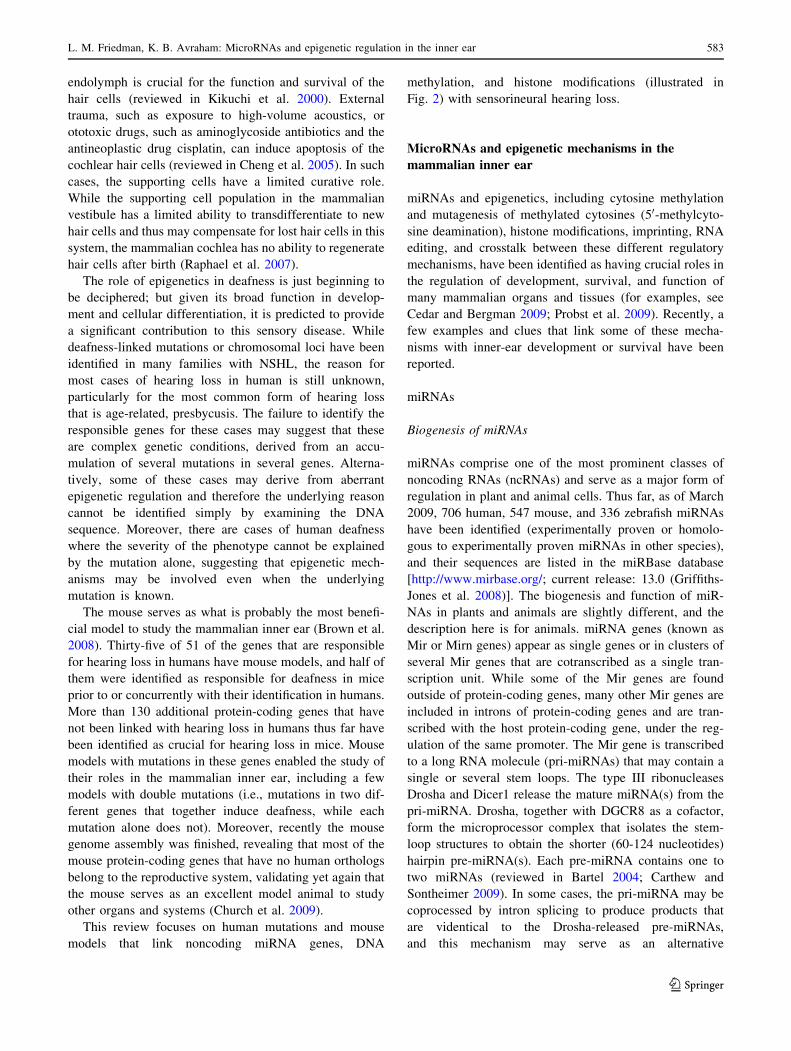
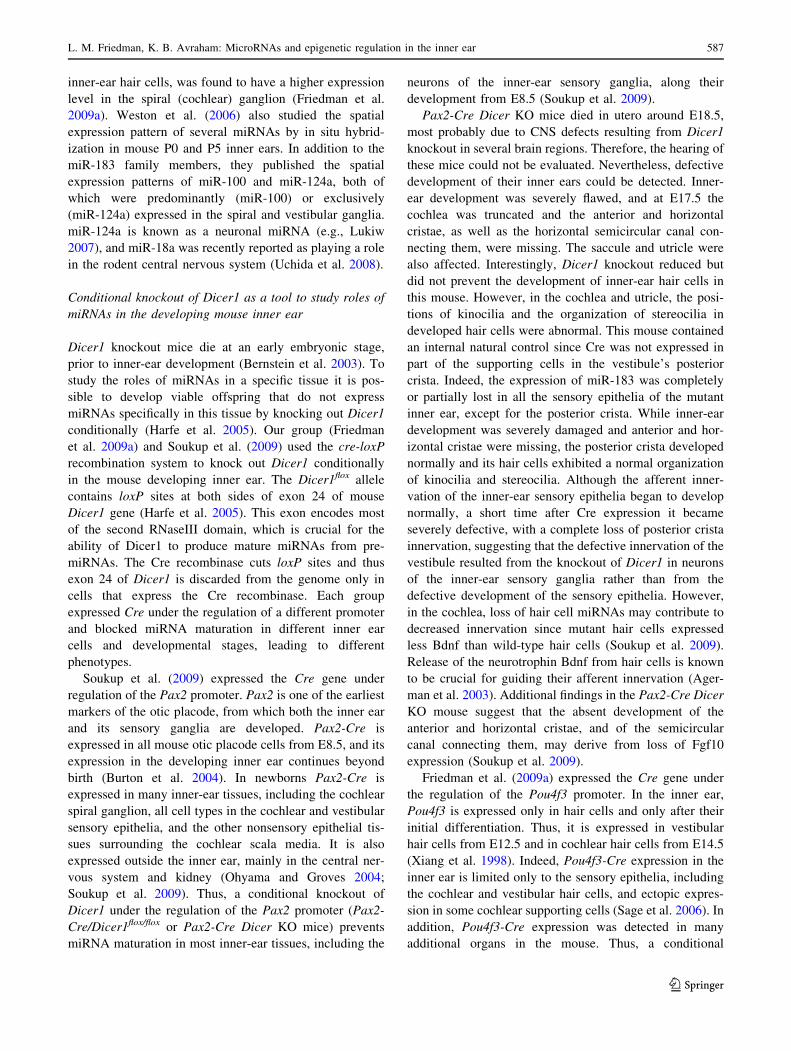
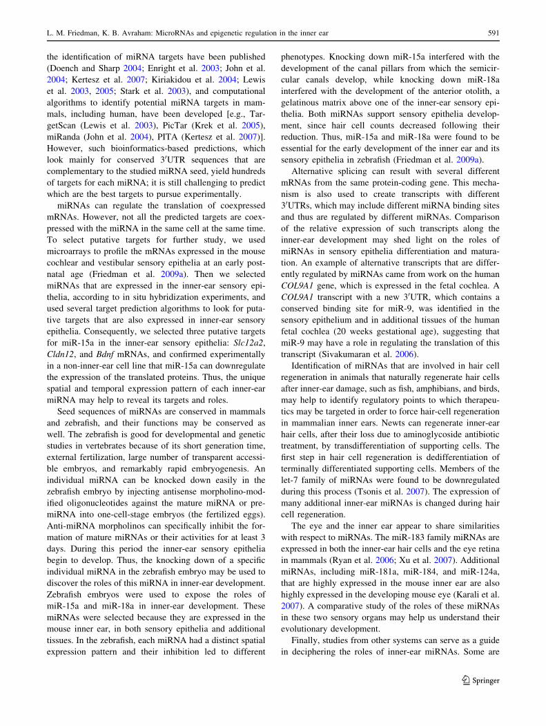
Fig. 1 Schematic illustration of the human ear. Left The ear is
composed of three parts: the external, middle, and inner ear. The inner
ear is composed of two organs, the snail-like cochlea and the
vestibule. Middle Cross section of the cochlear duct of the snail-
shaped cochlea. The cochlear sensory epithelium is illustrated, with
the tectorial membrane (TM) above. Right Enlarged illustration of the
hair and supporting cells in the cochlear sensory epithelium, the organ
of Corti. In the right panel: green, hair cells; other colors, supporting
cells
582 L. M. Friedman, K. B. Avraham: MicroRNAs and epigenetic regulation in the inner ear
123
endolymph is crucial for the function and survival of the
hair cells (reviewed in Kikuchi et al. 2000). External
trauma, such as exposure to high-volume acoustics, or
ototoxic drugs, such as aminoglycoside antibiotics and the
antineoplastic drug cisplatin, can induce apoptosis of the
cochlear hair cells (reviewed in Cheng et al. 2005). In such
cases, the supporting cells have a limited curative role.
While the supporting cell population in the mammalian
vestibule has a limited ability to transdifferentiate to new
hair cells and thus may compensate for lost hair cells in this
system, the mammalian cochlea has no ability to regenerate
hair cells after birth (Raphael et al. 2007).
The role of epigenetics in deafness is just beginning to
be deciphered; but given its broad function in develop-
ment and cellular differentiation, it is predicted to provide
a significant contribution to this sensory disease. While
deafness-linked mutations or chromosomal loci have been
identified in many families with NSHL, the reason for
most cases of hearing loss in human is still unknown,
particularly for the most common form of hearing loss
that is age-related, presbycusis. The failure to identify the
responsible genes for these cases may suggest that these
are complex genetic conditions, derived from an accu-
mulation of several mutations in several genes. Alterna-
tively, some of these cases may derive from aberrant
epigenetic regulation and therefore the underlying reason
cannot be identified simply by examining the DNA
sequence. Moreover, there are cases of human deafness
where the severity of the phenotype cannot be explained
by the mutation alone, suggesting that epigenetic mech-
anisms may be involved even when the underlying
mutation is known.
The mouse serves as what is probably the most benefi-
cial model to study the mammalian inner ear (Brown et al.
2008). Thirty-five of 51 of the genes that are responsible
for hearing loss in humans have mouse models, and half of
them were identified as responsible for deafness in mice
prior to or concurrently with their identification in humans.
More than 130 additional protein-coding genes that have
not been linked with hearing loss in humans thus far have
been identified as crucial for hearing loss in mice. Mouse
models with mutations in these genes enabled the study of
their roles in the mammalian inner ear, including a few
models with double mutations (i.e., mutations in two dif-
ferent genes that together induce deafness, while each
mutation alone does not). Moreover, recently the mouse
genome assembly was finished, revealing that most of the
mouse protein-coding genes that have no human orthologs
belong to the reproductive system, validating yet again that
the mouse serves as an excellent model animal to study
other organs and systems (Church et al. 2009).
This review focuses on human mutations and mouse
models that link noncoding miRNA genes, DNA
methylation, and histone modifications (illustrated in
Fig. 2) with sensorineural hearing loss.
MicroRNAs and epigenetic mechanisms in the
mammalian inner ear
miRNAs and epigenetics, including cytosine methylation
and mutagenesis of methylated cytosines (50-methylcyto-
sine deamination), histone modifications, imprinting, RNA
editing, and crosstalk between these different regulatory
mechanisms, have been identified as having crucial roles in
the regulation of development, survival, and function of
many mammalian organs and tissues (for examples, see
Cedar and Bergman 2009; Probst et al. 2009). Recently, a
few examples and clues that link some of these mecha-
nisms with inner-ear development or survival have been
reported.
miRNAs
Biogenesis of miRNAs
miRNAs comprise one of the most prominent classes of
noncoding RNAs (ncRNAs) and serve as a major form of
regulation in plant and animal cells. Thus far, as of March
2009, 706 human, 547 mouse, and 336 zebrafish miRNAs
have been identified (experimentally proven or homolo-
gous to experimentally proven miRNAs in other species),
and their sequences are listed in the miRBase database
[http://www.mirbase.org/; current release: 13.0 (Griffiths-
Jones et al. 2008)]. The biogenesis and function of miR-
NAs in plants and animals are slightly different, and the
description here is for animals. miRNA genes (known as
Mir or Mirn genes) appear as single genes or in clusters of
several Mir genes that are cotranscribed as a single tran-
scription unit. While some of the Mir genes are found
outside of protein-coding genes, many other Mir genes are
included in introns of protein-coding genes and are tran-
scribed with the host protein-coding gene, under the reg-
ulation of the same promoter. The Mir gene is transcribed
to a long RNA molecule (pri-miRNAs) that may contain a
single or several stem loops. The type III ribonucleases
Drosha and Dicer1 release the mature miRNA(s) from the
pri-miRNA. Drosha, together with DGCR8 as a cofactor,
form the microprocessor complex that isolates the stem-
loop structures to obtain the shorter (60-124 nucleotides)
hairpin pre-miRNA(s). Each pre-miRNA contains one to
two miRNAs (reviewed in Bartel 2004; Carthew and
Sontheimer 2009). In some cases, the pri-miRNA may be
coprocessed by intron splicing to produce products that
are videntical to the Drosha-released pre-miRNAs,
and this mechanism may serve as an alternative
L. M. Friedman, K. B. Avraham: MicroRNAs and epigenetic regulation in the inner ear 583
123
Drosha-independent way to release pre-miRNAs (Ruby
et al. 2007). Then, pre-miRNAs are exported from the
nucleus to the cytoplasm, and Dicer1 excises the mature 16-
29-nucleotide-long miRNA (average length = 22 nucleo-
tides) from the pre-miRNA (illustrated in Fig. 2). Thus,
Dicer1 is essential for the production of mature and func-
tional miRNAs (reviewed in Bartel 2004; Carthew and
Sontheimer 2009). Dicer1 is incorporated in a complex of
proteins, and therefore the released mature miRNA is bound
to this riboprotein complex, termed miRISC (miRNA-
induced silencing complex). miRISC includes additional
proteins that are required for miRNA binding and miRNA-
induced regulation of gene transcription and mRNA trans-
lation, such as an argonaute (Ago) protein [Ago2/Eif2c2 is
the common argonaute in the miRISC complex in
mammals, but any of the other three argonautes may
supersede it (Su et al. 2009)], TARBP2 (previously known
as TRBP) (Chendrimada et al. 2005), GW182, and addi-
tional proteins (reviewed in Carthew and Sontheimer 2009).
Functions of miRNAs
miRNAs, incorporated in miRISC, regulate expression of
genes that contain cis complementary sequence(s), most
often in their 30 untranslated region (30UTR). The most
established function of miRNAs is translational suppres-
sion of mRNAs with imperfect complementary sequences
and cleavage of mRNAs with a perfect match (reviewed in
Bartel 2004). It has been suggested that at least 60% of the
vertebrate protein-coding mRNAs are direct targets of
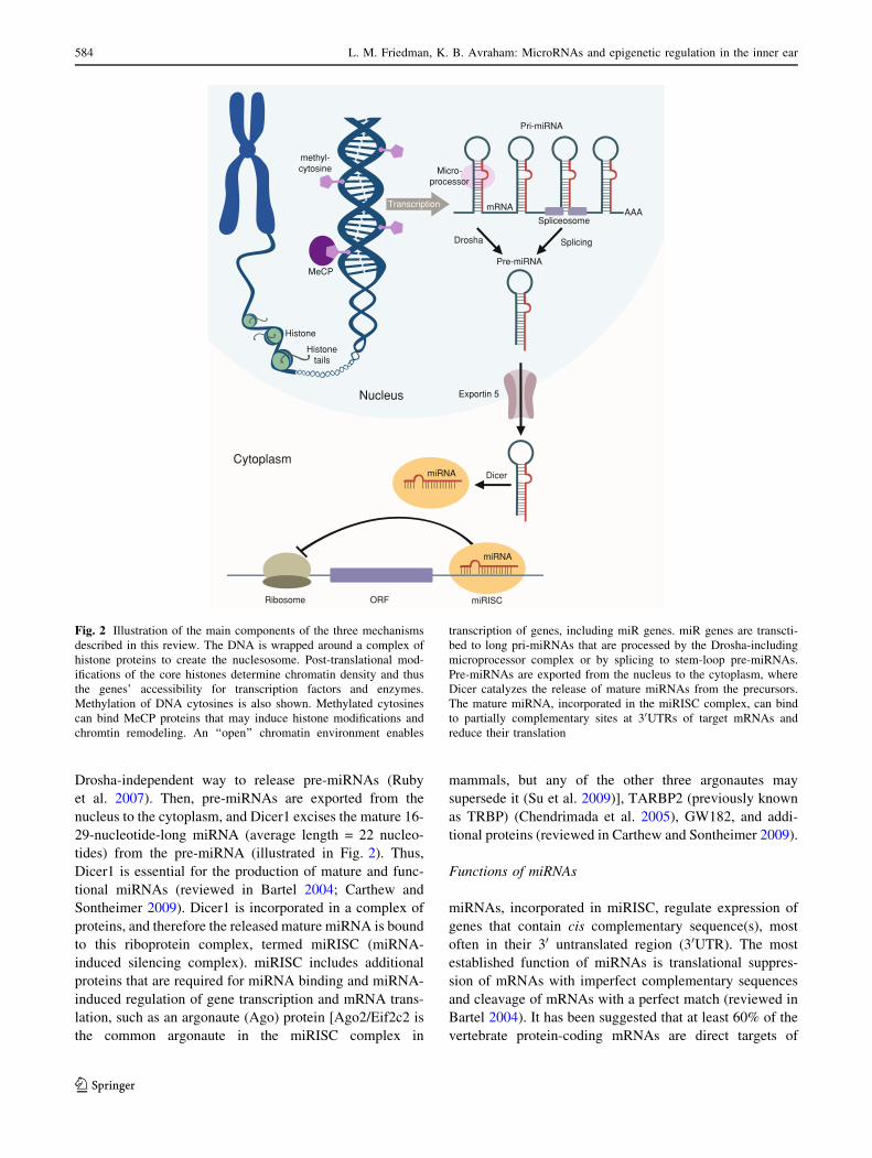
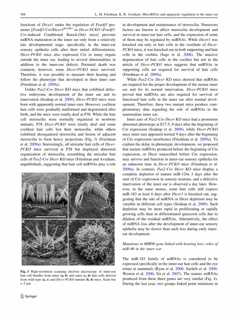
Pre-miRNA
Pri-miRNA
mRNA
miRNA
miRNA
ORFRibosome
Spliceosome
Micro-processor
AAATranscription
Drosha Splicing
Exportin 5
Dicer
miRISC
Histone
Histonetails
MeCP
Nucleus
Cytoplasm
methyl-cytosine
Fig. 2 Illustration of the main components of the three mechanisms
described in this review. The DNA is wrapped around a complex of
histone proteins to create the nuclesosome. Post-translational mod-
ifications of the core histones determine chromatin density and thus
the genes’ accessibility for transcription factors and enzymes.
Methylation of DNA cytosines is also shown. Methylated cytosines
can bind MeCP proteins that may induce histone modifications and
chromtin remodeling. An ‘‘open’’ chromatin environment enables
transcription of genes, including miR genes. miR genes are transcti-
bed to long pri-miRNAs that are processed by the Drosha-including
microprocessor complex or by splicing to stem-loop pre-miRNAs.
Pre-miRNAs are exported from the nucleus to the cytoplasm, where
Dicer catalyzes the release of mature miRNAs from the precursors.
The mature miRNA, incorporated in the miRISC complex, can bind
to partially complementary sites at 30UTRs of target mRNAs and
reduce their translation
584 L. M. Friedman, K. B. Avraham: MicroRNAs and epigenetic regulation in the inner ear
123
miRNAs (Friedman et al. 2009c; Miranda et al. 2006) and
that miRNAs affect, directly or indirectly, virtually all
cellular and organismal processes.
In mammals, most of the target mRNAs for miRNAs
contain imperfect complementary sequences in their
30UTRs, and binding of a miRNA to this site suppresses the
mRNA translation. When the target mRNA contains sev-
eral binding sites for the same miRNA, its translation is
more suppressed. However, sometimes miRNAs have the
reverse effect and upregulate the translation of their target
mRNAs. This may depend on the cell cycle stage, since
two recent studies proposed that while miRNAs inhibit the
translation of their target mRNAs in proliferating human
cell lines, they enhance the translation of the same targets
in quiescent cells (Vasudevan et al. 2007, 2008). Although
in most cases miRNAs bind to complementary (or partial
complementary) sequences at the 30UTRs of their target
mRNAs, miRNA binding sites were found also in other
locations, such as 50UTRs. The miRNA effect may depend
on the location of its binding site. While translation
repression is mediated by the binding of miRNAs to the
mRNA 30UTRs, a case in which miRNA binds to its target
50UTR and upregulates its translation was recently reported
(Orom et al. 2008).
miRNAs are associated with many developmental pro-
cesses and diseases and have been found to be crucial for
normal development in all animals tested (Zhang et al.
2007). They are differentially expressed in different tissues
(Landgraf et al. 2007), and certain miRNAs are known as
having a role in the development of specific organs and
tissues (reviewed in Stefani and Slack 2008; Zhang et al.
2007). In mammals, miRNAs are also essential for early
embryogenesis and viability, and Dicer1 knockout mice
arrest miRNA maturation and die at an early embryonic
stage, before embryonic day 9.5 (E9.5) (Bernstein et al.
2003). In the zebrafish, Dicer and the production of
miRNAs are required at later stages and, thus, Dicer
knockout in whole zebrafish embryos enables the evalua-
tion of miRNA importance for brain formation, somato-
genesis, heart development, and general organogenesis
(Giraldez et al. 2005).
Expression of miR-183 family miRNAs in animals
The study of miRNAs in the inner ear began only in 2005,
when an atlas of in situ hybridization results in zebrafish
embryos, demonstrating the expression of miRNAs, was
published (Wienholds et al. 2005). Some of the miRNAs
were found to be expressed in the inner ear of the zebrafish
embryo. Three miRNAs (miR-182, miR-96, and miR-183)
that share similar sequences are coexpressed in zebrafish
embryos. Since their genes are clustered within 4 kb in the
mouse and human genomes and are most probably
transcribed together as a polycistronic gene, these three
miRNAs are commonly referred to as the miR-183 cluster or
family. Their expression in zebrafish embryos is most
prominent in the hair cells of the inner ear and the lateral line
neuromasts, and in the sensory cells of the eye and the nose,
but they are also expressed in cranial ganglia and epiphysis.
The zebrafish lateral line neuromasts contain sensory hair
cells that are similar to inner-ear hair cells (Nicolson 2005).
These miRNAs are also expressed in inner-ear hair cells and
eye retina in chick embryos (Darnell et al. 2006).
The expression of the miR-183 family miRNAs in
zebrafish embryos attracted the attention of inner-ear and
eye researchers, who used in situ hybridization to detect the
expression of these three miRNAs in the mouse inner ear
and eye. Indeed, this triad of miRNAs was found to be
expressed specifically in the inner-ear hair cells, in both the
cochlea and the vestibule (Weston et al. 2006), and in the
photoreceptors, bipolar cells, and amacrine cells in the eye
retina (Ryan et al. 2006; Xu et al. 2007) of postnatal mice.
A recent study (Sacheli et al. 2009) described the expres-
sion of these miRNAs along mouse inner-ear development,
which begins around E9.5 when the otic vesicle (also
termed otocyst) is invaginated from the otic placode. Along
inner-ear development, miR-182 and miR-183 expression
patterns are similar, while miR-96 is not always coex-
pressed with them. After birth (P0-P2), the expression of
these miRNAs is reduced, and their cochlear expression is
limited to the hair cells and the spiral ganglion. As the
postnatal cochlea matures, the expression of these miRNAs
gradually disappears from cochlear hair cells and moves to
other cochlear tissues, the inner sulcus and the spiral lim-
bus. Overall, the dynamic expression of these miRNAs in
the developing mouse inner ear, with the higher expression
level in differentiating hair cell, suggests that these
miRNAs are associated with hair cell differentiation and
maturation.
Further study revealed that the miR-183 family miRNAs
are expressed in the hair cells of the inner ear and neuro-
masts of additional vertebrates and have orthologous
miRNAs that are specifically expressed in neurosensory
cells in invertebrate deuterostomes and in ciliated sensory
cells of the fruit fly Drosophila melanogaster. How-
ever, the miR-183 orthologous miRNA in the nematode
Caenorhabditis elegans may also be expressed in noncili-
ated glial or supporting cells in sensory organs (Pierce et al.
2008). Thus, miR-183 family miRNAs are considered to be
specifically expressed in neurosensory organs, and in most
cases in ciliated or stereociliated neurosensory cells.
Although it is widely believed that miR-183 family
miRNAs are expressed in postnatal mammals only in the
inner ear and the retina, a recent report detected these
miRNAs in several additional organs from adult mice, in
particular, the submandibular gland (Jin et al. 2009).
L. M. Friedman, K. B. Avraham: MicroRNAs and epigenetic regulation in the inner ear 585
123
Additional miRNAs expressed in the mouse inner ear
miRNA microarrays have been used to detect additional
miRNAs in the whole mouse inner ear. Weston et al.
(2006) used microarrays to profile the expression of pre-
miRNAs or mature miRNAs in postnatal whole mouse
inner ear at several ages from newborn to adult. The
expression level of several miRNAs changed along inner-
ear development. In a subsequent report, our group
(Friedman et al. 2009a) used microarrays to compare the
expression profiles of mature miRNAs in separated
cochleae and vestibules from newborn mice. Because
cochlear and vestibular sensory epithelia are similar but not
identical, we hoped to identify miRNAs that may be
responsible for some of these differences. However, most
miRNAs exhibited similar expression levels in whole
cochleae and whole vestibules, and the few that were
expressed differentially showed only relatively small dif-
ferences. The cochlea and vestibule are complex organs
that include many different tissues and cell types, and the
expression level detected for each miRNA was actually an
average of its different expression levels in the different
cell populations. This was emphasized by the detailed in
situ hybridization expression analysis of several miRNAs
(Friedman et al. 2009a). The differences in miRNA
expression in whole inner ears along postnatal inner-ear
development, reported by Weston et al. (2006), were also
small, most probably for the same reasons. Overall, these
microarray experiments demonstrated that more then 100
different miRNAs are expressed in the mouse inner ear.
Weston et al. (2006) proposed that if the miRNA pop-
ulation in embryonic inner ears is compared to the post-
natal population, the differences may be greater. Although
the newborn mouse inner ear is still not functional and
continues to develop, most of inner-ear development does
occur before birth during embryogenesis. To address
this question, we selected six miRNAs [miR-15a, miR-18a,
miR-30b, miR-99a, miR-199a-3p (also known as
miR-199a), and miR-182] with different expression levels
in the newborn whole cochleae and vestibules, according to
microarray results, and followed their expression levels
along inner-ear development from embryonic day 16.5
(E16.5) to postnatal day 30 (P30) by the more sensitive and
accurate qRT-PCR assay (Friedman et al. 2009a). At P30,
the inner ear is fully developed and functional. We found
significant differences in the levels of each miRNA along
development. The level of some miRNAs was highest in
embryonic (miR-18a) or newborn (miR-99a, miR-199a-3p,
and miR-182, a member of the miR-183 cluster described
above) developing inner ears, while others peaked in the
fully developed P30 inner ears (miR-15a and miR-30b).
Differences were also found between cochlear and vestib-
ular levels at the same age for some miRNAs at selected
time points. Thus, the relatively low sensitivity and high
background of microarrays may mask differences in
miRNA levels that may be detected by qRT-PCR. The
differences in temporal expression patterns suggest that
each of these miRNAs has a different role in the inner ear.
miRNAs that are highly expressed before birth may play a
role in early inner-ear development; miRNAs that peak in
the newborn inner ear (such as miR-182) may regulate the
final differentiation of inner-ear tissues; while miRNAs
with a higher expression level in P30 inner ears may play a
role in cell maintenance and function. However, most
miRNAs may play a role during inner-ear development
because the miRNA populations in P100 inner ears
(Weston et al. 2006) and P30 separated cochleae and
vestibules (Friedman and Avraham, unpublished) are much
smaller than the miRNA populations at P0.
Quantitative assays to determine miRNA levels in whole
organs are not sufficient to understand the role of each
miRNA. In complex organs like the cochlea and the ves-
tibule it is very important to know the exact cells that
express each miRNA. We showed, by in situ hybridization
of the mouse newborn inner ear, that miRNAs with similar
average expression levels (in whole cochleae and vesti-
bules) are actually expressed in different cell types and,
thus, may have different roles. For example, miR-182 and
miR-199a-3p, which have similar temporal expression
patterns in the developing mouse vestibule and cochlea and
are very highly expressed in the newborn inner ear, are
actually expressed in different cell types. While miR-182 is
expressed exclusively in the inner-ear hair cells and the
cochlear spiral ganglion neurons, miR-199a-3p is expres-
sed in many other inner-ear tissues but not in these tissues.
Although recently miR-199a-3p was reported as highly
expressed in neuronal tissues in rats (Hua et al. 2009), its
expression was not limited to the spiral or vestibular gan-
glia in our newborn mouse inner ears. Other miRNAs, with
a lower expression level in the whole inner ear (i.e.,
miR-15a, miR-18a, miR-30b, and miR-99a), are expressed
in many inner-ear tissues, including the sensory epithelia.
In addition, the distribution of some miRNAs may be dif-
ferent in the cochlea and the vestibule. For example, miR-
30b, which has a similar expression level in the newborn
mouse cochlea and vestibule, is expressed in both cochlear
hair and supporting cells, but in the vestibule it is expressed
only in hair cells. We also found two miRNAs, miR-99a
and miR-199a-3p, with a mirroring spatial expression,
suggesting that these two miRNAs are involved in a neg-
ative control loop, most probably indirectly. All the
miRNAs detected in inner-ear hair cells were also
expressed in the spiral ganglion, the cochlear sensory nerve
cells. This result is not surprising since hair cells are
neurosensory cells that exhibit some neuronal-like char-
acteristics. However, miR-18a, which is also expressed in
586 L. M. Friedman, K. B. Avraham: MicroRNAs and epigenetic regulation in the inner ear
123
inner-ear hair cells, was found to have a higher expression
level in the spiral (cochlear) ganglion (Friedman et al.
2009a). Weston et al. (2006) also studied the spatial
expression pattern of several miRNAs by in situ hybrid-
ization in mouse P0 and P5 inner ears. In addition to the
miR-183 family members, they published the spatial
expression patterns of miR-100 and miR-124a, both of
which were predominantly (miR-100) or exclusively
(miR-124a) expressed in the spiral and vestibular ganglia.
miR-124a is known as a neuronal miRNA (e.g., Lukiw
2007), and miR-18a was recently reported as playing a role
in the rodent central nervous system (Uchida et al. 2008).
Conditional knockout of Dicer1 as a tool to study roles of
miRNAs in the developing mouse inner ear
Dicer1 knockout mice die at an early embryonic stage,
prior to inner-ear development (Bernstein et al. 2003). To
study the roles of miRNAs in a specific tissue it is pos-
sible to develop viable offspring that do not express
miRNAs specifically in this tissue by knocking out Dicer1
conditionally (Harfe et al. 2005). Our group (Friedman
et al. 2009a) and Soukup et al. (2009) used the cre-loxP
recombination system to knock out Dicer1 conditionally
in the mouse developing inner ear. The Dicer1flox allele
contains loxP sites at both sides of exon 24 of mouse
Dicer1 gene (Harfe et al. 2005). This exon encodes most
of the second RNaseIII domain, which is crucial for the
ability of Dicer1 to produce mature miRNAs from pre-
miRNAs. The Cre recombinase cuts loxP sites and thus
exon 24 of Dicer1 is discarded from the genome only in
cells that express the Cre recombinase. Each group
expressed Cre under the regulation of a different promoter
and blocked miRNA maturation in different inner ear
cells and developmental stages, leading to different
phenotypes.
Soukup et al. (2009) expressed the Cre gene under
regulation of the Pax2 promoter. Pax2 is one of the earliest
markers of the otic placode, from which both the inner ear
and its sensory ganglia are developed. Pax2-Cre is
expressed in all mouse otic placode cells from E8.5, and its
expression in the developing inner ear continues beyond
birth (Burton et al. 2004). In newborns Pax2-Cre is
expressed in many inner-ear tissues, including the cochlear
spiral ganglion, all cell types in the cochlear and vestibular
sensory epithelia, and the other nonsensory epithelial tis-
sues surrounding the cochlear scala media. It is also
expressed outside the inner ear, mainly in the central ner-
vous system and kidney (Ohyama and Groves 2004;
Soukup et al. 2009). Thus, a conditional knockout of
Dicer1 under the regulation of the Pax2 promoter (Pax2-
Cre/Dicer1flox/flox or Pax2-Cre Dicer KO mice) prevents
miRNA maturation in most inner-ear tissues, including the
neurons of the inner-ear sensory ganglia, along their
development from E8.5 (Soukup et al. 2009).
Pax2-Cre Dicer KO mice died in utero around E18.5,
most probably due to CNS defects resulting from Dicer1
knockout in several brain regions. Therefore, the hearing of
these mice could not be evaluated. Nevertheless, defective
development of their inner ears could be detected. Inner-
ear development was severely flawed, and at E17.5 the
cochlea was truncated and the anterior and horizontal
cristae, as well as the horizontal semicircular canal con-
necting them, were missing. The saccule and utricle were
also affected. Interestingly, Dicer1 knockout reduced but
did not prevent the development of inner-ear hair cells in
this mouse. However, in the cochlea and utricle, the posi-
tions of kinocilia and the organization of stereocilia in
developed hair cells were abnormal. This mouse contained
an internal natural control since Cre was not expressed in
part of the supporting cells in the vestibule’s posterior
crista. Indeed, the expression of miR-183 was completely
or partially lost in all the sensory epithelia of the mutant
inner ear, except for the posterior crista. While inner-ear
development was severely damaged and anterior and hor-
izontal cristae were missing, the posterior crista developed
normally and its hair cells exhibited a normal organization
of kinocilia and stereocilia. Although the afferent inner-
vation of the inner-ear sensory epithelia began to develop
normally, a short time after Cre expression it became
severely defective, with a complete loss of posterior crista
innervation, suggesting that the defective innervation of the
vestibule resulted from the knockout of Dicer1 in neurons
of the inner-ear sensory ganglia rather than from the
defective development of the sensory epithelia. However,
in the cochlea, loss of hair cell miRNAs may contribute to
decreased innervation since mutant hair cells expressed
less Bdnf than wild-type hair cells (Soukup et al. 2009).
Release of the neurotrophin Bdnf from hair cells is known
to be crucial for guiding their afferent innervation (Ager-
man et al. 2003). Additional findings in the Pax2-Cre Dicer
KO mouse suggest that the absent development of the
anterior and horizontal cristae, and of the semicircular
canal connecting them, may derive from loss of Fgf10
expression (Soukup et al. 2009).
Friedman et al. (2009a) expressed the Cre gene under
the regulation of the Pou4f3 promoter. In the inner ear,
Pou4f3 is expressed only in hair cells and only after their
initial differentiation. Thus, it is expressed in vestibular
hair cells from E12.5 and in cochlear hair cells from E14.5
(Xiang et al. 1998). Indeed, Pou4f3-Cre expression in the
inner ear is limited only to the sensory epithelia, including
the cochlear and vestibular hair cells, and ectopic expres-
sion in some cochlear supporting cells (Sage et al. 2006). In
addition, Pou4f3-Cre expression was detected in many
additional organs in the mouse. Thus, a conditional
L. M. Friedman, K. B. Avraham: MicroRNAs and epigenetic regulation in the inner ear 587
123
knockout of Dicer1 under the regulation of Pou4f3 pro-
moter [Pou4f3-Cre/Dicer1flox/flox or Dicer-PCKO (Pou4f3-
Cre-induced Conditional Knock-Out) mice] prevents
miRNA maturation in the inner ear only from a relatively
late developmental stage, specifically in the inner-ear
sensory epithelia cells after their initial differentiation.
Dicer-PCKO mice also expressed Cre in many organs
outside the inner ear, leading to several abnormalities in
addition to the inner-ear defects. Perinatal death was
common; however, some Dicer-PCKO mice survived.
Therefore, it was possible to measure their hearing and
follow the phenotype that developed in their inner ears
(Friedman et al. 2009a).
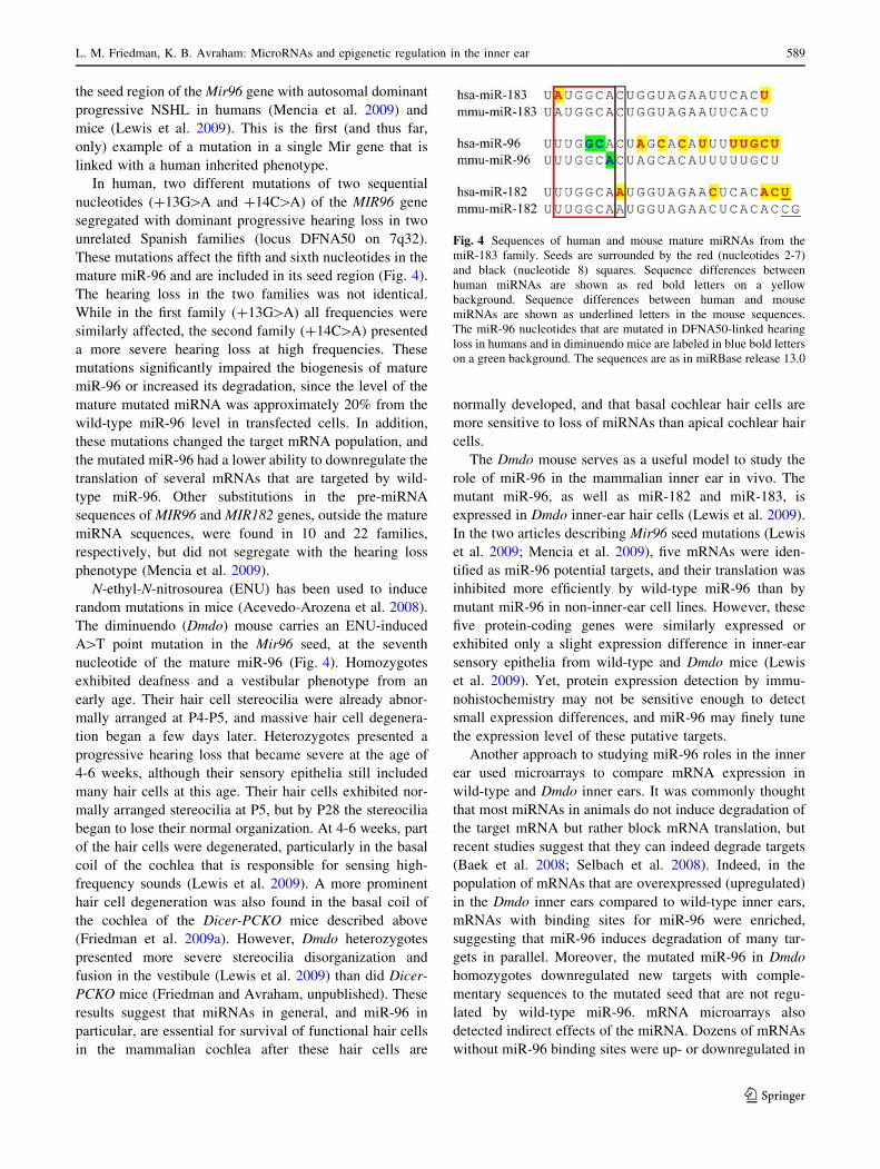
Unlike Pax2-Cre Dicer KO mice that exhibited defec-
tive embryonic development of the inner ear and its
innervation (Soukup et al. 2009), Dicer-PCKO mice were
born with apparently normal inner ears. However, cochlear
hair cells were gradually malformed and degenerated after
birth, and the mice were totally deaf at P38. While the hair
cell stereocilia were normally organized in newborn
mutants, P38 Dicer-PCKO were totally deaf and some
cochlear hair cells lost their stereocilia, while others
exhibited disorganized stereocilia and fusion of adjacent
stereocilia to form heavy projections (Fig. 3) (Friedman
et al. 2009a). Interestingly, all utricular hair cells of Dicer-
PCKO mice survived at P38 but displayed abnormal
organization of stereocilia, resembling the utricular hair
cells of Pax2-Cre Dicer KO mice (Friedman and Avraham,
unpublished), suggesting that hair cell miRNAs play a role
in development and maintenance of stereocilia. Numerous
factors are known to affect stereocilia development and
survival in inner-ear hair cells, and the expression of some
of them may be regulated by miRNAs. While Dicer1 was
knocked out only in hair cells in the vestibule of Dicer-
PCKO mice, it was knocked out in both supporting and hair
cells in the cochlea (Sage et al. 2006). The massive
degeneration of hair cells in the cochlea but not in the
utricle of Dicer-PCKO mice suggests that miRNAs in
supporting cells are required for survival of hair cells
(Friedman et al. 2009a).
While Pax2-Cre Dicer KO mice showed that miRNAs
are required for the proper development of the mouse inner
ear and for its normal innervation, Dicer-PCKO mice
proved that miRNAs are also required for survival of
functional hair cells in the inner ear after normal devel-
opment. Therefore, these two mutant mice produce com-
plementary data regarding the role of miRNAs in the
mammalian inner ear.
Inner ears of Pax2-Cre Dicer KO mice had a prominent
abnormal phenotype at E17.5, 9 days after the beginning of
Cre expression (Soukup et al. 2009), while Dicer-PCKO
mice inner ears appeared normal 9 days after the beginning
of Cre expression (newborns) (Friedman et al. 2009a). To
explain the delay in phenotypic development, we proposed
that mature miRNAs produced before the beginning of Cre
expression, or Dicer transcribed before Cre expression,
may survive and function in inner-ear sensory epithelia for
an unknown time in Dicer-PCKO mice (Friedman et al.
2009a). In contrast, Pax2-Cre Dicer KO mice display a
complete depletion of mature miR-124a 3 days after the
start of Cre expression in sensory neurons, and a defective
innervation of the inner ear is observed a day later. How-
ever, in the same mouse, some hair cells still express
miR-183 at least 6 days after Dicer1 is knocked out, sug-
gesting that the rate of miRNA or Dicer depletion may be
variable in different cell types (Soukup et al. 2009). Such
depletion may be more rapid in proliferating or rapidly
growing cells than in differentiated quiescent cells due to
dilution of the residual miRNAs. Alternatively, the effect
of miRNA loss after the development of inner-ear sensory
epithelia may be slower than such loss during early inner-
ear development.
Mutations in MIR96 gene linked with hearing loss: roles of
miR-96 in the inner ear
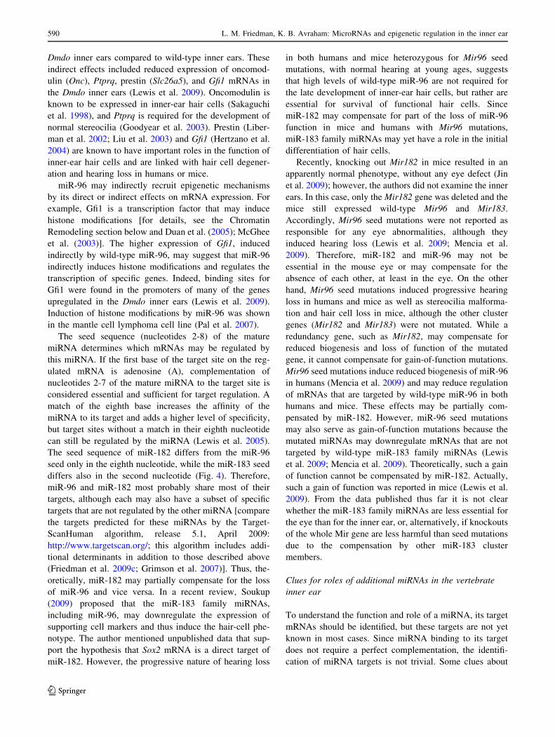
The miR-183 family of miRNAs is considered to be
expressed specifically in the inner-ear hair cells and the eye
retina in mammals (Ryan et al. 2006; Sacheli et al. 2009;
Weston et al. 2006; Xu et al. 2007). The mature miRNAs
produced from these three genes are very similar (Fig. 4).
During the last year, two groups linked point mutations in
Fig. 3 High-resolution scanning electron microscopy of inner-ear
hair cell bundles from inner (a, b) and outer (c, d) hair cells derived
from wild-type (a, c) and Dicer-PCKO mutant (b, d) mice. Scale bar
= 5 lm
588 L. M. Friedman, K. B. Avraham: MicroRNAs and epigenetic regulation in the inner ear
123
the seed region of the Mir96 gene with autosomal dominant
progressive NSHL in humans (Mencia et al. 2009) and
mice (Lewis et al. 2009). This is the first (and thus far,
only) example of a mutation in a single Mir gene that is
linked with a human inherited phenotype.
In human, two different mutations of two sequential
nucleotides (?13G[A and ?14C[A) of the MIR96 gene
segregated with dominant progressive hearing loss in two
unrelated Spanish families (locus DFNA50 on 7q32).
These mutations affect the fifth and sixth nucleotides in the
mature miR-96 and are included in its seed region (Fig. 4).
The hearing loss in the two families was not identical.
While in the first family (?13G[A) all frequencies were
similarly affected, the second family (?14C[A) presented
a more severe hearing loss at high frequencies. These
mutations significantly impaired the biogenesis of mature
miR-96 or increased its degradation, since the level of the
mature mutated miRNA was approximately 20% from the
wild-type miR-96 level in transfected cells. In addition,
these mutations changed the target mRNA population, and
the mutated miR-96 had a lower ability to downregulate the
translation of several mRNAs that are targeted by wild-
type miR-96. Other substitutions in the pre-miRNA
sequences of MIR96 and MIR182 genes, outside the mature
miRNA sequences, were found in 10 and 22 families,
respectively, but did not segregate with the hearing loss
phenotype (Mencia et al. 2009).
N-ethyl-N-nitrosourea (ENU) has been used to induce
random mutations in mice (Acevedo-Arozena et al. 2008).
The diminuendo (Dmdo) mouse carries an ENU-induced
A[T point mutation in the Mir96 seed, at the seventh
nucleotide of the mature miR-96 (Fig. 4). Homozygotes
exhibited deafness and a vestibular phenotype from an
early age. Their hair cell stereocilia were already abnor-
mally arranged at P4-P5, and massive hair cell degenera-
tion began a few days later. Heterozygotes presented a
progressive hearing loss that became severe at the age of
4-6 weeks, although their sensory epithelia still included
many hair cells at this age. Their hair cells exhibited nor-
mally arranged stereocilia at P5, but by P28 the stereocilia
began to lose their normal organization. At 4-6 weeks, part
of the hair cells were degenerated, particularly in the basal
coil of the cochlea that is responsible for sensing high-
frequency sounds (Lewis et al. 2009). A more prominent
hair cell degeneration was also found in the basal coil of
the cochlea of the Dicer-PCKO mice described above
(Friedman et al. 2009a). However, Dmdo heterozygotes
presented more severe stereocilia disorganization and
fusion in the vestibule (Lewis et al. 2009) than did Dicer-
PCKO mice (Friedman and Avraham, unpublished). These
results suggest that miRNAs in general, and miR-96 in
particular, are essential for survival of functional hair cells
in the mammalian cochlea after these hair cells are
normally developed, and that basal cochlear hair cells are
more sensitive to loss of miRNAs than apical cochlear hair
cells.
The Dmdo mouse serves as a useful model to study the
role of miR-96 in the mammalian inner ear in vivo. The
mutant miR-96, as well as miR-182 and miR-183, is
expressed in Dmdo inner-ear hair cells (Lewis et al. 2009).
In the two articles describing Mir96 seed mutations (Lewis
et al. 2009; Mencia et al. 2009), five mRNAs were iden-
tified as miR-96 potential targets, and their translation was
inhibited more efficiently by wild-type miR-96 than by
mutant miR-96 in non-inner-ear cell lines. However, these
five protein-coding genes were similarly expressed or
exhibited only a slight expression difference in inner-ear
sensory epithelia from wild-type and Dmdo mice (Lewis
et al. 2009). Yet, protein expression detection by immu-
nohistochemistry may not be sensitive enough to detect
small expression differences, and miR-96 may finely tune
the expression level of these putative targets.
Another approach to studying miR-96 roles in the inner
ear used microarrays to compare mRNA expression in
wild-type and Dmdo inner ears. It was commonly thought
that most miRNAs in animals do not induce degradation of
the target mRNA but rather block mRNA translation, but
recent studies suggest that they can indeed degrade targets
(Baek et al. 2008; Selbach et al. 2008). Indeed, in the
population of mRNAs that are overexpressed (upregulated)
in the Dmdo inner ears compared to wild-type inner ears,
mRNAs with binding sites for miR-96 were enriched,
suggesting that miR-96 induces degradation of many tar-
gets in parallel. Moreover, the mutated miR-96 in Dmdo
homozygotes downregulated new targets with comple-
mentary sequences to the mutated seed that are not regu-
lated by wild-type miR-96. mRNA microarrays also
detected indirect effects of the miRNA. Dozens of mRNAs
without miR-96 binding sites were up- or downregulated in
Fig. 4 Sequences of human and mouse mature miRNAs from the
miR-183 family. Seeds are surrounded by the red (nucleotides 2-7)
and black (nucleotide 8) squares. Sequence differences between
human miRNAs are shown as red bold letters on a yellow
background. Sequence differences between human and mouse
miRNAs are shown as underlined letters in the mouse sequences.
The miR-96 nucleotides that are mutated in DFNA50-linked hearing
loss in humans and in diminuendo mice are labeled in blue bold letters
on a green background. The sequences are as in miRBase release 13.0
L. M. Friedman, K. B. Avraham: MicroRNAs and epigenetic regulation in the inner ear 589
123
Dmdo inner ears compared to wild-type inner ears. These
indirect effects included reduced expression of oncomod-
ulin (Onc), Ptprq, prestin (Slc26a5), and Gfi1 mRNAs in
the Dmdo inner ears (Lewis et al. 2009). Oncomodulin is
known to be expressed in inner-ear hair cells (Sakaguchi
et al. 1998), and Ptprq is required for the development of
normal stereocilia (Goodyear et al. 2003). Prestin (Liber-
man et al. 2002; Liu et al. 2003) and Gfi1 (Hertzano et al.
2004) are known to have important roles in the function of
inner-ear hair cells and are linked with hair cell degener-
ation and hearing loss in humans or mice.
miR-96 may indirectly recruit epigenetic mechanisms
by its direct or indirect effects on mRNA expression. For
example, Gfi1 is a transcription factor that may induce
histone modifications [for details, see the Chromatin
Remodeling section below and Duan et al. (2005); McGhee
et al. (2003)]. The higher expression of Gfi1, induced
indirectly by wild-type miR-96, may suggest that miR-96
indirectly induces histone modifications and regulates the
transcription of specific genes. Indeed, binding sites for
Gfi1 were found in the promoters of many of the genes
upregulated in the Dmdo inner ears (Lewis et al. 2009).
Induction of histone modifications by miR-96 was shown
in the mantle cell lymphoma cell line (Pal et al. 2007).
The seed sequence (nucleotides 2-8) of the mature
miRNA determines which mRNAs may be regulated by
this miRNA. If the first base of the target site on the reg-
ulated mRNA is adenosine (A), complementation of
nucleotides 2-7 of the mature miRNA to the target site is
considered essential and sufficient for target regulation. A
match of the eighth base increases the affinity of the
miRNA to its target and adds a higher level of specificity,
but target sites without a match in their eighth nucleotide
can still be regulated by the miRNA (Lewis et al. 2005).
The seed sequence of miR-182 differs from the miR-96
seed only in the eighth nucleotide, while the miR-183 seed
differs also in the second nucleotide (Fig. 4). Therefore,
miR-96 and miR-182 most probably share most of their
targets, although each may also have a subset of specific
targets that are not regulated by the other miRNA [compare
the targets predicted for these miRNAs by the Target-
ScanHuman algorithm, release 5.1, April 2009:
http://www.targetscan.org/; this algorithm includes addi-
tional determinants in addition to those described above
(Friedman et al. 2009c; Grimson et al. 2007)]. Thus, the-
oretically, miR-182 may partially compensate for the loss
of miR-96 and vice versa. In a recent review, Soukup
(2009) proposed that the miR-183 family miRNAs,
including miR-96, may downregulate the expression of
supporting cell markers and thus induce the hair-cell phe-
notype. The author mentioned unpublished data that sup-
port the hypothesis that Sox2 mRNA is a direct target of
miR-182. However, the progressive nature of hearing loss
in both humans and mice heterozygous for Mir96 seed
mutations, with normal hearing at young ages, suggests
that high levels of wild-type miR-96 are not required for
the late development of inner-ear hair cells, but rather are
essential for survival of functional hair cells. Since
miR-182 may compensate for part of the loss of miR-96
function in mice and humans with Mir96 mutations,
miR-183 family miRNAs may yet have a role in the initial
differentiation of hair cells.
Recently, knocking out Mir182 in mice resulted in an
apparently normal phenotype, without any eye defect (Jin
et al. 2009); however, the authors did not examine the inner
ears. In this case, only the Mir182 gene was deleted and the
mice still expressed wild-type Mir96 and Mir183.
Accordingly, Mir96 seed mutations were not reported as
responsible for any eye abnormalities, although they
induced hearing loss (Lewis et al. 2009; Mencia et al.
2009). Therefore, miR-182 and miR-96 may not be
essential in the mouse eye or may compensate for the
absence of each other, at least in the eye. On the other
hand, Mir96 seed mutations induced progressive hearing
loss in humans and mice as well as stereocilia malforma-
tion and hair cell loss in mice, although the other cluster
genes (Mir182 and Mir183) were not mutated. While a
redundancy gene, such as Mir182, may compensate for
reduced biogenesis and loss of function of the mutated
gene, it cannot compensate for gain-of-function mutations.
Mir96 seed mutations induce reduced biogenesis of miR-96
in humans (Mencia et al. 2009) and may reduce regulation
of mRNAs that are targeted by wild-type miR-96 in both
humans and mice. These effects may be partially com-
pensated by miR-182. However, miR-96 seed mutations
may also serve as gain-of-function mutations because the
mutated miRNAs may downregulate mRNAs that are not
targeted by wild-type miR-183 family miRNAs (Lewis
et al. 2009; Mencia et al. 2009). Theoretically, such a gain
of function cannot be compensated by miR-182. Actually,
such a gain of function was reported in mice (Lewis et al.
2009). From the data published thus far it is not clear
whether the miR-183 family miRNAs are less essential for
the eye than for the inner ear, or, alternatively, if knockouts
of the whole Mir gene are less harmful than seed mutations
due to the compensation by other miR-183 cluster
members.
Clues for roles of additional miRNAs in the vertebrate
inner ear
To understand the function and role of a miRNA, its target
mRNAs should be identified, but these targets are not yet
known in most cases. Since miRNA binding to its target
does not require a perfect complementation, the identifi-
cation of miRNA targets is not trivial. Some clues about
590 L. M. Friedman, K. B. Avraham: MicroRNAs and epigenetic regulation in the inner ear
123
the identification of miRNA targets have been published
(Doench and Sharp 2004; Enright et al. 2003; John et al.
2004; Kertesz et al. 2007; Kiriakidou et al. 2004; Lewis
et al. 2003, 2005; Stark et al. 2003), and computational
algorithms to identify potential miRNA targets in mam-
mals, including human, have been developed [e.g., Tar-
getScan (Lewis et al. 2003), PicTar (Krek et al. 2005),
miRanda (John et al. 2004), PITA (Kertesz et al. 2007)].
However, such bioinformatics-based predictions, which
look mainly for conserved 30UTR sequences that are
complementary to the studied miRNA seed, yield hundreds
of targets for each miRNA; it is still challenging to predict
which are the best targets to pursue experimentally.
miRNAs can regulate the translation of coexpressed
mRNAs. However, not all the predicted targets are coex-
pressed with the miRNA in the same cell at the same time.
To select putative targets for further study, we used
microarrays to profile the mRNAs expressed in the mouse
cochlear and vestibular sensory epithelia at an early post-
natal age (Friedman et al. 2009a). Then we selected
miRNAs that are expressed in the inner-ear sensory epi-
thelia, according to in situ hybridization experiments, and
used several target prediction algorithms to look for puta-
tive targets that are also expressed in inner-ear sensory
epithelia. Consequently, we selected three putative targets
for miR-15a in the inner-ear sensory epithelia: Slc12a2,
Cldn12, and Bdnf mRNAs, and confirmed experimentally
in a non-inner-ear cell line that miR-15a can downregulate
the expression of the translated proteins. Thus, the unique
spatial and temporal expression pattern of each inner-ear
miRNA may help to reveal its targets and roles.
Seed sequences of miRNAs are conserved in mammals
and zebrafish, and their functions may be conserved as
well. The zebrafish is good for developmental and genetic
studies in vertebrates because of its short generation time,
external fertilization, large number of transparent accessi-
ble embryos, and remarkably rapid embryogenesis. An
individual miRNA can be knocked down easily in the
zebrafish embryo by injecting antisense morpholino-mod-
ified oligonucleotides against the mature miRNA or pre-
miRNA into one-cell-stage embryos (the fertilized eggs).
Anti-miRNA morpholinos can specifically inhibit the for-
mation of mature miRNAs or their activities for at least 3
days. During this period the inner-ear sensory epithelia
begin to develop. Thus, the knocking down of a specific
individual miRNA in the zebrafish embryo may be used to
discover the roles of this miRNA in inner-ear development.
Zebrafish embryos were used to expose the roles of
miR-15a and miR-18a in inner-ear development. These
miRNAs were selected because they are expressed in the
mouse inner ear, in both sensory epithelia and additional
tissues. In the zebrafish, each miRNA had a distinct spatial
expression pattern and their inhibition led to different
phenotypes. Knocking down miR-15a interfered with the
development of the canal pillars from which the semicir-
cular canals develop, while knocking down miR-18a
interfered with the development of the anterior otolith, a
gelatinous matrix above one of the inner-ear sensory epi-
thelia. Both miRNAs support sensory epithelia develop-
ment, since hair cell counts decreased following their
reduction. Thus, miR-15a and miR-18a were found to be
essential for the early development of the inner ear and its
sensory epithelia in zebrafish (Friedman et al. 2009a).
Alternative splicing can result with several different
mRNAs from the same protein-coding gene. This mecha-
nism is also used to create transcripts with different
30UTRs, which may include different miRNA binding sites
and thus are regulated by different miRNAs. Comparison
of the relative expression of such transcripts along the
inner-ear development may shed light on the roles of
miRNAs in sensory epithelia differentiation and matura-
tion. An example of alternative transcripts that are differ-
ently regulated by miRNAs came from work on the human
COL9A1 gene, which is expressed in the fetal cochlea. A
COL9A1 transcript with a new 30UTR, which contains a
conserved binding site for miR-9, was identified in the
sensory epithelium and in additional tissues of the human
fetal cochlea (20 weeks gestational age), suggesting that
miR-9 may have a role in regulating the translation of this
transcript (Sivakumaran et al. 2006).
Identification of miRNAs that are involved in hair cell
regeneration in animals that naturally regenerate hair cells
after inner-ear damage, such as fish, amphibians, and birds,
may help to identify regulatory points to which therapeu-
tics may be targeted in order to force hair-cell regeneration
in mammalian inner ears. Newts can regenerate inner-ear
hair cells, after their loss due to aminoglycoside antibiotic
treatment, by transdifferentiation of supporting cells. The
first step in hair cell regeneration is dedifferentiation of
terminally differentiated supporting cells. Members of the
let-7 family of miRNAs were found to be downregulated
during this process (Tsonis et al. 2007). The expression of
many additional inner-ear miRNAs is changed during hair
cell regeneration.
The eye and the inner ear appear to share similarities
with respect to miRNAs. The miR-183 family miRNAs are
expressed in both the inner-ear hair cells and the eye retina
in mammals (Ryan et al. 2006; Xu et al. 2007). Additional
miRNAs, including miR-181a, miR-184, and miR-124a,
that are highly expressed in the mouse inner ear are also
highly expressed in the developing mouse eye (Karali et al.
2007). A comparative study of the roles of these miRNAs
in these two sensory organs may help us understand their
evolutionary development.
Finally, studies from other systems can serve as a guide
in deciphering the roles of inner-ear miRNAs. Some are
L. M. Friedman, K. B. Avraham: MicroRNAs and epigenetic regulation in the inner ear 591
123
upregulated in cancers and may induce proliferation and
angiogenesis [e.g., the miR-17-92 cluster that includes
miR-18a (Mendell 2008)], while others are downregulated
following exposure to carcinogens [miR-99a (Izzotti et al.
2008) and miR-199a-3p (Kalscheuer et al. 2008)]. miR-15a
and miR-199a-3p are known to have anticarcinogenic and
apoptotic effects (Cimmino et al. 2005; Rane et al. 2009),
but in other cases miR-199a was linked with carcinogen-
esis (Garzon et al. 2008; Lee et al. 2008). However, for
most miRNAs expressed in the mouse inner ear, a role in
normal development and function has not been extensively
studied. miR-15a has been linked to pancreas regeneration
(Joglekar et al. 2007). miR-18a is downregulated during
differentiation of chondrocytic cells to bone cells (Oh-
gawara et al. 2009). Both miR-18a and miR-124a down-
regulate the translation of the glucocorticoid receptor and
were linked to the response of neurons to stress (Vreug-
denhil et al. 2009). miR-199a-3p was reported to regulate
the response of cardiac myocytes to hypoxia (Rane et al.
2009). A knockout of the Mir199a-2 cluster, induced by
replacement of its gene (Dnm30s) by a lacZ gene, affected
normal skeletal development and body and muscle growth.
The inner ears of these mice have not been examined thus
far (Watanabe et al. 2008).
DNA methylation and 5-methyl-cytosine spontaneous
deamination
Methylation of DNA at position 5 of the cytosine ring is
catalyzed by DNA methyltransferases, and 5-methyl-cy-
tosines comprise 1-6% of the nucleotides in the mamma-
lian genome. CpG dinucleotides are preferred substrates of
DNA methyltransferases, and in the mammalian genome
cytosine methylation occurs at most CpG dinucleotides
(Lister and Ecker 2009). 5-Methyl-cytosines in the DNA
are mutational hotspots and therefore genetically unstable
due to the high rate of spontaneous [or mutagenic-induced
(Pfeifer 2006)] deamination of this base to thymine and
ammonia, resulting in a G/T mismatch. While deamination
of unmethylated cytosine creates uracil, which is recog-
nized and removed from the DNA by uracil DNA glyco-
sylase, a C?T mutation is not easily recognized and
repaired, leading to depletion of methylated CpG sequen-
ces from the genome (Walsh and Xu 2006). Therefore,
CpG sequences are rarer than expected in the genome.
Nevertheless, clusters of CpG sequences, termed CpG
islands, appear in the genome, particularly in promoter
regions of protein-coding genes, and sometimes flank the
first exon. Approximately half of the human protein-coding
genes contain CpG islands in their promoters. Although
most of the CpG dinucleotides in the mammalian genome
are methylated, methylation of CpG islands is rare and
strictly regulated (Cross and Bird 1995). Cytosine
methylation of CpG islands in gene promoters represses
gene transcription by inhibition of transcription factors
binding to the methylated promoter, by recruitment of
methyl-CpG-binding proteins (MeCP), or by inducing
histone modifications and chromatin remodeling to a more
condensed chromatin, leading to interference with tran-
scription initiation or elongation (reviewed in Colot and
Rossignol 1999; Klose and Bird 2006). The methylation
level of CpG islands in some gene promoters changes
during embryogenesis, development (e.g., Ginder et al.
2008), aging, and disease (e.g., Richardson 2002; Wilson
et al. 2007), and thus may have an important role as an
epigenetic marker that silences transcription in specific cell
types and developmental stages. DNA hypermethylation
and histone modifications also cooperate to induce
imprinting, silencing of specific genes and loci from
maternal or paternal origin, and inactivation of one of the
two X chromosomes in mammalian female cells (reviewed
in LaSalle 2007). Cytosine methylation is important not
only for regulation of gene expression but also for main-
tenance of DNA stability because it normally occurs
mainly in areas of repeated DNA elements, including
transposons, endogenous retroviruses (mainly LTRs), ret-
rotransposons (including LINEs and SINEs), simple short
repeats arranged in tandem (termed DNA satellites), and
additional repetitive sequences. At these sites, methylation
prevents transcription of the transposable elements, induces
C?T mutations that destroy many transposons, and indu-
ces chromatin conformation that prevents chromosomal
rearrangements and facilitates normal replication (Dillon
and Festenstein 2002; Hassan et al. 2001; Vilain et al.
2000).
Although cytosine methylation has important conse-
quences, the identification of all the methylated cytosines
in the genome became technically possible only recently
(reviewed in Lister and Ecker 2009). A systematic study of
cytosine methylation in inner-ear tissues has not been
performed yet, and aberrant DNA methylation has not been
found to be associated with NSHL. Recently, the promoter
region of DFNA5, a NSHL-related gene (Van Laer et al.
1998), was found to be methylated in several cancers,
leading to its silencing (Akino et al. 2007; Kim et al.
2008a, b). DFNA5 is ubiquitously expressed, although
mutations in DFNA5 were linked to autosomal dominant
progressive NSHL without any additional phenotype. All
of the deafness-linked DFNA5 mutations result in the
skipping of exon 8, suggesting a specific gain-of-function
effect. Indeed, transfection of mammalian cells with
DFNA5 led to cell death (Van Laer et al. 2004, 2007),
while knockout mice for this gene displayed normal hear-
ing but with an abnormal number of hair cells (Van Laer
et al. 2005). Nevertheless, aberrant methylation of this gene
has been studied only in cancers thus far, and it is unknown
592 L. M. Friedman, K. B. Avraham: MicroRNAs and epigenetic regulation in the inner ear
123
if cytosine methylation of the DFNA5 promoter plays a role
in the inner ear and hearing.
Aberrant CpG methylation has been linked to a few
inherited syndromes that include hearing loss (reviewed in
Provenzano and Domann 2007). Although genes that
control cytosine methylation are expected to be expressed
and crucial in all the mammalian body cells, surprisingly,
mutations in such genes do not affect all body systems and
they may induce different phenotypes. There are two
syndromes that involve CpG methylation and include
sensorineural hearing loss, at least in some patients.
Stickler syndrome type I (STL1; OMIM #108300) may
involve deamination of methylated cytosines (Wilkin et al.
2000). This syndrome (Stickler et al. 1965) includes pro-
gressive sensorineural hearing loss, progressive myopathy,
blindness due to vitreoretinal degeneration and retinal
detachment, premature degenerative changes in various
joints with abnormal epiphyseal development, vertebral
abnormalities, osteoarthritis, and sometimes unusual facial
features and cleft palate. Stickler syndrome type I derives
from mutations in the COL2A1 (collagen type II, alpha-1)
gene (reviewed in Donoso et al. 2003). COL2A1 is an
important component of the inner ear’s tectorial mem-
brane, the eye’s vitreous, and cartilage. Since the devel-
oping inner ear has a cartilage cover, which plays an
important role in its embryogenesis, COL2A1 mutations
affect not only the inner ear’s tectorial membrane but also
its global structure and development (Berggren et al. 1997;
Maddox et al. 1998). Many different dominant mutations in
the COL2A1 gene have been linked with Stickler syndrome
type I thus far. Most of them are point mutations resulting
in a premature TGA stop codon (e.g., Ahmad et al. 1993;
Freddi et al. 2000) or deletions of single nucleotides
resulting in a frameshift and a premature TGA stop codon
(e.g., Ahmad et al. 1995; Brown et al. 1992; Freddi et al.
2000; Ritvaniemi et al. 1993); however, point mutations
responsible for replacement of a single amino acid by
another or longer deletions were also reported. Because this
syndrome is characterized by extensive intrafamilial and
interfamilial phenotypic variability, epigenetic regulation
was proposed. Due to the relatively large incidence of
CGA-to-TGA point mutations in Stickler syndrome type I,
Wilkin et al. (2000) proposed that these highly mutated
sites are methylated cytosines that are deaminated to thy-
mines. To evaluate their hypothesis, they sequenced the ten
in-frame CGA codons in COL2A1 in 40 unrelated Stickler
syndrome type I patients. Twenty percent of the patients
exhibited C-to-T transition in five different CGA codons,
leading to a premature TGA stop codon. Four of these
patients also exhibited sensorineural hearing loss (Wilkin
et al. 2000). These results suggest that methylated CGA
codons are mutational hotspots in the COL2A1 gene.
Thus, circumstantial evidence supports the theory that
deamination of methylated cytosines is responsible for a
significant fraction of Stickler syndrome type I cases.
Aberrant hypomethylation of a specific DNA repeat
array may be part of facioscapulohumeral muscular dys-
trophy type 1A (FSHMD1A; OMIM #158900). The most
prominent phenotype of this autosomal dominant syndrome
is muscle weakness due to dystrophy, typically beginning
in face, shoulder, and upper-arm muscles but can descend
later to hip and leg muscles. This syndrome also includes,
as integral parts, sensorineural hearing loss and abnor-
malities of retinal vessels, although these phenotypes do
not appear in all patients. In most cases, hearing is lost only
at high frequencies (mainly between 4000 and 6000 Hz),
but in some cases the hearing loss is progressive and with
time lower frequencies may be also affected (Brouwer
et al. 1991; Voit et al. 1986). The exact mechanism for
sensorineural hearing loss in this syndrome is not yet clear.
FSHMD1A is characterized by DNA hypomethylation and
contraction of the polymorphic D4Z4 tandem repeat array
at the subtelomeric human chromosomal locus 4qter
(4q35). This array contains many repeats of the D4Z4
sequence, 3.3 kb in size. Each repeat contains a single open
reading frame, DUX4, encoding a putative double
homeobox gene (reviewed in Tawil 2008), but DUX4
transcription has not been detected yet in standard cDNA
libraries. While healthy subjects exhibit 11-100 D4Z4
repeats in both chromosome 4qter loci, FSHMD1A patients
carry a shorter array of 1-10 units on one of their 4qter.
Similar contractions of a homologous D4Z4 array in the
subtelomeric region of chromosome 10 are not pathogenic
(Lemmers et al. 2002), but an ectopic recombination
between chromosome 4 and chromosome 10 arrays was
suggested as being responsible for chromosome 4 D4Z4
array contraction (van der Maarel et al. 2000; van Deute-
kom et al. 1996). The severity of FSHMD1A syndrome is
related not only to the length of chromosome 4 D4Z4 array
but also to its methylation level, with a more severe phe-
notype and earlier onset in patients displaying a more
prominent D4Z4 hypomethylation and array sizes of 10-20
kb (i.e., \ 6 repeats) (van Overveld et al. 2003, 2005). It
has been proposed that contraction of the chromosome 4
array leads to its demethylation and that hypomethylation
rather than array shortening is responsible for the pheno-
type since the array contraction is not sufficient to induce
the syndrome (Lemmers et al. 2002), and affected indi-
viduals that carry the same size deletion, even within the
same family, can exhibit different phenotype severities
(Tupler et al. 1996). Moreover, the FSHMD1A-like phe-
notype appears in individuals that exhibit hypomethylation
of the D4Z4 array in chromosome 4 without array con-
traction (van Overveld et al. 2003). A 10-20-kb polymor-
phic segment exists immediately distal to the D4Z4 array at
4qter, and two possible alleles (A and B) with almost equal
L. M. Friedman, K. B. Avraham: MicroRNAs and epigenetic regulation in the inner ear 593
123
distributions were reported for this segment. However, only
allele A was found distally to contracted chromosome 4
D4Z4 arrays in FSHMD1A patients, while asymptomatic
individuals with chromosome 4 D4Z4 array contractions
displayed both alleles (Lemmers et al. 2002). Therefore, an
epigenetic mechanism involving D4Z4 hypomethylation
that controls or is regulated by an unknown gene in allele A
may be suggested. However, the contribution of this distal
DNA segment to the FSHMD1A phenotype is not yet
understood, and the reason for D4Z4 hypomethylation in
FSHMD1A has not yet been elucidated.
Chromatin remodeling
A role for histone acetylation in regeneration and loss
of inner-ear hair cells
The nucleosome consists of approximately 147 bp of DNA
wrapped around a histone octamer, consisting of two
copies each of the core histones H2A, H2B, H3, and H4.
Dozens of different post-translational modifications of
histone proteins are known, including acetylation, meth-
ylation, phosphorylation, and ubiquitination of their tails or
cores. In addition, two of the histones (H2A and H3) can be
replaced by different variants. These modifications alter
chromatin structure and the organization of the nucleo-
some, serve as docking sites for recruitment of chromatin-
associating proteins, affect the DNA accessibility for
DNA-binding enzymes and proteins, and, as a result, affect
gene transcription and regulation. Acetylation of histone
tails is particularly enriched around promoter and tran-
scription start site (TSS) regions of genes, which are
thought to regulate transcriptional initiation. Acetylated
histones are linked with a ‘‘relaxed’’ chromatin configura-
tion and increase the DNA accessibility for transcription or
for binding of additional proteins (reviewed in Bhaumik
et al. 2007; Jiang and Pugh 2009).
The sensory epithelia of the newborn mouse inner ear
retain many progenitor cells, which compose a subpopu-
lation of the supporting cells. These progenitor cells can
proliferate and transdifferentiate into new hair cells in vitro
and may compensate for lost hair cells in vivo. Unfortu-
nately, the ability to regenerate hair cells declines sharply a
few days after birth, and the adult murine inner ear has no
such ability in the cochlea and only limited regeneration
ability in the vestibule (Gu et al. 2007; Savary et al. 2007).
Primary organ cultures of the utricle, one of the five sen-
sory epithelium patches of the vestibule, are commonly
used to study vestibular hair and supporting cells. Inhibi-
tors of histone deacetylases increase histone acetylation
and gene transcription and are known to inhibit prolifera-
tion and cell cycling in many cell types. Recently, it was
suggested that histone deacetylation is required for
proliferation of utricular supporting cells from mouse (Lu
and Corwin 2008) and bird (Slattery et al. 2009) inner ears.
Sodium butyrate, an inhibitor of histone deacetylases,
sharply decreases the proliferation rate of supporting cells
cultured from mouse P3 utricles (Lu and Corwin 2008).
The transcription factor Snail can recruit histone deacety-
lases and corepressor Sin3A to target promoters and thus
downregulate expression of target genes (Peinado et al.
2004). Indirect evidence suggests that Snail may down-
regulate E-cadherin (Cdh1) expression in mouse utricular
supporting cells by inducing histone deacetylation of the
Cdh1 promoter. Reduction of Snail level and upregulation
of E-cadherin accompanied the loss of ability of utricular
supporting cells to proliferate, perhaps due to enhanced
histone acetylation at the Cdh1 promoter (Lu and Corwin
2008). However, additional sites of histone deacetylation
may be crucial for maintenance of progenitor cell pheno-
type in the mouse inner ear.
While the ability of inner-ear hair cells to regenerate is
very limited in mammals, avian inner ears regenerate lost
hair cells quickly. Similarly, the activity of histone
deacetylases was reported as essential for normal prolif-
eration of supporting cells in primary cultures from avian
utricles. Following treatment of cultures with the ototoxic
antibiotic streptomycin, many hair cells die and the number
of proliferating supporting cells increases dramatically in
order to regenerate lost hair cells. Histone deacetylase
inhibitors inhibited both the basal low proliferation and the
post-damage high proliferation rates. Inhibition of class I
histone deacetylases was sufficient to prevent proliferation,
and high doses of inhibitors even induced apoptosis of
supporting cells. However, while histone deacetylases were
found to be required for proliferation of supporting cells,
proper activity of these enzymes was not required for dif-
ferentiation of new hair cells from existing supporting cells
(Slattery et al. 2009).
Although the main role of histone deacetylases is to
decrease acetylation of histones, these enzymes have
additional roles. They can also regulate the activity of
transcription factors by inducing their deacetylation
(reviewed in Bolden et al. 2006). Therefore, the results
described above suggest that histone deacetylases are
required for proliferation of progenitor cells in the mouse
and avian utricles, but they do not prove that histone
acetylation is responsible for the reduced proliferation.
Alternatively, histone deacetylation may contribute to
the ototoxic damage induced by aminoglycoside antibiot-
ics, leading to death of inner-ear hair cells. In the experi-
ments discussed below, the levels of acetylated histone
proteins were measured directly by specific antibodies.
A systematic administration of kanamycin to mice in
vivo induced downregulation of phosphatidylinositol-
3,4,5-trisphosphate (PIP3) expression and upregulation of
594 L. M. Friedman, K. B. Avraham: MicroRNAs and epigenetic regulation in the inner ear
123
phosphatidylinositol-4,5-bisphosphate (PIP2) in cochlear
outer hair cells (OHC). Some of the upregulated PIP2 was
translocated to the cells’ nuclei, where it was immuno-
precipitated with histone H3 and reduced its acetylation
(Jiang et al. 2006). In another study, gentamicin-induced
deacetylation of many histone proteins in vitro, followed
by hair cell death, was reported in primary organ cultures
of mouse cochlear sensory epithelia. This effect was
mediated by overexpression of several histone deacety-
lases, while histone acetyltransferases (the enzymes that
acetylate histone proteins) were not affected. Moreover,
inhibition of histone deacetylases attenuated hair cell death
(Chen et al. 2009). Not only aminoglycosides insult hair
cells by inducing histone deacetylation. The ototoxic effect
of the anticancer drug cisplatin may also result from his-
tone deacetylation in hair cells, since inhibition by sodium
butyrate of histone deacetylases in guinea pigs in vivo
completely prevented hearing loss after cisplatin adminis-
tration. Since histone deacetylase inhibitors are also anti-
cancer drugs, they may be suitable candidates for cotherapy
with cisplatin in order to prevent ototoxic damage to
patients (Drottar et al. 2006).
One candidate for upstream regulation of histone acet-
ylation in inner-ear hair cells is the transcription factor Gfi1,
known as a transcriptional repressor that promotes cell
proliferation and prevents apoptosis (Duan and Horwitz
2003). Our group reported that in the mouse inner ear, Gfi1
expression may be regulated by the deafness-related tran-
scription factor Pou4f3 (Vahava et al. 1998), and both
Pou4f3 and Gfi1 expression in the mouse inner ear is limited
to hair cells. A knockout of Gfi1 in mice led to progressive
deformation and loss of outer hair cells, while residual hair
cells lost their stereocilia or displayed abnormal or imma-
ture stereocilia (Hertzano et al. 2004). Although Gfi1-
induced histone modifications have not been studied in the
inner ear yet, it was shown in other human cell lines that
Gfi1 can recruit histone deacetylases and induce histone
deacetylation (Duan et al. 2005; McGhee et al. 2003), and it
can recruit histone lysine methyltransferase G9a and induce
methylation of histone H3 (Duan et al. 2005). Moreover,
Gfi1 was found in a complex with histone lysine methyl-
transferase G9a and histone deacetylase 1 on the promoter
of CDKN1A (p21Cip/WAF1), which in known to induce cell
cycle arrest. As a result, Gfi1 repressed CDKN1A expres-
sion in a transfected human cell line (Duan et al. 2005).
Thus, Gfi1 can repress transcription of target genes by
inducing histone modifications at their promoter sites.
Other chromatin-related proteins linked with syndromic
sensorineural hearing loss
Townes-Brocks syndrome (TBS; OMIM #107480) is an
autosomal dominant syndrome with sensorineural hearing
loss and several body malformations of the anus, thumbs,
hands, feet, and ears. This syndrome derives from muta-
tions in the SALL1 (Sal-like 1) gene that encodes for a
putative transcription factor. Localization of the GFP-
SALL-1 fusion protein in transiently transfected NIH-3T3
cells revealed that this protein is colocalized with hetero-
chromatin, particularly in chromocenters (dense clusters of
pericentromeric heterochromatin), but also in smaller het-
erochromatin foci. Heterochromatin is tightly packed
chromatin, and the transcription of genes packaged in
heterochromatin is known to be repressed. Indeed, SALL1
was found to be a strong transcriptional repressor in
transfected cells. Moreover, its effect does not depend on
histone acetylation since treatment of transfected cells with
histone deacetylase inhibitor did not relieve the transcrip-
tional suppression. Therefore, it was proposed that SALL1
plays a role in regulating chromatin structure (Netzer et al.
2001).
CHARGE syndrome (OMIM #214800) includes colo-
boma of the eye, heart defects, atresia of the choanae,
retardation of mental and somatic development, micro-
phallus, external ear abnormalities, and sensorineural
hearing loss. In most cases, CHARGE syndrome results
from mutations in the gene CHD7 (chromodomain helicase
DNA-binding protein-7) (Vissers et al. 2004), which
encodes for an ATP-dependent chromatin-remodeling
enzyme. Recently, chromatin immunoprecipitation of
CHD7 and hybridization of precipitated sequences with
tilling microarrays were used to profile the binding sites of
CHD7 in several human cell lines (but not in inner-ear
cells). CHD7 was found to be a specific chromatin-binding
protein because it binds specifically to histone H3 meth-
ylated at lysine 4 (H3K4me). CHD7 binding sites are dis-
crete in different cell types, and most of them are distal to
transcription start sites (TSS) and hypersensitive to DNase
I digestion, suggesting that they are located in an open
chromatin environment. Finally, CHD7 binding was asso-
ciated with increased gene expression. Thus, the H3K4me-
binding CHD7 may function as an enhancer that upregu-
lates transcription of genes labeled with the histone mod-
ification H3K4me, and CHARGE syndrome may result
from alterations in transcription of tissue-specific genes
regulated by CHD7 (Schnetz et al. 2009).
Hearing-related genes implicated in both DNA
methylation and histone modifications
DNA methylation and histone modification are dynamic
epigenetic marks that change extensively during cellular
differentiation and cooperate to induce transcription or
gene silencing. DNA methylation labels sites at which the
histone should be modified and the chromatin should be
L. M. Friedman, K. B. Avraham: MicroRNAs and epigenetic regulation in the inner ear 595
123
rearranged (Klose and Bird 2006; Meissner et al. 2008).
For example, the maintenance DNA methyltransferase
DNMT1, which is responsible for placing methyl groups in
the daughter DNA stand during replication only in sites
that are methylated in the original strand, interacts with
histone methyltransferases and methyl-CpG-binding pro-
teins (MeCPs) that mediate chromatin remodeling
(reviewed in Klose and Bird 2006). Two genes crucial for
normal hearing in humans and mice, MECP2 and Rb1, link
DNA methylation and histone modifications in the mam-
malian inner ear.
Rett syndrome (OMIM #312750), a dominant X-linked
neurodevelopmental disorder, is one of the most common
causes of mental retardation in girls and often lethal in
males due to encephalopathy (Ellaway and Christodoulou
1999). Approximately 85% of cases derive from mutations
in the methyl-CpG-binding protein 2 (MECP2) gene at
chromosomal locus Xq28 (Amir et al. 1999), and more
than 200 different mutations in this gene have been iden-
tified in Rett patients (Hoffbuhr et al. 2002; Matijevic et al.
2009). Most common mutations are C-to-T transitions at
eight CpG dinucleotides that may derive from deamination
of methylated cytosines (Matijevic et al. 2009). Typical
Rett patients with MECP2 mutations develop normally
until they are 6-18 months old, then their development is
arrested, leading to regression of acquired skills, loss of
speech, stereotypical movements (classically of the hands),
microcephaly, seizures, and mental retardation (Hagberg
1995). In mice, deficiency of MeCP2 after the normal
development of immature neurons results in defective
postnatal neuronal maturation, synaptogenesis, and main-
tenance of synapses and neurons, leading to dysfunction-
ality and apoptosis of neurons (Francke 2006; Palmer et al.
2008; Smrt et al. 2007). Sensorineural hearing loss is rel-
atively common in Rett patients (*17%; Pillion et al.
2003), suggesting that MeCP2 is necessary for the devel-
opment, maintenance, or function of both the nervous
system and the inner ear. MeCP2 binds selectively to
methylated CpG dinucleotides and acts as a transcriptional
repressor (Nan et al. 1997) that induces histone deacety-
lation, mostly of core histones H3 and H4, by recruiting the
transcriptional silencer Sin3A and histone deacetylase 1
(HDAC1) to the methylated CpG site (Jones et al. 1998;
Nan et al. 1998). Indeed, Rett MECP2 mutations induce
histone hyperacetylation in blood cells (Wan et al. 2001).
MeCP2 induces additional histone modifications indirectly,
in particular, methylation of core histone H3 (Fuks et al.
2003; Kaufmann et al. 2005) mainly during the critical
postnatal stage of neuronal maturation (Thatcher and
LaSalle 2006). Thus, MeCP2 links two epigenetic modifi-
cations, DNA methylation and histone methylation or
deacetylation, and reinforces a repressive chromatin state.
MeCP2 has additional epigenetic roles (reviewed in Singh
et al. 2008): The MeCPG/Sin3A/HDAC1 silencing com-
plex can bind transcription factors and thus competitively
inhibit their binding to promoters (Suzuki et al. 2003); it
interacts with transcription factor IIB (TFIIB) to prevent
assembly of the preinitiation complex (Kaludov and
Wolffe 2000); and it may target transcripts in addition to
methylated DNA (Buschdorf and Stratling 2004). More-
over, MeCP2 may affect X chromosome inactivation
(XCI). Since the MECP2 gene is located on chromosome
X, heterozygote females that carry mutations in this gene
may be protected from disease by nonrandom (skewed)
inactivation of the X chromosome carrying the mutant
MECP2. However, XCI is random in most Rett cases,
leading to many female patients, and different mosaicism
of XCI may result in different phenotypes in female indi-
viduals that carry the same mutation, even in the same
family. However, skewed XCI was found in patients that
exhibit a less severe phenotype compared to their mono-
zygotic twins (Hoffbuhr et al. 2001; Ishii et al. 2001;
Migeon et al. 1995), and some healthy women that carry
the mutation responsible for Rett syndrome in their
daughters may also derive from skewed XCI (Hoffbuhr
et al. 2002; Sirianni et al. 1998). These findings suggest
that MeCP2 may affect the XCI mechanism. However,
some other cases with phenotype differences in the same
family could not be explained by skewed XCI, suggesting
that additional mechanisms may be responsible for phe-
notypical differences (Takahashi et al. 2008; Xinhua et al.
2008). XCI requires hypermethylation of the inactivated
chromosome and maintenance of this hypermethylation.
Although it is not clear yet whether MeCP2 participates
directly or indirectly in the XCI machinery, recent evidence
suggests that MeCP2 is required for parental imprinting of
two specific genes, UBE3A (Makedonski et al. 2005) and
DLX5 (Horike et al. 2005), while it is not required for
imprinting of other imprinted genes (Balmer et al. 2002).
The possibility that MeCP2 is required for imprinting of
UBE3A is particularly interesting since UBE3A is known to
be imprinted in neurons only after birth. Studies suggesting
that MeCP2 induces XCI or imprinting of specific genes
should be taken with caution because most of them detected
XCI or imprinting in blood leukocytes from Rett patients,
and the effect of MeCP2 on a specific target gene may be
different at different cells or tissues (LaSalle 2007), such as
the nervous system or the inner-ear sensory epithelia.
MeCP2 expression peaks in mature neurons and it regulates
the expression of a relatively small set of genes (Francke
2006; Singh et al. 2008; Tudor et al. 2002), including BDNF
(brain-derived neurotrophic factor). The effect of MeCP2
on BDNF expression is different in different tissues. For
example, MeCP2 binds selectively to BDNF promoter III
and represses BDNF expression in neuronal cultures (Chen
et al. 2003), while it enhances Bdnf expression in mouse
596 L. M. Friedman, K. B. Avraham: MicroRNAs and epigenetic regulation in the inner ear
123
brain (Chang et al. 2006). The gene Bdnf was linked to
hereditary hearing loss in mice (Bianchi et al. 1996) and is
expressed in vestibular hair cells and in cochlear hair and
supporting cells in newborn rodents (Farinas et al. 2001;
Pirvola et al. 1992). Bdnf is released from inner-ear hair
cells to guide their afferent innervation (Agerman et al.
2003), which is required for survival of vestibular and spiral
ganglia neurons, and has a role protecting spiral ganglion
cells following exposure to ototoxic antibiotics and acoustic
trauma (Staecker et al. 1998).
The second protein known to affect both DNA methyl-
ation and chromatin remodeling of inner-ear genes is the
transcriptional repressor Rb1 (retinoblastoma 1). Rb1 can
directly interact with histone modification enzymes and
induce chromatin remodeling (reviewed in Giacinti and
Giordano 2006). In addition, the Rb1/E2F1 complex can
bind the DNMT1 promoter and increase the expression of
this DNA methylation enzyme (McCabe et al. 2005). Rb1
expression itself is regulated by histone modifications
(reviewed in Moss and Wallrath 2007) and DNA methyl-
ation (Morey et al. 2006) and is required for normal
hearing in mice. A conditional knockout of Rb1 in the
mouse inner-ear sensory epithelia induced degeneration of
outer hair cells in the cochlea, and, as a result, the organ of
Corti collapsed and all the cochlear hair cells were
degenerated, inducing hearing loss that gradually pro-
gressed to profound deafness in adult mice. Thus, Rb1 is
required for maturation and survival of cochlear outer hair
cells. In contrast, most of the vestibular hair cells survived
and functioned normally in these mice (Sage et al. 2006).
Two alternative hypotheses may explain these results.
First, Rb1 was knocked out from both supporting and hair
cells in the cochlea, but only from hair cells in the vesti-
bule. Perhaps its expression in the supporting cells is
required for hair cell survival. Alternatively, this protein
may be less crucial in the vestibular sensory epithelia.
Integration between miRNAs and epigenetic
mechanisms
There are several interactions between miRNAs and epi-
genetic mechanisms. miRNAs can inhibit the translation of
enzymes required for DNA methylation or histone modi-
fications and thus affect gene transcription indirectly. For
example, it was suggested that the miR-29 family of
miRNAs directly inhibits the translation of DNA methyl-
transferases DNMT3A and DNMT3B and thus inhibits
both maintenance of methylation and de novo DNA
methylation (Fabbri et al. 2007). In another example, the
miR-290 cluster miRNAs indirectly affect histone modifi-
cations and reduce the transcription of DNA methyltrans-
ferases (Benetti et al. 2008; Sinkkonen et al. 2008). Such
transcriptional regulation may be considered a more per-
manent method of gene silencing than the post-transcrip-
tional effect. Direct effects of miRNAs or other Dicer
products on gene transcription were also suggested. Dicer
knockout cells exhibit reduced expression of DNA meth-
yltransferases, reduced DNA methylation, and aberrant
phenotype (reviewed in Guil and Esteller 2009). However,
the level of DNA methyltransferases was not reduced in
other Dicer knockdown cells (with a defective hypomor-
phic Dicer gene). These cells exhibited demethylation of
promoters of specific genes rather than along the whole
genome, with an unknown mechanism (Ting et al. 2008).
Dicer is responsible for production of both miRNAs and
natural short interfering RNAs (siRNAs). siRNAs (at least
exogenous siRNAs) are known to control DNA methyla-
tion, histone modifications, and transcription of specific
genes in mammals (Kawasaki and Taira 2004; Morris et al.
2004 and reviewed in Morris 2005). Recently, it was pro-
posed that miRNAs may also function as siRNAs and
affect DNA methylation and histone modifications of
specific sites in the genome. The proposed mechanism
includes binding of siRNAs and perhaps also miRNAs to
complementary sequences on the DNA [or on the tran-
scribed mRNA during transcription, as reported in plants
(Bao et al. 2004)] and induction of chromatin modification
(Verdel et al. 2004) or de novo DNA methylation
(reviewed in Bayne and Allshire 2005) downstream to the
binding sites. As a result, the transcription of the target
gene is inhibited. This mechanism was reported initially in
plants and yeasts and subsequently in mammals [siRNAs
(Bayne and Allshire 2005) and miRNAs (Tan et al. 2009)].
Mir genes are also subjected to epigenetic regulation. The
transcription of a Mir gene may also be regulated by cytosine
methylation in its promoter region (Guil and Esteller 2009;
Saito and Jones 2006) or by histone acetylation (e.g., Lee
et al. 2009), and aberrant methylation of Mir genes was
shown to contribute to tumorigenesis and metastasis
(Lujambio et al. 2008). Chromatin-modifying drugs were
also reported to affect miRNA expression (Saito et al. 2006).
Moreover, DNA methylation of Mir gene promoters is
associated with histone modifications that underlie a closed
chromatin structure and repressed expression of the encoded
miRNA. Such epigenetic regulation of cancer-related
miRNAs was associated with survival rates in humans
(Roman-Gomez et al. 2009). The interactions between
miRNAs, DNA methylation, and chromatin remodeling
were recently reviewed (Guil and Esteller 2009).
Conclusions
The complex sensory transduction of the inner ear, and pri-
marily the cochlea, the organ of hearing, is dependent on a
L. M. Friedman, K. B. Avraham: MicroRNAs and epigenetic regulation in the inner ear 597
123
tremendous amount of synchronized processes and mecha-
nisms that require a large battery of protein-coding genes and
regulatory elements. While in many cases NSHL is heredi-
tary, hearing loss may also result from nonhereditary causes
such as degeneration of inner-ear hair cells following
exposure to intense auditory stimuli. However, the sensi-
tivity to these stimuli may also be influenced by genetic or
epigenetic factors. Study of the factors and pathways that
lead to malformation or degeneration of the inner-ear sen-
sory epithelia may help to identify regulatory intersections to
which therapies can be directed. For example, gene therapy
may be used to induce cochlear supporting cells to trans-
differentiate to hair cells in adult mammals, although such a
process does not occur naturally (Izumikawa et al. 2005).
While the protein-coding genes that have been implicated in
hearing and deafness are widely studied, there are only a few
reports describing hearing loss-related miRNA or epigenetic
regulation in the mammalian inner ear.
A systematic study of the expression and roles of
miRNAs in the mammalian inner ear has begun in the last 3
years, as described above. However, technical difficulties
have precluded a systematic study of cytosine methylation
or histone modification sites in inner-ear tissues. The pro-
filing of cytosine methylation and histone modification
sites in the mammalian inner ear and the comparison of
these epigenetic marks in wild-type and deaf animals is
complicated by the fact that the inner ear is a very complex
organ, composed of many different tissues and cells. For
example, the mouse cochlear sensory epithelium includes
up to 15,000 hair cells and several times more supporting
cells, arranged in a way that it is very difficult to separate
surgically (see Fig. 1). A strict separation of different cell
types is required to compare methylation or histone mod-
ification sites in deaf and hearing mice, since these epi-
genetic markers are expected to differ in different cell
types, and if genomes from several cell types are pooled
together, important hair cell-specific markers may be
masked. Although recent articles describe flow cytometry-
based separation of hair and supporting cells from dis-
sected inner-ear sensory epithelia (Lumpkin et al. 2003;
Savary et al. 2007; White et al. 2006), it is difficult to
obtain with these techniques a sufficient number of sepa-
rated cells for profiling epigenetic markers. With the
development of new technologies for the efficient separa-
tion of inner-ear cell types and for the screening of DNA
methylation and histone modifications in their genome,
hopefully it will become possible in the near future to study
these epigenetic mechanisms in the inner ear. Such studies
may open new horizons for understanding inner-ear normal
function, development, and pathology.
Acknowledgments Research done in the Avraham Laboratory
was funded by NIH R01 DC005641, the Israel Science Foundation
1486/07, and the European Commission FP6 Integrated Projects
EuroHear LSHG-CT-20054-512063 and Eumodic 037188. We thank
Amiel Dror for drawing the figures and for the data in Fig. 3.
References
Acevedo-Arozena A, Wells S, Potter P, Kelly M, Cox RD et al (2008)
ENU mutagenesis, a way forward to understand gene function.
Annu Rev Genomics Hum Genet 9:49–69
Agerman K, Hjerling-Leffler J, Blanchard MP, Scarfone E, Canlon B
et al (2003) BDNF gene replacement reveals multiple mecha-
nisms for establishing neurotrophin specificity during sensory
nervous system development. Development 130:1479–1491
Ahmad NN, McDonald-McGinn DM, Zackai EH, Knowlton RG,
LaRossa D et al (1993) A second mutation in the type II
procollagen gene (COL2AI) causing Stickler syndrome (arthro-
ophthalmopathy) is also a premature termination codon. Am J
Hum Genet 52:39–45
Ahmad NN, Dimascio J, Knowlton RG, Tasman WS (1995) Stickler
syndrome. A mutation in the nonhelical 30 end of type II
procollagen gene. Arch Ophthalmol 113:1454–1457
Ahmed ZM, Masmoudi S, Kalay E, Belyantseva IA, Mosrati MA et al
(2008) Mutations of LRTOMT, a fusion gene with alternative
reading frames, cause nonsyndromic deafness in humans. Nat
Genet 40:1335–1340
Akino K, Toyota M, Suzuki H, Imai T, Maruyama R et al (2007)
Identification of DFNA5 as a target of epigenetic inactivation in
gastric cancer. Cancer Sci 98:88–95
Amir RE, Van den Veyver IB, Wan M, Tran CQ, Francke U et al
(1999) Rett syndrome is caused by mutations in X-linked
MECP2, encoding methyl-CpG-binding protein 2. Nat Genet
23:185–188
Baek D, Villen J, Shin C, Camargo FD, Gygi SP et al (2008) The
impact of microRNAs on protein output. Nature 455:64–71
Balmer D, Arredondo J, Samaco RC, LaSalle JM (2002) MECP2mutations in Rett syndrome adversely affect lymphocyte growth,
but do not affect imprinted gene expression in blood or brain.
Hum Genet 110:545–552
Bao N, Lye KW, Barton MK (2004) MicroRNA binding sites in
Arabidopsis class III HD-ZIP mRNAs are required for methyl-
ation of the template chromosome. Dev Cell 7:653–662
Bartel DP (2004) MicroRNAs: genomics, biogenesis, mechanism, and
function. Cell 116:281–297
Bayne EH, Allshire RC (2005) RNA-directed transcriptional gene
silencing in mammals. Trends Genet 21:370–373
Benetti R, Gonzalo S, Jaco I, Munoz P, Gonzalez S et al (2008) A
mammalian microRNA cluster controls DNA methylation and
telomere recombination via Rbl2-dependent regulation of DNA
methyltransferases. Nat Struct Mol Biol 15:268–279
Berggren D, Frenz D, Galinovic-Schwartz V, Van de Water TR
(1997) Fine structure of extracellular matrix and basal laminae in
two types of abnormal collagen production: L-proline analog-
treated otocyst cultures and disproportionate micromelia (Dmm/
Dmm) mutants. Hear Res 107:125–135
Bernstein E, Kim SY, Carmell MA, Murchison EP, Alcorn H et al
(2003) Dicer is essential for mouse development. Nat Genet
35:215–217
Bhaumik SR, Smith E, Shilatifard A (2007) Covalent modifications of
histones during development and disease pathogenesis. Nat
Struct Mol Biol 14:1008–1016
Bianchi LM, Conover JC, Fritzsch B, DeChiara T, Lindsay RM et al
(1996) Degeneration of vestibular neurons in late embryogenesis
of both heterozygous and homozygous BDNF null mutant mice.
Development 122:1965–1973
598 L. M. Friedman, K. B. Avraham: MicroRNAs and epigenetic regulation in the inner ear
123
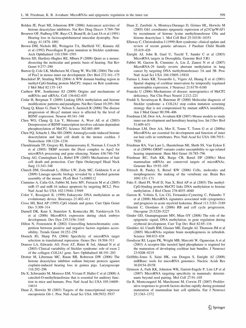
Bolden JE, Peart MJ, Johnstone RW (2006) Anticancer activities of
histone deacetylase inhibitors. Nat Rev Drug Discov 5:769–784
Brouwer OF, Padberg GW, Ruys CJ, Brand R, de Laat JA et al (1991)
Hearing loss in facioscapulohumeral muscular dystrophy. Neu-
rology 41:1878–1881
Brown DM, Nichols BE, Weingeist TA, Sheffield VC, Kimura AE
et al (1992) Procollagen II gene mutation in Stickler syndrome.
Arch Ophthalmol 110:1589–1593
Brown SD, Hardisty-Hughes RE, Mburu P (2008) Quiet as a mouse:
dissecting the molecular and genetic basis of hearing. Nat Rev
Genet 9:277–290
Burton Q, Cole LK, Mulheisen M, Chang W, Wu DK (2004) The role
of Pax2 in mouse inner ear development. Dev Biol 272:161–175
Buschdorf JP, Stratling WH (2004) A WW domain binding region in
methyl-CpG-binding protein MeCP2: impact on Rett syndrome.
J Mol Med 82:135–143
Carthew RW, Sontheimer EJ (2009) Origins and mechanisms of
miRNAs and siRNAs. Cell 136:642–655
Cedar H, Bergman Y (2009) Linking DNA methylation and histone
modification: patterns and paradigms. Nat Rev Genet 10:295–304
Chang Q, Khare G, Dani V, Nelson S, Jaenisch R (2006) The disease
progression of Mecp2 mutant mice is affected by the level of
BDNF expression. Neuron 49:341–348
Chen WG, Chang Q, Lin Y, Meissner A, West AE et al (2003)
Derepression of BDNF transcription involves calcium-dependent
phosphorylation of MeCP2. Science 302:885–889
Chen FQ, Schacht J, Sha SH (2009) Aminoglycoside-induced histone
deacetylation and hair cell death in the mouse cochlea. J
Neurochem 108:1226–1236
Chendrimada TP, Gregory RI, Kumaraswamy E, Norman J, Cooch N
et al (2005) TRBP recruits the Dicer complex to Ago2 for
microRNA processing and gene silencing. Nature 436:740–744
Cheng AG, Cunningham LL, Rubel EW (2005) Mechanisms of hair
cell death and protection. Curr Opin Otolaryngol Head Neck
Surg 13:343–348
Church DM, Goodstadt L, Hillier LW, Zody MC, Goldstein S et al
(2009) Lineage-specific biology revealed by a finished genome
assembly of the mouse. PLoS Biol 7:e1000112
Cimmino A, Calin GA, Fabbri M, Iorio MV, Ferracin M et al (2005)
miR-15 and miR-16 induce apoptosis by targeting BCL2. Proc
Natl Acad Sci USA 102:13944–13949
Colot V, Rossignol JL (1999) Eukaryotic DNA methylation as an
evolutionary device. Bioessays 21:402–411
Cross SH, Bird AP (1995) CpG islands and genes. Curr Opin Genet
Dev 5:309–314
Darnell DK, Kaur S, Stanislaw S, Konieczka JH, Yatskievych TA
et al (2006) MicroRNA expression during chick embryo
development. Dev Dyn 235:3156–3165
Dillon N, Festenstein R (2002) Unravelling heterochromatin: com-
petition between positive and negative factors regulates acces-
sibility. Trends Genet 18:252–258
Doench JG, Sharp PA (2004) Specificity of microRNA target
selection in translational repression. Genes Dev 18:504–511
Donoso LA, Edwards AO, Frost AT, Ritter R 3rd, Ahmad N et al
(2003) Clinical variability of Stickler syndrome: role of exon 2
of the collagen COL2A1 gene. Surv Ophthalmol 48:191–203
Drottar M, Liberman MC, Ratan RR, Roberson DW (2006) The
histone deacetylase inhibitor sodium butyrate protects against
cisplatin-induced hearing loss in guinea pigs. Laryngoscope
116:292–296
Du X, Schwander M, Moresco EM, Viviani P, Haller C et al (2008) A
catechol-O-methyltransferase that is essential for auditory func-
tion in mice and humans. Proc Natl Acad Sci USA 105:14609–
14614
Duan Z, Horwitz M (2003) Targets of the transcriptional repressor
oncoprotein Gfi-1. Proc Natl Acad Sci USA 100:5932–5937
Duan Z, Zarebski A, Montoya-Durango D, Grimes HL, Horwitz M
(2005) Gfi1 coordinates epigenetic repression of p21Cip/WAF1
by recruitment of histone lysine methyltransferase G9a and
histone deacetylase 1. Mol Cell Biol 25:10338–10351
Ellaway C, Christodoulou J (1999) Rett syndrome: clinical update and
review of recent genetic advances. J Paediatr Child Health
35:419–426
Enright AJ, John B, Gaul U, Tuschl T, Sander C et al (2003)
MicroRNA targets in Drosophila. Genome Biol 5:R1
Fabbri M, Garzon R, Cimmino A, Liu Z, Zanesi N et al (2007)
MicroRNA-29 family reverts aberrant methylation in lung
cancer by targeting DNA methyltransferases 3A and 3B. Proc
Natl Acad Sci USA 104:15805–15810
Farinas I, Jones KR, Tessarollo L, Vigers AJ, Huang E et al (2001)
Spatial shaping of cochlear innervation by temporally regulated
neurotrophin expression. J Neurosci 21:6170–6180
Francke U (2006) Mechanisms of disease: neurogenetics of MeCP2
deficiency. Nat Clin Pract Neurol 2:212–221
Freddi S, Savarirayan R, Bateman JF (2000) Molecular diagnosis of
Stickler syndrome: a COL2A1 stop codon mutation screening
strategy that is not compromised by mutant mRNA instability.
Am J Med Genet 90:398–406
Friedman LM, Dror AA, Avraham KB (2007) Mouse models to study
inner ear development and hereditary hearing loss. Int J Dev Biol
51:609–631
Friedman LM, Dror AA, Mor E, Tenne T, Toren G et al (2009a)
MicroRNAs are essential for development and function of inner
ear hair cells in vertebrates. Proc Natl Acad Sci USA 106:7915–
7920
Friedman RA, Van Laer L, Huentelman MJ, Sheth SS, Van Eyken E
et al (2009b) GRM7 variants confer susceptibility to age-related
hearing impairment. Hum Mol Genet 18:785–796
Friedman RC, Farh KK, Burge CB, Bartel DP (2009c) Most
mammalian mRNAs are conserved targets of microRNAs.
Genome Res 19:92–105
Fritzsch B, Pauley S, Beisel KW (2006) Cells, molecules and
morphogenesis: the making of the vertebrate ear. Brain Res
1091:151–171
Fuks F, Hurd PJ, Wolf D, Nan X, Bird AP et al (2003) The methyl-
CpG-binding protein MeCP2 links DNA methylation to histone
methylation. J Biol Chem 278:4035–4040
Garzon R, Volinia S, Liu CG, Fernandez-Cymering C, Palumbo T
et al (2008) MicroRNA signatures associated with cytogenetics
and prognosis in acute myeloid leukemia. Blood 111:3183–3189
Giacinti C, Giordano A (2006) RB and cell cycle progression.
Oncogene 25:5220–5227
Ginder GD, Gnanapragasam MN, Mian OY (2008) The role of the
epigenetic signal, DNA methylation, in gene regulation during
erythroid development. Curr Top Dev Biol 82:85–116
Giraldez AJ, Cinalli RM, Glasner ME, Enright AJ, Thomson JM et al
(2005) MicroRNAs regulate brain morphogenesis in zebrafish.
Science 308:833–838
Goodyear RJ, Legan PK, Wright MB, Marcotti W, Oganesian A et al
(2003) A receptor-like inositol lipid phosphatase is required for
the maturation of developing cochlear hair bundles. J Neurosci
23:9208–9219
Griffiths-Jones S, Saini HK, van Dongen S, Enright AJ (2008)
miRBase: tools for microRNA genomics. Nucleic Acids Res
36:D154–D158
Grimson A, Farh KK, Johnston WK, Garrett-Engele P, Lim LP et al
(2007) MicroRNA targeting specificity in mammals: determi-
nants beyond seed pairing. Mol Cell 27:91–105
Gu R, Montcouquiol M, Marchionni M, Corwin JT (2007) Prolifer-
ative responses to growth factors decline rapidly during postnatal
maturation of mammalian hair cell epithelia. Eur J Neurosci
25:1363–1372
L. M. Friedman, K. B. Avraham: MicroRNAs and epigenetic regulation in the inner ear 599
123
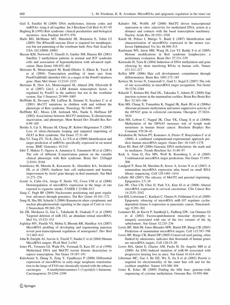
Guil S, Esteller M (2009) DNA methylomes, histone codes and
miRNAs: tying it all together. Int J Biochem Cell Biol 41:87–95
Hagberg B (1995) Rett syndrome: clinical peculiarities and biological
mysteries. Acta Paediatr 84:971–976
Harfe BD, McManus MT, Mansfield JH, Hornstein E, Tabin CJ
(2005) The RNaseIII enzyme Dicer is required for morphogen-
esis but not patterning of the vertebrate limb. Proc Natl Acad Sci
USA 102:10898–10903
Hassan KM, Norwood T, Gimelli G, Gartler SM, Hansen RS (2001)
Satellite 2 methylation patterns in normal and ICF syndrome
cells and association of hypomethylation with advanced repli-
cation. Hum Genet 109:452–462
Hertzano R, Montcouquiol M, Rashi-Elkeles S, Elkon R, Yucel R
et al (2004) Transcription profiling of inner ears from
Pou4f3(ddl/ddl) identifies Gfi1 as a target of the Pou4f3 deafness
gene. Hum Mol Genet 13:2143–2153
Hertzano R, Dror AA, Montcouquiol M, Ahmed ZM, Ellsworth B
et al (2007) Lhx3, a LIM domain transcription factor, is
regulated by Pou4f3 in the auditory but not in the vestibular
system. Eur J Neurosci 25:999–1005
Hoffbuhr K, Devaney JM, LaFleur B, Sirianni N, Scacheri C et al
(2001) MeCP2 mutations in children with and without the
phenotype of Rett syndrome. Neurology 56:1486–1495
Hoffbuhr KC, Moses LM, Jerdonek MA, Naidu S, Hoffman EP
(2002) Associations between MeCP2 mutations, X-chromosome
inactivation, and phenotype. Ment Retard Dev Disabil Res Rev
8:99–105
Horike S, Cai S, Miyano M, Cheng JF, Kohwi-Shigematsu T (2005)
Loss of silent-chromatin looping and impaired imprinting of
DLX5 in Rett syndrome. Nat Genet 37:31–40
Hua YJ, Tang ZY, Tu K, Zhu L, Li YX et al (2009) Identification and
target prediction of miRNAs specifically expressed in rat neural
tissue. BMC Genomics 10:214
Ishii T, Makita Y, Ogawa A, Amamiya S, Yamamoto M et al (2001)
The role of different X-inactivation pattern on the variable
clinical phenotype with Rett syndrome. Brain Dev 23(Suppl
1):S161–S164
Izumikawa M, Minoda R, Kawamoto K, Abrashkin KA, Swiderski
DL et al (2005) Auditory hair cell replacement and hearing
improvement by Atoh1 gene therapy in deaf mammals. Nat Med
11:271–276
Izzotti A, Calin GA, Arrigo P, Steele VE, Croce CM et al (2008)
Downregulation of microRNA expression in the lungs of rats
exposed to cigarette smoke. FASEB J 23:806–812
Jiang C, Pugh BF (2009) Nucleosome positioning and gene regula-
tion: advances through genomics. Nat Rev Genet 10:161–172
Jiang H, Sha SH, Schacht J (2006) Kanamycin alters cytoplasmic and
nuclear phosphoinositide signaling in the organ of Corti in vivo.
J Neurochem 99:269–276
Jin ZB, Hirokawa G, Gui L, Takahashi R, Osakada F et al (2009)
Targeted deletion of miR-182, an abundant retinal microRNA.
Mol Vis 15:523–533
Joglekar MV, Parekh VS, Mehta S, Bhonde RR, Hardikar AA (2007)
MicroRNA profiling of developing and regenerating pancreas
reveal post-transcriptional regulation of neurogenin3. Dev Biol
311:603–612
John B, Enright AJ, Aravin A, Tuschl T, Sander C et al (2004) Human
MicroRNA targets. PLoS Biol 2:e363
Jones PL, Veenstra GJ, Wade PA, Vermaak D, Kass SU et al (1998)
Methylated DNA and MeCP2 recruit histone deacetylase to
repress transcription. Nat Genet 19:187–191
Kalscheuer S, Zhang X, Zeng Y, Upadhyaya P (2008) Differential
expression of microRNAs in early-stage neoplastic transforma-
tion in the lungs of F344 rats chronically treated with the tobacco
carcinogen 4-(methylnitrosamino)-1-(3-pyridyl)-1-butanone.
Carcinogenesis 29:2394–2399
Kaludov NK, Wolffe AP (2000) MeCP2 driven transcriptional
repression in vitro: selectivity for methylated DNA, action at a
distance and contacts with the basal transcription machinery.
Nucleic Acids Res 28:1921–1928
Karali M, Peluso I, Marigo V, Banfi S (2007) Identification and
characterization of microRNAs expressed in the mouse eye.
Invest Ophthalmol Vis Sci 48:509–515
Kaufmann WE, Jarrar MH, Wang JS, Lee YJ, Reddy S et al (2005)
Histone modifications in Rett syndrome lymphocytes: a
preliminary evaluation. Brain Dev 27:331–339
Kawasaki H, Taira K (2004) Induction of DNA methylation and gene
silencing by short interfering RNAs in human cells. Nature
431:211–217
Kelley MW (2006) Hair cell development: commitment through
differentiation. Brain Res 1091:172–185
Kertesz M, Iovino N, Unnerstall U, Gaul U, Segal E (2007) The role
of site accessibility in microRNA target recognition. Nat Genet
39:1278–1284
Kikuchi T, Kimura RS, Paul DL, Takasaka T, Adams JC (2000) Gap
junction systems in the mammalian cochlea. Brain Res Brain Res
Rev 32:163–166
Kim MS, Chang X, Yamashita K, Nagpal JK, Baek JH et al (2008a)
Aberrant promoter methylation and tumor suppressive activity of
the DFNA5 gene in colorectal carcinoma. Oncogene 27:3624–
3634
Kim MS, Lebron C, Nagpal JK, Chae YK, Chang X et al (2008b)
Methylation of the DFNA5 increases risk of lymph node
metastasis in human breast cancer. Biochem Biophys Res
Commun 370:38–43
Kiriakidou M, Nelson PT, Kouranov A, Fitziev P, Bouyioukos C et al
(2004) A combined computational-experimental approach pre-
dicts human microRNA targets. Genes Dev 18:1165–1178
Klose RJ, Bird AP (2006) Genomic DNA methylation: the mark and
its mediators. Trends Biochem Sci 31:89–97
Krek A, Grun D, Poy MN, Wolf R, Rosenberg L et al (2005)
Combinatorial microRNA target predictions. Nat Genet 37:495–
500
Landgraf P, Rusu M, Sheridan R, Sewer A, Iovino N et al (2007) A
mammalian microRNA expression atlas based on small RNA
library sequencing. Cell 129:1401–1414
LaSalle JM (2007) The odyssey of MeCP2 and parental imprinting.
Epigenetics 2:5–10
Lee JW, Choi CH, Choi JJ, Park YA, Kim SJ et al (2008) Altered
microRNA expression in cervical carcinomas. Clin Cancer Res
14:2535–2542
Lee KH, Lotterman C, Karikari C, Omura N, Feldmann G et al (2009)
Epigenetic silencing of microRNA miR-107 regulates cyclin-
dependent kinase 6 expression in pancreatic cancer. Pancreatol-
ogy 9:293–301
Lemmers RJ, de Kievit P, Sandkuijl L, Padberg GW, van Ommen GJ
et al (2002) Facioscapulohumeral muscular dystrophy is
uniquely associated with one of the two variants of the 4q
subtelomere. Nat Genet 32:235–236
Lewis BP, Shih IH, Jones-Rhoades MW, Bartel DP, Burge CB (2003)
Prediction of mammalian microRNA targets. Cell 115:787–798
Lewis BP, Burge CB, Bartel DP (2005) Conserved seed pairing, often
flanked by adenosines, indicates that thousands of human genes
are microRNA targets. Cell 120:15–20
Lewis MA, Quint E, Glazier AM, Fuchs H, De Angelis MH et al
(2009) An ENU-induced mutation of miR-96 associated with
progressive hearing loss in mice. Nat Genet 41:614–618
Liberman MC, Gao J, He DZ, Wu X, Jia S et al (2002) Prestin is
required for electromotility of the outer hair cell and for the
cochlear amplifier. Nature 419:300–304
Lister R, Ecker JR (2009) Finding the fifth base: genome-wide
sequencing of cytosine methylation. Genome Res 19:959–966
600 L. M. Friedman, K. B. Avraham: MicroRNAs and epigenetic regulation in the inner ear
123
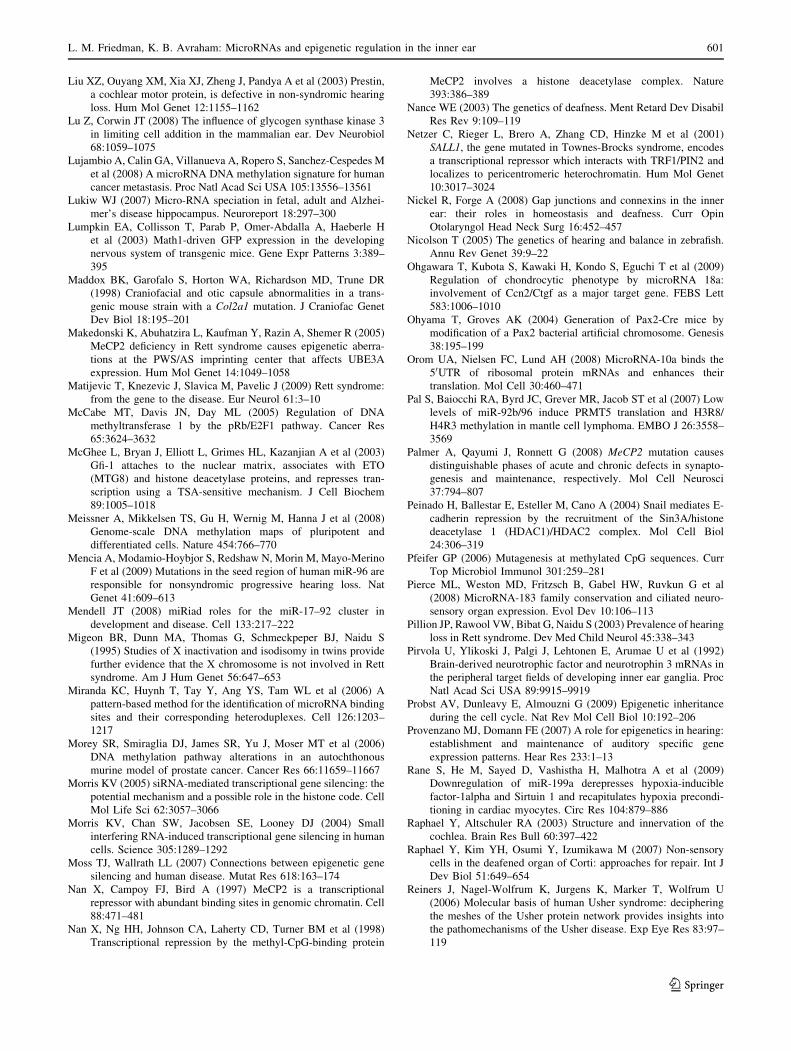
Liu XZ, Ouyang XM, Xia XJ, Zheng J, Pandya A et al (2003) Prestin,
a cochlear motor protein, is defective in non-syndromic hearing
loss. Hum Mol Genet 12:1155–1162
Lu Z, Corwin JT (2008) The influence of glycogen synthase kinase 3
in limiting cell addition in the mammalian ear. Dev Neurobiol
68:1059–1075
Lujambio A, Calin GA, Villanueva A, Ropero S, Sanchez-Cespedes M
et al (2008) A microRNA DNA methylation signature for human
cancer metastasis. Proc Natl Acad Sci USA 105:13556–13561
Lukiw WJ (2007) Micro-RNA speciation in fetal, adult and Alzhei-
mer’s disease hippocampus. Neuroreport 18:297–300
Lumpkin EA, Collisson T, Parab P, Omer-Abdalla A, Haeberle H
et al (2003) Math1-driven GFP expression in the developing
nervous system of transgenic mice. Gene Expr Patterns 3:389–
395
Maddox BK, Garofalo S, Horton WA, Richardson MD, Trune DR
(1998) Craniofacial and otic capsule abnormalities in a trans-
genic mouse strain with a Col2a1 mutation. J Craniofac Genet
Dev Biol 18:195–201
Makedonski K, Abuhatzira L, Kaufman Y, Razin A, Shemer R (2005)
MeCP2 deficiency in Rett syndrome causes epigenetic aberra-
tions at the PWS/AS imprinting center that affects UBE3A
expression. Hum Mol Genet 14:1049–1058
Matijevic T, Knezevic J, Slavica M, Pavelic J (2009) Rett syndrome:
from the gene to the disease. Eur Neurol 61:3–10
McCabe MT, Davis JN, Day ML (2005) Regulation of DNA
methyltransferase 1 by the pRb/E2F1 pathway. Cancer Res
65:3624–3632
McGhee L, Bryan J, Elliott L, Grimes HL, Kazanjian A et al (2003)
Gfi-1 attaches to the nuclear matrix, associates with ETO
(MTG8) and histone deacetylase proteins, and represses tran-
scription using a TSA-sensitive mechanism. J Cell Biochem
89:1005–1018
Meissner A, Mikkelsen TS, Gu H, Wernig M, Hanna J et al (2008)
Genome-scale DNA methylation maps of pluripotent and
differentiated cells. Nature 454:766–770
Mencia A, Modamio-Hoybjor S, Redshaw N, Morin M, Mayo-Merino
F et al (2009) Mutations in the seed region of human miR-96 are
responsible for nonsyndromic progressive hearing loss. Nat
Genet 41:609–613
Mendell JT (2008) miRiad roles for the miR-17–92 cluster in
development and disease. Cell 133:217–222
Migeon BR, Dunn MA, Thomas G, Schmeckpeper BJ, Naidu S
(1995) Studies of X inactivation and isodisomy in twins provide
further evidence that the X chromosome is not involved in Rett
syndrome. Am J Hum Genet 56:647–653
Miranda KC, Huynh T, Tay Y, Ang YS, Tam WL et al (2006) A
pattern-based method for the identification of microRNA binding
sites and their corresponding heteroduplexes. Cell 126:1203–
1217
Morey SR, Smiraglia DJ, James SR, Yu J, Moser MT et al (2006)
DNA methylation pathway alterations in an autochthonous
murine model of prostate cancer. Cancer Res 66:11659–11667
Morris KV (2005) siRNA-mediated transcriptional gene silencing: the
potential mechanism and a possible role in the histone code. Cell
Mol Life Sci 62:3057–3066
Morris KV, Chan SW, Jacobsen SE, Looney DJ (2004) Small
interfering RNA-induced transcriptional gene silencing in human
cells. Science 305:1289–1292
Moss TJ, Wallrath LL (2007) Connections between epigenetic gene
silencing and human disease. Mutat Res 618:163–174
Nan X, Campoy FJ, Bird A (1997) MeCP2 is a transcriptional
repressor with abundant binding sites in genomic chromatin. Cell
88:471–481
Nan X, Ng HH, Johnson CA, Laherty CD, Turner BM et al (1998)
Transcriptional repression by the methyl-CpG-binding protein
MeCP2 involves a histone deacetylase complex. Nature
393:386–389
Nance WE (2003) The genetics of deafness. Ment Retard Dev Disabil
Res Rev 9:109–119
Netzer C, Rieger L, Brero A, Zhang CD, Hinzke M et al (2001)
SALL1, the gene mutated in Townes-Brocks syndrome, encodes
a transcriptional repressor which interacts with TRF1/PIN2 and
localizes to pericentromeric heterochromatin. Hum Mol Genet
10:3017–3024
Nickel R, Forge A (2008) Gap junctions and connexins in the inner
ear: their roles in homeostasis and deafness. Curr Opin
Otolaryngol Head Neck Surg 16:452–457
Nicolson T (2005) The genetics of hearing and balance in zebrafish.
Annu Rev Genet 39:9–22
Ohgawara T, Kubota S, Kawaki H, Kondo S, Eguchi T et al (2009)
Regulation of chondrocytic phenotype by microRNA 18a:
involvement of Ccn2/Ctgf as a major target gene. FEBS Lett
583:1006–1010
Ohyama T, Groves AK (2004) Generation of Pax2-Cre mice by
modification of a Pax2 bacterial artificial chromosome. Genesis
38:195–199
Orom UA, Nielsen FC, Lund AH (2008) MicroRNA-10a binds the
50UTR of ribosomal protein mRNAs and enhances their
translation. Mol Cell 30:460–471
Pal S, Baiocchi RA, Byrd JC, Grever MR, Jacob ST et al (2007) Low
levels of miR-92b/96 induce PRMT5 translation and H3R8/
H4R3 methylation in mantle cell lymphoma. EMBO J 26:3558–
3569
Palmer A, Qayumi J, Ronnett G (2008) MeCP2 mutation causes
distinguishable phases of acute and chronic defects in synapto-
genesis and maintenance, respectively. Mol Cell Neurosci
37:794–807
Peinado H, Ballestar E, Esteller M, Cano A (2004) Snail mediates E-
cadherin repression by the recruitment of the Sin3A/histone
deacetylase 1 (HDAC1)/HDAC2 complex. Mol Cell Biol
24:306–319
Pfeifer GP (2006) Mutagenesis at methylated CpG sequences. Curr
Top Microbiol Immunol 301:259–281
Pierce ML, Weston MD, Fritzsch B, Gabel HW, Ruvkun G et al
(2008) MicroRNA-183 family conservation and ciliated neuro-
sensory organ expression. Evol Dev 10:106–113
Pillion JP, Rawool VW, Bibat G, Naidu S (2003) Prevalence of hearing
loss in Rett syndrome. Dev Med Child Neurol 45:338–343
Pirvola U, Ylikoski J, Palgi J, Lehtonen E, Arumae U et al (1992)
Brain-derived neurotrophic factor and neurotrophin 3 mRNAs in
the peripheral target fields of developing inner ear ganglia. Proc
Natl Acad Sci USA 89:9915–9919
Probst AV, Dunleavy E, Almouzni G (2009) Epigenetic inheritance
during the cell cycle. Nat Rev Mol Cell Biol 10:192–206
Provenzano MJ, Domann FE (2007) A role for epigenetics in hearing:
establishment and maintenance of auditory specific gene
expression patterns. Hear Res 233:1–13
Rane S, He M, Sayed D, Vashistha H, Malhotra A et al (2009)
Downregulation of miR-199a derepresses hypoxia-inducible
factor-1alpha and Sirtuin 1 and recapitulates hypoxia precondi-
tioning in cardiac myocytes. Circ Res 104:879–886
Raphael Y, Altschuler RA (2003) Structure and innervation of the
cochlea. Brain Res Bull 60:397–422
Raphael Y, Kim YH, Osumi Y, Izumikawa M (2007) Non-sensory
cells in the deafened organ of Corti: approaches for repair. Int J
Dev Biol 51:649–654
Reiners J, Nagel-Wolfrum K, Jurgens K, Marker T, Wolfrum U
(2006) Molecular basis of human Usher syndrome: deciphering
the meshes of the Usher protein network provides insights into
the pathomechanisms of the Usher disease. Exp Eye Res 83:97–
119
L. M. Friedman, K. B. Avraham: MicroRNAs and epigenetic regulation in the inner ear 601
123
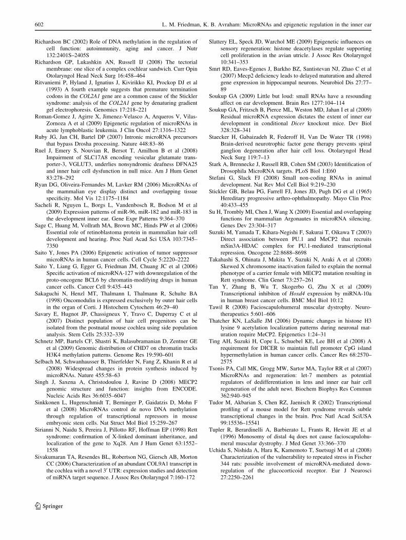
Richardson BC (2002) Role of DNA methylation in the regulation of
cell function: autoimmunity, aging and cancer. J Nutr
132:2401S–2405S
Richardson GP, Lukashkin AN, Russell IJ (2008) The tectorial
membrane: one slice of a complex cochlear sandwich. Curr Opin
Otolaryngol Head Neck Surg 16:458–464
Ritvaniemi P, Hyland J, Ignatius J, Kivirikko KI, Prockop DJ et al
(1993) A fourth example suggests that premature termination
codons in the COL2A1 gene are a common cause of the Stickler
syndrome: analysis of the COL2A1 gene by denaturing gradient
gel electrophoresis. Genomics 17:218–221
Roman-Gomez J, Agirre X, Jimenez-Velasco A, Arqueros V, Vilas-
Zornoza A et al (2009) Epigenetic regulation of microRNAs in
acute lymphoblastic leukemia. J Clin Oncol 27:1316–1322
Ruby JG, Jan CH, Bartel DP (2007) Intronic microRNA precursors
that bypass Drosha processing. Nature 448:83–86
Ruel J, Emery S, Nouvian R, Bersot T, Amilhon B et al (2008)
Impairment of SLC17A8 encoding vesicular glutamate trans-
porter-3, VGLUT3, underlies nonsyndromic deafness DFNA25
and inner hair cell dysfunction in null mice. Am J Hum Genet
83:278–292
Ryan DG, Oliveira-Fernandes M, Lavker RM (2006) MicroRNAs of
the mammalian eye display distinct and overlapping tissue
specificity. Mol Vis 12:1175–1184
Sacheli R, Nguyen L, Borgs L, Vandenbosch R, Bodson M et al
(2009) Expression patterns of miR-96, miR-182 and miR-183 in
the development inner ear. Gene Expr Patterns 9:364–370
Sage C, Huang M, Vollrath MA, Brown MC, Hinds PW et al (2006)
Essential role of retinoblastoma protein in mammalian hair cell
development and hearing. Proc Natl Acad Sci USA 103:7345–
7350
Saito Y, Jones PA (2006) Epigenetic activation of tumor suppressor
microRNAs in human cancer cells. Cell Cycle 5:2220–2222
Saito Y, Liang G, Egger G, Friedman JM, Chuang JC et al (2006)
Specific activation of microRNA-127 with downregulation of the
proto-oncogene BCL6 by chromatin-modifying drugs in human
cancer cells. Cancer Cell 9:435–443
Sakaguchi N, Henzl MT, Thalmann I, Thalmann R, Schulte BA
(1998) Oncomodulin is expressed exclusively by outer hair cells
in the organ of Corti. J Histochem Cytochem 46:29–40
Savary E, Hugnot JP, Chassigneux Y, Travo C, Duperray C et al
(2007) Distinct population of hair cell progenitors can be
isolated from the postnatal mouse cochlea using side population
analysis. Stem Cells 25:332–339
Schnetz MP, Bartels CF, Shastri K, Balasubramanian D, Zentner GE
et al (2009) Genomic distribution of CHD7 on chromatin tracks
H3K4 methylation patterns. Genome Res 19:590–601
Selbach M, Schwanhausser B, Thierfelder N, Fang Z, Khanin R et al
(2008) Widespread changes in protein synthesis induced by
microRNAs. Nature 455:58–63
Singh J, Saxena A, Christodoulou J, Ravine D (2008) MECP2
genomic structure and function: insights from ENCODE.
Nucleic Acids Res 36:6035–6047
Sinkkonen L, Hugenschmidt T, Berninger P, Gaidatzis D, Mohn F
et al (2008) MicroRNAs control de novo DNA methylation
through regulation of transcriptional repressors in mouse
embryonic stem cells. Nat Struct Mol Biol 15:259–267
Sirianni N, Naidu S, Pereira J, Pillotto RF, Hoffman EP (1998) Rett
syndrome: confirmation of X-linked dominant inheritance, and
localization of the gene to Xq28. Am J Hum Genet 63:1552–
1558
Sivakumaran TA, Resendes BL, Robertson NG, Giersch AB, Morton
CC (2006) Characterization of an abundant COL9A1 transcript in
the cochlea with a novel 30 UTR: expression studies and detection
of miRNA target sequence. J Assoc Res Otolaryngol 7:160–172
Slattery EL, Speck JD, Warchol ME (2009) Epigenetic influences on
sensory regeneration: histone deacetylases regulate supporting
cell proliferation in the avian utricle. J Assoc Res Otolaryngol
10:341–353
Smrt RD, Eaves-Egenes J, Barkho BZ, Santistevan NJ, Zhao C et al
(2007) Mecp2 deficiency leads to delayed maturation and altered
gene expression in hippocampal neurons. Neurobiol Dis 27:77–
89
Soukup GA (2009) Little but loud: small RNAs have a resounding
affect on ear development. Brain Res 1277:104–114
Soukup GA, Fritzsch B, Pierce ML, Weston MD, Jahan I et al (2009)
Residual microRNA expression dictates the extent of inner ear
development in conditional Dicer knockout mice. Dev Biol
328:328–341
Staecker H, Gabaizadeh R, Federoff H, Van De Water TR (1998)
Brain-derived neurotrophic factor gene therapy prevents spiral
ganglion degeneration after hair cell loss. Otolaryngol Head
Neck Surg 119:7–13
Stark A, Brennecke J, Russell RB, Cohen SM (2003) Identification of
Drosophila MicroRNA targets. PLoS Biol 1:E60
Stefani G, Slack FJ (2008) Small non-coding RNAs in animal
development. Nat Rev Mol Cell Biol 9:219–230
Stickler GB, Belau PG, Farrell FJ, Jones JD, Pugh DG et al (1965)
Hereditary progressive arthro-ophthalmopathy. Mayo Clin Proc
40:433–455
Su H, Trombly MI, Chen J, Wang X (2009) Essential and overlapping
functions for mammalian Argonautes in microRNA silencing.
Genes Dev 23:304–317
Suzuki M, Yamada T, Kihara-Negishi F, Sakurai T, Oikawa T (2003)
Direct association between PU.1 and MeCP2 that recruits
mSin3A-HDAC complex for PU.1-mediated transcriptional
repression. Oncogene 22:8688–8698
Takahashi S, Ohinata J, Makita Y, Suzuki N, Araki A et al (2008)
Skewed X chromosome inactivation failed to explain the normal
phenotype of a carrier female with MECP2 mutation resulting in
Rett syndrome. Clin Genet 73:257–261
Tan Y, Zhang B, Wu T, Skogerbo G, Zhu X et al (2009)
Transcriptional inhibiton of Hoxd4 expression by miRNA-10a
in human breast cancer cells. BMC Mol Biol 10:12
Tawil R (2008) Facioscapulohumeral muscular dystrophy. Neuro-
therapeutics 5:601–606
Thatcher KN, LaSalle JM (2006) Dynamic changes in histone H3
lysine 9 acetylation localization patterns during neuronal mat-
uration require MeCP2. Epigenetics 1:24–31
Ting AH, Suzuki H, Cope L, Schuebel KE, Lee BH et al (2008) A
requirement for DICER to maintain full promoter CpG island
hypermethylation in human cancer cells. Cancer Res 68:2570–
2575
Tsonis PA, Call MK, Grogg MW, Sartor MA, Taylor RR et al (2007)
MicroRNAs and regeneration: let-7 members as potential
regulators of dedifferentiation in lens and inner ear hair cell
regeneration of the adult newt. Biochem Biophys Res Commun
362:940–945
Tudor M, Akbarian S, Chen RZ, Jaenisch R (2002) Transcriptional
profiling of a mouse model for Rett syndrome reveals subtle
transcriptional changes in the brain. Proc Natl Acad SciUSA
99:15536–15541
Tupler R, Berardinelli A, Barbierato L, Frants R, Hewitt JE et al
(1996) Monosomy of distal 4q does not cause facioscapulohu-
meral muscular dystrophy. J Med Genet 33:366–370
Uchida S, Nishida A, Hara K, Kamemoto T, Suetsugi M et al (2008)
Characterization of the vulnerability to repeated stress in Fischer
344 rats: possible involvement of microRNA-mediated down-
regulation of the glucocorticoid receptor. Eur J Neurosci
27:2250–2261
602 L. M. Friedman, K. B. Avraham: MicroRNAs and epigenetic regulation in the inner ear
123
Uchida Y, Sugiura S, Nakashima T, Ando F, Shimokata H (2009)
Endothelin-1 gene polymorphism and hearing impairment in
elderly Japanese. Laryngoscope 119:938–943
Vahava O, Morell R, Lynch ED, Weiss S, Kagan ME et al (1998)
Mutation in transcription factor POU4F3 associated with inher-
ited progressive hearing loss in humans. Science 279:1950–1954
van der Maarel SM, Deidda G, Lemmers RJ, van Overveld PG, van
der Wielen M et al (2000) De novo facioscapulohumeral
muscular dystrophy: frequent somatic mosaicism, sex-dependent
phenotype, and the role of mitotic transchromosomal repeat
interaction between chromosomes 4 and 10. Am J Hum Genet
66:26–35
van Deutekom JC, Bakker E, Lemmers RJ, van der Wielen MJ, Bik E
et al (1996) Evidence for subtelomeric exchange of 3.3 kb
tandemly repeated units between chromosomes 4q35 and 10q26:
implications for genetic counselling and etiology of FSHD1.
Hum Mol Genet 5:1997–2003
Van Laer L, Huizing EH, Verstreken M, van Zuijlen D, Wauters JG
et al (1998) Nonsyndromic hearing impairment is associated
with a mutation in DFNA5. Nat Genet 20:194–197
Van Laer L, Vrijens K, Thys S, Van Tendeloo VF, Smith RJ et al
(2004) DFNA5: hearing impairment exon instead of hearing
impairment gene? J Med Genet 41:401–406
Van Laer L, Pfister M, Thys S, Vrijens K, Mueller M et al (2005)
Mice lacking Dfna5 show a diverging number of cochlear fourth
row outer hair cells. Neurobiol Dis 19:386–399
Van Laer L, Meyer NC, Malekpour M, Riazalhosseini Y, Moghan-
nibashi M et al (2007) A novel DFNA5 mutation does not cause
hearing loss in an Iranian family. J Hum Genet 52:549–552
van Overveld PG, Lemmers RJ, Sandkuijl LA, Enthoven L, Winokur
ST et al (2003) Hypomethylation of D4Z4 in 4q-linked and non-
4q-linked facioscapulohumeral muscular dystrophy. Nat Genet
35:315–317
van Overveld PG, Enthoven L, Ricci E, Rossi M, Felicetti L et al
(2005) Variable hypomethylation of D4Z4 in facioscapulohu-
meral muscular dystrophy. Ann Neurol 58:569–576
Vasudevan S, Tong Y, Steitz JA (2007) Switching from repression to
activation: microRNAs can up-regulate translation. Science
318:1931–1934
Vasudevan S, Tong Y, Steitz JA (2008) Cell-cycle control of
microRNA-mediated translation regulation. Cell Cycle 7:1545–
1549
Verdel A, Jia S, Gerber S, Sugiyama T, Gygi S et al (2004) RNAi-
mediated targeting of heterochromatin by the RITS complex.
Science 303:672–676
Vilain A, Bernardino J, Gerbault-Seureau M, Vogt N, Niveleau A
et al (2000) DNA methylation and chromosome instability in
lymphoblastoid cell lines. Cytogenet Cell Genet 90:93–101
Vissers LE, van Ravenswaaij CM, Admiraal R, Hurst JA, de Vries BB
et al (2004) Mutations in a new member of the chromodomain
gene family cause CHARGE syndrome. Nat Genet 36:955–957
Voit T, Lamprecht A, Lenard HG, Goebel HH (1986) Hearing loss in
facioscapulohumeral dystrophy. Eur J Pediatr 145:280–285
Vreugdenhil E, Verissimo CS, Mariman R, Kamphorst JT, Barbosa JS
et al (2009) MicroRNA 18 and 124a down-regulate the
glucocorticoid receptor: implications for glucocorticoid respon-
siveness in the brain. Endocrinology 150:2220–2228
Walsh CP, Xu GL (2006) Cytosine methylation and DNA repair. Curr
Top Microbiol Immunol 301:283–315
Wan M, Zhao K, Lee SS, Francke U (2001) MECP2 truncating
mutations cause histone H4 hyperacetylation in Rett syndrome.
Hum Mol Genet 10:1085–1092
Watanabe T, Sato T, Amano T, Kawamura Y, Kawamura N et al
(2008) Dnm3os, a non-coding RNA, is required for normal
growth and skeletal development in mice. Dev Dyn 237:3738–
3748
Weston MD, Pierce ML, Rocha-Sanchez S, Beisel KW, Soukup GA
(2006) MicroRNA gene expression in the mouse inner ear. Brain
Res 1111:95–104
White PM, Doetzlhofer A, Lee YS, Groves AK, Segil N (2006)
Mammalian cochlear supporting cells can divide and trans-
differentiate into hair cells. Nature 441:984–987
Whitfield TT (2002) Zebrafish as a model for hearing and deafness. J
Neurobiol 53:157–171
Wienholds E, Kloosterman WP, Miska E, Alvarez-Saavedra E,
Berezikov E et al (2005) MicroRNA expression in zebrafish
embryonic development. Science 309:310–311
Wilkin DJ, Liberfarb R, Davis J, Levy HP, Cole WG et al (2000)
Rapid determination of COL2A1 mutations in individuals with
Stickler syndrome: analysis of potential premature termination
codons. Am J Med Genet 94:141–148
Wilson AS, Power BE, Molloy PL (2007) DNA hypomethylation and
human diseases. Biochim Biophys Acta 1775:138–162
Xiang M, Gao WQ, Hasson T, Shin JJ (1998) Requirement for Brn-3c
in maturation and survival, but not in fate determination of inner
ear hair cells. Development 125:3935–3946
Xinhua B, Shengling J, Fuying S, Hong P, Meirong L et al (2008) X
chromosome inactivation in Rett Syndrome and its correlations
with MECP2 mutations and phenotype. J Child Neurol 23:22–25
Xu S, Witmer PD, Lumayag S, Kovacs B, Valle D (2007) MicroRNA
(miRNA) transcriptome of mouse retina and identification of a
sensory organ-specific miRNA cluster. J Biol Chem 282:25053–
25066
Yang T, Gurrola JG 2nd, Wu H, Chiu SM, Wangemann P et al (2009)
Mutations of KCNJ10 together with mutations of SLC26A4cause digenic nonsyndromic hearing loss associated with
enlarged vestibular aqueduct syndrome. Am J Hum Genet
84:651–657
Zhang B, Wang Q, Pan X (2007) MicroRNAs and their regulatory
roles in animals and plants. J Cell Physiol 210:279–289
L. M. Friedman, K. B. Avraham: MicroRNAs and epigenetic regulation in the inner ear 603
123