Embed Size (px)
Citation preview
MicroRNA Profiling of BRCA1/2 Mutation-Carrying andNon-Mutation-Carrying High-Grade Serous Carcinomasof OvaryCheng-Han Lee1*, Subbaya Subramanian2, Andrew H. Beck3, Inigo Espinosa3, Janine Senz4, Shirley X.
Zhu3, David Huntsman4, Matt van de Rijn3, C. Blake Gilks1
1 Department of Pathology & Laboratory Medicine, University of British Columbia, Vancouver General Hospital, Vancouver, British Columbia, Canada, 2 Department of
Laboratory Medicine and Pathology, University of Minnesota, Minneapolis, Minnesota, United States of America, 3 Department of Pathology, Stanford University Medical
Center, Stanford, California, United States of America, 4 Department of Pathology & Laboratory Medicine, British Columbia Cancer Agency, Vancouver, British Columbia,
Canada
Abstract
Background: MicroRNAs (miRNA) are 20,25 nucleotide non-coding RNAs that inhibit the translation of targeted mRNA,and they have been implicated in the development of human malignancies. High grade serous ovarian carcinomas, themost common and lethal subtype of ovarian cancer, can occur sporadically or in the setting of BRCA1/2 syndromes. Little isknown regarding the miRNA expression profiles of high grade serous carcinoma in relation to BRCA1/2 status, andcompared to normal tubal epithelium, the putative tissue of origin for high grade serous carcinomas.
Methodology/Principal Findings: Global miRNA expression profiling was performed on a series of 33 high grade serouscarcinomas, characterized with respect to BRCA1/2 status (mutation, epigenetic silencing with loss of expression or normal),and with clinical follow-up, together with 2 low grade serous carcinomas, 2 serous borderline tumors, and 3 normal fallopiantube samples, using miRNA microarrays (328 human miRNA). Unsupervised hierarchical clustering based on miRNAexpression profiles showed no clear separation between the groups of carcinomas with different BRCA1/2 status. Therewere relatively few miRNAs that were differentially expressed between the genotypic subgroups. Comparison of 33 highgrade serous carcinomas to 3 normal fallopian tube samples identified several dysregulated miRNAs (false discovery rate,5%), including miR-422b and miR-34c. Quantitative RT-PCR analysis performed on selected miRNAs confirmed the patternof differential expression shown by microarray analysis. Prognostically, lower level miR-422b and miR-34c in high gradeserous carcinomas were both associated with decreased disease-specific survival by Kaplan-Meier analysis (p,0.05).
Conclusions/Significance: High grade serous ovarian carcinomas with and without BRCA1/2 abnormalities demonstratevery similar miRNA expression profiles. High grade serous carcinomas as a group exhibit significant miRNA dysregulation incomparison to tubal epithelium and the levels of miR-34c and miR-422b appear to be prognostically important.
Citation: Lee C-H, Subramanian S, Beck AH, Espinosa I, Senz J, et al. (2009) MicroRNA Profiling of BRCA1/2 Mutation-Carrying and Non-Mutation-Carrying High-Grade Serous Carcinomas of Ovary. PLoS ONE 4(10): e7314. doi:10.1371/journal.pone.0007314
Editor: Amanda Ewart Toland, Ohio State University Medical Center, United States of America
Received December 14, 2008; Accepted September 11, 2009; Published October 2, 2009
Copyright: � 2009 Lee et al. This is an open-access article distributed under the terms of the Creative Commons Attribution License, which permits unrestricteduse, distribution, and reproduction in any medium, provided the original author and source are credited.
Funding: This study is funded by the National Cancer Institute of Canada (#017051) and the Michael Smith Foundation for Health Research Unit Grant(#INRUA006045). The funders had no role in study design, data collection and analysis, decision to publish, or preparation of the manuscript.
Competing Interests: The authors have declared that no competing interests exist.
* E-mail: [email protected]
Introduction
Ovarian carcinomas are the leading cause of death among
tumors of the female reproductive tract and high grade serous
carcinomas are the most aggressive subtype of ovarian carcinomas
[1]. High grade serous carcinomas can occur in both the familial
and sporadic settings. Women with germ-line BRCA1 or BRCA2
mutation are at increased risk of developing ovarian serous
carcinoma while a subset of non-familial ovarian serous carcino-
mas also demonstrate loss of BRCA1 through either somatic
mutations or promoter methylation with transcriptional silencing
[2]. More than half of high grade serous carcinomas overall
possess some abnormality of BRCA1 or BRCA2 [3,4,5]. The
majority of high grade serous carcinomas also show mutation and/
or loss of functional p53 [6,7,8]. Though typically showing the
greatest tumor burden in the ovaries, there is increasing evidence
that high grade serous carcinomas originate from the epithelium of
the tubal fimbriae and mullerian type epithelial inclusions of the
ovary in the majority of the cases [9,10,11].
MicroRNAs (miRNA) are 20,25 nucleotide, evolutionarily
conserved, non-coding RNAs that are important in post-
transcriptional gene regulation [12,13]. By binding to the 39
UTR region of targeted genes, miRNA can rapidly inhibit the
translation of the mRNA transcript and subsequently, through
formation of RNA-induced silencing complex, cause degradation
of the transcript [12]. In some instances, miRNA can also promote
the degradation of the targeted mRNA [14]. This genetic
regulation by miRNA is important in the fundamental processes
PLoS ONE | www.plosone.org 1 October 2009 | Volume 4 | Issue 10 | e7314
of cell growth and differentiation. There is also emerging evidence
to suggest that quantitative and qualitative (mutational) changes in
miRNA and their target binding sites can promote the
development and progression of tumors [12,13,15,16,17,18,19].
miRNA profiling studies have revealed differential expression of
miRNA in various carcinomas compared to their normal tissue
counterparts [13,16]. Some of the differentially expressed miRNA
have further been linked to the repression of tumor suppressor
genes or the upregulation of oncogenes at the protein product level
[20,21,22,23].
Recently, the miRNA expression profiles of a number of
ovarian surface epithelial tumors, including high grade serous
carcinomas have been described and several differentially
expressed miRNAs have been identified in high grade serous
carcinomas compared to normal ovarian tissue or cell lines derived
from ovarian surface epithelium [24,25,26,27,28,29]. However, a
number of important questions remain unaddressed. Firstly, it is
unclear whether high grade serous carcinomas with BRCA1/2
mutation differ in their miRNA expression patterns from non-
mutation carrying cases; it is plausible that etiologically relevant
differences in miRNA expression may be present. Secondly, with
emerging evidence to suggest tubal epithelium as the tissue of
origin for many high grade serous carcinomas [9,10], it is likely
that comparison to tubal epithelium will reveal a more
representative and accurate set of dysregulated miRNA for high
grade serous carcinomas, particularly given that the choice of
comparator group is known to significantly influence the results of
comparative gene profiling analysis [30].
In the current study, we examined the miRNA expression
profiles of 328 human miRNAs in a series of ovarian serous
tumors, molecularly characterized with respect to their BRCA1/2
status, focusing on high grade serous carcinomas. Comparisons
were made between subgroups with different BRCA1/2 status, to
a series of normal fallopian tube samples, as well to a previously
reported series of normal tissue samples and tumors of
mesenchymal origin.
Results
Clinicopathologic profiles of tumor/tissue samplesThe series of ovarian serous tumors has been described
previously [2]. These samples were extensively characterized with
respect to BRCA1 and BRCA2 status with mutational and mRNA
expression analysis and, in the case of BRCA1, promoter
methylation and protein expression levels were also analyzed.
Thirty-three high grade serous carcinomas, 2 low grade serous
carcinomas and 2 serous borderline tumors, as well as 3 normal
fallopian tube samples derived from the fimbriated end were
included in the current analysis (Table 1). The average age of the
patients at the time of surgery was 62 years with a range from 39
to 85 years. The majority of the patients with high grade serous
carcinoma in the current series presented with advanced-stage
disease (FIGO stage III or IV). Among the 33 high grade serous
carcinomas, 9 cases carry mutations in BRCA1 (8 germline and 1
somatic), 3 have mutations in BRCA2 (2 germline and 1 somatic),
10 show BRCA1 epigenetic loss (promoter methylation and
decreased expression), while 11 cases show no demonstrable
BRCA1 or BRCA2 loss (no mutation in BRCA1/BRCA2 and no
promoter methylation with decreased BRCA1 mRNA or protein).
Patient follow-up was available for all 33 cases of high grade serous
carcinomas, with a median follow-up period of 3.4 years (range
from 0.8 to 5.8 years). All patients with high grade serous
carcinomas received combination platinum-taxane chemotherapy
after surgical debulking of tumor.
Hierarchical clustering based on microRNA expressionprofiles
To better appreciate the miRNA expression signature of
ovarian serous tumors, miRNA expression of the 37 ovarian
serous tumors and 3 normal fallopian tube samples in our current
series were analyzed together with the miRNA expression from a
previously reported series of soft tissue tumors and non-neoplastic
soft tissue samples [31]. Unsupervised hierarchical clustering based
on 61 stringently filtered human miRNA showed a clear
separation between ovarian serous tumors, normal fallopian tubes
and soft tissue tumor/normal muscle samples (Figure 1). The
complete filtered dataset is available in Table S1. Ovarian serous
tumors, as a group, showed upregulation of a group of 21 miRNA
illustrated by the heatmap in Figure 1 (highlighted in blue). Among
this cluster of 21 miRNA, miR-200c, miR-141 and miR-203 were
previously reported to be expressed at relatively high levels in
tissues of the male and female reproductive system [32]. In
comparison, soft tissue tumors and non-neoplastic muscle tissues
showed uniformly higher level expression of miR-517a and miR-
517b (highlighted in red in Figure 1). The 3 normal fallopian tube
samples clustered closely together and showed a highly uniform
miRNA expression profile that is distinct from that of either
ovarian carcinomas or the soft tissue tumors. No apparent
separation between high grade ovarian serous carcinomas of
different BRCA1/2 status was noted.
High grade ovarian serous carcinomas of differentBRCA1/2 status
For the comparative analysis between high grade serous
carcinomas of different BRCA1/2 status, a less stringent set of
gene filtering criteria was employed for the 37 serous tumors and 3
fallopian tube samples in an attempt to identify miRNA showing
more subtle variation that may be of biologic interest. A total of 80
miRNA passed the filtering criteria and the complete filtered dataset
is available in Table S2, with the accompanying unsupervised
hierarchical clustering dendrogram depicted in Figure 2. While the
3 fallopian tube samples and 3 of the 4 serous borderline tumors/
low grade serous carcinoma samples clustered together, there was
again no apparent separation seen among the high grade ovarian
serous carcinomas with respect to BRCA1/2 status or tumor stage.
Significance analysis of microarrays (SAM) was performed to
identify differences in miRNA expression between groups of high
grade serous carcinomas with mutations in BRCA1, with mutations
in BRCA2, with BRCA1 epigenetic loss and with no demonstrable
BRCA1 or BRCA2 loss. Very few miRNA were found to be
differentially expressed with a false-discovery rate (FDR) ,5%
between the BRCA1/2 subgroups (Table 2). Increased expression
of miR-29a and miR-29b was seen in the combined group of
carcinomas with any demonstrable BRCA1/2 loss compared to the
group with no demonstrable BRCA1/2 abnormalities. A list of
predicted target genes of miR-29a, miR-29b and miR-214 (www.
targetscan.org/) is shown in Table S3 and it includes known tumor
suppressors such as PTEN, a putative target of miR-29a. BRCA1
and BRCA2 were not among the predicted targets of miR-29a,
miR-29b or miR-214.
High grade ovarian serous carcinomas versus Normalfallopian tubes
Given that a significant proportion of high grade ovarian serous
carcinomas are currently believed to be derived from the tubal
epithelium, a comparison was made between high grade serous
carcinomas and normal fallopian tube, sampled from the
fimbriated end. The fimbriated end of the fallopian tube is the
MiRNA in Ovarian Serous Cancer
PLoS ONE | www.plosone.org 2 October 2009 | Volume 4 | Issue 10 | e7314
site where primary tubal carcinoma occurs in BRCA mutation
carriers [9,10]. The use of tissue from the fimbriated end of the
fallopian tube was also done to increase the representation of the
epithelial elements while minimizing the presence of mesenchymal
elements, as epithelial cells predominate in this region of the tube.
Even though only 3 fallopian tube samples were included in the
current study, they exhibited highly similar patterns of miRNA
expression that is likely an accurate reflection of the miRNA levels
in the fallopian tube. SAM comparison of the 33 high grade serous
carcinomas to the 3 normal fallopian tubes identified 19
upregulated miRNA and 9 downregulated miRNA with a FDR
,5% (Table 2). While all 9 downregulated miRNA identified in
our current study have been previously reported to be downreg-
ulated in ovarian carcinomas in series composed predominantly or
exclusively of serous carcinomas compared to normal ovarian
tissue/normal ovarian surface epithelial cell lines, only 8 of the 19
upregulated miRNA identified in our current study have been
reported in these prior comparisons (Table S4) [24,25,26,27,28].
Low grade serous carcinoma/serous borderline tumorsIn comparison to normal fallopian tubes, low grade serous
carcinomas and serous borderline tumors as a group showed
Table 1. Summary of the clinicopathologic features of the study cases.
VOA STT Age* Histopathology Grade Stage** Primary site*** BRCA1 and BRCA2 status
186 5035 49 Serous/undifferentiated carcinoma 3 4 Bilateral Ovaries BRCA1 Mutations
223 5043 60 Serous carcinoma 3 2C Right Ovary BRCA1 Mutations
329 5060 47 Serous carcinoma 3 3B Bilateral Ovaries BRCA1 Mutations
293 5053 45 Serous carcinoma 3 3B Ovarian NOS BRCA1 Mutations{
283 5052 57 Serous carcinoma 3 3C Left Ovary BRCA1 Mutations
239 5045 44 Serous carcinoma 3 3C Bilateral Ovaries BRCA1 Mutations
336 5064 42 Serous/undifferentiated carcinoma 3 3B Bilateral Ovaries BRCA1 Mutations
327 5059 61 Serous carcinoma 3 3C Bilateral Ovaries BRCA1 Mutations
379 5071 64 Serous carcinoma 3 3C Bilateral Ovaries BRCA1 Mutations
163 5032 54 Serous carcinoma 3 3B Bilateral Ovaries BRCA2 Mutations
305 5055 55 Serous carcinoma 3 1A Right Ovary BRCA2 Mutations{
212 5038 54 Serous carcinoma 3 3C Bilateral Ovaries BRCA2 Mutations
217 5040 67 Serous carcinoma 3 3C Left Ovary BRCA1 Epigenetic Loss
330 5061 73 Serous carcinoma 3 1C Right Ovary BRCA1 Epigenetic Loss
332 5062 75 Serous carcinoma 3 2B Right Ovary BRCA1 Epigenetic Loss
388 5073 63 Serous carcinoma 3 3B Unable to determine BRCA1 Epigenetic Loss
201 5037 55 Serous/undifferentiated carcinoma 3 3C Bilateral Ovaries BRCA1 Epigenetic Loss
363 5068 64 Serous carcinoma 3 3C Omentum BRCA1 Epigenetic Loss
161 5031 76 Serous/undifferentiated carcinoma 3 2C Left Ovary BRCA1 Epigenetic Loss
344 5066 61 Serous carcinoma 3 3C Right Ovary/Possible Bilateral BRCA1 Epigenetic Loss
345 5067 69 Serous carcinoma 3 3B Right Ovary BRCA1 Epigenetic Loss
384 5072 50 Serous carcinoma 3 3C Unable to determine BRCA1 Epigenetic Loss
195 5036 85 Serous carcinoma 3 2C Left Ovary No BRCA1/2 abnormalities
236 5044 67 Serous carcinoma 3 3C Bilateral Ovaries No BRCA1/2 abnormalities
280 5050 56 Serous/undifferentiated carcinoma 3 3C Bilateral Ovaries No BRCA1/2 abnormalities
172 5033 68 Serous carcinoma 3 3C Right Ovary No BRCA1/2 abnormalities
254 5048 52 Serous carcinoma 3 3B Bilateral Ovaries No BRCA1/2 abnormalities
319 5057 76 Serous carcinoma 3 4 Bilateral Ovaries No BRCA1/2 abnormalities
372 5070 53 Serous carcinoma 3 3C Bilateral Ovaries No BRCA1/2 abnormalities
240 5046 54 Serous/undifferentiated carcinoma 3 3B Bilateral Ovaries No BRCA1/2 abnormalities
297 5054 78 Serous carcinoma 3 3B Bilateral Ovaries No BRCA1/2 abnormalities
366 5069 75 Serous carcinoma 3 3C Left Ovary No BRCA1/2 abnormalities
273 5049 82 Serous/undifferentiated carcinoma 3 2C Bilateral Ovaries No BRCA1/2 abnormalities
221 5042 68 Low-grade papillary serous carcinoma 1 3C Bilateral Ovaries No BRCA1/2 abnormalities
324 5058 39 Low-grade papillary serous carcinoma 1 3C Bilateral Ovaries No BRCA1/2 abnormalities
277 5132 81 Serous borderline tumor NA 1B Left Ovary ND
278 5133 48 Serous borderline tumor NA 3B Bilateral Ovaries ND
*Age at the time of surgery; **FIGO staging system; ***‘‘Primary site’’ refers to the site of the dominant tumor mass, if there was one, at the time of surgery; {Germlinemutation; VOA, Vancouver tumor bank number; STT, Stanford tumor bank number; NA, not applicable; ND, not done.
doi:10.1371/journal.pone.0007314.t001
MiRNA in Ovarian Serous Cancer
PLoS ONE | www.plosone.org 3 October 2009 | Volume 4 | Issue 10 | e7314
significant upregulation of 39 miRNA and downregulation of 13
miRNA with a FDR ,5% (Table 2). Low grade serous
carcinomas and serous borderline tumors were grouped together
in our current analysis because they are regarded as biologically
closely-related entities [6,33]. Comparison between high grade
serous carcinomas and low grade serous carcinomas/serous
borderline tumors revealed 12 miRNA that were more highly
expressed in low grade serous carcinomas/serous borderline
tumors with a FDR ,5% (Table 2).
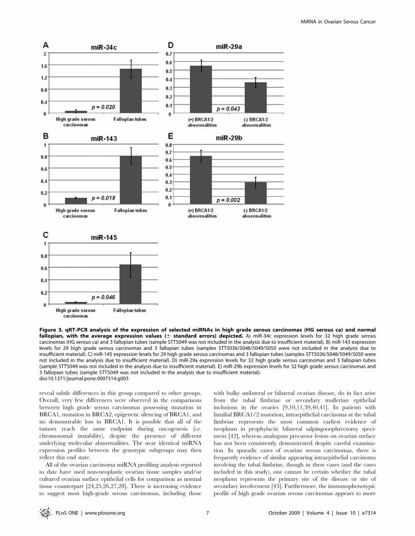
Validation of the miRNA expression patterns byquantitative RT-PCR
Quantitative RT-PCR (qRT-PCR) analysis was performed for
selected miRNA (miR-34c, miR-143, miR-145, miR-29a and miR-
29b) on the same series of high grade serous carcinoma and normal
fallopian tube samples, and the findings are depicted in Figure 3. In
accordance with the microarray data, miR-34c, miR-143 and miR-
145 showed significant downregulation in high grade serous
carcinomas compared to normal fallopian tubes by qRT-PCR
analysis. Similarly, among high grade serous carcinomas, the
expression of miR-29a and miR-29b were also significantly higher
in group with BRCA1/2 abnormalities compared to group lacking
demonstrable BRCA1/2 abnormalities. These qRT-PCR findings
support the microarray observations overall.
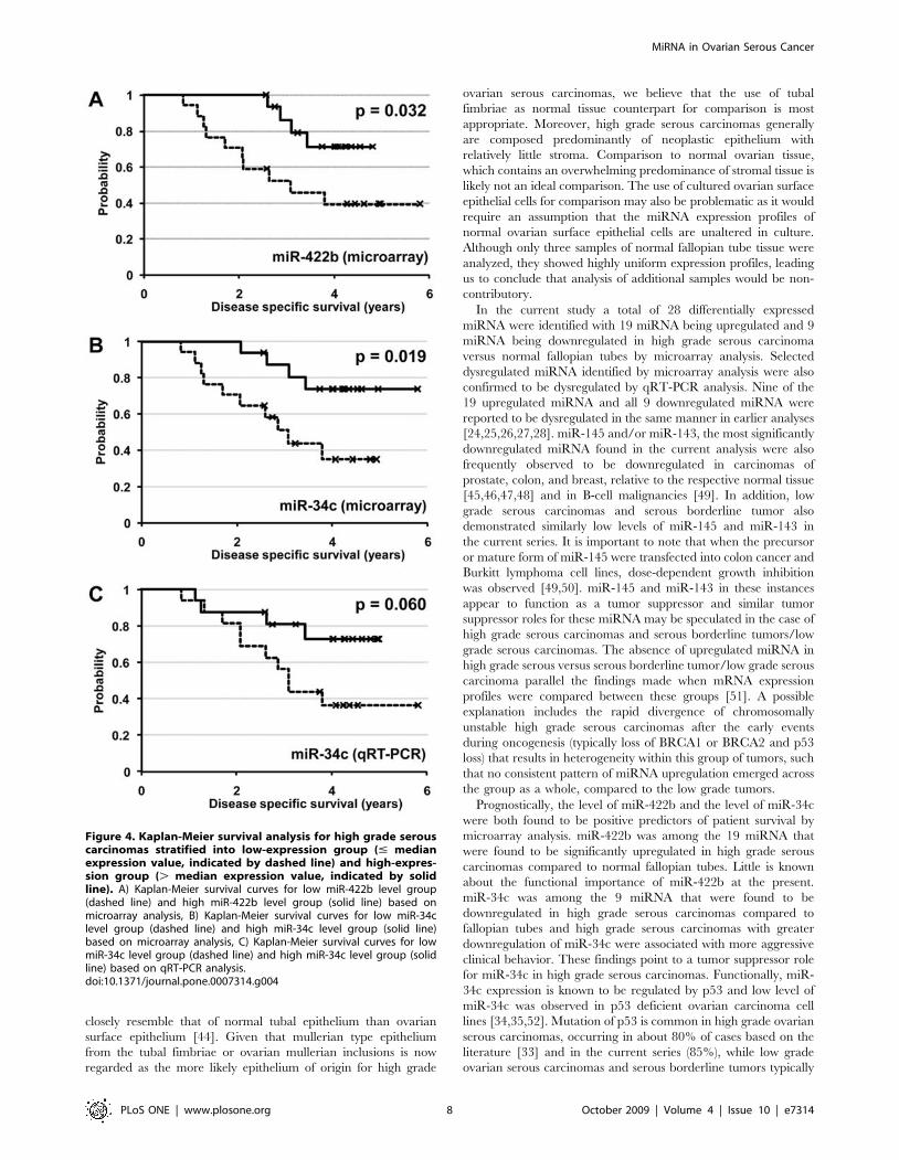
Prognostic significance of miR-34c and miR-422b highgrade ovarian serous carcinomas
Follow-up data on disease-specific survival were available for all
33 cases of high grade serous carcinomas, with a median follow-up
period of 3.4 years (range from 0.8 to 5.8 years). Because of the
relatively short period of follow-up for some of the cases in the
current series, we examined both recurrence-free survival and
disease-specific survival to identify prognostically important
miRNAs. Multivariate Cox regression analysis was performed on
the less stringently filtered dataset. miR-34c was found to be the
sole independent predictor of recurrence-free survival (HR = 0.29,
95% CI = 0.10–0.84; p = 0.02) and miR-422b was found to be the
sole independent predictor of disease-specific survival (HR = 0.21,
Figure 1. Unsupervised hierarchical clustering of 37 ovarian serous tumors, 3 normal fallopian tube and 34 soft tissue tumor/normal muscle samples based on the miRNA expression profiles of the 60 filtered human miRNA. * BRCA1 epigenetic loss; ** BRCA1mutation; *** BRCA2 mutation; SS, synovial sarcoma; LMS, leiomyosarcoma; GIST, gastrointestinal stromal tumor; ARMS, alveolar rhabdomyosar-comas; PRMS, pleomorphic rhabdomyosarcomas; ERMS, embryonal rhabdomyosarcomas.doi:10.1371/journal.pone.0007314.g001
MiRNA in Ovarian Serous Cancer
PLoS ONE | www.plosone.org 4 October 2009 | Volume 4 | Issue 10 | e7314
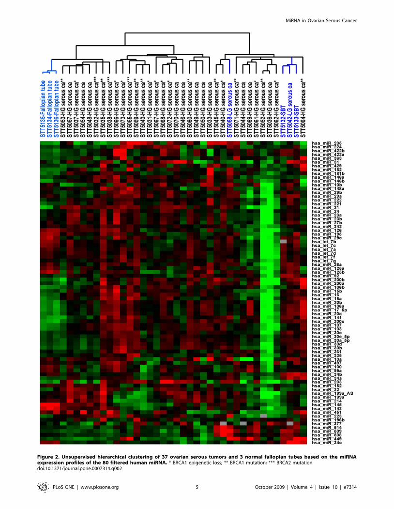
Figure 2. Unsupervised hierarchical clustering of 37 ovarian serous tumors and 3 normal fallopian tubes based on the miRNAexpression profiles of the 80 filtered human miRNA. * BRCA1 epigenetic loss; ** BRCA1 mutation; *** BRCA2 mutation.doi:10.1371/journal.pone.0007314.g002
MiRNA in Ovarian Serous Cancer
PLoS ONE | www.plosone.org 5 October 2009 | Volume 4 | Issue 10 | e7314
95% CI = 0.08–0.53; p = 0.001). Both miR-34c and miR-422b
were also among the miRNAs found to be significantly
dysregulated in high grade serous carcinomas compared to
fallopian tubes (Table 2).
Using the median expression values for miR-34c and miR-422b as
respective cutoffs, high grade serous carcinomas were separated into
high-expression group (expression values . median value) and low-
expression group (expression values # median value) for Kaplan-
Meier survival analysis (Figure 4). Based on microarray-derived
expression values, the group with lower level miR-34c expression had
decreased recurrence survival (p = 0.049) and decreased disease-
specific survival (p = 0.019) compared to the group with higher level
miR-34c expression. For miR-422b, the group with lower level miR-
422b expression was also associated with decreased disease-specific
survival (p = 0.032). When qRT-PCR derived expression values were
used for miR-34c, a similar trend between decreased miR-34c
expression and decreased disease-specific survival was seen though
statistical significance was not reached (p = 0.06). There was no
statistically significant correlation between the levels of miR-34c or
miR-422b and the initial tumor stages.
Because of the known functional link between p53 and miR-34c
[34,35,36], p53 mutation status was assessed and interpretable
data was available for 20 of the 33 high grade serous carcinomas.
17 of the 20 cases of high grade serous carcinomas demonstrated
p53 mutation with 11 missense mutation, 3 frame-shift mutation, 2
in frame deletion and one intronic deletion (Table S5). There was
no significant difference in miR-34c levels between mutation-
positive and mutation-negative tumors by either microarray or
qRT-PCR analyses.
Discussion
miRNA are non-coding RNA that play important roles in post-
transcriptional regulation. During oncogenesis, dysregulated or
dysfunctional miRNA can result in increased translation of
oncoprotein(s) and/or decreased translation of tumor suppressor
protein(s). In the current study, we used a previously validated
microarray-based methodology [31] to evaluate the expression
levels of 328 human miRNA in a series of extensively
characterized ovarian surface epithelial tumors and normal
fallopian tube samples. Several biologically important patterns
and correlations emerged from our analysis of the data. Ovarian
surface epithelial tumors as a group possessed a miRNA expression
profile that was highly dissimilar to the miRNA expression profiles
of soft tissue tumors. Given the documented roles of miRNA in
cellular differentiation [32,37], some of these observed differences
likely relate to underlying differences in differentiation into
different cellular lineages, i.e. epithelial versus mesenchymal.
While a number of recent studies have characterized the
expression profiles of miRNA in ovarian carcinomas including
high grade serous carcinomas, none has examined the miRNA
levels of high grade serous carcinomas with differing BRCA1/2
abnormalities. BRCA1 or BRCA2 abnormalities are present in
most high grade serous carcinomas and lead to chromosomal
instability through loss of the ability to repair double strand breaks
by homologous recombination [38]. In the current study, miR-29a
and miR-29b were found to be more highly upregulated in high
grade serous carcinomas with any demonstrable BRCA1/2 loss
compared to the group lacking demonstrable BRCA1/2 abnor-
malities by both microarray and qRT-PCR analyses. Notably,
high grade serous carcinomas carrying BRCA1 mutation were
previously reported by us to possess lower PTEN mRNA levels [2],
and PTEN is a among the top 20 predicted targets of miR-29a. It
is plausible that the high level of miR-29a may cause increased
degradation of PTEN mRNA, resulting in the decreased level of
PTEN mRNA observed in this group of tumors. It is important to
note that only a small number of BRCA2 mutation-carrying
tumors was included in this series and our analysis will likely not
Table 2. Summary of the SAM analysis comparing miRNA expression profiles between high grade serous carcinomas of differentBRCA1/2 status and between ovarian tumors of different histopathologic types, with a false-discovery rate (FDR) ,5% (miRNA inbold italic are associated with a FDR ,0.1%).
Tumor type Tumor type compared to Upregulated miRNA Downregulated miRNA
High grade serouscarcinoma
Fallopian tube 19 (miR-200c, miR-106b, miR-141, miR-106a,miR-17-5p, miR-107, miR-20b, miR-181b, miR-146b,miR-103, miR-205, miR-182, miR-203, miR-20a,miR-15a, miR-422b, miR-21, miR-363, miR-422a)
9 (miR-145, miR-143, miR-34c,miR-195, miR-29c, miR-125b,let-7b, miR-126, let-7c)
High grade serouscarcinoma
Low grade serouscarcinoma/serousborderline tumor
0 12 (miR-34b, miR-34c,miR-449, miR-509, miR-508,miR-34a, miR-92, miR-514, miR-29a, let-7b, miR-10a, miR-29c)
Low grade serouscarcinoma/serousborderline tumor
Fallopian tube 39 (miR-200c, miR-141, miR-20b, miR-20a, miR-106a,miR-16, miR-17-5p, miR-34a, miR-422b, miR-509, miR-449, miR-30b, miR-92, miR-15a, miR-422a, miR-107,miR-221, miR-30d, miR-103, miR-146b, miR-30a-5p, miR-15b, miR-30e-5p, miR-508, miR-514, miR-106b, miR-200b,miR-21, miR-30c, miR-29a, miR-200a, miR-377, miR-34b,miR-205, miR-224, miR-181b, let-7g, miR-222, miR-10a)
13 (miR-145, miR-143, miR-126,miR-100, miR-152, miR-214, miR-125b, miR-99a, miR-195, miR-29c,miR-451, miR-342, let-7b)
BRCA1 or BRCA2 loss (any) No BRCA1 loss 2 (miR-29a miR-29b) 0
BRCA1 loss (any) No BRCA1 loss 0 1 (miR-214)
BRCA1 mutation No BRCA1 loss 0 0
BRCA1 mutation BRCA2 mutation 0 0
BRCA1 mutation BRCA1 epigenetic loss 0 0
BRCA1 epigenetic loss No BRCA1 loss 0 0
doi:10.1371/journal.pone.0007314.t002
MiRNA in Ovarian Serous Cancer
PLoS ONE | www.plosone.org 6 October 2009 | Volume 4 | Issue 10 | e7314
reveal subtle differences in this group compared to other groups.
Overall, very few differences were observed in the comparisons
between high grade serous carcinomas possessing mutation in
BRCA1, mutation in BRCA2, epigenetic silencing of BRCA1, and
no demonstrable loss in BRCA1. It is possible that all of the
tumors reach the same endpoint during oncogenesis (i.e.
chromosomal instability), despite the presence of different
underlying molecular abnormalities. The near identical miRNA
expression profiles between the genotypic subgroups may then
reflect this end state.
All of the ovarian carcinoma miRNA profiling analysis reported
to date have used non-neoplastic ovarian tissue samples and/or
cultured ovarian surface epithelial cells for comparison as normal
tissue counterpart [24,25,26,27,28]. There is increasing evidence
to suggest most high-grade serous carcinomas, including those
with bulky unilateral or bilateral ovarian disease, do in fact arise
from the tubal fimbriae or secondary mullerian epithelial
inclusions in the ovaries [9,10,11,39,40,41]. In patients with
familial BRCA1/2 mutation, intraepithelial carcinoma at the tubal
fimbriae represents the most common earliest evidence of
neoplasm in prophylactic bilateral salpingooophrectomy speci-
mens [42], whereas analogous precursor lesion on ovarian surface
has not been consistently demonstrated despite careful examina-
tion. In sporadic cases of ovarian serous carcinomas, there is
frequently evidence of similar appearing intraepithelial carcinoma
involving the tubal fimbriae, though in these cases (and the cases
included in this study), one cannot be certain whether the tubal
neoplasm represents the primary site of the disease or site of
secondary involvement [43]. Furthermore, the immunophenotypic
profile of high grade ovarian serous carcinomas appears to more
Figure 3. qRT-PCR analysis of the expression of selected miRNAs in high grade serous carcinomas (HG serous ca) and normalfallopian, with the average expression values (6 standard errors) depicted. A) miR-34c expression levels for 32 high grade serouscarcinomas (HG serous ca) and 3 fallopian tubes (sample STT5049 was not included in the analysis due to insufficient material). B) miR-143 expressionlevels for 29 high grade serous carcinomas and 3 fallopian tubes (samples STT5036/5048/5049/5050 were not included in the analysis due toinsufficient material). C) miR-145 expression levels for 29 high grade serous carcinomas and 3 fallopian tubes (samples STT5036/5048/5049/5050 werenot included in the analysis due to insufficient material). D) miR-29a expression levels for 32 high grade serous carcinomas and 3 fallopian tubes(sample STT5049 was not included in the analysis due to insufficient material). E) miR-29b expression levels for 32 high grade serous carcinomas and3 fallopian tubes (sample STT5049 was not included in the analysis due to insufficient material).doi:10.1371/journal.pone.0007314.g003
MiRNA in Ovarian Serous Cancer
PLoS ONE | www.plosone.org 7 October 2009 | Volume 4 | Issue 10 | e7314
closely resemble that of normal tubal epithelium than ovarian
surface epithelium [44]. Given that mullerian type epithelium
from the tubal fimbriae or ovarian mullerian inclusions is now
regarded as the more likely epithelium of origin for high grade
ovarian serous carcinomas, we believe that the use of tubal
fimbriae as normal tissue counterpart for comparison is most
appropriate. Moreover, high grade serous carcinomas generally
are composed predominantly of neoplastic epithelium with
relatively little stroma. Comparison to normal ovarian tissue,
which contains an overwhelming predominance of stromal tissue is
likely not an ideal comparison. The use of cultured ovarian surface
epithelial cells for comparison may also be problematic as it would
require an assumption that the miRNA expression profiles of
normal ovarian surface epithelial cells are unaltered in culture.
Although only three samples of normal fallopian tube tissue were
analyzed, they showed highly uniform expression profiles, leading
us to conclude that analysis of additional samples would be non-
contributory.
In the current study a total of 28 differentially expressed
miRNA were identified with 19 miRNA being upregulated and 9
miRNA being downregulated in high grade serous carcinoma
versus normal fallopian tubes by microarray analysis. Selected
dysregulated miRNA identified by microarray analysis were also
confirmed to be dysregulated by qRT-PCR analysis. Nine of the
19 upregulated miRNA and all 9 downregulated miRNA were
reported to be dysregulated in the same manner in earlier analyses
[24,25,26,27,28]. miR-145 and/or miR-143, the most significantly
downregulated miRNA found in the current analysis were also
frequently observed to be downregulated in carcinomas of
prostate, colon, and breast, relative to the respective normal tissue
[45,46,47,48] and in B-cell malignancies [49]. In addition, low
grade serous carcinomas and serous borderline tumor also
demonstrated similarly low levels of miR-145 and miR-143 in
the current series. It is important to note that when the precursor
or mature form of miR-145 were transfected into colon cancer and
Burkitt lymphoma cell lines, dose-dependent growth inhibition
was observed [49,50]. miR-145 and miR-143 in these instances
appear to function as a tumor suppressor and similar tumor
suppressor roles for these miRNA may be speculated in the case of
high grade serous carcinomas and serous borderline tumors/low
grade serous carcinomas. The absence of upregulated miRNA in
high grade serous versus serous borderline tumor/low grade serous
carcinoma parallel the findings made when mRNA expression
profiles were compared between these groups [51]. A possible
explanation includes the rapid divergence of chromosomally
unstable high grade serous carcinomas after the early events
during oncogenesis (typically loss of BRCA1 or BRCA2 and p53
loss) that results in heterogeneity within this group of tumors, such
that no consistent pattern of miRNA upregulation emerged across
the group as a whole, compared to the low grade tumors.
Prognostically, the level of miR-422b and the level of miR-34c
were both found to be positive predictors of patient survival by
microarray analysis. miR-422b was among the 19 miRNA that
were found to be significantly upregulated in high grade serous
carcinomas compared to normal fallopian tubes. Little is known
about the functional importance of miR-422b at the present.
miR-34c was among the 9 miRNA that were found to be
downregulated in high grade serous carcinomas compared to
fallopian tubes and high grade serous carcinomas with greater
downregulation of miR-34c were associated with more aggressive
clinical behavior. These findings point to a tumor suppressor role
for miR-34c in high grade serous carcinomas. Functionally, miR-
34c expression is known to be regulated by p53 and low level of
miR-34c was observed in p53 deficient ovarian carcinoma cell
lines [34,35,52]. Mutation of p53 is common in high grade ovarian
serous carcinomas, occurring in about 80% of cases based on the
literature [33] and in the current series (85%), while low grade
ovarian serous carcinomas and serous borderline tumors typically
Figure 4. Kaplan-Meier survival analysis for high grade serouscarcinomas stratified into low-expression group (# medianexpression value, indicated by dashed line) and high-expres-sion group (. median expression value, indicated by solidline). A) Kaplan-Meier survival curves for low miR-422b level group(dashed line) and high miR-422b level group (solid line) based onmicroarray analysis, B) Kaplan-Meier survival curves for low miR-34clevel group (dashed line) and high miR-34c level group (solid line)based on microarray analysis, C) Kaplan-Meier survival curves for lowmiR-34c level group (dashed line) and high miR-34c level group (solidline) based on qRT-PCR analysis.doi:10.1371/journal.pone.0007314.g004
MiRNA in Ovarian Serous Cancer
PLoS ONE | www.plosone.org 8 October 2009 | Volume 4 | Issue 10 | e7314
do not possess p53 mutation [6,53]. However, the lack of
significant association between miR-34c downregulation and p53
mutation in our current series indicate that additional influences
are likely important in regulating miR-34c expression in high
grade serous carcinomas.
With regard to the prognostic significance of miR-34c, analysis
based on the microarray data demonstrated significant association
between low-level miR-34c expression and decreased disease-
specific survival, while analysis based on qRT-PCR data showed a
similar trend but did not reach statistical significance with a p-
value of 0.06. Because the input miRNA samples for both
microarray and qRT-PCR analysis were derived from the same
extraction batch, the observed discrepancies likely reflect differ-
ences in methodology. A number of studies/reviews have
addressed the issues of reproducibility between microarray and
qRT-PCR quantification [54,55,56]; while there is in general a
good correlation between microarray and qRT-PCR data, there is
some variability and the correlation is not perfect. In this study, the
downregulation of miR-34c in high grade serous carcinomas
shown by microarray analysis was confirmed by qRT-PCR
analysis but inter-method variability appears to have affected the
reproducibility of demonstration of a statistically significant miR-
34c prognostic association, indicating borderline prognostic
significance, based on this cohort. Further study will be needed
to verify the prognostic significance of miR-34c downregulation in
high grade serous carcinoma.
In summary, miRNA expression analysis of the current series of
ovarian serous tumors and normal fallopian tubes has provided us
with several important insights. High grade serous carcinomas of
different BRCA1/2 status exhibit highly similar miRNA expres-
sion patterns, with very few differences present. Comparison
between high grade serous carcinomas and normal tubal
epithelium revealed several dysregulated miRNA, including
miR-34c and miR-422b, the levels of which are both associated
with prognostic importance in our current series. This improved
understanding of the pattern and the significance of miRNA
dysregulation in high grade serous carcinoma will allow us to
better understand the oncobiology of this aggressive disease.
Materials and Methods
Patient tissue sample collectionForty fresh frozen tumor/tissue samples were obtained from
surgical specimens resected at Vancouver General Hospital
(Vancouver, BC, Canada) from 2003 to 2006 with consent from
the patients. This study was conducted according to the principles
expressed in the Declaration of Helsinki and was approved by the
institutional ethics review board at The University of British
Columbia (H02-61375 and R05–0119). All patients provided
written informed consent for the collection of samples and
subsequent analysis. The 40 samples include 33 high grade serous
carcinomas, 2 low grade serous carcinomas, 2 serous borderline
tumors and 3 normal fallopian tubes. Detailed clinicopathologic
features for the ovarian surface epithelial tumors were published
previously [2]. The clinicopathologic features and miRNA
expression data from the 34 soft tissue tumor and tissue samples
included for selected comparison were reported previously [31].
microRNA extraction and microRNA microarray studyThe details of miRNA extraction and microarray analysis were
published previously [31]. Briefly, frozen sections of the tumor/
tissue samples were performed to confirm the presence of specified
tumor/tissue types. The same frozen tumor samples were used to
isolate total RNA using mirVanaTM RNA isolation kit (Ambion,
Austin, TX, USA). Reference RNA (XpressRef Universal Total
RNA) was obtained from Super-Array (Frederick, MD, USA). The
microarrays contained a total of 668 probes spotted in duplicate,
representing 328 known human miRNAs, 113 mouse miRNAs, 45
rat miRNAs, 154 predicted human miRNAs and 28 control
probes (Ambion, Austin, TX, USA). After separating the miRNA
fraction from 25 ug of total RNA, miRNA from reference and
tissue samples were tailed and indirectly labeled using Cy3 and
Cy5 amine reactive dyes respectively (Amersham Biosciences,
Buckinghamshire, UK) using mirVana miRNA labeling kit
(Ambion, Austin, TX, USA). Hybridization was carried out at
42uC for 12–16 hrs. The arrays were washed and immediately
scanned using GenePiX 4000B array scanner (Axon Instruments,
Foster City, CA, USA) and fluorescence ratios (tumor/reference)
were calculated using GenePix software.
Analysis of microarray dataThe arrays were gridded using the GenePix program and
normalized images and data were uploaded to SMD where
complete raw data of all miRNA microarrays used in this study
can be found (http://genome-www5.stanford.edu/) [57]. Control
and empty spots on the arrays were not included for analysis, and
spots flagged as bad spots on visual inspection of the arrays were
also excluded. Only miRNA spots with a ratio of signal over
background of at least 1.5 in both the Cy3 and Cy5 channel, and
with at least 80% good data were included. For the combined
analysis of ovarian surface epithelial tumors and soft tissue tumors,
miRNAs were filtered retaining only those whose expression levels
differed by at least 9-fold in at least 3 samples, and for the analysis
of ovarian surface epithelial tumors and normal fallopian tubes,
miRNAs were filtered retaining only those whose expression levels
differed by at least 4-fold in at least 2 samples.
qRT-PCR miRNA analysisQuantitative RT-PCR analysis of miR-34c, miR-143, miR-145,
miR-29a and miR-29b was performed on the current series of high
grade serous carcinomas and fallopian tube samples, using the
same extracted total RNA used for the microarray analysis. cDNA
was reverse transcribed from individual total RNA samples (10 ng
input RNA) using TagMan MicroRNA Assays (Applied Biosys-
tems, USA) with miRNA specific primer and TagMan MicroRNA
Reverse Transcription Kit (Applied Biosystems, USA). A separate
reverse transcription was perfomed for endogenous control (18S
ribosomal RNA) using the SuperScript III First-Strand Synthesis
System (Invitrogen, USA). PCR products were amplified from the
cDNA samples using MicroRNA Assays and TagMan Universal
PCR Master Mix (Applied Biosystems, USA). The levels of the
endogenous control were used to normalize the expression levels of
each miRNA. The expression values depicted represent ratios
between the normalized level of individual sample and fallopian
tube sample STT5136.
P53 mutation analysisAll high grade serous carcinoma samples were screened for p53
mutation with exons 5 to 8 analyzed by direct sequencing in both
sense and antisense directions using the BigDye Terminator Cycle
Sequencing 3.1 Kit (Applied Biosystems). The sequencing
reactions were carried out according to the manufacturer’s
instructions.
StatisticsUnsupervised hierarchical clustering analysis and significance
analysis of microarrays (SAM) were then performed as described
MiRNA in Ovarian Serous Cancer
PLoS ONE | www.plosone.org 9 October 2009 | Volume 4 | Issue 10 | e7314
previously [58,59]. For SAM analysis, a false-discovery rate (FDR)
of less than 5% was considered significant in the current study.
Multivariate Cox regression analysis and Kaplan-Meier log-rank
method were used for survival data analysis using SPSS software
(SPSS, Chicago, IL, USA). TargetScan (www.targetscan.org)
release 4.2 was used to identify predicted miRNA targets [60,61].
Supporting Information
Table S1 Dataset containing the expression values for 60 filtered
human miRNA in ovarian surface serous tumor, normal fallopian
tube and soft tissue tumor/normal muscle samples.
Found at: doi:10.1371/journal.pone.0007314.s001 (0.09 MB
XLS)
Table S2 Dataset containing the expression values for 80 filtered
human miRNA in ovarian serous tumor and normal fallopian tube
samples.
Found at: doi:10.1371/journal.pone.0007314.s002 (0.07 MB
XLS)
Table S3 Predicted target genes of miR-29a, miR-29b and miR-
214 (www.targetscan.org/).
Found at: doi:10.1371/journal.pone.0007314.s003 (0.12 MB
XLS)
Table S4 Comparison of the miRNA profiling analysis results
between prior studies and the current study.
Found at: doi:10.1371/journal.pone.0007314.s004 (0.03 MB
DOC)
Table S5 Summary results of p53 mutation analysis findings in
high grade serous carcinomas.
Found at: doi:10.1371/journal.pone.0007314.s005 (0.03 MB
DOC)
Author Contributions
Conceived and designed the experiments: CHL SS DH MvdR CBG.
Performed the experiments: CHL SS IE JS SZ. Analyzed the data: CHL
AB JS. Contributed reagents/materials/analysis tools: DH MvdR CBG.
Wrote the paper: CHL DH MvdR CBG.
References
1. Cannistra SA (2004) Cancer of the ovary. N Engl J Med 351: 2519–2529.
2. Press JZ, De Luca A, Boyd N, Young S, Troussard A, et al. (2008) Ovarian
carcinomas with genetic and epigenetic BRCA1 loss have distinct molecularabnormalities. BMC Cancer 8: 17.
3. Esteller M, Silva JM, Dominguez G, Bonilla F, Matias-Guiu X, et al. (2000)Promoter hypermethylation and BRCA1 inactivation in sporadic breast and
ovarian tumors. J Natl Cancer Inst 92: 564–569.
4. Pal T, Permuth-Wey J, Betts JA, Krischer JP, Fiorica J, et al. (2005) BRCA1 and
BRCA2 mutations account for a large proportion of ovarian carcinoma cases.Cancer 104: 2807–2816.
5. Thrall M, Gallion HH, Kryscio R, Kapali M, Armstrong DK, et al. (2006)BRCA1 expression in a large series of sporadic ovarian carcinomas: a
Gynecologic Oncology Group study. Int J Gynecol Cancer 16 Suppl 1: 166–171.
6. Singer G, Stohr R, Cope L, Dehari R, Hartmann A, et al. (2005) Patterns of p53
mutations separate ovarian serous borderline tumors and low- and high-gradecarcinomas and provide support for a new model of ovarian carcinogenesis: a
mutational analysis with immunohistochemical correlation. Am J Surg Pathol
29: 218–224.
7. Kupryjanczyk J, Thor AD, Beauchamp R, Merritt V, Edgerton SM, et al. (1993)
p53 gene mutations and protein accumulation in human ovarian cancer. ProcNatl Acad Sci U S A 90: 4961–4965.
8. Milner BJ, Allan LA, Eccles DM, Kitchener HC, Leonard RC, et al. (1993) p53mutation is a common genetic event in ovarian carcinoma. Cancer Res 53:
2128–2132.
9. Crum CP, Drapkin R, Kindelberger D, Medeiros F, Miron A, et al. (2007)
Lessons from BRCA: the tubal fimbria emerges as an origin for pelvic serouscancer. Clin Med Res 5: 35–44.
10. Crum CP, Drapkin R, Miron A, Ince TA, Muto M, et al. (2007) The distalfallopian tube: a new model for pelvic serous carcinogenesis. Curr Opin Obstet
Gynecol 19: 3–9.
11. Salvador S, Rempel A, Soslow RA, Gilks B, Huntsman D, et al. (2008)
Chromosomal instability in fallopian tube precursor lesions of serous carcinomaand frequent monoclonality of synchronous ovarian and fallopian tube mucosal
serous carcinoma. Gynecol Oncol 110: 408–417.
12. Gregory RI, Shiekhattar R (2005) MicroRNA biogenesis and cancer. Cancer
Res 65: 3509–3512.
13. Zhang W, Dahlberg JE, Tam W (2007) MicroRNAs in tumorigenesis: a primer.
Am J Pathol 171: 728–738.
14. Valencia-Sanchez MA, Liu J, Hannon GJ, Parker R (2006) Control of
translation and mRNA degradation by miRNAs and siRNAs. Genes Dev 20:515–524.
15. Calin GA, Ferracin M, Cimmino A, Di Leva G, Shimizu M, et al. (2005) AMicroRNA signature associated with prognosis and progression in chronic
lymphocytic leukemia. N Engl J Med 353: 1793–1801.
16. Cho WC (2007) OncomiRs: the discovery and progress of microRNAs in
cancers. Mol Cancer 6: 60.
17. Esquela-Kerscher A, Slack FJ (2006) Oncomirs - microRNAs with a role in
cancer. Nat Rev Cancer 6: 259–269.
18. Asangani IA, Rasheed SA, Nikolova DA, Leupold JH, Colburn NH, et al. (2007)
MicroRNA-21 (miR-21) post-transcriptionally downregulates tumor suppressorPdcd4 and stimulates invasion, intravasation and metastasis in colorectal cancer.
Oncogene.
19. Shen J, Ambrosone CB, DiCioccio RA, Odunsi K, Lele SB, et al. (2008) A
functional polymorphism in the miR-146a gene and age of familial breast/
ovarian cancer diagnosis. Carcinogenesis 29: 1963–1966.
20. Mayr C, Hemann MT, Bartel DP (2007) Disrupting the pairing between let-7
and Hmga2 enhances oncogenic transformation. Science 315: 1576–1579.
21. Meng F, Henson R, Wehbe-Janek H, Ghoshal K, Jacob ST, et al. (2007)
MicroRNA-21 regulates expression of the PTEN tumor suppressor gene in
human hepatocellular cancer. Gastroenterology 133: 647–658.
22. Sampson VB, Rong NH, Han J, Yang Q, Aris V, et al. (2007) MicroRNA let-7a
down-regulates MYC and reverts MYC-induced growth in Burkitt lymphoma
cells. Cancer Res 67: 9762–9770.
23. Corney DC, Nikitin AY (2008) MicroRNA and ovarian cancer. Histol
Histopathol 23: 1161–1169.
24. Dahiya N, Sherman-Baust CA, Wang TL, Davidson B, Shih Ie M, et al. (2008)
MicroRNA expression and identification of putative miRNA targets in ovarian
cancer. PLoS ONE 3: e2436.
25. Iorio MV, Visone R, Di Leva G, Donati V, Petrocca F, et al. (2007) MicroRNA
signatures in human ovarian cancer. Cancer Res 67: 8699–8707.
26. Nam EJ, Yoon H, Kim SW, Kim H, Kim YT, et al. (2008) MicroRNA
expression profiles in serous ovarian carcinoma. Clin Cancer Res 14:
2690–2695.
27. Yang H, Kong W, He L, Zhao JJ, O’Donnell JD, et al. (2008) MicroRNA
expression profiling in human ovarian cancer: miR-214 induces cell survival and
cisplatin resistance by targeting PTEN. Cancer Res 68: 425–433.
28. Zhang L, Volinia S, Bonome T, Calin GA, Greshock J, et al. (2008) Genomic
and epigenetic alterations deregulate microRNA expression in human epithelial
ovarian cancer. Proc Natl Acad Sci U S A 105: 7004–7009.
29. Wyman SK, Parkin RK, Mitchell PS, Fritz BR, O’Briant K, et al. (2009)
Repertoire of microRNAs in epithelial ovarian cancer as determined by next
generation sequencing of small RNA cDNA libraries. PLoS One 4: e5311.
30. Zorn KK, Jazaeri AA, Awtrey CS, Gardner GJ, Mok SC, et al. (2003) Choice of
normal ovarian control influences determination of differentially expressed genes
in ovarian cancer expression profiling studies. Clin Cancer Res 9: 4811–4818.
31. Subramanian S, Lui WO, Lee CH, Espinosa I, Nielsen TO, et al. (2007)
MicroRNA expression signature of human sarcomas. Oncogene.
32. Landgraf P, Rusu M, Sheridan R, Sewer A, Iovino N, et al. (2007) A
mammalian microRNA expression atlas based on small RNA library
sequencing. Cell 129: 1401–1414.
33. Shih Ie M, Kurman RJ (2004) Ovarian tumorigenesis: a proposed model based
on morphological and molecular genetic analysis. Am J Pathol 164: 1511–1518.
34. Corney DC, Flesken-Nikitin A, Godwin AK, Wang W, Nikitin AY (2007)
MicroRNA-34b and MicroRNA-34c are targets of p53 and cooperate in control
of cell proliferation and adhesion-independent growth. Cancer Res 67:
8433–8438.
35. He L, He X, Lim LP, de Stanchina E, Xuan Z, et al. (2007) A microRNA
component of the p53 tumour suppressor network. Nature 447: 1130–1134.
36. Hermeking H (2007) p53 enters the microRNA world. Cancer Cell 12: 414–418.
37. Lu J, Getz G, Miska EA, Alvarez-Saavedra E, Lamb J, et al. (2005) MicroRNA
expression profiles classify human cancers. Nature 435: 834–838.
38. Kobel M, Huntsman D, Gilks CB (2008) Critical molecular abnormalities in
high-grade serous carcinoma of the ovary. Expert Rev Mol Med 10: e22.
39. Dubeau L (2008) The cell of origin of ovarian epithelial tumours. Lancet Oncol
9: 1191–1197.
40. Folkins AK, Jarboe EA, Roh MH, Crum CP (2009) Precursors to pelvic serous
carcinoma and their clinical implications. Gynecol Oncol 113: 391–396.
41. Levanon K, Crum C, Drapkin R (2008) New insights into the pathogenesis of
serous ovarian cancer and its clinical impact. J Clin Oncol 26: 5284–5293.
MiRNA in Ovarian Serous Cancer
PLoS ONE | www.plosone.org 10 October 2009 | Volume 4 | Issue 10 | e7314

42. Hirst JE, Gard GB, McIllroy K, Nevell D, Field M (2009) High rates of occult
fallopian tube cancer diagnosed at prophylactic bilateral salpingo-oophorecto-my. Int J Gynecol Cancer 19: 826–829.
43. Kindelberger DW, Lee Y, Miron A, Hirsch MS, Feltmate C, et al. (2007)
Intraepithelial carcinoma of the fimbria and pelvic serous carcinoma: Evidencefor a causal relationship. Am J Surg Pathol 31: 161–169.
44. Tong GX, Chiriboga L, Hamele-Bena D, Borczuk AC (2007) Expression ofPAX2 in papillary serous carcinoma of the ovary: immunohistochemical
evidence of fallopian tube or secondary Mullerian system origin? Mod Pathol 20:
856–863.45. Bandres E, Cubedo E, Agirre X, Malumbres R, Zarate R, et al. (2006)
Identification by Real-time PCR of 13 mature microRNAs differentiallyexpressed in colorectal cancer and non-tumoral tissues. Mol Cancer 5: 29.
46. Iorio MV, Ferracin M, Liu CG, Veronese A, Spizzo R, et al. (2005) MicroRNAgene expression deregulation in human breast cancer. Cancer Res 65:
7065–7070.
47. Michael MZ, SM OC, van Holst Pellekaan NG, Young GP, James RJ (2003)Reduced accumulation of specific microRNAs in colorectal neoplasia. Mol
Cancer Res 1: 882–891.48. Ozen M, Creighton CJ, Ozdemir M, Ittmann M (2007) Widespread
deregulation of microRNA expression in human prostate cancer. Oncogene.
49. Akao Y, Nakagawa Y, Kitade Y, Kinoshita T, Naoe T (2007) Downregulation ofmicroRNAs-143 and -145 in B-cell malignancies. Cancer Sci 98: 1914–1920.
50. Akao Y, Nakagawa Y, Naoe T (2006) MicroRNAs 143 and 145 are possiblecommon onco-microRNAs in human cancers. Oncol Rep 16: 845–850.
51. Gilks CB, Vanderhyden BC, Zhu S, van de Rijn M, Longacre TA (2005)Distinction between serous tumors of low malignant potential and serous
carcinomas based on global mRNA expression profiling. Gynecol Oncol 96:
684–694.
52. He L, He X, Lowe SW, Hannon GJ (2007) microRNAs join the p53 network–
another piece in the tumour-suppression puzzle. Nat Rev Cancer 7: 819–822.53. O’Neill CJ, Deavers MT, Malpica A, Foster H, McCluggage WG (2005) An
immunohistochemical comparison between low-grade and high-grade ovarian
serous carcinomas: significantly higher expression of p53, MIB1, BCL2, HER-2/neu, and C-KIT in high-grade neoplasms. Am J Surg Pathol 29: 1034–1041.
54. Chuaqui RF, Bonner RF, Best CJ, Gillespie JW, Flaig MJ, et al. (2002) Post-analysis follow-up and validation of microarray experiments. Nat Genet 32
Suppl: 509–514.
55. Etienne W, Meyer MH, Peppers J, Meyer RA, Jr. (2004) Comparison of mRNAgene expression by RT-PCR and DNA microarray. Biotechniques 36: 618–620,
622, 624–616.56. Morey JS, Ryan JC, Van Dolah FM (2006) Microarray validation: factors
influencing correlation between oligonucleotide microarrays and real-time PCR.Biol Proced Online 8: 175–193.
57. Sherlock G, Hernandez-Boussard T, Kasarskis A, Binkley G, Matese JC, et al.
(2001) The Stanford Microarray Database. Nucleic Acids Res 29: 152–155.58. Eisen MB, Spellman PT, Brown PO, Botstein D (1998) Cluster analysis and
display of genome-wide expression patterns. Proc Natl Acad Sci U S A 95:14863–14868.
59. Tusher VG, Tibshirani R, Chu G (2001) Significance analysis of microarrays
applied to the ionizing radiation response. Proc Natl Acad Sci U S A 98:5116–5121.
60. Lewis BP, Burge CB, Bartel DP (2005) Conserved seed pairing, often flanked byadenosines, indicates that thousands of human genes are microRNA targets. Cell
120: 15–20.61. Grimson A, Farh KK, Johnston WK, Garrett-Engele P, Lim LP, et al. (2007)
MicroRNA targeting specificity in mammals: determinants beyond seed pairing.
Mol Cell 27: 91–105.
MiRNA in Ovarian Serous Cancer
PLoS ONE | www.plosone.org 11 October 2009 | Volume 4 | Issue 10 | e7314