Embed Size (px)
Citation preview

Report
MicroRNA mimicry blocks pulmonary fibrosisRusty L Montgomery1,†, Guoying Yu2,†, Paul A Latimer1, Christianna Stack1, Kathryn Robinson1,
Christina M Dalby1, Naftali Kaminski2,* & Eva van Rooij1,3,**
Abstract
Over the last decade, great enthusiasm has evolved for microRNA(miRNA) therapeutics. Part of the excitement stems from the factthat a miRNA often regulates numerous related mRNAs. As such,modulation of a single miRNA allows for parallel regulation ofmultiple genes involved in a particular disease. While many studieshave shown therapeutic efficacy using miRNA inhibitors, efforts torestore or increase the function of a miRNA have been laggingbehind. The miR-29 family has gained a lot of attention for its clearfunction in tissue fibrosis. This fibroblast-enriched miRNA family isdownregulated in fibrotic diseases which induces a coordinateincrease of many extracellular matrix genes. Here, we show thatintravenous injection of synthetic RNA duplexes can increasemiR-29 levels in vivo for several days. Moreover, therapeutic deliv-ery of these miR-29 mimics during bleomycin-induced pulmonaryfibrosis restores endogenous miR-29 function whereby decreasingcollagen expression and blocking and reversing pulmonary fibrosis.Our data support the feasibility of using miRNA mimics to thera-peutically increase miRNAs and indicate miR-29 to be a potenttherapeutic miRNA for treating pulmonary fibrosis.
Keywords microRNA; mimic; miR-29; pulmonary fibrosis; therapeutics
Subject Categories Pharmacology & Drug Discovery; Respiratory System
DOI 10.15252/emmm.201303604 | Received 25 October 2013 | Revised 13
August 2014 | Accepted 20 August 2014 | Published online 19 September 2014
EMBO Mol Med (2014) 6: 1347–1356
Introduction
Based on gain- or loss-of-function data collected in animal disease
models using genetics or pharmacological modulation of microRNAs
(miRNAs), it is now well accepted that miRNAs are important play-
ers during disease. These studies, combined with recent positive
clinical efficacy data (Janssen et al, 2013), underscore the relevance
of miRNAs and the viability for miRNAs to become the next class of
therapeutics. Indeed, miRNAs have several advantages as therapeu-
tic intervention points in that they are small and comprise a known
sequence. Additionally, since a single miRNA can regulate numerous
target mRNAs within biological pathways, modulation of a miRNA
in principle allows for influencing an entire gene network and modi-
fying complex disease phenotypes (van Rooij & Olson, 2012).
While many studies have shown therapeutic efficacy using
single-stranded miRNA inhibitors called antimiRs, efforts to restore
or increase the function of a miRNA have been lagging behind (van
Rooij et al, 2012). Currently, miRNA function can be increased
either by viral overexpression or by using synthetic double-stranded
miRNAs. So far, the use of adeno-associated viruses (AAV) to drive
expression of a given miRNA for restoring its activity in vivo has
shown to be effective in a mouse model of hepatocellular and lung
carcinoma (Kota et al, 2009; Kasinski & Slack, 2012) and spinal and
bulbar muscular atrophy (Miyazaki et al, 2012), while the use of
unformulated synthetic oligonucleotide-based approaches to
increase miRNA levels has not been well explored.
The microRNA-29 (miR-29) family is well characterized for their
ability to regulate extracellular matrix proteins (He et al, 2013). The
family consists of miR-29a, miR-29b, and miR-29c, which are
expressed as 2 bicistronic clusters (miR-29a/miR-29b-1 and miR-
29b-2/miR-29c), and are largely homologous in sequence with only
a few mismatches between the different members in the 30 regionsof the mature miRNA (van Rooij et al, 2008). All three members are
reduced in different types of tissue fibrosis, and therapeutic benefit
of increasing miR-29 levels has been shown for heart (van Rooij
et al, 2008), kidney (Qin et al, 2011; Wang et al, 2012; Xiao et al,
2012), liver (Roderburg et al, 2011; Sekiya et al, 2011; Zhang et al,
2012), lung (Cushing et al, 2011; Xiao et al, 2012), and systemic
sclerosis (Maurer et al, 2010).
Our data indicate that miRNA mimics with modifications for
stability, and cellular uptake can be used to replicate endogenous
functions of miR-29. Systemic delivery of synthetic miR-29b mimic
increases miR-29b levels in vivo for several days without observable
side effects or effects on gene expression. However, therapeutic
treatment with miR-29b mimic in the setting of pulmonary fibrosis
restores the bleomycin-induced reduction of miR-29 and blocks and
reverses pulmonary fibrosis, which coincides with a repression of
miR-29 target genes that are induced during the disease process.
Our data support the feasibility of using miRNA mimics to thera-
peutically increase miRNAs and indicate miR-29 to be a potent
therapeutic miRNA as treatment for pulmonary fibrosis.
1 miRagen Therapeutics, Inc, Boulder, CO, USA2 Section of Pulmonary, Critical Care and Sleep Medicine, Yale School of Medicine, New Haven, CT, USA3 Hubrecht Institute, KNAW and University Medical Center Utrecht, Utrecht, The Netherlands
*Corresponding author. Tel: +1 203 7853508; E-mail: [email protected]**Corresponding author. Tel: +31 30 2121800; E-mail: [email protected]†Both authors contributed equally
ª 2014 miRagen Therapeutics. Published under the terms of the CC BY 4.0 license EMBO Molecular Medicine Vol 6 | No 10 | 2014 1347
Published online: September 19, 2014

Results and Discussion
miR-29 mimicry in vitro and in vivo
Synthetic RNA duplexes can be used to therapeutically mimic or
increase the level of a miRNA to enhance the endogenous activity of
the miRNA of interest. These miRNA mimics harbor chemical modifi-
cations for stability and cellular uptake. We designed double-stranded
miR-29 mimics utilizing lessons learned from antisense and siRNA
technologies. The “guide strand” or “antisense strand” is identical to
the miR-29b, with a UU overhang on the 30 end, modified to increase
stability, and chemically phosphorylated on the 50 end. Since the guidestrand has to function as a miRNA and the RISC machinery in the cell
needs to recognize it as such, the allowed chemical modifications are
limited. The 20-F modification helps to protect against exonucleases,
hence making the guide strand more stable, while it does not interfere
with RISC loading. The “passenger strand” or the “sense strand”
contains 20-O-Me modifications to prevent loading into RNA-induced
silencing complex (RISC) as well as increase stability and is linked to
cholesterol for enhanced cellular uptake. Several mismatches are
introduced to prevent this strand from functioning as an antimiR and
to lessen hybridization affinity for the guide strand (Fig 1A).
To test for functional efficacy, we transfected miR-29b mimic into
a mouse fibroblast cell line (NIH 3T3) and measured the effect on
Collagen1a1 (Col1a1) expression, a known direct target gene of
miR-29 (van Rooij et al, 2008). Increasing amount of miR-29b
mimic showed a dose-dependent decrease in Col1a1, compared to
either untreated or control oligo treated cells, indicating the miR-
29b mimic to be functional. An siRNA directly targeting Col1a1 was
taken along as a positive control (Fig 1B).
To start exploring the in vivo applicability and distribution of
miR-29 mimic, we injected mice intravenously with 10, 50, 100, or
125 mg per kg (mpk) and sacrificed them 4 days later. Northern
blot analysis on multiple tissues indicated little to no increase in
miR-29b in kidney or liver samples compared to saline control.
Cardiac distribution was detected; however, this appeared to be
quite variable and spleen delivery could be observed at the highest
dose only. In contrast, delivery to the lungs could be observed at all
3 of the highest doses 4 days after injection (Fig 1C). No effects on
liver function (transaminase, ALT) were observed in the plasma,
indicating that these miRNA mimics are well tolerated at these doses
(Supplementary Fig S1). Real-time PCR demonstrated similar results
with robust dose-dependent distribution of the miR-29b mimic to
the lung compared to saline-injected animals (Fig 1D). Additionally,
real-time PCR analysis of miR-29 targets showed no regulation at
the mRNA level in the treated animals except for Col3a1 at the high-
est dose in the spleen (Supplementary Fig S2). This suggests that
the target genes are either at steady state in non-stressed animals
and that mimics lower target genes when they are elevated, or that
functional delivery was inadequate or insufficient.
To gain more insights into the in vivo stability of miRNA mimics,
we injected 125 mpk of miR-29b mimic and sacrificed the mice 1, 2,
4, or 7 days later. Robust presence of miR-29b mimic could be
detected by both Northern blot and real-time PCR analysis 1 day
after injection in all tissues examined; however, tissue clearance
greatly differed thereafter (Fig 1E and F). Liver and kidney rapidly
cleared miR-29b mimic with minimal detection after day 1. Lung
and spleen demonstrated the most pronounced detection of miR-29b
mimic over time, which sustained at least 2–4 days post-treatment
(Fig 1E and F). The increase was specific for miR-29b without any
effect on miR-29a and miR-29c levels as measured by real-time PCR
(Supplementary Fig S3). Also, here real-time PCR analysis of miR-29
targets showed no downregulation at the mRNA level in non-
stressed animals (Supplementary Fig S4).
Together, these data indicate that unformulated miR-29b mimic
can increase the miRNA level with tissue-dependent clearance and
delivery efficiency, without any clear effect on gene expression
under baseline conditions.
miR-29b mimic blunts bleomycin-induced pulmonary fibrosis
Current treatments of tissue fibrosis mostly rely on targeting the
inflammatory response; however, these are ultimately ineffective in
preventing progression of the disease, underscoring the need for
new mechanistic insights and therapeutic approaches (Friedman
et al, 2013). Recent studies indicate the involvement of miRNAs in
pulmonary fibrosis (Pandit et al, 2011).
Due to the preferential lung distribution of our mimic, we set out
to explore whether stress and subsequent induction of target gene
expression would allow for detectable changes in mRNA target
genes and downstream therapeutic effects in response to miR-29b
mimic. To this end, we used the bleomycin-induced model of
pulmonary fibrosis as described (Pandit et al, 2010) and injected the
mice with 100 mpk miR-29b mimic, control or a comparable
volume of saline at two time-points: 3 and 10 days after bleomycin
treatment. As expected, 14 days after bleomycin treatment, miR-29
levels were reduced, while miR-29b mimic treatment resulted in the
increased detection of miR-29b levels compared to either control or
saline-injected animals as measured by real-time PCR, albeit with a
high level of variation (Fig 2A). It is currently unclear why the
increase in miR-29b levels is less than we detected in baseline mice
(Fig 1). A comparable decline in miR-29 levels was observed in
pulmonary biopsies of patients with idiopathic pulmonary fibrosis
(IPF) compared to normal controls (Fig 2B). Histological examina-
tion by trichrome staining showed a clear and robust fibrotic and
inflammatory reaction in response to bleomycin, which was blunted
by miR-29b mimic treatment (Fig 2C). Additionally, hydroxyproline
measurements to assay for total collagen content indicated a signifi-
cant increase following bleomycin treatment in both saline and
control-treated groups, while there was no statistical difference in
the miR-29 mimic-treated group between saline and bleomycin-
treated mice, indicating that miR-29b mimic treatment blunts
bleomycin-induced pulmonary fibrosis (Fig 2D).
Innate immune effector signaling pathways act as important drivers
of myofibroblast transdifferentiation by provoking fibrosis. To further
characterize the therapeutic effects of miR-29b mimic in the setting of
bleomycin-induced pulmonary fibrosis, we performed bronchoalveo-
lar lavage (BAL) on these mice and assessed cytokine levels. Signifi-
cantly higher concentrations of IL-12, IL-4, and G-CSF were detectable
in BAL fluids from lungs from bleomycin-treated mice, which were
reduced with miR-29b mimic (Fig 2E–G). Additionally, the bleomycin-
induced elevation of detectable immune cells in BAL fluids was signifi-
cantly reduced in the presence of miR-29b mimic (Fig 2H), indicating
an inhibitory effect on the immune response by miR-29b, which is
likely secondary to the antifibrotic-effect. To determine if miR-29
mimicry has a direct effect on macrophages, we transfected miR-29b
EMBO Molecular Medicine Vol 6 | No 10 | 2014 © 2014 miRagen Therapeutics
EMBO Molecular Medicine MicroRNA mimicry blocks pulmonary fibrosis Rusty L Montgomery et al
1348
Published online: September 19, 2014

sense
antisense
A
Chol
P
P
phosphodiester phosphorothioates
2´-O-Me 2´-F RNA mismatch monophosphate
U U
0
1.5
0.5
5 nM
0.5
nM50
nM
5 nM
0.5
nM50
nM
5 nM
0.5
nM50
nM
1.0
Col1a1
Rel
ativ
e ex
pres
sion
UntreatedMockNTCmiR-29b mimicCol1a1 siRNA
*
** ##
*
*
*#
#
#
B
Kidney
Spleen
Heart
Liver
100mpk 125mpk50mpk10mpkSalineC
Lung
U6
U6
U6
U6
U6
miR-29b
miR-29b
miR-29b
miR-29b
miR-29b
*
Lung
*
Heart Liver
*
*
Kidney Spleen0
10
50
30
2
4
6
40
20
miR-29b
Rel
ativ
e ex
pres
sion
Saline10mpk50mpk100mpk125mpk
*
*
D
Kidney
Spleen
Heart
Liver
Day 4 Day 7Day 2Day 1SalineE
Lung
U6
U6
U6
U6
U6
miR-29b
miR-29b
miR-29b
miR-29b
miR-29b
*
Lung
*
Heart
*
Liver
* *
Kidney Spleen0.1
10
10000
1
1000
100
miR-29b
Rel
ativ
e ex
pres
sion
SalineDay 1Day 2Day 4Day 7
*
F
Figure 1. Pharmacokinetic properties of miR-29b mimic.
A The double-stranded miR-29 mimics design contains a “guide strand” or “antisense strand” that is identical to the miR-29b, with a UU overhang on the 30 end,modified to increase stability, and chemically phosphorylated on the 50 end and a “passenger strand” or “sense strand” that contains 20-O-Me modifications toprevent loading into RNA-induced silencing complex (RISC) as well as increase stability and is linked to cholesterol for enhanced cellular uptake. Several mismatchesare introduced in the sense strand to prevent this strand from functioning as an antimiR.
B Transfection experiments in NIH 3T3 show a dose-dependent decrease in Col1a1 with increasing amount of miR-29b mimic compared to either untreated or mock-treated cells. An siRNA directly targeting Col1a1 was taken along as a positive control. *P < 0.05 versus mock, #P < 0.05 versus untreated.
C Northern blot analysis for miR-29b in different tissues 4 days after intravenous injection with 10, 50, 100, or 125 mpk miR-29b mimic indicates delivery to all tissuesat the highest dose, with the most effective delivery taking place to the lungs and spleen compared to saline-injected mice. U6 is used as a loading control.
D Real-time quantification of miR-29b mimicry indicates an increased level of miR-29b at the higher dose levels with the most efficient delivery to the lungs and spleen(n = 4 per group). *P < 0.05 versus saline-injected animals.
E Northern blot analysis for miR-29b in different tissues 1, 2, 4, and 7 days after intravenous injection with 125 mpk of mimic indicates the presence of miR-29b mimicin all tissues examined, with a longer detection in lung and spleen. U6 is used as a loading control.
F Real-time quantification of miR-29b mimicry indicates an increased level of miR-29b in all tissues measured which is maintained the longest in lungs and spleen(n = 4 per group). *P < 0.05 versus saline-injected animals.
© 2014 miRagen Therapeutics EMBO Molecular Medicine Vol 6 | No 10 | 2014
Rusty L Montgomery et al MicroRNA mimicry blocks pulmonary fibrosis EMBO Molecular Medicine
1349
Published online: September 19, 2014
miR-29bmimic
Controlmimic
Saline
Bleomycin
0
5
2
4
1
3
Fol
d ch
ange
miR-29amiR-29bmiR-29c
A
miR-29bmimic
Controlmimic
Saline
Saline
* *
0
5
2
4
1
3
Rel
ativ
e ex
pres
sion
NormalIPF
B
miR-29b miR-29cmiR-29a
* * *
C
Sal
ine
Ble
omyc
in
Control mimicSaline miR-29b mimic
0
2500
1000
2000
1500
500
II-12
(pg
/ml)
SalineBleomycin
E
miR-29bmimic
Controlmimic
Saline
*
ns
**
*
0
300
100
200
Hyd
roxy
prol
ine
(ug/
rt lu
ng)
SalineBleomycin
D
miR-29bmimic
Controlmimic
Saline
*
ns
**
0
250
100
200
150
50
II-4
(pg/
ml)
SalineBleomycin
F
miR-29bmimic
Controlmimic
Saline
*
ns
**
*
0
500
200
400
300
100
G-C
SF
(53
) (p
g/m
l)
SalineBleomycin
G
miR-29bmimic
Controlmimic
Saline
*
ns
**
*
- ++0
15
10
5Cel
ls/fi
eld
NeutrophilsLymphocytesMacrophages
H
+ --Bleo:miR-29b
mimicControlSaline
*^* ^^
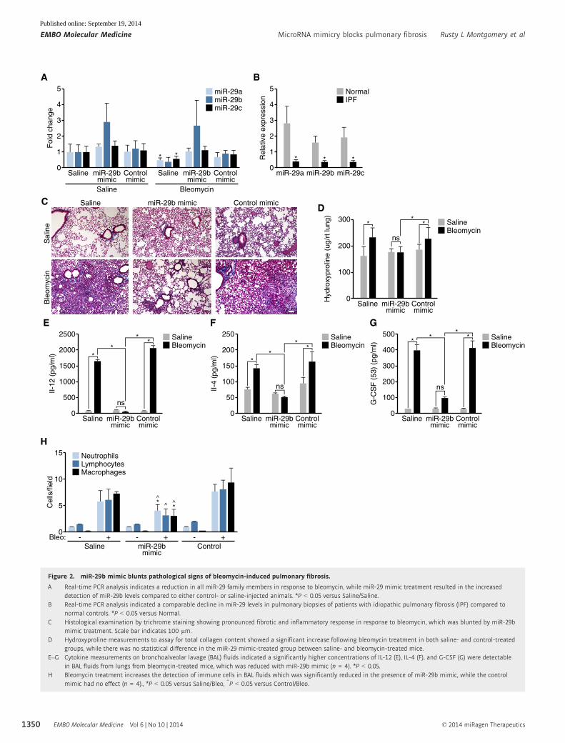
Figure 2. miR-29b mimic blunts pathological signs of bleomycin-induced pulmonary fibrosis.
A Real-time PCR analysis indicates a reduction in all miR-29 family members in response to bleomycin, while miR-29 mimic treatment resulted in the increaseddetection of miR-29b levels compared to either control- or saline-injected animals. *P < 0.05 versus Saline/Saline.
B Real-time PCR analysis indicated a comparable decline in miR-29 levels in pulmonary biopsies of patients with idiopathic pulmonary fibrosis (IPF) compared tonormal controls. *P < 0.05 versus Normal.
C Histological examination by trichrome staining showing pronounced fibrotic and inflammatory response in response to bleomycin, which was blunted by miR-29bmimic treatment. Scale bar indicates 100 lm.
D Hydroxyproline measurements to assay for total collagen content showed a significant increase following bleomycin treatment in both saline- and control-treatedgroups, while there was no statistical difference in the miR-29 mimic-treated group between saline- and bleomycin-treated mice.
E–G Cytokine measurements on bronchoalveolar lavage (BAL) fluids indicated a significantly higher concentrations of IL-12 (E), IL-4 (F), and G-CSF (G) were detectablein BAL fluids from lungs from bleomycin-treated mice, which was reduced with miR-29b mimic (n = 4). *P < 0.05.
H Bleomycin treatment increases the detection of immune cells in BAL fluids which was significantly reduced in the presence of miR-29b mimic, while the controlmimic had no effect (n = 4)., *P < 0.05 versus Saline/Bleo, ^P < 0.05 versus Control/Bleo.
EMBO Molecular Medicine Vol 6 | No 10 | 2014 © 2014 miRagen Therapeutics
EMBO Molecular Medicine MicroRNA mimicry blocks pulmonary fibrosis Rusty L Montgomery et al
1350
Published online: September 19, 2014

mimic and control into macrophage cells, RAW 264.7, and harvested
the supernatant at 24 and 48 h after transfection. IFN-r, IL-1B, IL-2,
IL-4, IL-5, IL-6, KC, IL-10, IL-12P70, and TNF-a were assessed, with no
significant differences observed between miR-29b mimic and control
(P. Latimer and R. Montgomery, unpublished data). By real-time PCR
analysis, there were no significant differences in Tgfb1, Ctgf, FGF1, or
PDGF expression; however, we did observe a significant difference in
Csf3, Igf1, and Kc expression (Supplementary Fig S5 and P. Latimer
and R. Montgomery, unpublished data).
Since it has been well validated that miR-29 functions through
the regulation of many different extracellular matrix related genes
(van Rooij & Olson, 2012), we confirmed the regulation of a subset
of these target genes. While a significant increase in Col1a1 and a
trend increase in Col3a1 expression were observed with bleomycin
treatment in both saline and control-treated groups, the detection of
Col1a1 and Col3a1 was significantly blunted in the presence of
miR-29b mimic in the bleomycin-treated mice (Fig 3A and B).
Interestingly, the increase in Igf1 levels in BAL fluids following
bleomycin treatment was significantly blunted in the presence of
miR-29 mimic compared to both saline and control-treated mice
(Fig 3C). Furthermore, immunohistochemistry for Igf1 demonstrated
robust reductions in Igf1 after bleomycin in miR-29b mimic-treated
groups compared to saline or controls (Fig 3D).
After establishing that early (days 3 and 10) miR-29 mimicry was
sufficient to prevent bleomycin-induced fibrosis, we sought to deter-
mine if miR-29 mimicry affects established fibrosis. For that
purpose, we started the miR-29b mimic administration at day 10
post-bleomycin and repeated the doses at days 14 and 17 after
which we harvested the lungs at day 21. Hydroxyproline assessment
of the right lung showed a significant increase with bleomycin in
both saline and control-treated lungs; however miR-29b mimic treat-
ment blunted this effect (Fig 4A). Furthermore, bleomycin treatment
resulted in significant increases in Col1a1 and Col3a1 expression,
which was also normalized with miR-29b mimic treatment (Fig 4B
and C). Histological assessment by trichrome staining corroborated
this effect, whereby bleomycin induced significant fibrosis with
saline or control treatment which was blunted with miR-29b
mimicry (Fig 4D).
While we believe these effects are mediated through regulation
of collagen production from lung fibroblasts, we are not able to rule
out effects from other collagen producing cells. To address this, we
assessed miR-29b mimic effects in vitro from different lung cells,
*
Controlmimic
*
ns
miR-29bmimic
ns
A
0
2.0
0.5
1.0
1.5
Col1a1
Fol
d ch
ange
*
B
Saline Controlmimic
*
miR-29bmimic
0
3
1
2
Col3a1
Fol
d ch
ange
Saline BleomycinSaline Bleomycin Saline Bleomycin
Saline
C
*
*
*
Controlmimic
**
miR-29bmimic
0
6000
2000
4000
IGF1 in BAL
Igf1
(pg
/ml)
Saline
D
Sal
ine
Ble
omyc
in
Control mimicSaline miR-29b mimic
Figure 3. In vivo mimicry of miR-29b represses the induction of miR-29 target genes during pulmonary fibrosis.
A, B Bleomycin treatment increases the expression of Col1a1 (A) and Col3a1 (B), and the presence of miR-29b mimic inhibits Col1a1 and Col3a1 as measured by real-time PCR. MiR-29b mimicry has no effect on target repression under baseline conditions. (n = 6–8), *P < 0.05.
C IGF1 levels in BAL fluids increase following bleomycin treatment which were significantly blunted in the presence of miR-29 mimic compared to both saline- andcontrol mimic-treated mice (n = 4). *P < 0.05.
D Immunohistochemistry demonstrated robust detection of IGF1 after bleomycin treatment, which was reduced in the miR-29b mimic-treated group compared tosaline- or control mimic-treated mice. Scale bar indicates 50 lm.
© 2014 miRagen Therapeutics EMBO Molecular Medicine Vol 6 | No 10 | 2014
Rusty L Montgomery et al MicroRNA mimicry blocks pulmonary fibrosis EMBO Molecular Medicine
1351
Published online: September 19, 2014

*
A
0
160
4020
6080
100120140
Hyd
roxy
prol
ine
(µg/
right
lung
)
**
+ +
***
D Bleo + Control mimicSaline Bleomycin Bleo + miR-29b mimic
++Saline:+ ++-Bleomycin:+ ---Control mimic:- +--miR-29b mimic:
**
B
0
2.0
0.5
1.0
1.5
Fol
d ch
ange
(Col
1a1
mR
NA
) **
+ +
****
++Saline:+ ++-Bleomycin:+ ---Control mimic:- +--miR-29b mimic:
*
C
0
2.0
0.5
1.0
1.5
Fol
d ch
ange
(Col
3a1
mR
NA
) *
+ +++Saline:+ ++-Bleomycin:+ ---Control mimic:- +--miR-29b mimic:
+TGF-β0
0.5
2.0
2.5
1.5
1.0
IPF COL1A1 24h
Rel
ativ
e ex
pres
sion
UntreatedMockControl mimic0.25nM miR-29b mimic
25nM miR-29b mimic2.5nM miR-29b mimic
E
*
#
*
+TGF-β0
0.5
2.0
1.5
1.0
IPF COL3A1 24h
Rel
ativ
e ex
pres
sion
UntreatedMockControl mimic0.25nM miR-29b mimic
25nM miR-29b mimic2.5nM miR-29b mimic
F
**
+TGF-β0
20
30
10
A549 Lung epithelial COL1A1 24h
Rel
ativ
e ex
pres
sion
UntreatedMockControl mimic0.25nM miR-29b mimic
25nM miR-29b mimic2.5nM miR-29b mimic
G
*
#
+TGF-β0
1
4
3
2
A549 Lung epithelial COL3A1 24h
Rel
ativ
e ex
pres
sion
UntreatedMockControl mimic0.25nM miR-29b mimic
25nM miR-29b mimic2.5nM miR-29b mimic
H
**
Col1a1 Col3a1
Figure 4. Therapeutic mimicry of miR-29 attenuates bleomycin-induced fibrosis.
A Hydroxyproline assessment showed a significant increase following bleomycin treatment in both saline- and control-treated groups; however, there was nostatistical difference in the miR-29 mimic-treated group between saline- and bleomycin-treated mice. *P < 0.05 (n = 8).
B, C Real-time PCR analysis showed a significant increase of Col1a1 (B) and Col3a1 (C) after bleomycin treatment. miR-29b mimic treatment normalized both Col1a1and Col3a1 to vehicle-treated expression levels. *P < 0.05 (n = 8).
D Histological examination by trichrome staining showing robust fibrosis in response to bleomycin, which was blunted by miR-29b mimic treatment. Scale barindicates 50 lm.
E, F Primary pulmonary fibroblasts from patients with IPF were treated with vehicle or TGF-b and transfected with control mimic or miR-29b mimic. Real-time PCRwas performed for Col1a1 (E) and Col3a1 (F). miR-29b mimic treatment showed a dose-dependent reduction in both collagens.
G, H A549 cells were treated with vehicle or TGF-b and transfected with control mimic or miR-29b mimic. Real-time PCR was performed for Col1a1 (G) and Col3a1 (H).miR-29b mimic treatment showed a dose-dependent reduction in expression of both Col1a1 and Col3a1.
EMBO Molecular Medicine Vol 6 | No 10 | 2014 © 2014 miRagen Therapeutics
EMBO Molecular Medicine MicroRNA mimicry blocks pulmonary fibrosis Rusty L Montgomery et al
1352
Published online: September 19, 2014
including primary fibroblasts from IPF patients and A549 cells, a
lung epithelial cell line. As expected, primary pulmonary fibroblasts
from IPF patients show an increase in Col1a1 and Col3a1 in
response to TGF-b (Fig 4E and F). This effect was dose-dependently
blunted with miR-29b mimic treatment at both 24 and 48 h (Fig 4E
and F and P. Latimer and R. Montgomery, unpublished data). Simi-
larly, A549 cells respond to TGF-b with robust increases in
Col1a1and Col3a1 expression (Fig 4G and H). Again, miR-29b
mimic treatment is able to block collagen induction, in both TGF-btreated as well as baseline conditions (Fig 4G and H). The effects on
collagen induction are much more robust in the A549 cells
compared to primary IPF cells; however, this is likely due to the
already high expression in primary fibroblasts from IPF patients.
While A549 cells are epithelial cells and the contribution of these
cells to pulmonary fibrosis is still debated, these data do show that
miR-29 mimicry can also block collagen induction in this cell type.
Additionally, we looked at miR-29 effects in the macrophage line,
Thp-1, but could not observe collagen expression in the cells,
regardless of stimulation (P. Latimer and R. Montgomery, unpub-
lished data). These data suggest miR-29b mimicry is able to blunt
collagen-induced expression in fibroblasts and epithelial cells. These
data are all in line with a recent paper by Xiao et al (2012), in which
they showed that gene transfer of miR-29 using a Sleeping
Beauty-transposon system was capable of preventing and treating
bleomycin-induced pulmonary fibrosis, further underscoring the
therapeutic potential for increasing miR-29.
Our data suggests the feasibility of using microRNA mimics to
restore the function of lost or downregulated miRNAs. However, it
is important to note that because RISC incorporation is required for
appropriate miRNA function, the allowed chemical modifications
are limited; thus, miRNA mimics are far less stable than antimiR
chemistries, and dosage and administration regiments need to be
worked out in detail as the doses used in most animal studies to date
are probably significantly higher than what would be therapeutically
acceptable. Another potential issue with miRNA replacement thera-
pies is the challenge of restoring the level of a downregulated
miRNA while preventing the introduction of supraphysiological
levels of the miRNA. Additionally, although a miRNA mimic can
have therapeutic use, potential off target effects of miRNA mimicry
can occur as a result of delivery to tissues or cells that do not
normally contain the miRNA of interest. Thus, targeting those
patients with exceedingly low levels of the miRNA and delivery to
the appropriate cell type or tissue are important aspects of effective
miRNA mimicry. In the case of pulmonary fibrosis, this may suggest
that direct delivery through the inhaled route may be an appealing
alternative to traditional routes of administration. Lastly, it should
also be noted that double-stranded miRNA mimics can potentially
induce a non-specific interferon response through toll-like receptors
(Peacock et al, 2011), and thus careful assessment of dosing and off
target effects will be required.
Despite the fact that the currently available mimic chemistries
require further optimization to increase stability and efficacy, our
data clearly support the notion that miRNA mimics can be used to
therapeutically increase miRNA levels and that miR-29 is a potent
therapeutic miRNA for treating pulmonary fibrosis. Validating these
data in additional models of pulmonary fibrosis will be important
before we can translate these data into a clinical setting. The lack of
observable effects on gene expression under baseline conditions
might relieve some of the concerns regarding the systemic gene
regulatory effects of miRNA mimics. Our data, combined with the
fact that the first synthetic formulated miRNA mimic for miR-34 is
currently entering a Phase 1 trial in patients with primary liver
cancer (Bouchie, 2013), provides great promise for mimics as novel
miRNA therapeutics.
Materials and Methods
Animals
All animal studies were reviewed and approved by the Animal Care
and Use Committee (IACUC) at miRagen Therapeutics, Inc. (murine
pharmacokinetic studies) or the University of Pittsburgh IACUC
(bleomycin study) and comply with Federal and State guidelines
concerning the use of animals in research and teaching as defined
by The Guide For the Care and Use of Laboratory Animals (NIH
Publication No. 80-23, revised 1985).
For the pharmacokinetic studies, n = 40 C57Bl/6 male mice of
7–8 weeks of age were used (Harlan). For the bleomycin studies,
n = 200 C57Bl/6 female mice of 8–11 weeks of age were used
(Toconic lab).
Synthesis and delivery of oligonucleotide chemistries
The oligonucleotides were synthesized at miRagen Therapeutics,
Inc. utilizing standard phosphoramidite solid phase synthesis. The
sense strands of both the miR-29b mimic and the non-targeting
control mimic were conjugated with cholesterol at the 30 end.
miRNA mimic duplexes were annealed at equimolar concentrations,
heated to 95�C, and then cooled to room temperature. The control
duplex sequence does not target any known murine or human
transcripts by BLAST analysis. Unless else indicated, in vivo delivery
of the oligonucleotide chemistries was achieved by low pressure
intravenous (i.v.) injections via the tail vein of either adult male
C56Bl6 mice (Harlan, Indianapolis). All chemistries were dissolved
and injected in a comparable end volume of saline after which the
animals were examined for obvious side effects of the chemistries.
Tissue samples were collected at the indicated timepoints for molec-
ular or histological examination.
In vitro experiments
NIH 3T3 cells were purchased from ATCC and cultured in
Dulbecco’s Minimum Essential Media (high glucose), (Hyclone)
supplemented with 4 mM L-Glutamine, 1 mM Sodium Pyruvate, and
10%BCS (CO serum company). Cells were transfectedwith 0.2 ll/well
(96 well) Dharmafect I (Thermofisher Scientific) as per the manu-
facturers’ instructions. Cells were harvested 48 h after transfection,
and gene expression was analyzed using qPCR (Life Tech).
A549 cells (ATCC CCL-185TM) were maintained in F-12K Medium
(ATCC Catalog No. 30-2004) with 10% FBS and kept in a 37°C incu-
bator with a 5% CO2 air atmosphere. Cells were transfected with
0.2 ll/well Dharmafect I (Thermofisher Scientific) as per the manu-
facturers’ protocol. TGF-b was added at the time of transfection.
Cells were harvested 24 and 48 h post-transfection, and RNA
expression was analyzed using qPCR (Life Technologies).
© 2014 miRagen Therapeutics EMBO Molecular Medicine Vol 6 | No 10 | 2014
Rusty L Montgomery et al MicroRNA mimicry blocks pulmonary fibrosis EMBO Molecular Medicine
1353
Published online: September 19, 2014
LL 29 (AnHa) (ATCC CCL-134TM) cells were maintained in Ham’s
F12K medium with 15% FBS and kept in a 37°C incubator with a
5% CO2 air atmosphere. Cells were transfected with 0.2 ll/well
Dharmafect I (Thermofisher Scientific) as per the manufacturers’
protocol. TGF-b was added at the time of transfection. Cells were
harvested 24 and 48 h post-transfection, and RNA expression was
analyzed using qPCR (Life Technologies).
Col1a1 Sense Strand: 50- GCAAGACAGUCAUCGAAUA
Col1a1 Antisense Strand: 30- CGUUCUGUCAGUAGCUUAU
Real-time PCR
For in vivo real-time PCR analysis, RNA was extracted from cardiac
tissue using Trizol (Invitrogen) after which 2 lg RNA from each
tissue sample was used to generate cDNA using Super Script II
reverse transcriptase per manufacturer’s specifications (Invitrogen).
Taqman MicroRNA assay (Applied Biosystems, ABI) was used to
detect changes in miRNAs or genes according the manufacturer’s
recommendations, using 10–100 ng of total RNA. U6 was used a
control for miRNA analysis, and Gapdh was used as a control for
gene analysis.
Northern blotting
Total RNA was isolated from cardiac tissue samples by using Trizol
reagent (Gibco/BRL). Northern blots (van Rooij et al, 2008) to
detect microRNAs were performed as described previously
described. A U6 probe served as a loading control (IDT). 10 ug of
total RNA from the indicated tissues was loaded on 20% acrylamide
denaturing gels and transferred to Zeta-probe GT genomic blotting
membranes (Bio-Rad) by electrophoresis. After transfer, the blots
were cross-linked and baked at 80°C for 1 h. To maximize the sensi-
tivity of miRNA detection, oligonucleotide probes were labeled with
the Starfire Oligos Kit (IDT, Coralville, IA) and a-32P dATP (Amer-
sham or Perkin Elmer). Probes were hybridized to the membranes
overnight at 39°C in Rapid-hyb buffer (Amersham), after which they
were washed twice for 10 min at 39°C with 0.5× SSC containing
0.1% SDS. The blots were exposed and quantified by PhosphorI
mager analysis (GE HealthCare Life Sciences) and a U6 probe served
as a loading control (ABI). The intensity of the radioactive signal
was used to quantify the fold change in expression using a phos-
phorimager and ImageQuant (Bio-Rad).
Bleomycin model for pulmonary fibrosis
Mice were anesthetized by placing them in a chamber having
paper towels soaked with 40% isoflurane solution. 0.0375 U of
bleomycin (Hospira, IL) was administered intratracheally in 50 llof 0.9% saline. Mimicry of miR-29b and scramble miR-29b were
administrated at dose of 100 mpk in tail with the control of 0.9%
saline. To determine that miR-29 mimicry could affect early fibro-
sis, we administered the mimic at days 3 and 10 after bleomycin
treatment and sacrificed the lungs at day 14. To demonstrate that
mimicry was effective against established fibrosis, we adminis-
tered the miR-29b mimic at days 10, 14, and 17 after bleomycin
or saline and sacrificed the mice at day 21. In both protocols, we
harvested the lungs for histological analysis, hydroxyproline
assay, and RNA extraction.
Idiopathic pulmonary fibrosis samples
De-identified lung tissue samples were obtained through the Univer-
sity of Pittsburgh Health Sciences Tissue Bank. Sixteen IPF lung
tissue samples were obtained from surgical remnants of biopsies or
lungs explanted from patients with IPF who underwent pulmonary
transplantation. All of the experiments have been approved by the
institutional Review Board at the University of Pittsburgh. The
experiments conformed to the principles set out in the WMA
Declaration of Helsinki (http://www.wma.net/en/30publications/
10policies/b3/) and the NIH Belmont Report (http://www.hhs.gov/
ohrp/humansubjects/guidance/belmont.html).
Histology
Tissue sections (4 lm) were stained with Masson Trichrome (colla-
gen/connective tissue), two slices per animal, two animals per
group. Immune staining was performed after paraffin removal,
hydration, and blocking, following the recommendation of the
manufacturer (ABC detection system form Vector’s lab, USA).
Sections were incubated overnight at 4°C with the primary antibody
(Igf1, diluted 1:100 in PBS) and during 1 h at room temperature
with the secondary antibodies (Invitrogen, USA). The sections were
counterstained with hematoxylin. The primary antibody was
replaced by non-immune serum for negative controls. Finally,
sections were mounted with mounting medium (DAKO, USA) and
analyzed using a Nikon microscope.
Hydroxyproline assay
Lung hydroxyproline was analyzed with hydroxyproline colorimet-
ric assay kit from Biovision (Milpitas, CA) following manufacturer’s
instruction. Briefly, the lungs from control and experimental mice
were dried until constant weight and hydrolyzed in 12 N HCl for 3 h
at 120°C. The digestions reacted with Chloramine T reagent and
The paper explained
ProblemMicroRNAs (miRNAs) are important regulator of gene expressionduring disease. Over the last decade, great enthusiasm has evolved formicroRNA (miRNA) therapeutics. However, while many studies haveshown therapeutic efficacy using miRNA inhibitors, efforts to restoreor increase the function of a miRNA have been lagging behind.
ResultsThe miR-29 family is a fibroblast-enriched miRNA family that is down-regulated in fibrotic diseases whereby leading to a coordinate increaseof many extracellular matrix genes. Here, we show that intravenousinjection of synthetic RNA duplexes can increase miR-29 levels in vivofor several days. Moreover, therapeutic delivery of these miR-29mimics during bleomycin-induced pulmonary fibrosis restores endoge-nous miR-29 function whereby decreasing collagen expression andblocking and reversing pulmonary fibrosis.
ImpactOur data provide great promise for the use of miRNA mimics to thera-peutically increase miRNA levels in vivo and indicate miR-29 to be apotent therapeutic miRNA for treating pulmonary fibrosis.
EMBO Molecular Medicine Vol 6 | No 10 | 2014 © 2014 miRagen Therapeutics
EMBO Molecular Medicine MicroRNA mimicry blocks pulmonary fibrosis Rusty L Montgomery et al
1354
Published online: September 19, 2014
visualized in DMAB reagent. The absorbance was measured at
560 nm in a microplate reader. Data are expressed as lg of hydroxy-
proline/right lung.
Determination cytokines/chemokines
A human Cytokine/Chemokine Panel from Bio-Rad was used for
simultaneous detection of analytes. The entire procedure was
performed following manufacturer’s instruction. Briefly, BALs were
diluted fivefold, and assay was performed in 96-well filter plates.
For the detection step, samples were incubated for 30 min with
streptavidin conjugated to R-phycoerythrin and analyzed in the Bio-
Plex suspension array system (Bio-Rad). Raw data were analyzed
using Bioplex Manager software 6.0 (Bio-Rad). The cytokine stan-
dards supplied by the manufacturer were used to calculate the
concentrations of the samples. The analytes that were below the
detection range were not included in date interpretation. Also,
samples that had a particular analyte below the detection range
were excluded while calculating the median value.
Statistical analysis
One-way ANOVA and Newman–Keuls multiple comparison post-test
or a t-test were used to determine significance. P < 0.05 was consid-
ered statistically significant. The exact P-values for each figure can
be found in Supplementary Table S1.
Supplementary information for this article is available online:
http://embomolmed.embopress.org
AcknowledgementsWe gratefully acknowledge the chemistry group at miRagen Therapeutics for
synthesis and purification of the oligonucleotides used in this study. The work
of NK and GY was funded by NIH grants U01HL108642 and R01HL095397.
Additionally, we are thankful to Jose Cabrera for graphics.
Author contributionsRLM, CD, NK, and EvR conceived the project and designed the experiments.
RLM, GY, CS, PAL, and KR performed experiments, and RLM, GY, NK, and EvR
analyzed the data and prepared the manuscript.
Conflict of interestRLM, CS, PAL, KR, and CD are employees, and EvR is scientific co-founder and
SAB member of miRagen Therapeutics, Inc. NK is a consultant for Biogen Idec,
InterMune, Boehringer-Ingelheim, MMI, Takeda, Vertex, Promedior Sanofi-
Aventis, and received investigator initiated grants from Gilead and Celgene
outside the submitted work.
For more informationhttp://www.miragentherapeutics.com
http://medicine.yale.edu/lab/kaminski
http://www.hubrecht.eu/research/vanrooij/index.html
http://www.mirnatherapeutics.com
References
Bouchie A (2013) First microRNA mimic enters clinic. Nat Biotechnol 31: 577
Cushing L, Kuang PP, Qian J, Shao F, Wu J, Little F, Thannickal VJ,
Cardoso WV, Lu J (2011) miR-29 is a major regulator of genes
associated with pulmonary fibrosis. Am J Respir Cell Mol Biol 45:
287 – 294
Friedman SL, Sheppard D, Duffield JS, Violette S (2013) Therapy for
fibrotic diseases: nearing the starting line. Sci Transl Med 5:
167sr161
He Y, Huang C, Lin X, Li J (2013) MicroRNA-29 family, a crucial therapeutic
target for fibrosis diseases. Biochimie 95: 1355 – 1359
Janssen HL, Reesink HW, Lawitz EJ, Zeuzem S, Rodriguez-Torres M, Patel K,
van der Meer AJ, Patick AK, Chen A, Zhou Y et al (2013) Treatment
of HCV infection by targeting microRNA. New Engl J Med 368:
1685 – 1694
Kasinski AL, Slack FJ (2012) miRNA-34 prevents cancer initiation and
progression in a therapeutically resistant K-ras and p53-induced mouse
model of lung adenocarcinoma. Cancer Res 72: 5576 – 5587
Kota J, Chivukula RR, O’Donnell KA, Wentzel EA, Montgomery CL,
Hwang HW, Chang TC, Vivekanandan P, Torbenson M, Clark KR et al
(2009) Therapeutic microRNA delivery suppresses tumorigenesis in a
murine liver cancer model. Cell 137: 1005 – 1017
Maurer B, Stanczyk J, Jungel A, Akhmetshina A, Trenkmann M, Brock M,
Kowal-Bielecka O, Gay RE, Michel BA, Distler JH et al (2010) MicroRNA-29,
a key regulator of collagen expression in systemic sclerosis. Arthritis
Rheum 62: 1733 – 1743
Miyazaki Y, Adachi H, Katsuno M, Minamiyama M, Jiang YM, Huang Z, Doi H,
Matsumoto S, Kondo N, Iida M et al (2012) Viral delivery of miR-196a
ameliorates the SBMA phenotype via the silencing of CELF2. Nat Med 18:
1136 – 1141.
Pandit KV, Corcoran D, Yousef H, Yarlagadda M, Tzouvelekis A, Gibson KF,
Konishi K, Yousem SA, Singh M, Handley D et al (2010) Inhibition and role
of let-7d in idiopathic pulmonary fibrosis. Am J Respir Crit Care Med 182:
220 – 229
Pandit KV, Milosevic J, Kaminski N (2011) MicroRNAs in idiopathic pulmonary
fibrosis. Transl Res 157: 191 – 199
Peacock H, Kannan A, Beal PA, Burrows CJ (2011) Chemical modification of
siRNA bases to probe and enhance RNA interference. J Org Chem 76:
7295 – 7300
Qin W, Chung AC, Huang XR, Meng XM, Hui DS, Yu CM, Sung JJ, Lan HY
(2011) TGF-beta/Smad3 signaling promotes renal fibrosis by inhibiting
miR-29. J Am Soc Nephrol 22: 1462 – 1474
Roderburg C, Urban GW, Bettermann K, Vucur M, Zimmermann H, Schmidt S,
Janssen J, Koppe C, Knolle P, Castoldi M et al (2011) Micro-RNA profiling
reveals a role for miR-29 in human and murine liver fibrosis. Hepatology
53: 209 – 218
van Rooij E, Sutherland LB, Thatcher JE, DiMaio JM, Naseem RH, Marshall
WS, Hill JA, Olson EN (2008) Dysregulation of microRNAs after myocardial
infarction reveals a role of miR-29 in cardiac fibrosis. Proc Natl Acad Sci
USA 105: 13027 – 13032
van Rooij E, Olson EN (2012) MicroRNA therapeutics for cardiovascular
disease: opportunities and obstacles. Nat Rev Drug Discov 11:
860 – 872
van Rooij E, Purcell AL, Levin AA (2012) Developing microRNA therapeutics.
Circ Res 110: 496 – 507
Sekiya Y, Ogawa T, Yoshizato K, Ikeda K, Kawada N (2011) Suppression of
hepatic stellate cell activation by microRNA-29b. Biochem Biophys Res
Commun 412: 74 – 79
Wang B, Komers R, Carew R, Winbanks CE, Xu B, Herman-Edelstein M, Koh P,
Thomas M, Jandeleit-Dahm K, Gregorevic P et al (2012) Suppression of
© 2014 miRagen Therapeutics EMBO Molecular Medicine Vol 6 | No 10 | 2014
Rusty L Montgomery et al MicroRNA mimicry blocks pulmonary fibrosis EMBO Molecular Medicine
1355
Published online: September 19, 2014

microRNA-29 expression by TGF-beta1 promotes collagen expression and
renal fibrosis. J Am Soc Nephrol 23: 252 – 265
Xiao J, Meng XM, Huang XR, Chung AC, Feng YL, Hui DS, Yu CM, Sung JJ, Lan
HY (2012) miR-29 inhibits bleomycin-induced pulmonary fibrosis in mice.
Mol Ther 20: 1251 – 1260
Zhang Y, Wu L, Wang Y, Zhang M, Li L, Zhu D, Li X, Gu H, Zhang CY, Zen K (2012)
Protective role of estrogen-induced miRNA-29 expression in
carbon tetrachloride-induced mouse liver injury. J Biol Chem 287:
14851 – 14862
License: This is an open access article under the terms
of the Creative Commons Attribution 4.0 License, which
permits use, distribution and reproduction in any
medium, provided the original work is properly cited.
EMBO Molecular Medicine Vol 6 | No 10 | 2014 © 2014 miRagen Therapeutics
EMBO Molecular Medicine MicroRNA mimicry blocks pulmonary fibrosis Rusty L Montgomery et al
1356
Published online: September 19, 2014


![miR-29b-3p regulated osteoblast differentiation via regulating ......to be a target gene of miR-29b [16, 17]. We speculated that miR-29b was respon-sive to mechanical strain applied](https://img.pdfslide.us/doc/110x75/609cc295ea74bc0eeb59d783/mir-29b-3p-regulated-osteoblast-differentiation-via-regulating-to-be-a-target.jpg)