Embed Size (px)
Citation preview
Microorganisms and Microbiology
1
Bacteria, such as thesescraped from the surface of ahuman tongue, are indepen-dant microorganisms that liveand interact with othermicroorganisms in microbialcommunities.
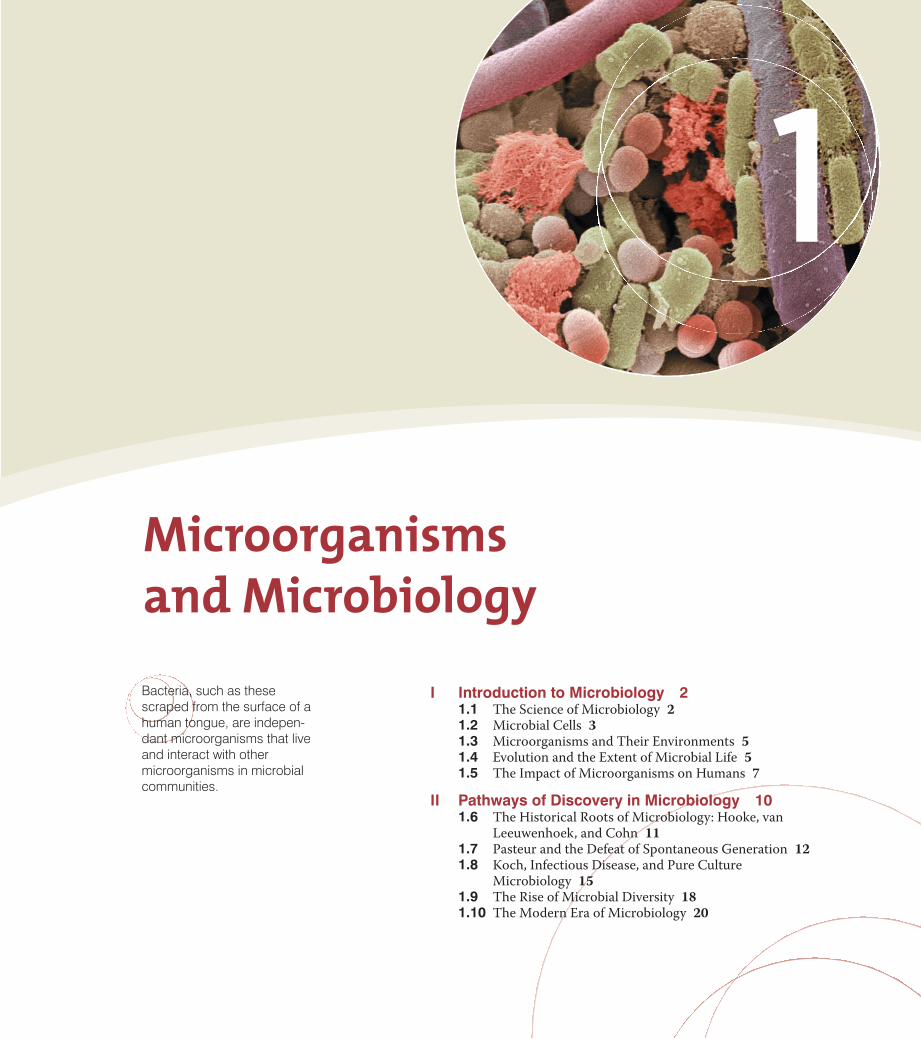
I Introduction to Microbiology 21.1 The Science of Microbiology 21.2 Microbial Cells 31.3 Microorganisms and Their Environments 51.4 Evolution and the Extent of Microbial Life 51.5 The Impact of Microorganisms on Humans 7
II Pathways of Discovery in Microbiology 101.6 The Historical Roots of Microbiology: Hooke, van
Leeuwenhoek, and Cohn 111.7 Pasteur and the Defeat of Spontaneous Generation 121.8 Koch, Infectious Disease, and Pure Culture
Microbiology 151.9 The Rise of Microbial Diversity 181.10 The Modern Era of Microbiology 20
UNIT 1 • Principles of Microbiology2
Pau
l V. D
unla
p
Pau
l V. D
unla
p
(a)
(b)
90 mm 0.01 mm (10 μm)
2 mm
(c)
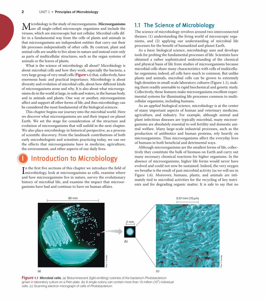
Figure 1.1 Microbial cells. (a) Bioluminescent (light-emitting) colonies of the bacterium Photobacteriumgrown in laboratory culture on a Petri plate. (b) A single colony can contain more than 10 million (107) individualcells. (c) Scanning electron micrograph of cells of Photobacterium.
Microbiology is the study of microorganisms. Microorganisms
are all single-celled microscopic organisms and include the
viruses, which are microscopic but not cellular. Microbial cells dif-
fer in a fundamental way from the cells of plants and animals in
that microorganisms are independent entities that carry out their
life processes independently of other cells. By contrast, plant and
animal cells are unable to live alone in nature and instead exist only
as parts of multicellular structures, such as the organ systems of
animals or the leaves of plants.
What is the science of microbiology all about? Microbiology is
about microbial cells and how they work, especially the bacteria, a
very large group of very small cells (Figure 1.1) that, collectively, have
enormous basic and practical importance. Microbiology is about
diversity and evolution of microbial cells, about how different kinds
of microorganisms arose and why. It is also about what microorga-
nisms do in the world at large, in soils and waters, in the human body,
and in animals and plants. One way or another, microorganisms
affect and support all other forms of life, and thus microbiology can
be considered the most fundamental of the biological sciences.
This chapter begins our journey into the microbial world. Here
we discover what microorganisms are and their impact on planet
Earth. We set the stage for consideration of the structure and
evolution of microorganisms that will unfold in the next chapter.
We also place microbiology in historical perspective, as a process
of scientific discovery. From the landmark contributions of both
early microbiologists and scientists practicing today, we can see
the effects that microorganisms have in medicine, agriculture,
the environment, and other aspects of our daily lives.
I Introduction to Microbiology
In the first five sections of this chapter we introduce the field of
microbiology, look at microorganisms as cells, examine where
and how microorganisms live in nature, survey the evolutionary
history of microbial life, and examine the impact that microor-
ganisms have had and continue to have on human affairs.
1.1 The Science of MicrobiologyThe science of microbiology revolves around two interconnected
themes: (1) understanding the living world of microscopic orga-
nisms, and (2) applying our understanding of microbial life
processes for the benefit of humankind and planet Earth.
As a basic biological science, microbiology uses and develops
tools for probing the fundamental processes of life. Scientists have
obtained a rather sophisticated understanding of the chemical
and physical basis of life from studies of microorganisms because
microbial cells share many characteristics with cells of multicellu-
lar organisms; indeed, all cells have much in common. But unlike
plants and animals, microbial cells can be grown to extremely
high densities in small-scale laboratory cultures (Figure 1.1), mak-
ing them readily amenable to rapid biochemical and genetic study.
Collectively, these features make microorganisms excellent exper-
imental systems for illuminating life processes common to multi-
cellular organisms, including humans.
As an applied biological science, microbiology is at the center
of many important aspects of human and veterinary medicine,
agriculture, and industry. For example, although animal and
plant infectious diseases are typically microbial, many microor-
ganisms are absolutely essential to soil fertility and domestic ani-
mal welfare. Many large-scale industrial processes, such as the
production of antibiotics and human proteins, rely heavily on
microorganisms. Thus microorganisms affect the everyday lives
of humans in both beneficial and detrimental ways.
Although microorganisms are the smallest forms of life, collec-
tively they constitute the bulk of biomass on Earth and carry out
many necessary chemical reactions for higher organisms. In the
absence of microorganisms, higher life forms would never have
evolved and could not now be sustained. Indeed, the very oxygen
we breathe is the result of past microbial activity (as we will see in
Figure 1.6). Moreover, humans, plants, and animals are inti-
mately tied to microbial activities for the recycling of key nutri-
ents and for degrading organic matter. It is safe to say that no
CHAPTER 1 • Microorganisms and Microbiology 3
other life forms are as important as microorganisms for the sup-
port and maintenance of life on Earth.
Microorganisms existed on Earth for billions of years before
plants and animals appeared, and we will see later that the
genetic and physiological diversity of microbial life greatly
exceeds that of the plants and animals. This huge diversity
accounts for some of the spectacular properties of microorga-
nisms. For example, we will see how microorganisms can live in
places that would kill other organisms and how the diverse physi-
ological capacities of microorganisms rank them as Earth’s pre-
mier chemists. We will also trace the evolutionary history of
microorganisms and see that three groups of cells can be distin-
guished by their evolutionary relationships. And finally, we will
see how microorganisms have established important relation-
ships with other organisms, some beneficial and some harmful.
We begin our study of microbiology with a consideration of
the cellular structure of microorganisms.
MiniQuiz• As they exist in nature, why can it be said that microbial cells
differ fundamentally from the cells of higher organisms?
• Why are microbial cells useful tools for basic science?
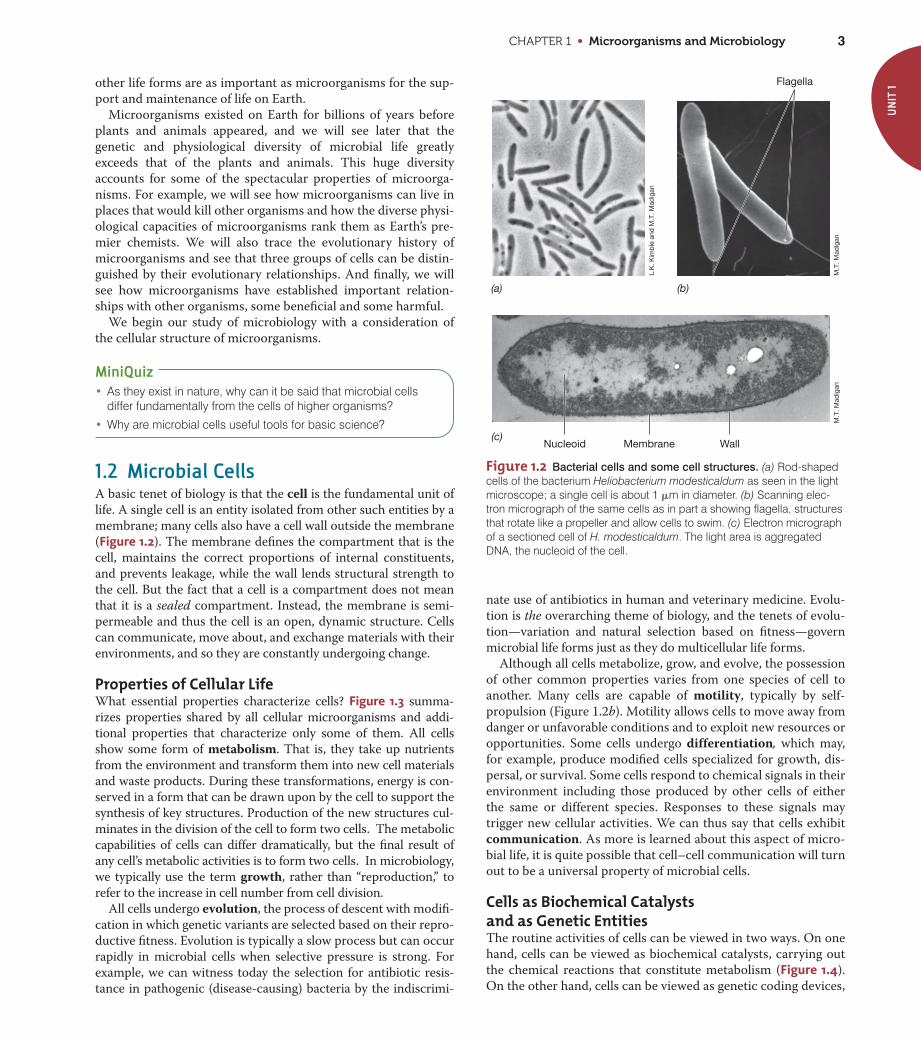
1.2 Microbial CellsA basic tenet of biology is that the cell is the fundamental unit of
life. A single cell is an entity isolated from other such entities by a
membrane; many cells also have a cell wall outside the membrane
(Figure 1.2). The membrane defines the compartment that is the
cell, maintains the correct proportions of internal constituents,
and prevents leakage, while the wall lends structural strength to
the cell. But the fact that a cell is a compartment does not mean
that it is a sealed compartment. Instead, the membrane is semi-
permeable and thus the cell is an open, dynamic structure. Cells
can communicate, move about, and exchange materials with their
environments, and so they are constantly undergoing change.
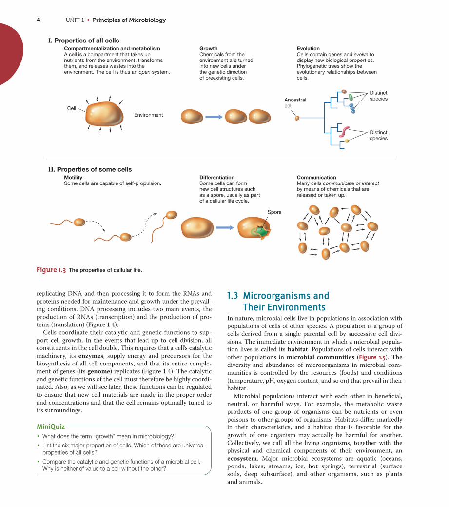
Properties of Cellular LifeWhat essential properties characterize cells? Figure 1.3 summa-
rizes properties shared by all cellular microorganisms and addi-
tional properties that characterize only some of them. All cells
show some form of metabolism. That is, they take up nutrients
from the environment and transform them into new cell materials
and waste products. During these transformations, energy is con-
served in a form that can be drawn upon by the cell to support the
synthesis of key structures. Production of the new structures cul-
minates in the division of the cell to form two cells. The metabolic
capabilities of cells can differ dramatically, but the final result of
any cell’s metabolic activities is to form two cells. In microbiology,
we typically use the term growth, rather than “reproduction,” to
refer to the increase in cell number from cell division.
All cells undergo evolution, the process of descent with modifi-
cation in which genetic variants are selected based on their repro-
ductive fitness. Evolution is typically a slow process but can occur
rapidly in microbial cells when selective pressure is strong. For
example, we can witness today the selection for antibiotic resis-
tance in pathogenic (disease-causing) bacteria by the indiscrimi-
nate use of antibiotics in human and veterinary medicine. Evolu-
tion is the overarching theme of biology, and the tenets of evolu-
tion—variation and natural selection based on fitness—govern
microbial life forms just as they do multicellular life forms.
Although all cells metabolize, grow, and evolve, the possession
of other common properties varies from one species of cell to
another. Many cells are capable of motility, typically by self-
propulsion (Figure 1.2b). Motility allows cells to move away from
danger or unfavorable conditions and to exploit new resources or
opportunities. Some cells undergo differentiation, which may,
for example, produce modified cells specialized for growth, dis-
persal, or survival. Some cells respond to chemical signals in their
environment including those produced by other cells of either
the same or different species. Responses to these signals may
trigger new cellular activities. We can thus say that cells exhibit
communication. As more is learned about this aspect of micro-
bial life, it is quite possible that cell–cell communication will turn
out to be a universal property of microbial cells.
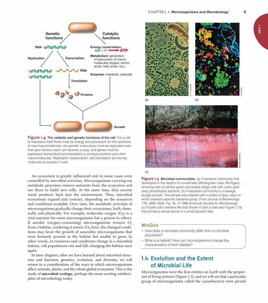
Cells as Biochemical Catalysts and as Genetic EntitiesThe routine activities of cells can be viewed in two ways. On one
hand, cells can be viewed as biochemical catalysts, carrying out
the chemical reactions that constitute metabolism (Figure 1.4).
On the other hand, cells can be viewed as genetic coding devices,
UN
IT 1
Flagella
Nucleoid Membrane Wall
L.K
. Kim
ble
and
M.T
. Mad
igan
M.T
. Mad
igan
M.T
. Mad
igan
(a) (b)
(c)
Figure 1.2 Bacterial cells and some cell structures. (a) Rod-shapedcells of the bacterium Heliobacterium modesticaldum as seen in the lightmicroscope; a single cell is about 1 �m in diameter. (b) Scanning elec-tron micrograph of the same cells as in part a showing flagella, structuresthat rotate like a propeller and allow cells to swim. (c) Electron micrographof a sectioned cell of H. modesticaldum. The light area is aggregatedDNA, the nucleoid of the cell.
UNIT 1 • Principles of Microbiology4
replicating DNA and then processing it to form the RNAs and
proteins needed for maintenance and growth under the prevail-
ing conditions. DNA processing includes two main events, the
production of RNAs (transcription) and the production of pro-
teins (translation) (Figure 1.4).
Cells coordinate their catalytic and genetic functions to sup-
port cell growth. In the events that lead up to cell division, all
constituents in the cell double. This requires that a cell’s catalytic
machinery, its enzymes, supply energy and precursors for the
biosynthesis of all cell components, and that its entire comple-
ment of genes (its genome) replicates (Figure 1.4). The catalytic
and genetic functions of the cell must therefore be highly coordi-
nated. Also, as we will see later, these functions can be regulated
to ensure that new cell materials are made in the proper order
and concentrations and that the cell remains optimally tuned to
its surroundings.
MiniQuiz• What does the term “growth” mean in microbiology?
• List the six major properties of cells. Which of these are universalproperties of all cells?
• Compare the catalytic and genetic functions of a microbial cell.Why is neither of value to a cell without the other?
Compartmentalization and metabolismA cell is a compartment that takes upnutrients from the environment, transformsthem, and releases wastes into theenvironment. The cell is thus an open system.
GrowthChemicals from the environment are turned into new cells under the genetic direction of preexisting cells.
DifferentiationSome cells can form new cell structures such as a spore, usually as part of a cellular life cycle.
CommunicationMany cells communicate or interact by means of chemicals that arereleased or taken up.
Distinctspecies
Distinctspecies
EvolutionCells contain genes and evolve to display new biological properties. Phylogenetic trees show the evolutionary relationships between cells.
CellEnvironment
Spore
MotilitySome cells are capable of self-propulsion.
Ancestralcell
I. Properties of all cells
II. Properties of some cells
Figure 1.3 The properties of cellular life.
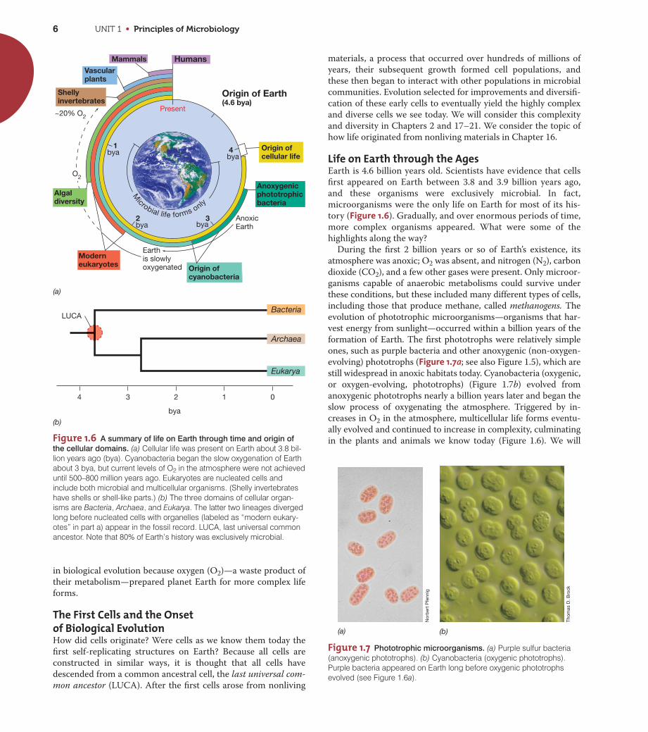
1.3 Microorganisms and Their Environments
In nature, microbial cells live in populations in association with
populations of cells of other species. A population is a group of
cells derived from a single parental cell by successive cell divi-
sions. The immediate environment in which a microbial popula-
tion lives is called its habitat. Populations of cells interact with
other populations in microbial communities (Figure 1.5). The
diversity and abundance of microorganisms in microbial com-
munities is controlled by the resources (foods) and conditions
(temperature, pH, oxygen content, and so on) that prevail in their
habitat.
Microbial populations interact with each other in beneficial,
neutral, or harmful ways. For example, the metabolic waste
products of one group of organisms can be nutrients or even
poisons to other groups of organisms. Habitats differ markedly
in their characteristics, and a habitat that is favorable for the
growth of one organism may actually be harmful for another.
Collectively, we call all the living organisms, together with the
physical and chemical components of their environment, an
ecosystem. Major microbial ecosystems are aquatic (oceans,
ponds, lakes, streams, ice, hot springs), terrestrial (surface
soils, deep subsurface), and other organisms, such as plants
and animals.
CHAPTER 1 • Microorganisms and Microbiology 5
MiniQuiz• How does a microbial community differ from a microbial
population?
• What is a habitat? How can microorganisms change thecharacteristics of their habitats?
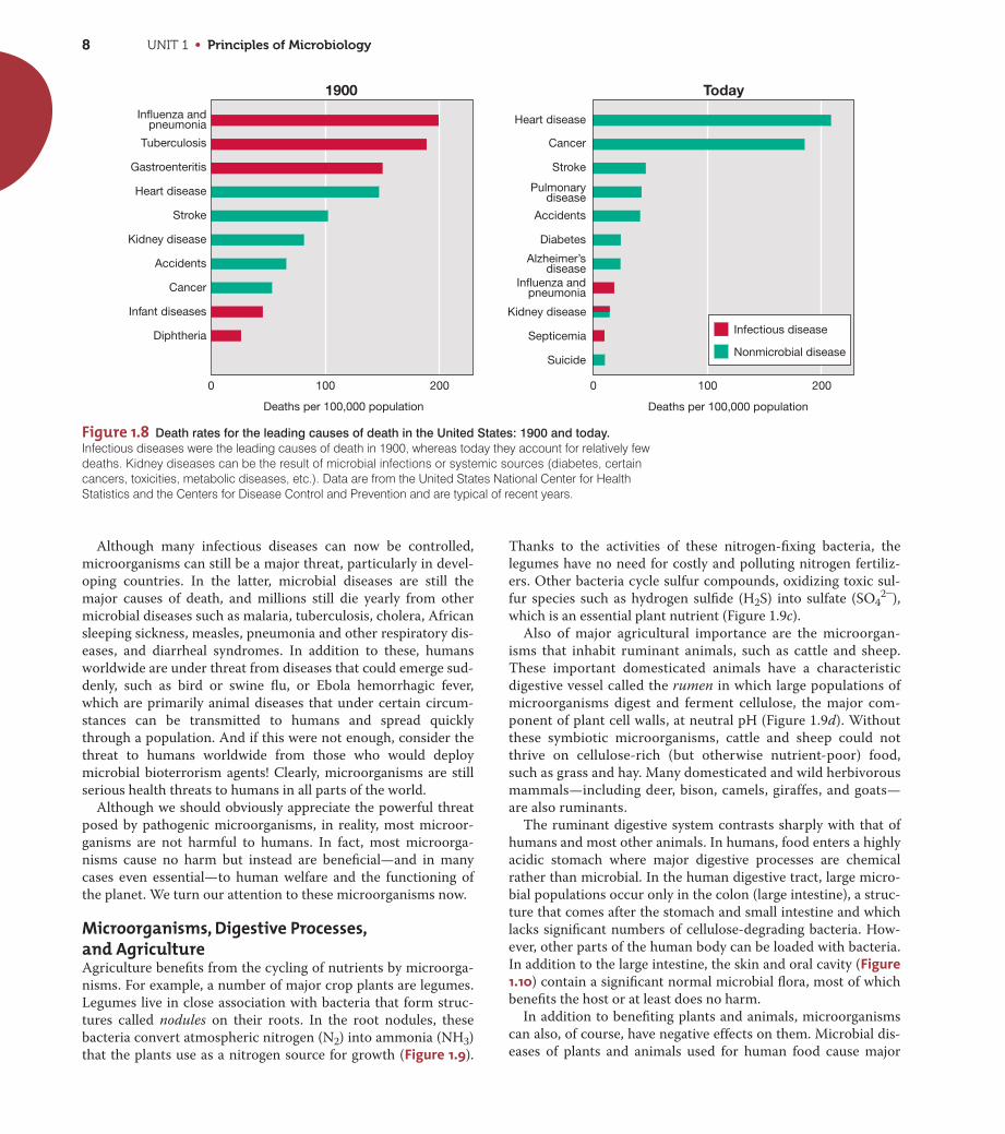
1.4 Evolution and the Extent of Microbial Life
Microorganisms were the first entities on Earth with the proper-
ties of living systems (Figure 1.3), and we will see that a particular
group of microorganisms called the cyanobacteria were pivotal
UN
IT 1
Growth
Catalyticfunctions
Geneticfunctions
Replication Transcription
Translation
RNA
Proteins
DNA Energy conservation: ADP + Pi ATPMetabolism: generation of precursors of macro- molecules (sugars, amino acids, fatty acids, etc.)
Enzymes: metabolic catalysts
Figure 1.4 The catalytic and genetic functions of the cell. For a cellto reproduce itself there must be energy and precursors for the synthesisof new macromolecules, the genetic instructions must be replicated suchthat upon division each cell receives a copy, and genes must beexpressed (transcribed and translated) to produce proteins and othermacromolecules. Replication, transcription, and translation are the keymolecular processes in cells.
(a) (b)
D. E
. Cal
dw
ell
Jiri
Sna
idr
Ric
ard
o G
uerr
ero
(c)
Figure 1.5 Microbial communities. (a) A bacterial community thatdeveloped in the depths of a small lake (Wintergreen Lake, Michigan),showing cells of various green and purple (large cells with sulfur gran-ules) phototrophic bacteria. (b) A bacterial community in a sewagesludge sample. The sample was stained with a series of dyes, each ofwhich stained a specific bacterial group. From Journal of Bacteriology178: 3496–3500, Fig. 2b. © 1996 American Society for Microbiology. (c) Purple sulfur bacteria like that shown in part a (see also Figure 1.7a)that formed a dense bloom in a small Spanish lake.
An ecosystem is greatly influenced and in some cases even
controlled by microbial activities. Microorganisms carrying out
metabolic processes remove nutrients from the ecosystem and
use them to build new cells. At the same time, they excrete
waste products back into the environment. Thus, microbial
ecosystems expand and contract, depending on the resources
and conditions available. Over time, the metabolic activities of
microorganisms gradually change their ecosystems, both chem-
ically and physically. For example, molecular oxygen (O2) is a
vital nutrient for some microorganisms but a poison to others.
If aerobic (oxygen-consuming) microorganisms remove O2
from a habitat, rendering it anoxic (O2 free), the changed condi-
tions may favor the growth of anaerobic microorganisms that
were formerly present in the habitat but unable to grow. In
other words, as resources and conditions change in a microbial
habitat, cell populations rise and fall, changing the habitat once
again.
In later chapters, after we have learned about microbial struc-
ture and function, genetics, evolution, and diversity, we will
return to a consideration of the ways in which microorganisms
affect animals, plants, and the whole global ecosystem. This is the
study of microbial ecology, perhaps the most exciting subdisci-
pline of microbiology today.
UNIT 1 • Principles of Microbiology6
in biological evolution because oxygen (O2)—a waste product of
their metabolism—prepared planet Earth for more complex life
forms.
The First Cells and the Onsetof Biological EvolutionHow did cells originate? Were cells as we know them today the
first self-replicating structures on Earth? Because all cells are
constructed in similar ways, it is thought that all cells have
descended from a common ancestral cell, the last universal com-
mon ancestor (LUCA). After the first cells arose from nonliving
materials, a process that occurred over hundreds of millions of
years, their subsequent growth formed cell populations, and
these then began to interact with other populations in microbial
communities. Evolution selected for improvements and diversifi-
cation of these early cells to eventually yield the highly complex
and diverse cells we see today. We will consider this complexity
and diversity in Chapters 2 and 17–21. We consider the topic of
how life originated from nonliving materials in Chapter 16.
Life on Earth through the AgesEarth is 4.6 billion years old. Scientists have evidence that cells
first appeared on Earth between 3.8 and 3.9 billion years ago,
and these organisms were exclusively microbial. In fact,
microorganisms were the only life on Earth for most of its his-
tory (Figure 1.6). Gradually, and over enormous periods of time,
more complex organisms appeared. What were some of the
highlights along the way?
During the first 2 billion years or so of Earth’s existence, its
atmosphere was anoxic; O2 was absent, and nitrogen (N2), carbon
dioxide (CO2), and a few other gases were present. Only microor-
ganisms capable of anaerobic metabolisms could survive under
these conditions, but these included many different types of cells,
including those that produce methane, called methanogens. The
evolution of phototrophic microorganisms—organisms that har-
vest energy from sunlight—occurred within a billion years of the
formation of Earth. The first phototrophs were relatively simple
ones, such as purple bacteria and other anoxygenic (non-oxygen-
evolving) phototrophs (Figure 1.7a; see also Figure 1.5), which are
still widespread in anoxic habitats today. Cyanobacteria (oxygenic,
or oxygen-evolving, phototrophs) (Figure 1.7b) evolved from
anoxygenic phototrophs nearly a billion years later and began the
slow process of oxygenating the atmosphere. Triggered by in-
creases in O2 in the atmosphere, multicellular life forms eventu-
ally evolved and continued to increase in complexity, culminating
in the plants and animals we know today (Figure 1.6). We will
Humans
Bacteria
Archaea
Eukarya
Shellyinvertebrates
Vascular plants
Mammals
Origin of Earth(4.6 bya)
Origin of cellular life
Anoxygenicphototrophicbacteria
Origin ofcyanobacteria
AnoxicEarth
OO22O2
~20% O2
Earth is slowlyoxygenated
LUCA
Moderneukaryotes
Algaldiversity
4bya
bya2 bya
1bya
3
Microbial life forms only
Present
(a)
(b)
4 3 2 1 0
bya
Figure 1.6 A summary of life on Earth through time and origin of
the cellular domains. (a) Cellular life was present on Earth about 3.8 bil-lion years ago (bya). Cyanobacteria began the slow oxygenation of Earthabout 3 bya, but current levels of O2 in the atmosphere were not achieveduntil 500–800 million years ago. Eukaryotes are nucleated cells andinclude both microbial and multicellular organisms. (Shelly invertebrateshave shells or shell-like parts.) (b) The three domains of cellular organ-isms are Bacteria, Archaea, and Eukarya. The latter two lineages divergedlong before nucleated cells with organelles (labeled as “modern eukary-otes” in part a) appear in the fossil record. LUCA, last universal commonancestor. Note that 80% of Earth’s history was exclusively microbial.
(b)
Thom
as D
. Bro
ck
Nor
ber
t P
fenn
ig
(a)
Figure 1.7 Phototrophic microorganisms. (a) Purple sulfur bacteria(anoxygenic phototrophs). (b) Cyanobacteria (oxygenic phototrophs).Purple bacteria appeared on Earth long before oxygenic phototrophsevolved (see Figure 1.6a).

CHAPTER 1 • Microorganisms and Microbiology 7
explore the evolutionary history of life later, but note here that the
events that unfolded beyond LUCA led to the evolution of three
major lineages of microbial cells, the Bacteria, the Archaea, and
the Eukarya (Figure 1.6b); microbial Eukarya were the ancestors
of the plants and animals.
How do we know that evolutionary events unfolded as sum-
marized in Figure 1.6? The answer is that we may never know
that all details in our description are correct. However, scientists
can reconstruct evolutionary transitions by using biomarkers,
specific molecules that are unique to particular groups in pres-
ent-day microorganisms. The presence or absence of a given bio-
marker in ancient rocks of a known age therefore reveals whether
that particular group was present at that time.
One way or the other and over enormous periods of time
(Figure 1.6), natural selection filled every suitable habitat on
Earth with one or more populations of microorganisms. This
brings us to the question of the current distribution of microbial
life on Earth. What do we know about this important topic?
The Extent of Microbial LifeMicrobial life is all around us. Examination of natural materials
such as soil or water invariably reveals microbial cells. But
unusual habitats such as boiling hot springs and glacial ice are
also teeming with microorganisms. Although widespread on
Earth, such tiny cells may seem inconsequential. But if we could
count them all, what number would we reach?
Estimates of total microbial cell numbers on Earth are on the
order of cells. The total amount of carbon present in
this very large number of very small cells equals that of all plants
on Earth (and plant carbon far exceeds animal carbon). But in
addition, the collective contents of nitrogen and phosphorus in
microbial cells is more than 10 times that in all plant biomass.
Thus, microbial cells, small as they are, constitute the major
fraction of biomass on Earth and are key reservoirs of essential
nutrients for life. Most microbial cells are found in just a few very
large habitats. For example, most microbial cells do not reside on
Earth’s surface but instead lie underground in the oceanic and
terrestrial subsurface (Table 1.1). Depths up to about 10 km under
Earth’s surface are clearly suitable for microbial life. We will see
later that subsurface microbial habitats support diverse popula-
tions of microbial cells that make their livings in unusual ways
and grow extremely slowly. By comparison to the subsurface, sur-
face soils and waters contain a relatively small percentage of the
total microbial cell numbers, and animals (including humans),
which can be heavily colonized with microorganisms (see Figure
1.10), collectively contain only a tiny fraction of the total micro-
bial cells on Earth (Table 1.1).
Because most of what we know about microbial life has come
from the study of surface-dwelling organisms, there is obviously
much left for future generations of microbiologists to discover
and understand about the life forms that dominate Earth’s biol-
ogy. And when we consider the fact that surface-dwelling orga-
nisms already show enormous diversity, the hunt for new
microorganisms in Earth’s unexplored habitats should yield some
exciting surprises.
2.5 * 1030
MiniQuiz• What is LUCA and what major lineages of cells evolved from
LUCA? Why were cyanobacteria so important in the evolution oflife on Earth?
• How old is Earth, and when did cellular life forms first appear?How can we use science to reconstruct the sequence oforganisms that appeared on Earth?
• Where are most microbial cells located on Earth?
1.5 The Impact of Microorganisms on Humans
Through the years microbiologists have had great success in dis-
covering how microorganisms work, and application of this
knowledge has greatly increased the beneficial effects of
microorganisms and curtailed many of their harmful effects.
Microbiology has thus greatly advanced human health and wel-
fare. Besides understanding microorganisms as agents of disease,
microbiology has made great advances in understanding the role
of microorganisms in food and agriculture, and in exploiting
microbial activities for producing valuable human products, gen-
erating energy, and cleaning up the environment.
Microorganisms as Agents of DiseaseThe statistics summarized in Figure 1.8 show microbiologists’
success in preventing infectious diseases since the beginning of
the twentieth century. These data compare today’s leading causes
of death in the United States with those of 100 years ago. At the
beginning of the twentieth century, the major causes of death in
humans were infectious diseases caused by microorganisms
called pathogens. Children and the aged in particular suc-
cumbed in large numbers to microbial diseases. Today, however,
infectious diseases are much less deadly, at least in developed
countries. Control of infectious disease has come from an
increased understanding of disease processes, improved sanitary
and public health practices, and the use of antimicrobial agents,
such as antibiotics. As we will see from the next sections, the
development of microbiology as a science can trace important
aspects of its roots to studies of infectious disease.
UN
IT 1Table 1.1 Distribution of microorganisms in and on Eartha
Habitat Percent of total
Marine subsurface 66
Terrestrial subsurface 26
Surface soil 4.8
Oceans 2.2
All other habitatsb 1.0
aData compiled by William Whitman, University of Georgia, USA; refer to total numbers(estimated to be about 2.5 1030 cells) of Bacteria and Archaea. This enormousnumber of cells contain, collectively, about grams of carbon.bIncludes, in order of decreasing numbers: freshwater and salt lakes, domesticated animals, sea ice, termites, humans, and domesticated birds.
5 * 1017*
UNIT 1 • Principles of Microbiology8
Although many infectious diseases can now be controlled,
microorganisms can still be a major threat, particularly in devel-
oping countries. In the latter, microbial diseases are still the
major causes of death, and millions still die yearly from other
microbial diseases such as malaria, tuberculosis, cholera, African
sleeping sickness, measles, pneumonia and other respiratory dis-
eases, and diarrheal syndromes. In addition to these, humans
worldwide are under threat from diseases that could emerge sud-
denly, such as bird or swine flu, or Ebola hemorrhagic fever,
which are primarily animal diseases that under certain circum-
stances can be transmitted to humans and spread quickly
through a population. And if this were not enough, consider the
threat to humans worldwide from those who would deploy
microbial bioterrorism agents! Clearly, microorganisms are still
serious health threats to humans in all parts of the world.
Although we should obviously appreciate the powerful threat
posed by pathogenic microorganisms, in reality, most microor-
ganisms are not harmful to humans. In fact, most microorga-
nisms cause no harm but instead are beneficial—and in many
cases even essential—to human welfare and the functioning of
the planet. We turn our attention to these microorganisms now.
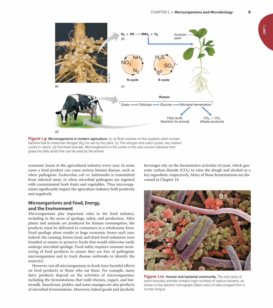
Microorganisms, Digestive Processes,and AgricultureAgriculture benefits from the cycling of nutrients by microorga-
nisms. For example, a number of major crop plants are legumes.
Legumes live in close association with bacteria that form struc-
tures called nodules on their roots. In the root nodules, these
bacteria convert atmospheric nitrogen (N2) into ammonia (NH3)
that the plants use as a nitrogen source for growth (Figure 1.9).
Thanks to the activities of these nitrogen-fixing bacteria, the
legumes have no need for costly and polluting nitrogen fertiliz-
ers. Other bacteria cycle sulfur compounds, oxidizing toxic sul-
fur species such as hydrogen sulfide (H2S) into sulfate (SO42-),
which is an essential plant nutrient (Figure 1.9c).
Also of major agricultural importance are the microorgan-
isms that inhabit ruminant animals, such as cattle and sheep.
These important domesticated animals have a characteristic
digestive vessel called the rumen in which large populations of
microorganisms digest and ferment cellulose, the major com-
ponent of plant cell walls, at neutral pH (Figure 1.9d). Without
these symbiotic microorganisms, cattle and sheep could not
thrive on cellulose-rich (but otherwise nutrient-poor) food,
such as grass and hay. Many domesticated and wild herbivorous
mammals—including deer, bison, camels, giraffes, and goats—
are also ruminants.
The ruminant digestive system contrasts sharply with that of
humans and most other animals. In humans, food enters a highly
acidic stomach where major digestive processes are chemical
rather than microbial. In the human digestive tract, large micro-
bial populations occur only in the colon (large intestine), a struc-
ture that comes after the stomach and small intestine and which
lacks significant numbers of cellulose-degrading bacteria. How-
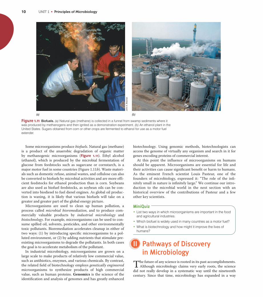
ever, other parts of the human body can be loaded with bacteria.
In addition to the large intestine, the skin and oral cavity (Figure1.10) contain a significant normal microbial flora, most of which
benefits the host or at least does no harm.
In addition to benefiting plants and animals, microorganisms
can also, of course, have negative effects on them. Microbial dis-
eases of plants and animals used for human food cause major
Figure 1.8 Death rates for the leading causes of death in the United States: 1900 and today.
Infectious diseases were the leading causes of death in 1900, whereas today they account for relatively fewdeaths. Kidney diseases can be the result of microbial infections or systemic sources (diabetes, certaincancers, toxicities, metabolic diseases, etc.). Data are from the United States National Center for HealthStatistics and the Centers for Disease Control and Prevention and are typical of recent years.
Deaths per 100,000 populationDeaths per 100,000 population
Kidney disease
Septicemia
Heart disease
Cancer
Stroke
Pulmonarydisease
Accidents
Diabetes
Alzheimer’sdisease
Influenza andpneumonia
Suicide
1900 Today
Influenza andpneumonia
Tuberculosis
Gastroenteritis
Heart disease
Stroke
Kidney disease
Accidents
Cancer
Infant diseases
Diphtheria
0 100 200 0 100 200
Infectious disease
Nonmicrobial disease
CHAPTER 1 • Microorganisms and Microbiology 9
economic losses in the agricultural industry every year. In some
cases a food product can cause serious human disease, such as
when pathogenic Escherichia coli or Salmonella is transmitted
from infected meat, or when microbial pathogens are ingested
with contaminated fresh fruits and vegetables. Thus microorga-
nisms significantly impact the agriculture industry both positively
and negatively.
Microorganisms and Food, Energy,and the EnvironmentMicroorganisms play important roles in the food industry,
including in the areas of spoilage, safety, and production. After
plants and animals are produced for human consumption, the
products must be delivered to consumers in a wholesome form.
Food spoilage alone results in huge economic losses each year.
Indeed, the canning, frozen food, and dried-food industries were
founded as means to preserve foods that would otherwise easily
undergo microbial spoilage. Food safety requires constant moni-
toring of food products to ensure they are free of pathogenic
microorganisms and to track disease outbreaks to identify the
source(s).
However, not all microorganisms in foods have harmful effects
on food products or those who eat them. For example, many
dairy products depend on the activities of microorganisms,
including the fermentations that yield cheeses, yogurt, and but-
termilk. Sauerkraut, pickles, and some sausages are also products
of microbial fermentations. Moreover, baked goods and alcoholic
beverages rely on the fermentative activities of yeast, which gen-
erate carbon dioxide (CO2) to raise the dough and alcohol as a
key ingredient, respectively. Many of these fermentations are dis-
cussed in Chapter 14.
UN
IT 1
(a) (c)
(b)
(d)
N2 + 8H 2NH3 + H2
N2
NH3
Rumen
Grass Cellulose Glucose Microbial fermentation
CO2 + CH4(Waste products)
Fatty acids(Nutrition for animal)
NO3–
H2S
S0SO4
2–
N-cycle S-cycle
Soybeanplant
Joe
Bur
ton
Figure 1.9 Microorganisms in modern agriculture. (a, b) Root nodules on this soybean plant containbacteria that fix molecular nitrogen (N2) for use by the plant. (c) The nitrogen and sulfur cycles, key nutrientcycles in nature. (d) Ruminant animals. Microorganisms in the rumen of the cow convert cellulose fromgrass into fatty acids that can be used by the animal.
Figure 1.10 Human oral bacterial community. The oral cavity ofwarm-blooded animals contains high numbers of various bacteria, asshown in this electron micrograph (false color) of cells scraped from ahuman tongue.
UNIT 1 • Principles of Microbiology10
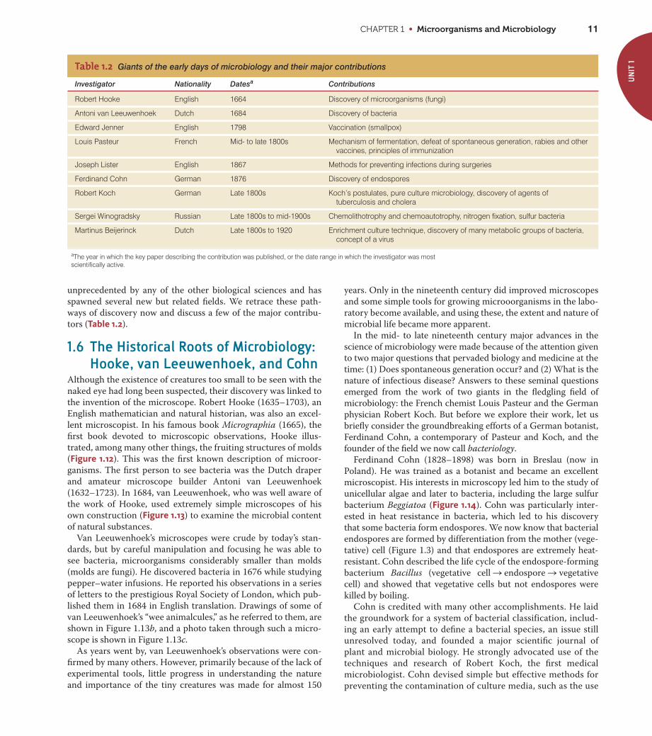
Some microorganisms produce biofuels. Natural gas (methane)
is a product of the anaerobic degradation of organic matter
by methanogenic microorganisms (Figure 1.11). Ethyl alcohol
(ethanol), which is produced by the microbial fermentation of
glucose from feedstocks such as sugarcane or cornstarch, is a
major motor fuel in some countries (Figure 1.11b). Waste materi-
als such as domestic refuse, animal wastes, and cellulose can also
be converted to biofuels by microbial activities and are more effi-
cient feedstocks for ethanol production than is corn. Soybeans
are also used as biofuel feedstocks, as soybean oils can be con-
verted into biodiesel to fuel diesel engines. As global oil produc-
tion is waning, it is likely that various biofuels will take on a
greater and greater part of the global energy picture.
Microorganisms are used to clean up human pollution, a
process called microbial bioremediation, and to produce com-
mercially valuable products by industrial microbiology and
biotechnology. For example, microorganisms can be used to con-
sume spilled oil, solvents, pesticides, and other environmentally
toxic pollutants. Bioremediation accelerates cleanup in either of
two ways: (1) by introducing specific microorganisms to a pol-
luted environment, or (2) by adding nutrients that stimulate pre-
existing microorganisms to degrade the pollutants. In both cases
the goal is to accelerate metabolism of the pollutant.
In industrial microbiology, microorganisms are grown on a
large scale to make products of relatively low commercial value,
such as antibiotics, enzymes, and various chemicals. By contrast,
the related field of biotechnology employs genetically engineered
microorganisms to synthesize products of high commercial
value, such as human proteins. Genomics is the science of the
identification and analysis of genomes and has greatly enhanced
biotechnology. Using genomic methods, biotechnologists can
access the genome of virtually any organism and search in it for
genes encoding proteins of commercial interest.
At this point the influence of microorganisms on humans
should be apparent. Microorganisms are essential for life and
their activities can cause significant benefit or harm to humans.
As the eminent French scientist Louis Pasteur, one of the
founders of microbiology, expressed it: “The role of the infi-
nitely small in nature is infinitely large.” We continue our intro-
duction to the microbial world in the next section with an
historical overview of the contributions of Pasteur and a few
other key scientists.
MiniQuiz• List two ways in which microorganisms are important in the food
and agricultural industries.
• Which biofuel is widely used in many countries as a motor fuel?
• What is biotechnology and how might it improve the lives ofhumans?
II Pathways of Discovery in Microbiology
The future of any science is rooted in its past accomplishments.
Although microbiology claims very early roots, the science
did not really develop in a systematic way until the nineteenth
century. Since that time, microbiology has expanded in a way
John
A. B
rezn
ak
(a) (b)
Figure 1.11 Biofuels. (a) Natural gas (methane) is collected in a funnel from swamp sediments where itwas produced by methanogens and then ignited as a demonstration experiment. (b) An ethanol plant in theUnited States. Sugars obtained from corn or other crops are fermented to ethanol for use as a motor fuelextender.
CHAPTER 1 • Microorganisms and Microbiology 11
unprecedented by any of the other biological sciences and has
spawned several new but related fields. We retrace these path-
ways of discovery now and discuss a few of the major contribu-
tors (Table 1.2).
1.6 The Historical Roots of Microbiology:Hooke, van Leeuwenhoek, and Cohn
Although the existence of creatures too small to be seen with the
naked eye had long been suspected, their discovery was linked to
the invention of the microscope. Robert Hooke (1635–1703), an
English mathematician and natural historian, was also an excel-
lent microscopist. In his famous book Micrographia (1665), the
first book devoted to microscopic observations, Hooke illus-
trated, among many other things, the fruiting structures of molds
(Figure 1.12). This was the first known description of microor-
ganisms. The first person to see bacteria was the Dutch draper
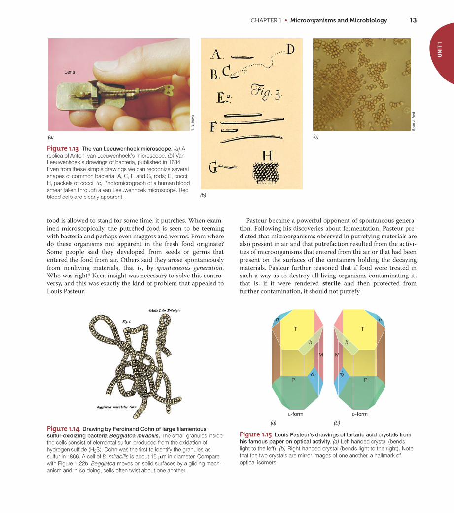
and amateur microscope builder Antoni van Leeuwenhoek
(1632–1723). In 1684, van Leeuwenhoek, who was well aware of
the work of Hooke, used extremely simple microscopes of his
own construction (Figure 1.13) to examine the microbial content
of natural substances.
Van Leeuwenhoek’s microscopes were crude by today’s stan-
dards, but by careful manipulation and focusing he was able to
see bacteria, microorganisms considerably smaller than molds
(molds are fungi). He discovered bacteria in 1676 while studying
pepper–water infusions. He reported his observations in a series
of letters to the prestigious Royal Society of London, which pub-
lished them in 1684 in English translation. Drawings of some of
van Leeuwenhoek’s “wee animalcules,” as he referred to them, are
shown in Figure 1.13b, and a photo taken through such a micro-
scope is shown in Figure 1.13c.
As years went by, van Leeuwenhoek’s observations were con-
firmed by many others. However, primarily because of the lack of
experimental tools, little progress in understanding the nature
and importance of the tiny creatures was made for almost 150
years. Only in the nineteenth century did improved microscopes
and some simple tools for growing microoorganisms in the labo-
ratory become available, and using these, the extent and nature of
microbial life became more apparent.
In the mid- to late nineteenth century major advances in the
science of microbiology were made because of the attention given
to two major questions that pervaded biology and medicine at the
time: (1) Does spontaneous generation occur? and (2) What is the
nature of infectious disease? Answers to these seminal questions
emerged from the work of two giants in the fledgling field of
microbiology: the French chemist Louis Pasteur and the German
physician Robert Koch. But before we explore their work, let us
briefly consider the groundbreaking efforts of a German botanist,
Ferdinand Cohn, a contemporary of Pasteur and Koch, and the
founder of the field we now call bacteriology.
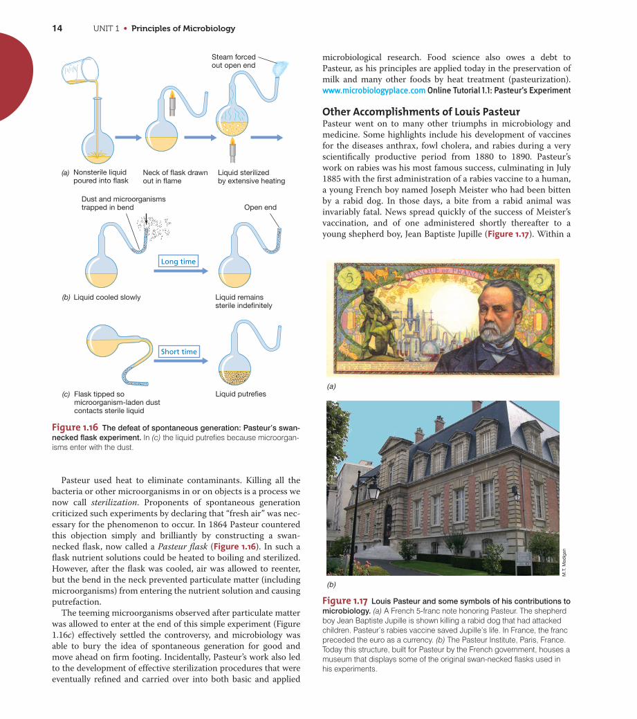
Ferdinand Cohn (1828–1898) was born in Breslau (now in
Poland). He was trained as a botanist and became an excellent
microscopist. His interests in microscopy led him to the study of
unicellular algae and later to bacteria, including the large sulfur
bacterium Beggiatoa (Figure 1.14). Cohn was particularly inter-
ested in heat resistance in bacteria, which led to his discovery
that some bacteria form endospores. We now know that bacterial
endospores are formed by differentiation from the mother (vege-
tative) cell (Figure 1.3) and that endospores are extremely heat-
resistant. Cohn described the life cycle of the endospore-forming
bacterium Bacillus (vegetative cell endospore vegetative
cell) and showed that vegetative cells but not endospores were
killed by boiling.
Cohn is credited with many other accomplishments. He laid
the groundwork for a system of bacterial classification, includ-
ing an early attempt to define a bacterial species, an issue still
unresolved today, and founded a major scientific journal of
plant and microbial biology. He strongly advocated use of the
techniques and research of Robert Koch, the first medical
microbiologist. Cohn devised simple but effective methods for
preventing the contamination of culture media, such as the use
SS
UN
IT 1Table 1.2 Giants of the early days of microbiology and their major contributions
Investigator Nationality Datesa Contributions
Robert Hooke English 1664 Discovery of microorganisms (fungi)
Antoni van Leeuwenhoek Dutch 1684 Discovery of bacteria
Edward Jenner English 1798 Vaccination (smallpox)
Louis Pasteur French Mid- to late 1800s Mechanism of fermentation, defeat of spontaneous generation, rabies and other vaccines, principles of immunization
Joseph Lister English 1867 Methods for preventing infections during surgeries
Ferdinand Cohn German 1876 Discovery of endospores
Robert Koch German Late 1800s Koch’s postulates, pure culture microbiology, discovery of agents of tuberculosis and cholera
Sergei Winogradsky Russian Late 1800s to mid-1900s Chemolithotrophy and chemoautotrophy, nitrogen fixation, sulfur bacteria
Martinus Beijerinck Dutch Late 1800s to 1920 Enrichment culture technique, discovery of many metabolic groups of bacteria, concept of a virus
aThe year in which the key paper describing the contribution was published, or the date range in which the investigator was mostscientifically active.
UNIT 1 • Principles of Microbiology12
of cotton for closing flasks and tubes. These methods were later
used by Koch and allowed him to make rapid progress in the
isolation and characterization of several disease-causing bacte-
ria (Section 1.8).
MiniQuiz• What prevented the science of microbiology from developing
before the era of Hooke and van Leeuwenhoek?
• What major discovery emerged from Cohn’s study of heat resis-tance in microorganisms?
1.7 Pasteur and the Defeat ofSpontaneous Generation
The late nineteenth century saw the science of microbiology
blossom. The theory of spontaneous generation was crushed by
the brilliant work of the Frenchman Louis Pasteur (1822–1895).
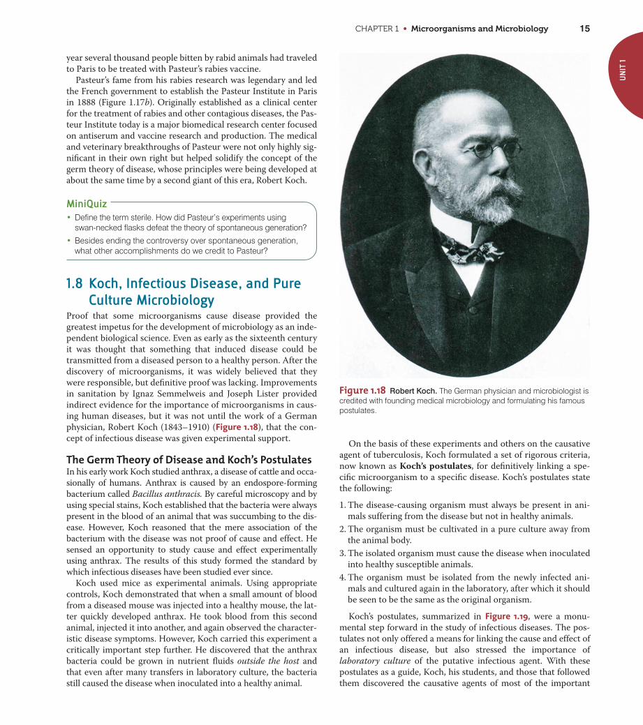
Optical Isomers and FermentationsPasteur was a chemist by training and was one of the first to rec-
ognize the significance of optical isomers. A molecule is optically
active if a pure solution or crystal diffracts light in only one direc-
tion. Pasteur studied crystals of tartaric acid that he separated by
hand into those that bent a beam of polarized light to the left and
those that bent the beam to the right (Figure 1.15). Pasteur found
that the mold Aspergillus metabolized D-tartrate, which bent light
to the right, but did not metabolize its optical isomer, L-tartrate.
The fact that a living organism could discriminate between opti-
cal isomers was of profound significance to Pasteur, and he began
to see living organisms as inherently asymmetric entities.
Pasteur’s thinking on the asymmetry of life carried over into
his work on fermentations and, eventually, spontaneous genera-
tion. At the invitation of a local industrialist who was having
problems making alcohol from the fermentation of beets, Pasteur
studied the mechanism of the alcoholic fermentation, at that
time thought to be a strictly chemical process. The yeast cells in
the fermenting broth were thought to be a complex chemical
substance formed by the fermentation. Although ethyl alcohol
does not form optical isomers, one of the side products of beet
fermentation is amyl alcohol, which does, and Pasteur tested the
fermenting juice and found the amyl alcohol to be of only one
optical isomer. From his work on tartrate metabolism this sug-
gested to Pasteur that the beet fermentation was a biological
process. Microscopic observations and other simple but rigorous
experiments convinced Pasteur that the alcoholic fermentation
was catalyzed by living organisms, the yeast cells. Indeed, in
Pasteur’s own words: “. . . fermentation is associated with the life
and structural integrity of the cells and not with their death and
decay.” From this foundation, Pasteur began a series of classic
experiments on spontaneous generation, experiments that are
forever linked to his name and to the science of microbiology.
Spontaneous GenerationThe concept of spontaneous generation had existed since bibli-
cal times and its basic tenet can be easily grasped. For example, if
(b)
(a)
Figure 1.12 Robert Hooke and early microscopy. (a) A drawing of themicroscope used by Robert Hooke in 1664. The lens was fitted at the endof an adjustable bellows (G) and light focused on the specimen by a sep-arate lens (1). (b) This drawing of a mold that was growing on the surfaceof leather, together with other drawings and accompanying text publishedby Robert Hooke in Micrographia in 1665, were the first descriptions ofmicroorganisms. The round structures contain spores of the mold.Compare Hooke’s microscope with that of van Leeuwenhoek’s shown in Figure 1.13.
CHAPTER 1 • Microorganisms and Microbiology 13
T. D
. Bro
ck
Bria
n J.
For
d
(a)
(b)
(c)
Lens
Figure 1.13 The van Leeuwenhoek microscope. (a) Areplica of Antoni van Leeuwenhoek’s microscope. (b) VanLeeuwenhoek’s drawings of bacteria, published in 1684.Even from these simple drawings we can recognize severalshapes of common bacteria: A, C, F, and G, rods; E, cocci;H, packets of cocci. (c) Photomicrograph of a human bloodsmear taken through a van Leeuwenhoek microscope. Redblood cells are clearly apparent.
Figure 1.14 Drawing by Ferdinand Cohn of large filamentous
sulfur-oxidizing bacteria Beggiatoa mirabilis. The small granules insidethe cells consist of elemental sulfur, produced from the oxidation ofhydrogen sulfide (H2S). Cohn was the first to identify the granules assulfur in 1866. A cell of B. mirabilis is about 15 �m in diameter. Comparewith Figure 1.22b. Beggiatoa moves on solid surfaces by a gliding mech-anism and in so doing, cells often twist about one another.
food is allowed to stand for some time, it putrefies. When exam-
ined microscopically, the putrefied food is seen to be teeming
with bacteria and perhaps even maggots and worms. From where
do these organisms not apparent in the fresh food originate?
Some people said they developed from seeds or germs that
entered the food from air. Others said they arose spontaneously
from nonliving materials, that is, by spontaneous generation.
Who was right? Keen insight was necessary to solve this contro-
versy, and this was exactly the kind of problem that appealed to
Louis Pasteur.
Pasteur became a powerful opponent of spontaneous genera-
tion. Following his discoveries about fermentation, Pasteur pre-
dicted that microorganisms observed in putrefying materials are
also present in air and that putrefaction resulted from the activi-
ties of microorganisms that entered from the air or that had been
present on the surfaces of the containers holding the decaying
materials. Pasteur further reasoned that if food were treated in
such a way as to destroy all living organisms contaminating it,
that is, if it were rendered sterile and then protected from
further contamination, it should not putrefy.
UN
IT 1
(a) (b)
T T
P
L-form D-form
M M
Pb' b'h
n n
h
Figure 1.15 Louis Pasteur’s drawings of tartaric acid crystals from
his famous paper on optical activity. (a) Left-handed crystal (bends light to the left). (b) Right-handed crystal (bends light to the right). Note that the two crystals are mirror images of one another, a hallmark ofoptical isomers.
UNIT 1 • Principles of Microbiology14
Steam forced out open end
(a)
(c)
(b)
Short time
Long time
Dust and microorganismstrapped in bend Open end
Nonsterile liquidpoured into flask
Neck of flask drawn out in flame
Liquid sterilizedby extensive heating
Flask tipped somicroorganism-laden dust contacts sterile liquid
Liquid putrefies
Liquid cooled slowly Liquid remainssterile indefinitely
Figure 1.16 The defeat of spontaneous generation: Pasteur’s swan-
necked flask experiment. In (c) the liquid putrefies because microorgan-isms enter with the dust.
M.T
. Mad
igan
(b)
(a)
Figure 1.17 Louis Pasteur and some symbols of his contributions to
microbiology. (a) A French 5-franc note honoring Pasteur. The shepherdboy Jean Baptiste Jupille is shown killing a rabid dog that had attackedchildren. Pasteur’s rabies vaccine saved Jupille’s life. In France, the francpreceded the euro as a currency. (b) The Pasteur Institute, Paris, France.Today this structure, built for Pasteur by the French government, houses amuseum that displays some of the original swan-necked flasks used inhis experiments.
Pasteur used heat to eliminate contaminants. Killing all the
bacteria or other microorganisms in or on objects is a process we
now call sterilization. Proponents of spontaneous generation
criticized such experiments by declaring that “fresh air” was nec-
essary for the phenomenon to occur. In 1864 Pasteur countered
this objection simply and brilliantly by constructing a swan-
necked flask, now called a Pasteur flask (Figure 1.16). In such a
flask nutrient solutions could be heated to boiling and sterilized.
However, after the flask was cooled, air was allowed to reenter,
but the bend in the neck prevented particulate matter (including
microorganisms) from entering the nutrient solution and causing
putrefaction.
The teeming microorganisms observed after particulate matter
was allowed to enter at the end of this simple experiment (Figure
1.16c) effectively settled the controversy, and microbiology was
able to bury the idea of spontaneous generation for good and
move ahead on firm footing. Incidentally, Pasteur’s work also led
to the development of effective sterilization procedures that were
eventually refined and carried over into both basic and applied
microbiological research. Food science also owes a debt to
Pasteur, as his principles are applied today in the preservation of
milk and many other foods by heat treatment (pasteurization).
www.microbiologyplace.com Online Tutorial 1.1: Pasteur’s Experiment
Other Accomplishments of Louis PasteurPasteur went on to many other triumphs in microbiology and
medicine. Some highlights include his development of vaccines
for the diseases anthrax, fowl cholera, and rabies during a very
scientifically productive period from 1880 to 1890. Pasteur’s
work on rabies was his most famous success, culminating in July
1885 with the first administration of a rabies vaccine to a human,
a young French boy named Joseph Meister who had been bitten
by a rabid dog. In those days, a bite from a rabid animal was
invariably fatal. News spread quickly of the success of Meister’s
vaccination, and of one administered shortly thereafter to a
young shepherd boy, Jean Baptiste Jupille (Figure 1.17). Within a
CHAPTER 1 • Microorganisms and Microbiology 15
UN
IT 1
Figure 1.18 Robert Koch. The German physician and microbiologist iscredited with founding medical microbiology and formulating his famouspostulates.
year several thousand people bitten by rabid animals had traveled
to Paris to be treated with Pasteur’s rabies vaccine.
Pasteur’s fame from his rabies research was legendary and led
the French government to establish the Pasteur Institute in Paris
in 1888 (Figure 1.17b). Originally established as a clinical center
for the treatment of rabies and other contagious diseases, the Pas-
teur Institute today is a major biomedical research center focused
on antiserum and vaccine research and production. The medical
and veterinary breakthroughs of Pasteur were not only highly sig-
nificant in their own right but helped solidify the concept of the
germ theory of disease, whose principles were being developed at
about the same time by a second giant of this era, Robert Koch.
MiniQuiz• Define the term sterile. How did Pasteur’s experiments using
swan-necked flasks defeat the theory of spontaneous generation?
• Besides ending the controversy over spontaneous generation,what other accomplishments do we credit to Pasteur?
1.8 Koch, Infectious Disease, and PureCulture Microbiology
Proof that some microorganisms cause disease provided the
greatest impetus for the development of microbiology as an inde-
pendent biological science. Even as early as the sixteenth century
it was thought that something that induced disease could be
transmitted from a diseased person to a healthy person. After the
discovery of microorganisms, it was widely believed that they
were responsible, but definitive proof was lacking. Improvements
in sanitation by Ignaz Semmelweis and Joseph Lister provided
indirect evidence for the importance of microorganisms in caus-
ing human diseases, but it was not until the work of a German
physician, Robert Koch (1843–1910) (Figure 1.18), that the con-
cept of infectious disease was given experimental support.
The Germ Theory of Disease and Koch’s PostulatesIn his early work Koch studied anthrax, a disease of cattle and occa-
sionally of humans. Anthrax is caused by an endospore-forming
bacterium called Bacillus anthracis. By careful microscopy and by
using special stains, Koch established that the bacteria were always
present in the blood of an animal that was succumbing to the dis-
ease. However, Koch reasoned that the mere association of the
bacterium with the disease was not proof of cause and effect. He
sensed an opportunity to study cause and effect experimentally
using anthrax. The results of this study formed the standard by
which infectious diseases have been studied ever since.
Koch used mice as experimental animals. Using appropriate
controls, Koch demonstrated that when a small amount of blood
from a diseased mouse was injected into a healthy mouse, the lat-
ter quickly developed anthrax. He took blood from this second
animal, injected it into another, and again observed the character-
istic disease symptoms. However, Koch carried this experiment a
critically important step further. He discovered that the anthrax
bacteria could be grown in nutrient fluids outside the host and
that even after many transfers in laboratory culture, the bacteria
still caused the disease when inoculated into a healthy animal.
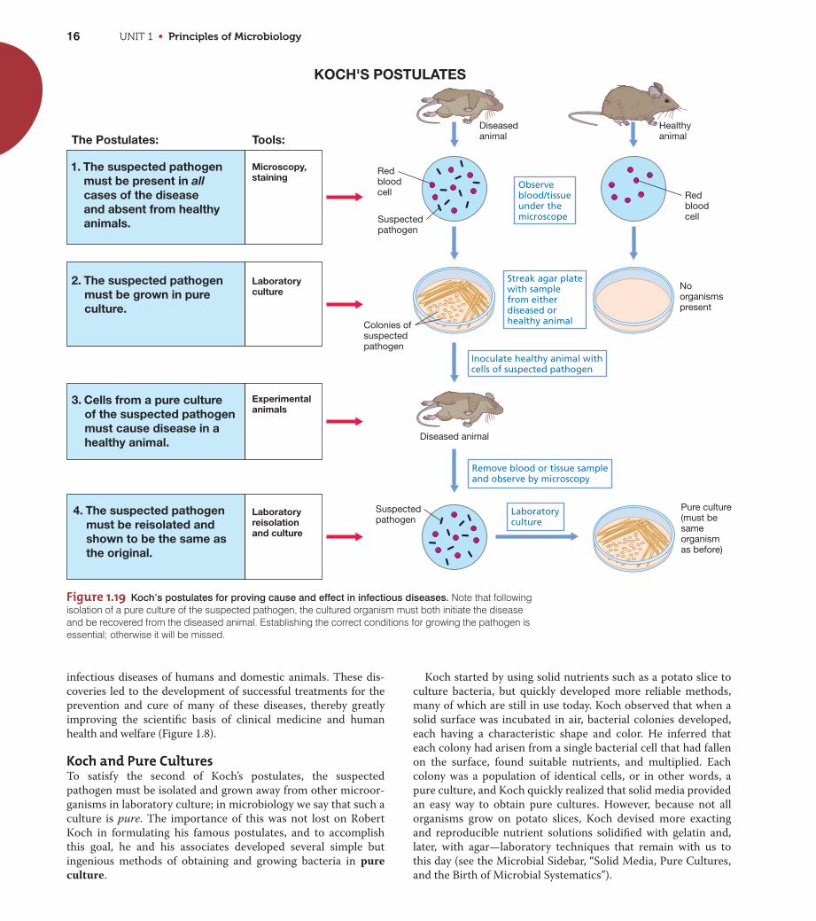
On the basis of these experiments and others on the causative
agent of tuberculosis, Koch formulated a set of rigorous criteria,
now known as Koch’s postulates, for definitively linking a spe-
cific microorganism to a specific disease. Koch’s postulates state
the following:
1. The disease-causing organism must always be present in ani-
mals suffering from the disease but not in healthy animals.
2. The organism must be cultivated in a pure culture away from
the animal body.
3. The isolated organism must cause the disease when inoculated
into healthy susceptible animals.
4. The organism must be isolated from the newly infected ani-
mals and cultured again in the laboratory, after which it should
be seen to be the same as the original organism.
Koch’s postulates, summarized in Figure 1.19, were a monu-
mental step forward in the study of infectious diseases. The pos-
tulates not only offered a means for linking the cause and effect of
an infectious disease, but also stressed the importance of
laboratory culture of the putative infectious agent. With these
postulates as a guide, Koch, his students, and those that followed
them discovered the causative agents of most of the important
UNIT 1 • Principles of Microbiology16
infectious diseases of humans and domestic animals. These dis-
coveries led to the development of successful treatments for the
prevention and cure of many of these diseases, thereby greatly
improving the scientific basis of clinical medicine and human
health and welfare (Figure 1.8).
Koch and Pure CulturesTo satisfy the second of Koch’s postulates, the suspected
pathogen must be isolated and grown away from other microor-
ganisms in laboratory culture; in microbiology we say that such a
culture is pure. The importance of this was not lost on Robert
Koch in formulating his famous postulates, and to accomplish
this goal, he and his associates developed several simple but
ingenious methods of obtaining and growing bacteria in pure
culture.
Koch started by using solid nutrients such as a potato slice to
culture bacteria, but quickly developed more reliable methods,
many of which are still in use today. Koch observed that when a
solid surface was incubated in air, bacterial colonies developed,
each having a characteristic shape and color. He inferred that
each colony had arisen from a single bacterial cell that had fallen
on the surface, found suitable nutrients, and multiplied. Each
colony was a population of identical cells, or in other words, a
pure culture, and Koch quickly realized that solid media provided
an easy way to obtain pure cultures. However, because not all
organisms grow on potato slices, Koch devised more exacting
and reproducible nutrient solutions solidified with gelatin and,
later, with agar—laboratory techniques that remain with us to
this day (see the Microbial Sidebar, “Solid Media, Pure Cultures,
and the Birth of Microbial Systematics”).
Diseasedanimal
Diseased animal
Observe blood/tissueunder the microscope
Streak agar platewith samplefrom eitherdiseased orhealthy animal
Suspectedpathogen
Suspectedpathogen
Laboratoryculture
Redbloodcell
Redbloodcell
Colonies ofsuspectedpathogen
Noorganismspresent
Pure culture(must be sameorganismas before)
Inoculate healthy animal withcells of suspected pathogen
Remove blood or tissue sampleand observe by microscopy
2. The suspected pathogenmust be grown in pure culture.
1. The suspected pathogenmust be present in allcases of the diseaseand absent from healthyanimals.
3. Cells from a pure culture of the suspected pathogenmust cause disease in a healthy animal.
4. The suspected pathogenmust be reisolated andshown to be the same asthe original.
KOCH'S POSTULATES
The Postulates: Tools:Healthyanimal
Microscopy,staining
Laboratory culture
Experimentalanimals
Laboratoryreisolationand culture
Figure 1.19 Koch’s postulates for proving cause and effect in infectious diseases. Note that followingisolation of a pure culture of the suspected pathogen, the cultured organism must both initiate the diseaseand be recovered from the diseased animal. Establishing the correct conditions for growing the pathogen isessential; otherwise it will be missed.
MICROBIAL SIDEBAR
Solid Media, Pure Cultures, and the Birth of Microbial Systematics
Robert Koch was the first to grow bacteriaon solid culture media. Koch’s early use
of potato slices as solid media was fraughtwith problems. Besides the problem that notall bacteria can grow on potatoes, the sliceswere frequently overgrown with molds. Kochthus needed a more reliable and repro-ducible means of growing bacteria on solidmedia, and he found the answer for solidify-ing his nutrient solutions in agar.
Koch initially employed gelatin as a solidi-fying agent for the various nutrient fluids heused to culture bacteria, and he kept hori-zontal slabs of solid gelatin free of contami-nation under a bell jar or in a glass box (seeFigure 1.20c). Nutrient-supplemented gelatinwas a good culture medium for the isolationand study of various bacteria, but it had sev-eral drawbacks, the most important of whichwas that it did not remain solid at 37°C, theoptimum temperature for growth of mosthuman pathogens. Thus, a different solidify-ing agent was needed.
Agar is a polysaccharide derived from redalgae. It was widely used in the nineteenthcentury as a gelling agent. Walter Hesse, anassociate of Koch, first used agar as a solidi-fying agent for bacteriological culture media(Figure 1). The actual suggestion that agarbe used instead of gelatin was made byHesse’s wife, Fannie. She had used agar tosolidify fruit jellies. When it was tried as asolidifying agent for microbial media, itssuperior gelling qualities were immediatelyevident. Hesse wrote to Koch about this dis-covery, and Koch quickly adapted agar to hisown studies, including his classic studies onthe isolation of the bacterium Mycobacteriumtuberculosis, the cause of the disease tuber-culosis (see text and Figure 1.20).
Agar has many other properties that makeit desirable as a gelling agent for microbialculture media. In particular, agar remainssolid at 37°C and, after melting during thesterilization process, remains liquid to about45°C, at which time it can be poured intosterile vessels. In addition, unlike gelatin,
agar is not degraded by most bacteria andtypically yields a transparent medium, mak-ing it easier to differentiate bacterial coloniesfrom inanimate particulate matter. For thesereasons, agar found its place early in theannals of microbiology and is still used todayfor obtaining and maintaining pure cultures.
In 1887 Richard Petri, a German bacteriol-ogist, published a brief paper describing amodification of Koch’s flat plate technique(Figure 1.20c). Petri’s enhancement, whichturned out to be amazingly useful, was thedevelopment of the transparent double-sideddishes that bear his name (Figure 2). Theadvantages of Petri dishes were immediatelyapparent. They could easily be stacked andsterilized separately from the medium, and,following the addition of molten culturemedium to the smaller of the two dishes, thelarger dish could be used as a cover to pre-vent contamination. Colonies that formed onthe surface of the agar in the Petri dishretained access to air without direct exposureto air and could easily be manipulated forfurther study. The original idea of Petri hasnot been improved on to this day, and thePetri dish, constructed of either glass orplastic, is a mainstay of the microbiologylaboratory.
Koch quickly grasped the significance ofpure cultures and was keenly aware of theimplications his pure culture methods had forclassifying microorganisms. He observedthat colonies that differed in color, morphol-ogy, size, and the like (see Figure 2) bredtrue and could be distinguished from oneanother. Cells from different colonies typicallydiffered in size and shape and often in theirtemperature or nutrient requirements as well.Koch realized that these differences amongmicroorganisms met all the requirements thatbiological taxonomists had established forthe classification of larger organisms, suchas plant and animal species. In Koch’s ownwords (translated from the German): “All bac-teria which maintain the characteristics whichdifferentiate one from another when they arecultured on the same medium and under thesame conditions, should be designated asspecies, varieties, forms, or other suitabledesignation.” Such insightful thinking wasimportant for the rapid acceptance of micro-biology as a new biological science, rootedas biology was in classification at the time ofKoch. It has since had a profound impact onthe diagnosis of infectious diseases and thefield of microbial diversity.
17
Pau
l V. D
unla
p
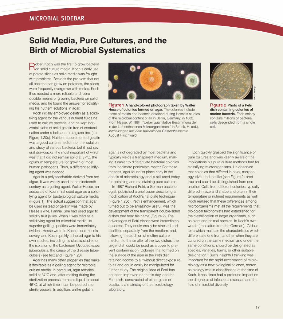
Figure 1 A hand-colored photograph taken by Walter
Hesse of colonies formed on agar. The colonies includethose of molds and bacteria obtained during Hesse’s studiesof the microbial content of air in Berlin, Germany, in 1882.From Hesse, W. 1884. “Ueber quantitative Bestimmung derin der Luft enthaltenen Mikroorganismen,” in Struck, H. (ed.),Mittheilungen aus dem Kaiserlichen Gesundheitsamte.August Hirschwald.
Figure 2 Photo of a Petri
dish containing colonies of
marine bacteria. Each colonycontains millions of bacterialcells descended from a singlecell.
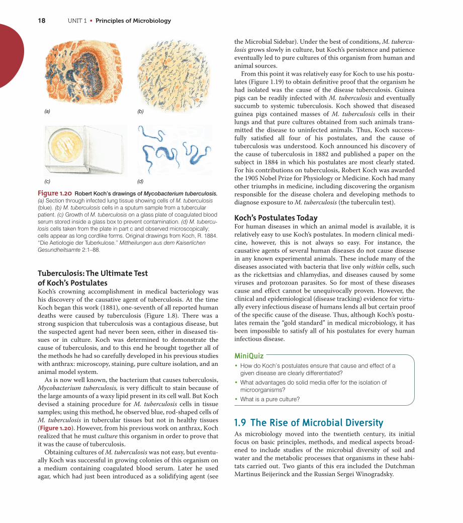
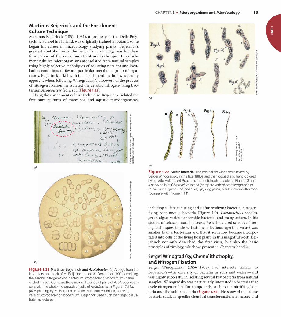
Figure 1.20 Robert Koch’s drawings of Mycobacterium tuberculosis.(a) Section through infected lung tissue showing cells of M. tuberculosis(blue). (b) M. tuberculosis cells in a sputum sample from a tubercularpatient. (c) Growth of M. tuberculosis on a glass plate of coagulated bloodserum stored inside a glass box to prevent contamination. (d) M. tubercu-losis cells taken from the plate in part c and observed microscopically;cells appear as long cordlike forms. Original drawings from Koch, R. 1884.“Die Aetiologie der Tuberkulose.” Mittheilungen aus dem KaiserlichenGesundheitsamte 2:1–88.
UNIT 1 • Principles of Microbiology18
Tuberculosis: The Ultimate Testof Koch’s PostulatesKoch’s crowning accomplishment in medical bacteriology was
his discovery of the causative agent of tuberculosis. At the time
Koch began this work (1881), one-seventh of all reported human
deaths were caused by tuberculosis (Figure 1.8). There was a
strong suspicion that tuberculosis was a contagious disease, but
the suspected agent had never been seen, either in diseased tis-
sues or in culture. Koch was determined to demonstrate the
cause of tuberculosis, and to this end he brought together all of
the methods he had so carefully developed in his previous studies
with anthrax: microscopy, staining, pure culture isolation, and an
animal model system.
As is now well known, the bacterium that causes tuberculosis,
Mycobacterium tuberculosis, is very difficult to stain because of
the large amounts of a waxy lipid present in its cell wall. But Koch
devised a staining procedure for M. tuberculosis cells in tissue
samples; using this method, he observed blue, rod-shaped cells of
M. tuberculosis in tubercular tissues but not in healthy tissues
(Figure 1.20). However, from his previous work on anthrax, Koch
realized that he must culture this organism in order to prove that
it was the cause of tuberculosis.
Obtaining cultures of M. tuberculosis was not easy, but eventu-
ally Koch was successful in growing colonies of this organism on
a medium containing coagulated blood serum. Later he used
agar, which had just been introduced as a solidifying agent (see
the Microbial Sidebar). Under the best of conditions, M. tubercu-
losis grows slowly in culture, but Koch’s persistence and patience
eventually led to pure cultures of this organism from human and
animal sources.
From this point it was relatively easy for Koch to use his postu-
lates (Figure 1.19) to obtain definitive proof that the organism he
had isolated was the cause of the disease tuberculosis. Guinea
pigs can be readily infected with M. tuberculosis and eventually
succumb to systemic tuberculosis. Koch showed that diseased
guinea pigs contained masses of M. tuberculosis cells in their
lungs and that pure cultures obtained from such animals trans-
mitted the disease to uninfected animals. Thus, Koch success-
fully satisfied all four of his postulates, and the cause of
tuberculosis was understood. Koch announced his discovery of
the cause of tuberculosis in 1882 and published a paper on the
subject in 1884 in which his postulates are most clearly stated.
For his contributions on tuberculosis, Robert Koch was awarded
the 1905 Nobel Prize for Physiology or Medicine. Koch had many
other triumphs in medicine, including discovering the organism
responsible for the disease cholera and developing methods to
diagnose exposure to M. tuberculosis (the tuberculin test).
Koch’s Postulates TodayFor human diseases in which an animal model is available, it is
relatively easy to use Koch’s postulates. In modern clinical medi-
cine, however, this is not always so easy. For instance, the
causative agents of several human diseases do not cause disease
in any known experimental animals. These include many of the
diseases associated with bacteria that live only within cells, such
as the rickettsias and chlamydias, and diseases caused by some
viruses and protozoan parasites. So for most of these diseases
cause and effect cannot be unequivocally proven. However, the
clinical and epidemiological (disease tracking) evidence for virtu-
ally every infectious disease of humans lends all but certain proof
of the specific cause of the disease. Thus, although Koch’s postu-
lates remain the “gold standard” in medical microbiology, it has
been impossible to satisfy all of his postulates for every human
infectious disease.
MiniQuiz• How do Koch’s postulates ensure that cause and effect of a
given disease are clearly differentiated?
• What advantages do solid media offer for the isolation ofmicroorganisms?
• What is a pure culture?
1.9 The Rise of Microbial DiversityAs microbiology moved into the twentieth century, its initial
focus on basic principles, methods, and medical aspects broad-
ened to include studies of the microbial diversity of soil and
water and the metabolic processes that organisms in these habi-
tats carried out. Two giants of this era included the Dutchman
Martinus Beijerinck and the Russian Sergei Winogradsky.
(a) (b)
(c) (d)
CHAPTER 1 • Microorganisms and Microbiology 19
UN
IT 1
Figure 1.21 Martinus Beijerinck and Azotobacter. (a) A page from thelaboratory notebook of M. Beijerinck dated 31 December 1900 describingthe aerobic nitrogen-fixing bacterium Azotobacter chroococcum (namecircled in red). Compare Beijerinck’s drawings of pairs of A. chroococcumcells with the photomicrograph of cells of Azotobacter in Figure 17.18a.(b) A painting by M. Beijerinck’s sister, Henriëtte Beijerinck, showing cells of Azotobacter chroococcum. Beijerinck used such paintings to illus-trate his lectures.
(a)
(b)
Lesl
ey R
ober
tson
and
the
Klu
yver
Lab
orat
ory
Mus
eum
,D
elft
Uni
vers
ity o
f Tec
hnol
ogy
Lesl
ey R
ober
tson
and
the
Klu
yver
Lab
orat
ory
Mus
eum
, Del
ft U
nive
rsity
of T
echn
olog
y
Martinus Beijerinck and the EnrichmentCulture TechniqueMartinus Beijerinck (1851–1931), a professor at the Delft Poly-
technic School in Holland, was originally trained in botany, so he
began his career in microbiology studying plants. Beijerinck’s
greatest contribution to the field of microbiology was his clear
formulation of the enrichment culture technique. In enrich-
ment cultures microorganisms are isolated from natural samples
using highly selective techniques of adjusting nutrient and incu-
bation conditions to favor a particular metabolic group of orga-
nisms. Beijerinck’s skill with the enrichment method was readily
apparent when, following Winogradsky’s discovery of the process
of nitrogen fixation, he isolated the aerobic nitrogen-fixing bac-
terium Azotobacter from soil (Figure 1.21).
Using the enrichment culture technique, Beijerinck isolated the
first pure cultures of many soil and aquatic microorganisms,
including sulfate-reducing and sulfur-oxidizing bacteria, nitrogen-
fixing root nodule bacteria (Figure 1.9), Lactobacillus species,
green algae, various anaerobic bacteria, and many others. In his
studies of tobacco mosaic disease, Beijerinck used selective filter-
ing techniques to show that the infectious agent (a virus) was
smaller than a bacterium and that it somehow became incorpo-
rated into cells of the living host plant. In this insightful work, Bei-
jerinck not only described the first virus, but also the basic
principles of virology, which we present in Chapters 9 and 21.
Sergei Winogradsky, Chemolithotrophy,and Nitrogen FixationSergei Winogradsky (1856–1953) had interests similar to
Beijerinck’s—the diversity of bacteria in soils and waters—and
was highly successful in isolating several key bacteria from natural
samples. Winogradsky was particularly interested in bacteria that
cycle nitrogen and sulfur compounds, such as the nitrifying bac-
teria and the sulfur bacteria (Figure 1.22). He showed that these
bacteria catalyze specific chemical transformations in nature and
Figure 1.22 Sulfur bacteria. The original drawings were made bySergei Winogradsky in the late 1880s and then copied and hand-coloredby his wife Hèléne. (a) Purple sulfur phototrophic bacteria. Figures 3 and4 show cells of Chromatium okenii (compare with photomicrographs of C. okenii in Figures 1.5a and 1.7a). (b) Beggiatoa, a sulfur chemolithotroph(compare with Figure 1.14).
From
Mic
rob
iolo
gie
du
Sol
, use
d w
ith p
erm
issi
on
From
Win
ogra
dsk
y, S
. 194
9.M
icro
bio
logi
e d
u S
ol.
Mas
son,
Par
is.
(a)
(b)
UNIT 1 • Principles of Microbiology20
proposed the important concept of chemolithotrophy, the oxida-
tion of inorganic compounds to yield energy. Winogradsky further
showed that these organisms, which he called chemolithotrophs,
obtained their carbon from CO2. Winogradsky thus revealed that,
like phototrophic organisms, chemolithotrophic bacteria were
autotrophs.
Winogradsky performed the first isolation of a nitrogen-fixing
bacterium, the anaerobe Clostridium pasteurianum, and as just
mentioned, Beijerinck used this discovery to guide his isolation
of aerobic nitrogen-fixing bacteria years later (Figure 1.21).
Winogradsky lived to be almost 100, publishing many scientific
papers and a major monograph, Microbiologie du Sol (Soil Micro-
biology). This work, a milestone in microbiology, contains draw-
ings of many of the organisms Winogradsky studied during his
lengthy career (Figure 1.22).
MiniQuiz• What is meant by the term “enrichment culture”?
• What is meant by the term “chemolithotrophy”? In what way arechemolithotrophs like plants?
1.10 The Modern Era of MicrobiologyIn the twentieth century, the field of microbiology developed rap-
idly in two different yet complementary directions—applied and
basic. During this period a host of new laboratory tools became
available, and the science of microbiology began to mature and
spawn new subdisciplines. Few of these subdisciplines were
purely applied or purely basic. Instead, most had both discovery
(basic) and problem-solving (applied) components. Table 1.3summarizes these major subdisciplines of microbiology that
arose in the twentieth century.
Several microbiologists are remembered for their key contri-
butions during this period. In the early twentieth century many
remained focused on medical aspects of microbiology, and even
today, many dedicated microbiologists grapple with the impacts
of microorganisms on human, animal, and plant disease. But fol-
lowing World War II, an exciting new emphasis began to take
hold with studies of the genetic properties of microorganisms.
From roots in microbial genetics has emerged “modern biology,”
driven by molecular biology, genetic engineering, and genomics.
This molecular approach has revolutionized scientific thinking in
the life sciences and has driven experimental approaches to the
most compelling problems in biology. Some key Nobel laureates
and their contributions to the molecular era of microbiology are
listed in Table 1.4.
Many of the advances in microbiology today are fueled by the
genomics revolution; that is, we are clearly in the era of “molecu-
lar microbiology.” Rapid progress in DNA sequencing technology
and improved computational power have yielded huge amounts
of genomic information that have supported major advances in
medicine, agriculture, biotechnology, and microbial ecology. For
example, to obtain the sequence of the entire genome of a bac-
terium takes only a few hours (although sequence analysis is a
much more time-consuming process). The fast-paced field of
genomics has itself spawned highly focused new subdisciplines,
such as transcriptomics, proteomics, and metabolomics, which
explore, respectively, the patterns of RNA, protein, and meta-
bolic expression in cells. The concepts of genomics, transcrip-
tomics, proteomics, and metabolomics are all developed in
Chapter 12.
All signs point to a continued maturation of molecular micro-
biology as we enter a period where technology is almost ahead of
our ability to formulate exciting scientific questions. In fact,
microbial research today is very close to defining the minimalist
genome—the minimum complement of genes necessary for a liv-
ing cell. When such a genetic blueprint is available, microbiolo-
gists should be able to define, at least in biochemical terms, the
prerequisites for life. When that day arrives, can the laboratory
creation of an actual living cell from nonliving components, that
is, spontaneous generation under controlled laboratory condi-
tions, be far off? Almost certainly not. Stay tuned, as much excit-
ing science is on the way!
MiniQuiz• For each of the following topics, name the subdiscipline of
microbiology that focuses on it: metabolism, enzymology,nucleic acid and protein synthesis, microorganisms and theirnatural environments, microbial classification, inheritance ofcharacteristics.
Table 1.3 The major subdisciplines of microbiology
Subdiscipline Focus
I. Basic emphasesa
Microbial physiology Nutrition, metabolism
Microbial genetics Genes, heredity, and genetic variation
Microbial biochemistry Enzymes and chemical reactions in cells
Microbial systematics Classification and nomenclature
Virology Viruses and subviral particles
Molecular biology Nucleic acids and protein
Microbial ecology Microbial diversity and activity in natural habitats; biogeochemistry
II. Applied emphasesa
Medical microbiology Infectious disease
Immunology Immune systems
Agricultural/soil microbiology Microbial diversity and processes in soil
Industrial microbiology Large-scale production of antibiotics, alcohol, and other chemicals
Biotechnology Production of human proteins by genetically engineered microorganisms
Aquatic microbiology Microbial processes in waters and wastewaters, drinking water safety
aNone of these subdisciplines are devoted entirely to basic science or applied science.However, the subdisciplines listed in I tend to be more focused on discovery and thosein II more focused on solving specific problems or synthesizing commercial productsfrom microbial sources.
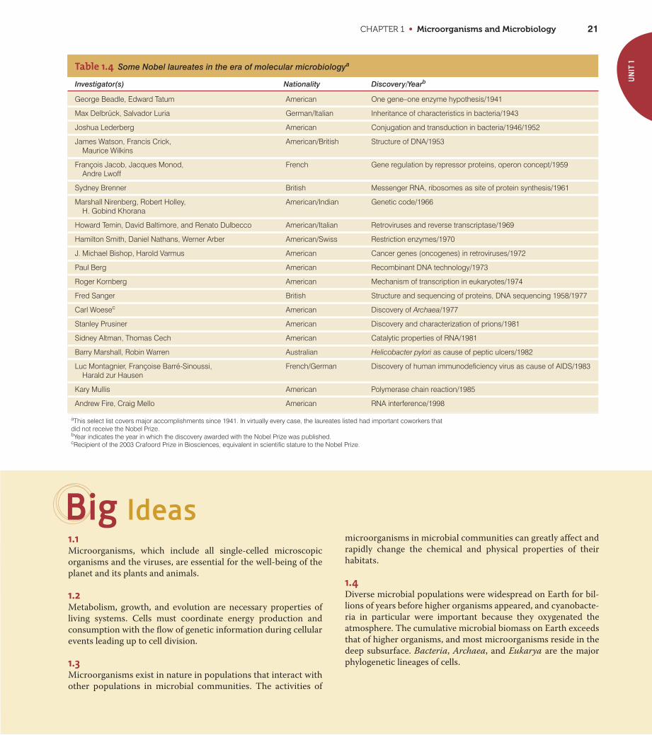
CHAPTER 1 • Microorganisms and Microbiology 21
UN
IT 1Table 1.4 Some Nobel laureates in the era of molecular microbiologya
Investigator(s) Nationality Discovery/Yearb
George Beadle, Edward Tatum American One gene–one enzyme hypothesis/1941
Max Delbrück, Salvador Luria German/Italian Inheritance of characteristics in bacteria/1943
Joshua Lederberg American Conjugation and transduction in bacteria/1946/1952
James Watson, Francis Crick, American/British Structure of DNA/1953Maurice Wilkins
François Jacob, Jacques Monod, French Gene regulation by repressor proteins, operon concept/1959Andre Lwoff
Sydney Brenner British Messenger RNA, ribosomes as site of protein synthesis/1961
Marshall Nirenberg, Robert Holley, American/Indian Genetic code/1966H. Gobind Khorana
Howard Temin, David Baltimore, and Renato Dulbecco American/Italian Retroviruses and reverse transcriptase/1969
Hamilton Smith, Daniel Nathans, Werner Arber American/Swiss Restriction enzymes/1970
J. Michael Bishop, Harold Varmus American Cancer genes (oncogenes) in retroviruses/1972
Paul Berg American Recombinant DNA technology/1973
Roger Kornberg American Mechanism of transcription in eukaryotes/1974
Fred Sanger British Structure and sequencing of proteins, DNA sequencing 1958/1977
Carl Woesec American Discovery of Archaea/1977
Stanley Prusiner American Discovery and characterization of prions/1981
Sidney Altman, Thomas Cech American Catalytic properties of RNA/1981
Barry Marshall, Robin Warren Australian Helicobacter pylori as cause of peptic ulcers/1982
Luc Montagnier, Françoise Barré-Sinoussi, Harald zur Hausen
French/German Discovery of human immunodeficiency virus as cause of AIDS/1983
Kary Mullis American Polymerase chain reaction/1985
Andrew Fire, Craig Mello American RNA interference/1998
aThis select list covers major accomplishments since 1941. In virtually every case, the laureates listed had important coworkers thatdid not receive the Nobel Prize.bYear indicates the year in which the discovery awarded with the Nobel Prize was published.cRecipient of the 2003 Crafoord Prize in Biosciences, equivalent in scientific stature to the Nobel Prize.
Big Ideas1.1Microorganisms, which include all single-celled microscopic
organisms and the viruses, are essential for the well-being of the
planet and its plants and animals.
1.2Metabolism, growth, and evolution are necessary properties of
living systems. Cells must coordinate energy production and
consumption with the flow of genetic information during cellular
events leading up to cell division.
1.3 Microorganisms exist in nature in populations that interact with
other populations in microbial communities. The activities of
microorganisms in microbial communities can greatly affect and
rapidly change the chemical and physical properties of their
habitats.
1.4 Diverse microbial populations were widespread on Earth for bil-
lions of years before higher organisms appeared, and cyanobacte-
ria in particular were important because they oxygenated the
atmosphere. The cumulative microbial biomass on Earth exceeds
that of higher organisms, and most microorganisms reside in the
deep subsurface. Bacteria, Archaea, and Eukarya are the major
phylogenetic lineages of cells.
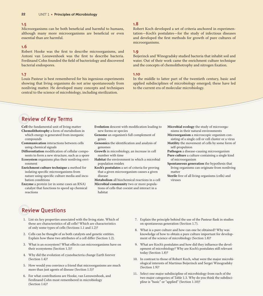
Cell the fundamental unit of living matter
Chemolithotrophy a form of metabolism in
which energy is generated from inorganic
compounds
Communication interactions between cells
using chemical signals
Differentiation modification of cellular compo-
nents to form a new structure, such as a spore
Ecosystem organisms plus their nonliving envi-
ronment
Enrichment culture technique a method for
isolating specific microorganisms from
nature using specific culture media and incu-
bation conditions
Enzyme a protein (or in some cases an RNA)
catalyst that functions to speed up chemical
reactions
Evolution descent with modification leading to
new forms or species
Genome an organism’s full complement of
genes
Genomics the identification and analysis of
genomes
Growth in microbiology, an increase in cell
number with time
Habitat the environment in which a microbial
population resides
Koch’s postulates a set of criteria for proving
that a given microorganism causes a given
disease
Metabolism all biochemical reactions in a cell
Microbial community two or more popula-
tions of cells that coexist and interact in a
habitat
Microbial ecology the study of microorga-
nisms in their natural environments
Microorganism a microscopic organism con-
sisting of a single cell or cell cluster or a virus
Motility the movement of cells by some form of
self-propulsion
Pathogen a disease-causing microorganism
Pure culture a culture containing a single kind
of microorganism
Spontaneous generation the hypothesis that
living organisms can originate from nonliving
matter
Sterile free of all living organisms (cells) and
viruses
Review of Key Terms
1.5 Microorganisms can be both beneficial and harmful to humans,
although many more microorganisms are beneficial or even
essential than are harmful.
1.6 Robert Hooke was the first to describe microorganisms, and
Antoni van Leeuwenhoek was the first to describe bacteria.
Ferdinand Cohn founded the field of bacteriology and discovered
bacterial endospores.
1.7 Louis Pasteur is best remembered for his ingenious experiments
showing that living organisms do not arise spontaneously from
nonliving matter. He developed many concepts and techniques
central to the science of microbiology, including sterilization.
1.8 Robert Koch developed a set of criteria anchored in experimen-
tation—Koch’s postulates—for the study of infectious diseases
and developed the first methods for growth of pure cultures of
microorganisms.
1.9 Beijerinck and Winogradsky studied bacteria that inhabit soil and
water. Out of their work came the enrichment culture technique
and the concepts of chemolithotrophy and nitrogen fixation.
1.10 In the middle to latter part of the twentieth century, basic and
applied subdisciplines of microbiology emerged; these have led
to the current era of molecular microbiology.
1. List six key properties associated with the living state. Which of
these are characteristics of all cells? Which are characteristics
of only some types of cells (Sections 1.1 and 1.2)?
2. Cells can be thought of as both catalysts and genetic entities.
Explain how these two attributes of a cell differ (Section 1.2).
3. What is an ecosystem? What effects can microorganisms have on
their ecosystems (Section 1.3)?
4. Why did the evolution of cyanobacteria change Earth forever
(Section 1.4)?
5. How would you convince a friend that microorganisms are much
more than just agents of disease (Section 1.5)?
6. For what contributions are Hooke, van Leeuwenhoek, and
Ferdinand Cohn most remembered in microbiology
(Section 1.6)?
7. Explain the principle behind the use of the Pasteur flask in studies
on spontaneous generation (Section 1.7).
8. What is a pure culture and how can one be obtained? Why was
knowledge of how to obtain a pure culture important for develop-
ment of the science of microbiology (Section 1.8)?
9. What are Koch’s postulates and how did they influence the devel-
opment of microbiology? Why are Koch’s postulates still relevant
today (Section 1.8)?
10. In contrast to those of Robert Koch, what were the major microbi-
ological interests of Martinus Beijerinck and Sergei Winogradsky
(Section 1.9)?
11. Select one major subdiscipline of microbiology from each of the
two major categories of Table 1.3. Why do you think the subdisci-
pline is “basic” or “applied” (Section 1.10)?
Review Questions
UNIT 1 • Principles of Microbiology22