Embed Size (px)
Citation preview
MICROFLUIDIC DEVICE FOR SUPER-FAST EVALUATION OF MEMBRANE PROTEIN
CRYSTALLIZATION Hsin-Jui Wu1*, Tamara Basta1, Mary Morphew1, D. C. Rees2, Michael H. B. Stowell1, and Y. C. Lee1
1Univeristy of Colorado, Boulder, Colorado, USA 2California Institute of Technology, Pasadena, California, USA
*Corresponding Author: Hsin-Jui Wu, [email protected]
Abstract—Membrane proteins embedded in bi-layer lipids of cell membrane have unique functions including inter-cell communication, ions/molecules transport. And there is more than 50% of drug design emphasizes on membrane proteins specifically studying on their structure and formation. Recently we reported the structural and functional studies of membrane protein lipid nanoparticles in native biological membrane. This virus-like nanoparticle formed by a self-assembly crystallization process of membrane protein and lipids is critical to pharmaceutical industrial. These nanoparticles have a variety of potential applications in drug delivery and drug design that can carry specific the membrane protein on aim or release control. The previous studies stay on an inefficient method with a standard dialysis process that has low-throughput, time consumption, and protein sample waste. However, the interdisciplinary cooperation between in biology and Micro electro mechanical systems (MEMS) has been tremendous developed such as Bio-MEMS and Lab-on-a-chip technologies. Here we demonstrate a new concept with a high-throughput membraneless microfluidic device to fast produce the reconstitution of membrane protein nanoparticles. The reconstitution process in continuous micro flow dominated by convection-diffusion phenomena in microfluidic channel can be completed in seconds to form protein/lipid particles under multiple conditions applied. The controllable syringe pumps is used to test a combination of conditions rather than using inefficient hand pipette. Moreover this novel microfluidic device can save protein sample consumption down to only nanoliter or picoliter. By using this device, we have an ability to rapidly form uniform membrane protein lipid nanoparticles and we believe this new method will make a transformative impact to commercial applications in variety of areas from biology to pharmacology.
Keywords- Membrane proteins, nanoparticles, Bio-MEMS.
I. INTRODUCTION
Membrane proteins represent more than 30% of the proteins encoded in the all genomes and play a major rule in
physiological environment controlling irons/molecules, energy and information through cell-to-cell membrane bilayer. The first structure of membrane protein was found 25 years ago but currently only around 754 membrane protein structures with 266 unique types of membrane proteins have been identified and recorded. That is only around 1% of entire protein structure in Protein Data Bank [1]. Therefore to investigate and understand the structure of membrane proteins become an important research area.
The reason for this slow development of membrane protein analysis technique is because the inefficient standard method [2]. The standard method to reconstitute membrane proteins in a native bilayer lipid environment consists of a sample well, a buffer solution well and a dialysis membrane in the middle. The reconstitution process to obtain structure of membrane proteins in standard method is inefficient because it driven by slow diffusion to remove higher concentration of detergents from protein/lipid/detergent complex sample well through adialysis membrane to lower concentration of buffer solution well. The designed pores of dialysis membrane only allowed a small molecular such as detergents to pass through and remind a large one such as membrane proteins in sample well. Once the detergents fully removed by diffusion process the membrane protein aggregated with native lipid bilayer structure to from membrane protein nanoparticles [3].However this standard method with dialysis membrane by using slow diffusion process is inconvenient and inefficient that takes 7 - 14 days for removing detergents to achieve the reconstitution process of membrane protein nanoparticle. Furthermore it is difficult to optimize and find the correct
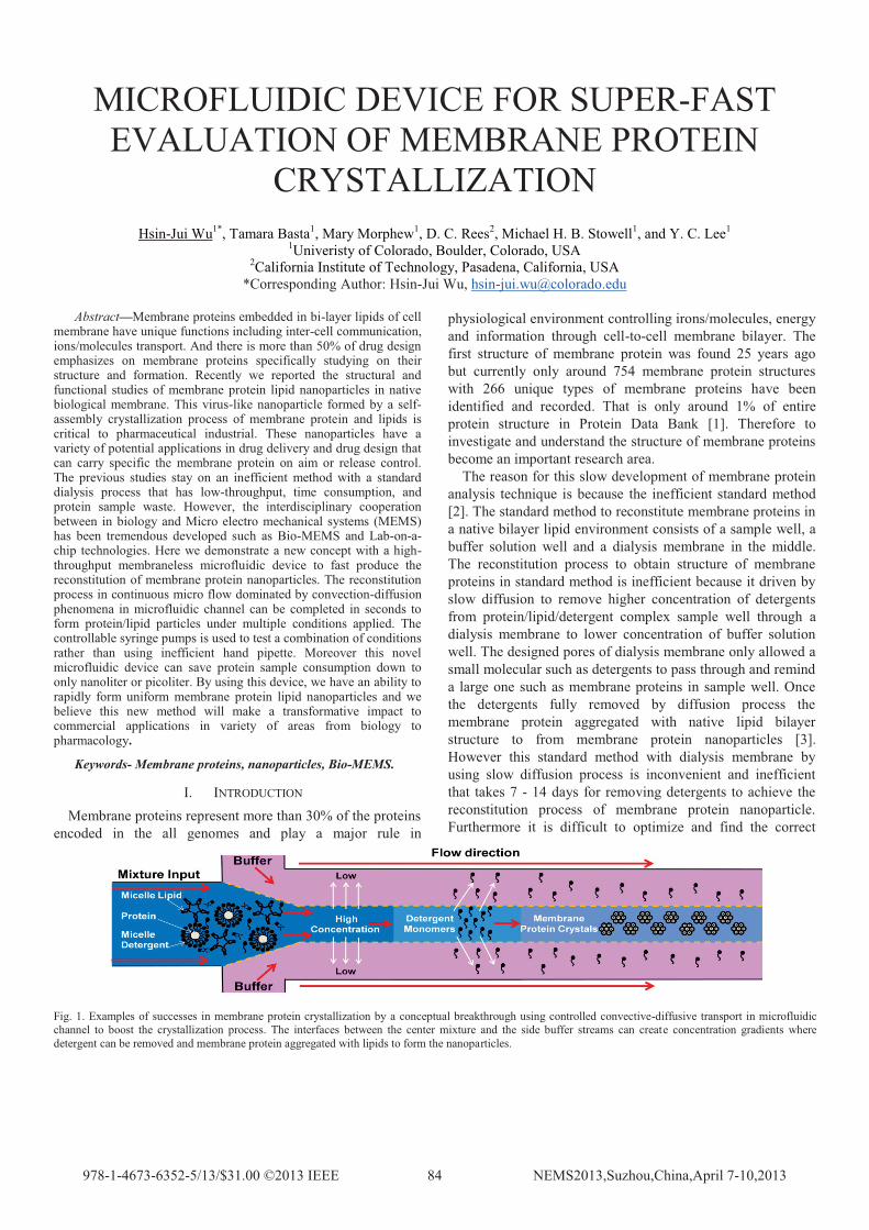
Fig. 1. Examples of successes in membrane protein crystallization by a conceptual breakthrough using controlled convective-diffusive transport in microfluidic channel to boost the crystallization process. The interfaces between the center mixture and the side buffer streams can create concentration gradients where detergent can be removed and membrane protein aggregated with lipids to form the nanoparticles.
combination to obtain the membrane protein from few controllable parameters such as protein to lipid ratio, ph, and NaCl concentration.
Recently the interdisciplinary collaboration has been developing on diversity of fields especially within engineering and biology. Microfluidic and Bio-MEMS are good examples for this such interesting topic [4,5]. In the past few decades Microfluidic has been rapidly applied into the biology area to manipulate and control a small volume for both simple or complex experiments such as mixing, and diluting processes. Sequentially it can enhance the efficiency of biological experiments. In this small scale, typically the characteristic channel size of microfluidic device is from several micrometers to few hundred micrometers, there is some advantages of Microfluidic including super low Reynold number, less sample required, and fast chemical reaction [6,7].Based on these benefits we can obtain a stable laminar flow pattern in microfluidic channels where allow people to have a solid device without any influences [8]. Figure 1. shows our new concept for super-fast evaluation of membrane protein crystallization and we demonstrate a successful microfluidic device converted a traditional dialysis membrane method to a novel method that can achieve the fast response time for reconstitution of membrane protein nanoparticle from days to seconds with nanoliter or less sample required.
II. FABRICATION PROCESS
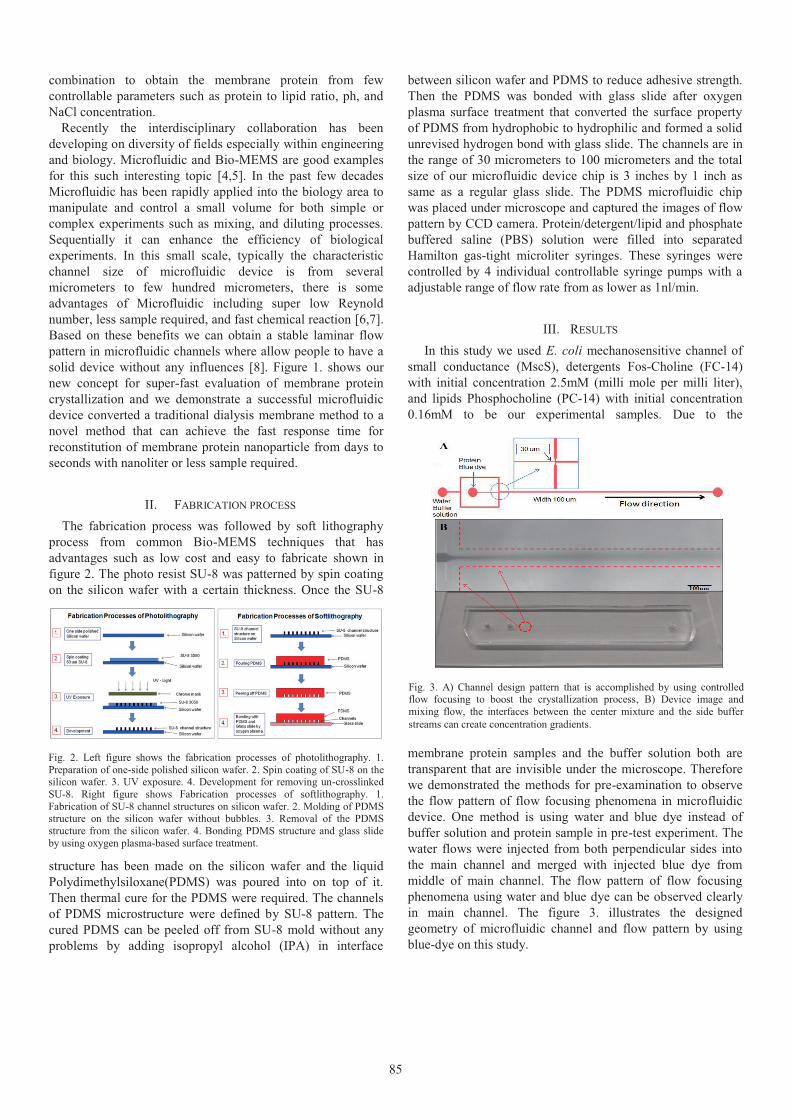
The fabrication process was followed by soft lithography process from common Bio-MEMS techniques that has advantages such as low cost and easy to fabricate shown in figure 2. The photo resist SU-8 was patterned by spin coating on the silicon wafer with a certain thickness. Once the SU-8
structure has been made on the silicon wafer and the liquid Polydimethylsiloxane(PDMS) was poured into on top of it. Then thermal cure for the PDMS were required. The channels of PDMS microstructure were defined by SU-8 pattern. The cured PDMS can be peeled off from SU-8 mold without any problems by adding isopropyl alcohol (IPA) in interface
between silicon wafer and PDMS to reduce adhesive strength. Then the PDMS was bonded with glass slide after oxygen plasma surface treatment that converted the surface property of PDMS from hydrophobic to hydrophilic and formed a solid unrevised hydrogen bond with glass slide. The channels are in the range of 30 micrometers to 100 micrometers and the total size of our microfluidic device chip is 3 inches by 1 inch as same as a regular glass slide. The PDMS microfluidic chip was placed under microscope and captured the images of flow pattern by CCD camera. Protein/detergent/lipid and phosphate buffered saline (PBS) solution were filled into separated Hamilton gas-tight microliter syringes. These syringes were controlled by 4 individual controllable syringe pumps with a adjustable range of flow rate from as lower as 1nl/min.
III. RESULTS
In this study we used E. coli mechanosensitive channel of small conductance (MscS), detergents Fos-Choline (FC-14) with initial concentration 2.5mM (milli mole per milli liter), and lipids Phosphocholine (PC-14) with initial concentration 0.16mM to be our experimental samples. Due to the
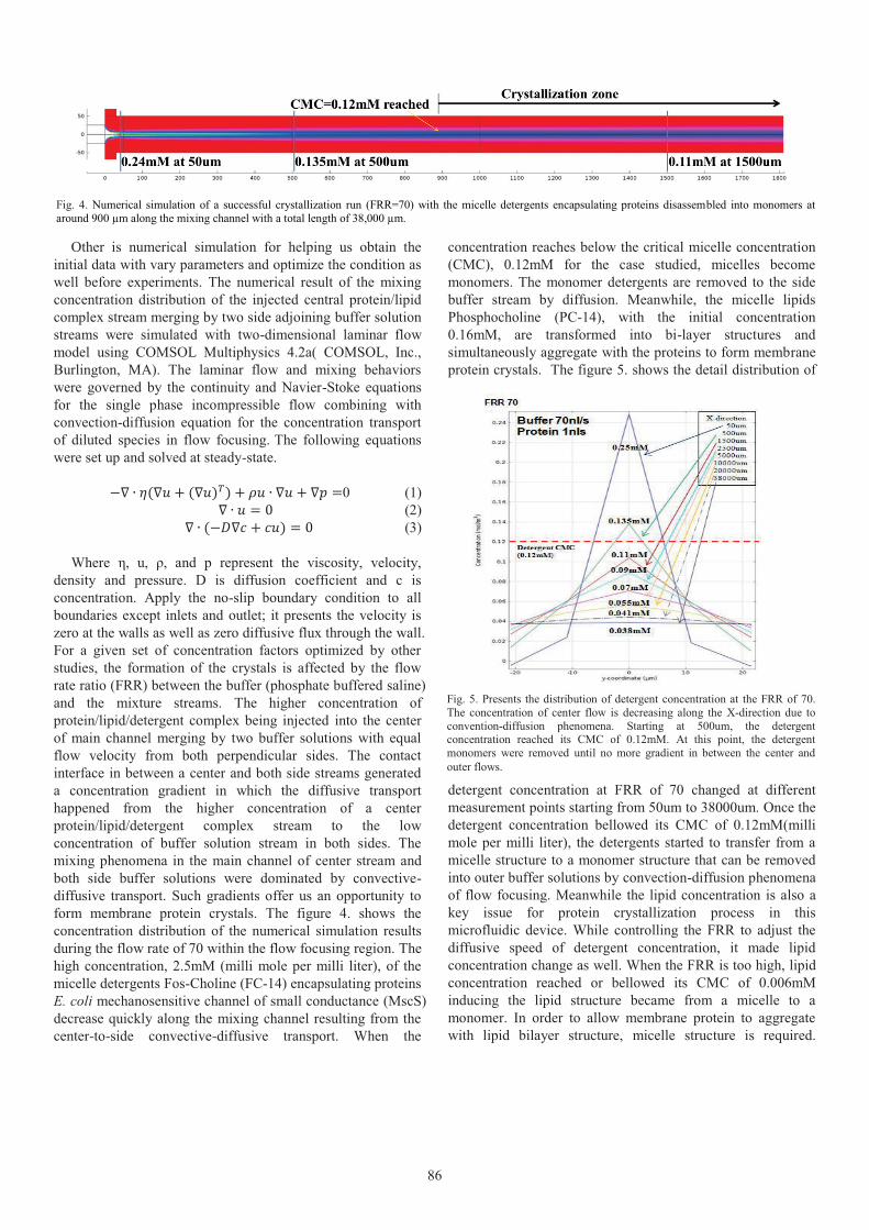
membrane protein samples and the buffer solution both are transparent that are invisible under the microscope. Therefore we demonstrated the methods for pre-examination to observe the flow pattern of flow focusing phenomena in microfluidic device. One method is using water and blue dye instead of buffer solution and protein sample in pre-test experiment. The water flows were injected from both perpendicular sides into the main channel and merged with injected blue dye from middle of main channel. The flow pattern of flow focusing phenomena using water and blue dye can be observed clearly in main channel. The figure 3. illustrates the designed geometry of microfluidic channel and flow pattern by using blue-dye on this study.
Fig. 3. A) Channel design pattern that is accomplished by using controlledflow focusing to boost the crystallization process, B) Device image and mixing flow, the interfaces between the center mixture and the side buffer streams can create concentration gradients.
Fig. 2. Left figure shows the fabrication processes of photolithography. 1. Preparation of one-side polished silicon wafer. 2. Spin coating of SU-8 on thesilicon wafer. 3. UV exposure. 4. Development for removing un-crosslinkedSU-8. Right figure shows Fabrication processes of softlithography. 1. Fabrication of SU-8 channel structures on silicon wafer. 2. Molding of PDMS structure on the silicon wafer without bubbles. 3. Removal of the PDMS structure from the silicon wafer. 4. Bonding PDMS structure and glass slide by using oxygen plasma-based surface treatment.
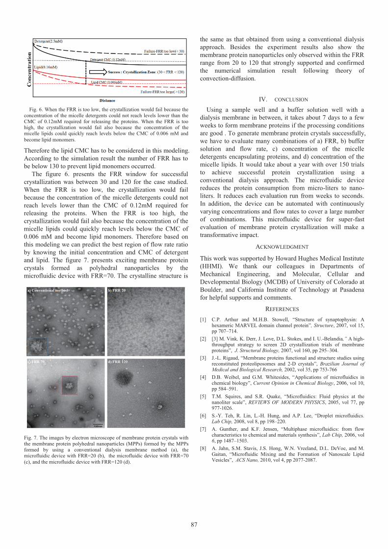
Other is numerical simulation for helping us obtain the initial data with vary parameters and optimize the condition as well before experiments. The numerical result of the mixing concentration distribution of the injected central protein/lipid complex stream merging by two side adjoining buffer solution streams were simulated with two-dimensional laminar flow model using COMSOL Multiphysics 4.2a( COMSOL, Inc., Burlington, MA). The laminar flow and mixing behaviors were governed by the continuity and Navier-Stoke equations for the single phase incompressible flow combining with convection-diffusion equation for the concentration transport of diluted species in flow focusing. The following equations were set up and solved at steady-state.
0 (1) (2)
(3)
Where η, u, ρ, and p represent the viscosity, velocity, density and pressure. D is diffusion coefficient and c is concentration. Apply the no-slip boundary condition to all boundaries except inlets and outlet; it presents the velocity is zero at the walls as well as zero diffusive flux through the wall. For a given set of concentration factors optimized by other studies, the formation of the crystals is affected by the flow rate ratio (FRR) between the buffer (phosphate buffered saline) and the mixture streams. The higher concentration of protein/lipid/detergent complex being injected into the center of main channel merging by two buffer solutions with equal flow velocity from both perpendicular sides. The contact interface in between a center and both side streams generated a concentration gradient in which the diffusive transport happened from the higher concentration of a center protein/lipid/detergent complex stream to the low concentration of buffer solution stream in both sides. The mixing phenomena in the main channel of center stream and both side buffer solutions were dominated by convective-diffusive transport. Such gradients offer us an opportunity to form membrane protein crystals. The figure 4. shows the concentration distribution of the numerical simulation results during the flow rate of 70 within the flow focusing region. The high concentration, 2.5mM (milli mole per milli liter), of the micelle detergents Fos-Choline (FC-14) encapsulating proteins E. coli mechanosensitive channel of small conductance (MscS)decrease quickly along the mixing channel resulting from the center-to-side convective-diffusive transport. When the
concentration reaches below the critical micelle concentration (CMC), 0.12mM for the case studied, micelles become monomers. The monomer detergents are removed to the side buffer stream by diffusion. Meanwhile, the micelle lipids Phosphocholine (PC-14), with the initial concentration 0.16mM, are transformed into bi-layer structures and simultaneously aggregate with the proteins to form membrane protein crystals. The figure 5. shows the detail distribution of
detergent concentration at FRR of 70 changed at different measurement points starting from 50um to 38000um. Once the detergent concentration bellowed its CMC of 0.12mM(milli mole per milli liter), the detergents started to transfer from a micelle structure to a monomer structure that can be removed into outer buffer solutions by convection-diffusion phenomena of flow focusing. Meanwhile the lipid concentration is also a key issue for protein crystallization process in this microfluidic device. While controlling the FRR to adjust the diffusive speed of detergent concentration, it made lipid concentration change as well. When the FRR is too high, lipid concentration reached or bellowed its CMC of 0.006mM inducing the lipid structure became from a micelle to a monomer. In order to allow membrane protein to aggregate with lipid bilayer structure, micelle structure is required.
Fig. 4. Numerical simulation of a successful crystallization run (FRR=70) with the micelle detergents encapsulating proteins disassembled into monomers at around 900 µm along the mixing channel with a total length of 38,000 µm.
Fig. 5. Presents the distribution of detergent concentration at the FRR of 70.The concentration of center flow is decreasing along the X-direction due toconvention-diffusion phenomena. Starting at 500um, the detergentconcentration reached its CMC of 0.12mM. At this point, the detergentmonomers were removed until no more gradient in between the center and outer flows.
Therefore the lipid CMC has to be considered in this modeling. According to the simulation result the number of FRR has to be below 130 to prevent lipid monomers occurred.
The figure 6. presents the FRR window for successful crystallization was between 30 and 120 for the case studied. When the FRR is too low, the crystallization would fail because the concentration of the micelle detergents could not reach levels lower than the CMC of 0.12mM required for releasing the proteins. When the FRR is too high, the crystallization would fail also because the concentration of the micelle lipids could quickly reach levels below the CMC of 0.006 mM and become lipid monomers. Therefore based on this modeling we can predict the best region of flow rate ratio by knowing the initial concentration and CMC of detergent and lipid. The figure 7. presents exciting membrane protein crystals formed as polyhedral nanoparticles by the microfluidic device with FRR=70. The crystalline structure is
the same as that obtained from using a conventional dialysis approach. Besides the experiment results also show the membrane protein nanoparticles only observed within the FRR range from 20 to 120 that strongly supported and confirmed the numerical simulation result following theory of convection-diffusion.
IV. CONCLUSION
Using a sample well and a buffer solution well with a dialysis membrane in between, it takes about 7 days to a few weeks to form membrane proteins if the processing conditions are good . To generate membrane protein crystals successfully, we have to evaluate many combinations of a) FRR, b) buffer solution and flow rate, c) concentration of the micelle detergents encapsulating proteins, and d) concentration of the micelle lipids. It would take about a year with over 150 trials to achieve successful protein crystallization using a conventional dialysis approach. The microfluidic device reduces the protein consumption from micro-liters to nano-liters. It reduces each evaluation run from weeks to seconds. In addition, the device can be automated with continuously varying concentrations and flow rates to cover a large number of combinations. This microfluidic device for super-fast evaluation of membrane protein crystallization will make a transformative impact.
ACKNOWLEDGMENT
This work was supported by Howard Hughes Medical Institute (HHMI). We thank our colleagues in Departments of Mechanical Engineering, and Molecular, Cellular and Developmental Biology (MCDB) of University of Colorado at Boulder, and California Institute of Technology at Pasadena for helpful supports and comments.
REFERENCES
[1] C.P. Arthur and M.H.B. Stowell, “Structure of synaptophysin: A hexameric MARVEL domain channel protein”, Structure, 2007, vol 15,pp 707–714.
[2] [3] M. Vink, K. Derr, J. Love, D.L. Stokes, and I. U.-Belandia,” A high-throughput strategy to screen 2D crystallization trials of membrane proteins”, J. Structural Biology, 2007, vol 160, pp 295–304.
[3] J.-L. Rigaud, “Membrane proteins functional and structure studies using reconstituted proteoliposomes and 2-D crystals”, Brazilian Journal of Medical and Biological Research, 2002, vol 35, pp 753-766
[4] D.B. Weibel, and G.M. Whitesides, “Applications of microfluidics in chemical biology”, Current Opinion in Chemical Biology, 2006, vol 10,pp 584–591.
[5] T.M. Squires, and S.R. Quake, “Microfluidics: Fluid physics at the nanoliter scale”, REVIEWS OF MODERN PHYSICS, 2005, vol 77, pp977-1026.
[6] S.-Y. Teh, R. Lin, L.-H. Hung, and A.P. Lee, “Droplet microfluidics. Lab Chip, 2008, vol 8, pp 198–220.
[7] A. Gunther, and K.F. Jensen, “Multiphase microfluidics: from flow characteristics to chemical and materials synthesis”, Lab Chip, 2006, vol 6, pp 1487–1503.
[8] A. Jahn, S.M. Stavis, J.S. Hong, W.N. Vreeland, D.L. DeVoe, and M. Gaitan, “Microfluidic Mixing and the Formation of Nanoscale Lipid Vesicles”, ACS Nano, 2010, vol 4, pp 2077-2087.
Fig. 6. When the FRR is too low, the crystallization would fail because the concentration of the micelle detergents could not reach levels lower than the CMC of 0.12mM required for releasing the proteins. When the FRR is too high, the crystallization would fail also because the concentration of the micelle lipids could quickly reach levels below the CMC of 0.006 mM and become lipid monomers.
Fig. 7. The images by electron microscope of membrane protein crystals with the membrane protein polyhedral nanoparticles (MPPs) formed by the MPPs formed by using a conventional dialysis membrane method (a), themicrofluidic device with FRR=20 (b), the microfluidic device with FRR=70(c), and the microfluidic device with FRR=120 (d).
![Published By: HIVE [karachi]€¦ · Ahl-e-Hadith Wafaq al -Madaris al-Salafiya Shia Wafaq al -Madaris al-Shia ... (JUI-F) and Maulana Samiul Haq (JUI-S) run more than 65 per cent](https://img.pdfslide.us/doc/110x75/610bf96e97f2096f8260b027/published-by-hive-karachi-ahl-e-hadith-wafaq-al-madaris-al-salafiya-shia-wafaq.jpg)


![ponts en maçonnerie. constitution et stabilité [3 tomes] (jui 1982)_2](https://img.pdfslide.us/doc/110x75/5572008c49795991699f9fd5/ponts-en-maconnerie-constitution-et-stabilite-3-tomes-jui-19822-55ab58c50db9d.jpg)


![THE ROSICRUCIAN FELLOWSHIP › ssoc › 1909__heindel___why_i_am_a_rosicrucian.pdf · JUI]v 3](https://img.pdfslide.us/doc/110x75/60cd02fa40fb275f0a1377d9/the-rosicrucian-a-ssoc-a-1909heindelwhyiamarosicrucianpdf-juiv.jpg)
