Embed Size (px)
Citation preview

Seediscussions,stats,andauthorprofilesforthispublicationat:https://www.researchgate.net/publication/316239408
Citation:MicrobiotaIsInvolvedinPost-resectionAdaptationinHumanswithShortBowelSyndrome
ArticleinFrontiersinPhysiology·May2017
DOI:10.3389/fphys.2017.00224
CITATIONS
0
READS
24
9authors,including:
CamilleMayeur
FrenchNationalInstituteforAgriculturalRes…
62PUBLICATIONS746CITATIONS
SEEPROFILE
VeRobert
FrenchNationalInstituteforAgriculturalRes…
28PUBLICATIONS529CITATIONS
SEEPROFILE
JohanneLeBeyec
UMRSU1149-CRI
46PUBLICATIONS487CITATIONS
SEEPROFILE
MurielThomas
FrenchNationalInstituteforAgriculturalRes…
70PUBLICATIONS1,292CITATIONS
SEEPROFILE
AllcontentfollowingthispagewasuploadedbyMurielThomason19April2017.
Theuserhasrequestedenhancementofthedownloadedfile.Allin-textreferencesunderlinedinblueareaddedtotheoriginaldocument
andarelinkedtopublicationsonResearchGate,lettingyouaccessandreadthemimmediately.
ORIGINAL RESEARCHpublished: 19 April 2017
doi: 10.3389/fphys.2017.00224
Frontiers in Physiology | www.frontiersin.org 1 April 2017 | Volume 8 | Article 224
Edited by:
Atsushi Masamune,
Tohoku University, Japan
Reviewed by:
Tomohiro Watanabe,
Kindai University, Japan
Takayuki Kogure,
Tohoku Medical and Pharmaceutical
University, Japan
*Correspondence:
Muriel Thomas
†These co-senior authors have
contributed equally to this work.
Specialty section:
This article was submitted to
Gastrointestinal Sciences,
a section of the journal
Frontiers in Physiology
Received: 12 December 2016
Accepted: 28 March 2017
Published: 19 April 2017
Citation:
Gillard L, Mayeur C, Robert V,
Pingenot I, Le Beyec J, Bado A,
Lepage P, Thomas M and Joly F
(2017) Microbiota Is Involved in
Post-resection Adaptation in Humans
with Short Bowel Syndrome.
Front. Physiol. 8:224.
doi: 10.3389/fphys.2017.00224
Microbiota Is Involved inPost-resection Adaptation inHumans with Short Bowel SyndromeLaura Gillard 1, Camille Mayeur 2, Véronique Robert 2, Isabelle Pingenot 3,
Johanne Le Beyec 1, 4, 5, André Bado 1, Patricia Lepage 2, Muriel Thomas 2*† and
Francisca Joly 1, 3 †
1 Institut National de la Santé et de la Recherche Médicale UMR 1149, L’Unité de Formation de Recherche de Médecine
Paris Diderot, Université Paris Diderot, Sorbonne Paris Cité, Assistance Publique – Hôpitaux de Paris, Départements
Hospitalo Universitaires Unity, Paris, France, 2Micalis Institute, Institut National de la Recherche Agronomique,
AgroParisTech, Université Paris-Saclay, Jouy-en-Josas, France, 3 Service de Gastroenterologie et Assistance Nutritive,
Hôpital Beaujon, Clichy, France, 4 Assistance Publique – Hôpitaux de Paris, Hôpital Pitié-Salpêtrière-Charles Foix, Biochimie
Endocrinienne et Oncologique, Paris, France, 5Université Pierre et Marie Curie, Sorbonne Université, Paris, France
Short bowel syndrome (SBS) is characterized by severe intestinal malabsorption following
restrictive surgery. The objective of this study was to determine the functional contribution
of SBS-microbiota after resection. It is well-known that SBS-microbiota displayed
specific features with a prevalence of Lactobacillus, a low amount of some anaerobic
microbes (Clostridium leptum) and an accumulation of fecal lactate in some patients.
Patients with jejuno-colonic anastomosis were stratified according to the presence
of lactate in their feces and, we observe that the lactate-producing bacteria were
predominant in the sub-group of patients accumulating fecal lactate. One case of
D-encephalopathy crisis occurred when the D-lactate isoform accumulated in the
feces and plasma bicarbonate levels decreased. The fecal sample at the time of
the encephalopathy was transferred to germ free rats (SBS-H rats). The SBS-H
microbiota conserved some characteristics of the SBS donnor, predominated by
lactate-producing bacteria (mainly Lactobacillus), a low level of lactate-consuming
bacteria and undetectable C. leptum. However, lactate did not accumulate in feces
of recipient rats and the D-encephalopathy was not reproduced in SBS-H rats. This
suggests that the intact small bowel of the recipient rats protected them from lactate
accumulation and that D-lactate encephalopathy can occur only in the absence of small
intestine. After fecal transfer, we also show that gnotobiotic rats exhibited high levels
of circulating GLP-1 and ghrelin, two hormones that are known to be induced in SBS
patients. Therefore, the microbiota of SBS is a reservoir of biological signals involved in
post-resection adaptation.
Keywords: entero-endocrine hormones, resection, dysbiosis, lactate, microbiome, Glucagon-like peptide, short
chain fatty acids
INTRODUCTION
Short bowel syndrome (SBS) results from extensive surgical resection leaving a small bowelremnant length of <200 cm (Pironi et al., 2015). SBS is a rare disease, with a prevalence of1.4 cases per million people in Europe. The large reduction of gut absorptive area results in theabsorption of macronutrients, water, and electrolytes that is below the minimum necessary to meet

Gillard et al. Microbiota after a Small Bowel Resection
the nutritional needs of the patient. As a consequence, patientsrequire parenteral nutrition (PN). The severity of SBS is related toits etiology, the length of resection, and the degree of adaptationof the remnant bowel (Pironi et al., 2015; Mayeur et al., 2016).Some adaptive processes occur during the first 2 years afterthe surgery that improve the absorption of hydro-electrolytesand energy with a decrease of diarrhea (Nordgaard et al.,1996). PN may be stopped in SBS patients when the adaptiveprocess compensates for the malabsorption (Jeppesen, 2014). InSBS patients with a colon in continuity, i.e., following jejuno-colonic or jejuno-ileal anastomosis, the remnant colon displaysmorphological, endocrine, and microbiological adaptations: (i)an increase in the absorptive area, with a higher number ofcells and deeper crypts (Joly et al., 2009), (ii) an increasein Glucagon-like peptide GLP-1, GLP-2 and plasma ghrelinlevels (Jeppesen et al., 2000, 2001; Gillard et al., 2016) andfinally, (iii) modification of the fecal microbiota of SBS patients,characterized by the high prevalence of Lactobacillus, sub-dominance of Bacteroidetes, major depletion of Clostridiumcoccoides, and the absence of Clostridium leptum (Joly et al.,2010). Concomitant with these local intestinal adaptations, 70%of adult SBS patients develop hyperphagia and spontaneouslyincrease their food intake (Messing et al., 1991; Crenn et al.,2004). The administration of GLP-1 and GLP-2 hormones(or analogs) is efficient to enhance the natural adaptationprocess and reduce PN (Drucker et al., 1996; Jeppesen, 2012;Madsen et al., 2013). Thus, the adaptation following intestinalresection includes spontaneous, complex intestinal, and systemiccompensatory processes that could be improved by therapeutictreatments.
The intestinal microbiota plays a key role in energy salvageand digestive functions (Cherbuy et al., 2010; Tomas et al.,2015; Verbeke et al., 2015; Deschemin et al., 2016); but theSBS-microbiota is especially efficient in energy recovery as itprovides more energy (up to 1000 Kcal) than the microbiota ofhealthy humans (up to 200 Kcal; McNeil, 1984; Nordgaard et al.,1994, 1996; Briet et al., 1995). The SBS-microbiota, since rich inLactobacillus, leads to the accumulation of fecal lactate in somepatients.
Lactate does not accumulate in healthy human feces becauseit is absorbed by intestinal cells or used by lactate consumingbacteria. In some SBS patients, the high amount of lactatefound in feces indicates that production exceeds absorptioncapacities by host or by lactate consuming bacteria. The L-and D-forms of lactate can be produced by micro-organisms.L-lactate is rapidly metabolized and its accumulation seemsnot to be linked to specific disorders. In contrast, D-lactateenantiomer is neurotoxic even if the mechanisms underlyingits toxicity are not well-understood. Some SBS patients developsevere, symptomatic D-lactic acidosis, with metabolic acidosisand neurological disorders (Kowlgi and Chhabra, 2015). The D-acidosis is observed only in some patients with a large smallbowel resection with a part of the colon in continuity and some
Abbreviations: SBS, short bowel syndrome; SCFAs, short chain fatty acids; Aqp-
3, aquaporin 3; SGLT-1, Sodium-glucose co-transporter 1; PEPT1, proton-coupled
oligopeptide transporter;MCT1,monocarboxylate transporter 1; SMCT1, sodium-
coupled monocarboxylate transporter 1; NHE3, Na(+/) H(+) exchanger 3.
cases are also described in patients with bypass (Narula et al.,2000). The impact of this complication is important becauseunknown by clinicians and without specific preventive andcurative therapy. The clinical presentation is often characterizedby episodes of unusual comportment, altered mental functions,weakness, and/or impaired motor coordination. Hostile andaggressive behaviors have been also described. The correctionof neurological symptoms by fasting is an additional diagnosticelement. These neuropsychiatric disorders are associated withsevere metabolic acidosis. The occurrence of D-lactic acidosisremains sporadic and non-predictable in SBS patients and isoften difficult to diagnose since measurement of serum D-lactateconcentration is not routinely done in hospitals. We observethat the D/L lactate ratio in feces is a relevant index for D-encephalopathy risk and we encourage monitoring of the D/L-fecal lactates, when patients are suspected to be at risk (Mayeuret al., 2013).
The biological signals arising from the SBS-microbiota needto be better understood as they are both beneficial (with a highability to recover energy) and deleterious (with a potential tooverproduce D-lactate). Here, we describe the gut microbialcomposition and fermentative activity of SBS patients classifiedeither as lactate accumulator (LA) or non-lactate accumulator(NLA). The gut microbial composition and fermentative activitywere also followed for 1 year in a patient known to developsevere recurrent and non-predictable D-encephalopathy crises.We tested the contribution of the microbiota to the physio-pathological characteristics of SBS by performing bulk fecaltransfer from a patient at the time of D-encephalopathy intorecipient germ-free rats. We thus highlight how gut remodeling(due to a surgery) can affect the microbiota that in turncontributes to the clinical outcome, i.e., susceptibility to D-acidosis and post-resection adaptation.
MATERIALS AND METHODS
Selection and Clinical Characteristics ofPatients with Short Bowel SyndromeEthics Statement and Informed ConsentThe Human Investigations Committee of the Saint-LouisHospital in Paris approved the study (no. 031048,) in January2004. All patients gave their written informed consent toparticipate in this study. Inclusion took place between January2006 and March 2013. Patients were monitored in the nutritionsupport unit, an approved center for intestinal failure andhome parenteral nutrition, located at the Lariboisiere andBeaujon Hospitals (Paris and Clichy, respectively). Inclusionand exclusion criteria of the SBS patients were similar tothose published in Joly et al. (2009). Finally, 17 patients wereincluded (S1–S17) and the last one, with severe recurrent D-lactic encephalopathy crises, was followed and provided samplesduring 1 year (S17).
Clinical Characteristics of Patients and GLP-1
DosagesThe clinical and nutritional data from the LA and NLA groups(Table 1) were collected as described in Mayeur et al. (2013).
Frontiers in Physiology | www.frontiersin.org 2 April 2017 | Volume 8 | Article 224

Gillard et al. Microbiota after a Small Bowel Resection
TABLE1|Clinicalcharacteristicsandfoodintakeofpatients.
SBSpatient
Sex(n)
Ageat
Tim
eto
Remnant
Remnant
BMI(kg/m
2)
Bicarbonate
Oral
Proteins(g)
Fats
(g)
Carbohydrates(g)
Fiber(g)
HPN
(%)
study(y)
reestablishment
smallbowel
colon(%
)(m
mol/l)
intake
ofcontinuity(y)
length
(cm)
(Kcal/day)
NLA(n
=7)
F(n
=3),M
(n=
4)
64±
813±
642±
21
65±
15
21±
323±
42,412±
659
91±
23
95±
22
252±
79
14±
440±
24
LA(n
=9)
F(n
=4),M
(n=
5)
50±
14
8±
750±
33
49±
17
21±
217±
22,856±
921
103±
39
114±
40
296±
83
14±
335±
27
S17
Over1year
M(n
=1)
21
22
35
75
17.5
±0.4
27±
23,199
122
133
315
–0
Acidosiscrisis
22
17
22
––
––
–0
SixteenpatientswithSBSweredividedinto
twogroups(LA,Lactateaccumulator;NLA,non-lactateaccumulator),dependingonthepresence(LA,n=9)orabsence(NLA,n=7)oflactateinthefecesatthetimeofthestudy.One
patient(S17)wasfollowedfor1yearuntilhavinganacidosiscrisis.Theageatthedateofsamplingandthetimesincethereestablishmentofcontinuityareexpressedinyears(y).Thelengthofremainingcolonincontinuityisexpressed
asthepercentageoftheusuallength.BMI,bodymassindex(kg/m
2).Fastingplasmabicarbonatelevelsareexpressedasmmol/L.Totaloralintake
isexpressedinkcal/day.Therepartitionofproteins,fat,carbohydrates,andfiberare
expressedasthegramsoftotaldailyoralintake
(g/day).HPN,supplybyhomeparenteralnutritioninfusionsexpressedasthepercentageoftotalintake
perweek.Valuesareexpressedasthemean±SD.F,female;M,male.
Human GLP-1 was measured in fasting blood samplescontaining DPPIV inhibitor (DPPIV-010, Millipore) by ELISA(EDITM Total GLP-1 ELISA kit, KT 876, Epitope diagnostics,France). Samples were centrifuged at 3000 rpm for 10 min.Aliquoted plasma was stored at−80◦C until analysis.
Establishment ofSBS-Patient-Gut-Microbiota-HumanizedRatsApproval and AccordanceAll procedures were carried out according to Europeanguidelines for the care and use of laboratory animals withpermission from the French Veterinary Services dedicated toM. T (DAP 14_37).
Animals and Study DesignAll germ free (GF, males, Fisher 344) animals were born and bredat the Institut National de la Recherche Agronomique (Anaxem,INRA, Jouy-en-Josas, France). To establish the humanized-SBSrats (SBS-H group, n = 6), we performed one oral gavage inrecipient GF rats with 1ml of S17 fecal sample freshly diluted 2.5-fold in reduced phosphate buffered saline (PBS) in an anaerobicchamber. GF control rats (n = 4) were mock inoculated withsterile reduced PBS. GF and SBS-H rats were 5 weeks old wheninoculated by gavage. Conventional (CV, n = 6) male Fisher344 rats (Charles Rivers), were reared in a specific pathogen free(SPF) facility. All animals were fed ad libitum with a standarddiet (R03; SAFE, Augy, France) sterilized by irradiation. Tochallenge the in vivo SBS fecal microbiota functional activity, CV,and SBS-H rats received 45 g/L lactose in water ad libitum forthe last 5 days before sacrifice. Fecal samples from SBS-H ratswere collected daily for the first 3 days and twice a week for 30days after gavage. Fecal samples (200mg fractions) were frozenat −80◦C for future analyses. All rats were 9 weeks old whensacrificed.
Composition of Fecal Microbiota from SBSPatients and H-SBS, GF, and CV RatsDNA Extraction from FecesTotal DNA was extracted from aliquots of 200–250mg offecal samples as described previously (Mayeur et al., 2013).DNA extracts were stored at −20◦C for real-time quantitativePCR (qPCR) analysis of the 16S ribosomal genes and 454pyrosequencing.
Evaluation of Total Bacterial Counts by Real-Time
qPCR Analysis of Bacterial 16s rRNA GeneThe total load of bacteria and C. leptum present in the microbiotaof SBS patients and in each group of rats was evaluated by qPCRanalyses targeting “all bacteria” and the “C. leptum group”16SrRNA genes. We used universal primers (F-bact1369 and R-prok1492), a P-TM1389F probe for “all bacteria” and (F-Clept09and R-Clept08) a P-Clep 01probe for the C. leptum group. TheqPCR amplification as previously described (Mayeur et al., 2013).PCR inhibition was tested with fecal DNA dilutions using theTaqMan exogenous internal positive control (Applied Biosytems,Carlsbad, CA, USA). No inhibition was detected using 10−3
Frontiers in Physiology | www.frontiersin.org 3 April 2017 | Volume 8 | Article 224

Gillard et al. Microbiota after a Small Bowel Resection
dilutions of fecal DNA; consequently, this dilution was used forall PCR amplifications.
Evaluation of Microbiota Composition by 454
PyrosequencingMicrobiota composition and diversity were analyzed using454 pyrosequencing targeting the V3–V4 region of the 16SrRNA gene (V3fwd: 5′ TACGGRAGGCAGCAG 3′, V4rev:5′ GGACTACCAGGGTATCTAAT 3′), as previously described(Le Roy et al., 2013). DNA samples were sequenced atGenoscreen (Genoscreen, Lille, France) using GS-FLX-Titaniumtechnology following the manufacturer’s instructions (Roche).Briefly, sequences were trimmed for adaptor and PCR primerremoval and binned for a minimal sequence length of 300 bases.The minimal base quality threshold was set at 27 and 15% oftolerated N. Using QIIME, sequences were further clustered inOperational Taxonomic Units (OTUs) at 97% identity using Cd-hit (Li and Godzik, 2006; Caporaso et al., 2010). RepresentativeOTUs were assigned to different taxonomic levels (from phylumto genus) and closest relative bacterial species using SEQMATCHand an up-to-date 16S rRNA gene RDP database. Estimates ofOTU diversity were calculated according to the Shannon andSimpson indices.
Fermentative Activity Analyses of Fecal Microbiota
from SBS Patients and H-SBS, GF, and CV RatsThe concentrations of short-chain fatty acids (acetate,propionate, and butyrate) in the fecal content were analyzedafter water extraction of acidified samples using gas-liquidchromatography (Nelson 1020; Perkin-Elmer, St. Quentin enYvelines, France), as described previously (Lan et al., 2008). FecalSCFA concentrations are expressed as millimolar (mM). Theconcentration of D and L-Lactates was measured in fecal samplesusing the Biosentec D/L lactic acid enzymatic kit according tothe manufacturer’s instructions (Biosentec, Toulouse, France)and as we described previously (Rul et al., 2011). Fecal D andL-Lactate concentrations are expressed as millimolar (mM).
Blood Collection and AnalysisThe day of sacrifice (30 days after colonization), all animalswere fasted overnight but allowed ad libitum access to water.Blood was collected from the tail in chilled Heparine-coated (IU)tubes containing 1/1,000 DPP-4 inhibitor (DPP-IV-010MilliporeMillipore, France). Total rat ghrelin was measured by RIA(MI-GHRT-89HK Merck Millipore, Saint-Quentin en Yvelines,France). The determination of the plasma leptin and GLP-1concentration was performed in duplicate for each rat usingthe Milliplex map rat metabolic hormones magnetic bead panel(RMHMAG-84K) according to the manufacturer’s instructions.Results are expressed in pg/mL. The total GLP-1 concentrationof SBS patients was determined by ELISA (Epitope Diagnostics,Eurobio, France) in the department of Endocrine and OncologicBiochemistry at the Pitié-Salpetrière hospital (Paris).
Rat Tissue Collection and PreservationThe day of sacrifice, the colon was removed and rinsed twice inPBS. A segment was used for histological analyses. The remainingpart of the colon was opened and the mucosa was scrapped off
and immediately frozen in liquid nitrogen and stored at –80◦Cuntil mRNA preparation.
Histology and Immunochemistry AnalysesParaffin-embedded 4% PAF-fixed rat colons were sectioned at5 µm. The sections were stained with Hematoxylin PhloxineSaffron and an average of 12 crypt depths was measured peranimal. For immunohistochemistry, the sections were immuno-labeled with mouse monoclonal anti-GLP-1 antibody ([8G9]Abcam) and rat monoclonal anti-Ki67 antibody (DAKO, LesUlis, France) using a detection kit (Bond Polymer Refinedetection; DS9800; Leica Microsystems). The primary antibodywas substituted by PBS as a negative control. The number of GLP-1-positive cells was determined by counting positive cells perµm2 (one labeled slide per animal) of mucosa and the number ofKi67-positive cells was determined by counting the positive cellsper crypt (10 per animal) using Calopix image analysis software(TRIBVN, Chatillon, France). The results are expressed as thenumber of GLP-1-positive epithelial cells per µm2 and as thepercentage of Ki67-positive epithelial cells per crypt.
RNA Isolation and Real-Time PCRTotal RNA was extracted from frozen colon mucosa scrapingsusing Trizol reagent (Life Technologies, Saint Autin, France).The RNA yield was quantified by spectrophotometry, and thequality determined by Agilent 2100 Bioanalyzer analysis usingthe RNA 6000 nanoassay kit (Agilent Technologies, SantaClara, CA, USA). The obtained RNA integrity number (RIN)indicated good RNA quality for all samples (average RIN of 8).Reverse transcription was performed with 8 µg of total RNAusing a high-capacity cDNA reverse transcription kit (AppliedBiosytems) following the manufacturer’s instructions. Real-timeqPCR was performed using the ABI PRISM 7000 sequencedetection system and TaqMan universal PCR technology. Probeswere from Applied Biosytems: SGLT-1 (Rn01640634_m1),PepT-1 (Rn01466071_m1), SMCT-1 (Rn01503812-m1), MCT-1 (Rn00562332-m1), Nhe3 (Rn00709709-m1), Aquaporin-3(Rn00581754-m1), and HCO3−/− (Rn00709709-m1). Wequantified mitochondrial ribosomal RNA RPL19 (Rn00821265-g1) as a control gene cDNA. The fold induction was calculatedusing the comparative 2−1Ct method. Results were obtainedusing the 7000 system software, version 1.2.3 (AppliedBiosytems).
Statistical AnalysisAll statistical analysis for DNA sequences were performed usingR. Principal component analyses (PCA) were computed andstatistically assessed using aMonte Carlo rank test. TheWilcoxontest was applied to assess statistical significance in bacterialcomposition between the different samples.
All values are expressed as themean± S.E.M. Non-parametrictests were used: The Kruskal-Wallis test followed by Dunn’sadjustedmultiple comparisons to comparemore than two groupsor Spearman test to correlate two parameters were performedusing GraphPad Prism version 5.0 for Windows (GraphPadSoftware, San Diego, CA, USA). A value of P < 0.05 wasconsidered to be statistically significant.
Frontiers in Physiology | www.frontiersin.org 4 April 2017 | Volume 8 | Article 224

Gillard et al. Microbiota after a Small Bowel Resection
RESULTS
SBS Patient Clinical Characteristics andDescription of the SBS MicrobiotaSeventeen patients with jejuno-colonic anastomosis wereenrolled, amongst whom 16 (S1–S16) were sampled once andone (S17) was followed during 1 year with five samplings(Table 1). The clinical and nutritional data of each S1–S16patient were described in Mayeur et al. (2013). Among S1–S16,seven had no lactate in their feces and were classified as NLA andnine had lactate in their feces and were classified as LA (Table 1and Figure 1A). We have the opportunity to measure fastingplasma GLP-1 levels in only three patients of the cohort: theywere 1.92 (NLA group); 8.54 (NLA group), and 33.3 pmol/L (LAgroup). Two patients, belonging either to LA or NLA, displayedhigher GLP-1 than normal levels of healthy subjects (1.4–5.1pmo/L; Jeppesen et al., 2000; Gillard et al., 2016).
Patient S17 was followed for 1 year, during which time hewas hospitalized once for a D-lactic acidosis crisis at the end ofthe period (AC; T0; Table 1). During the AC, S17 had a reducedlevel of plasma bicarbonate (HCO3− from 27 to 22 mmol/L,Table 1) and accumulated fecal lactate with a D/L lactate ratioof >2 (Figure 1A). S17 had a fecal pH that was similar to that ofthe other SBS patients (NLA or LA) but it dropped to 4.1 duringthe AC (T0; Figure 1A).
The relative proportion of SCFA in the fecal samples of theNLA and LA sub-groups was acetate > propionate > butyrate.LA and NLA fecal samples had similar levels of acetate andbutyrate (Figure 1B), but samples from the LA subgroup hadlower propionate levels (3.5± 2.8mmol/L) than those of theNLAsub-group (13.5 ± 6.8 mmol/L, P < 0.05). The total fecal SCFAconcentration of both the LA subgroup and S17 (all samples)was lower than that of the NLA subgroup (Figure 1B). The levelof fecal acetate was lower in S17 than in all other patients. Thedifference in fecal SCFA concentrations between the LA andNLA subgroups and patient S17 was not related to differences intotal bacterial load or diversity (Figures 1C,D and SupplementalFigure 1).
The microbiota composition at the phylum (Figure 1E) andfamily (Figure 1F) levels was further analyzed by 16S rRNAgene sequencing. Firmicutes was the most highly representedphylum comprising 65 and 63% of total bacteria in the NLAand LA sub-groups, respectively (Figure 1E). Proteobacteria wasthe second most dominant phylum (16% in the NLA sub-groupand 21% in the LA sub-group) followed by Bacteroidetes (11%in the NLA sub-group and 5% in LA the sub-group), andlast by Actinobacteria (8% in the NLA sub-group and 12% inthe LA sub-group). Firmicutes was also the most abundantphylum in S17 except at T-9 (Figure 1E). The microbiota of allpatients was dominated by the Lactobacillaceae (Figure 1F) inaccordance with previous results (Kaneko et al., 1997; Mayeuret al., 2013). In the LA sub-group, the lactate-producing bacteria(Lactobacillaceae, Enterobacteriaceae, and Bifidobacteriaceae)were dominant (>80%), whereas the lactate-consumingbacteria (Veillonellaceae, Bacteroidaceae, Sutterellaceae, andAcidaminococcacea) were under-represented (<20%). In NLApatients the lactate-consuming bacteria were dominant (40%).
Thus, the LA/NLA classification based on the fecal accumulationof lactates reflects the relative abundance of lactate producing-and lactate-consuming bacteria.
The relative abundance of lactate consuming bacteriain S17 was similar to or higher than that in the NLAsubgroup except at T0, during the AC. The lactate producinggroups (>70%, Lactobacillaceae and Bifidobacteriaceae) becamedominant during the AC (Figure 1F). Overall, in patient S17,the crisis was concomitant with the accumulation of lactate(with a predominance of the D-isoform), a high level of lactate-producing bacteria in feces, and a low level of plasma bicarbonate.
Fecal Transfer from Humans to RecipientGerm Free RatsThe fecal sample of S17, at the time of the AC (T0), was preparedand diluted in an anaerobic chamber as inoculum (HI) andtransferred by oral gavage into recipient germ free (GF) rats.We assessed the fermentative activity and microbial compositionin rats harboring an SBS-derived microbiota (SBS-H) 1 (D1), 2(D2), and 30 days (D30) after gavage (Figures 2, 3). The amountof total bacteria reached 109–1010 bacteria/g of feces, similar tothe load of the inoculum (1010), indicating proper colonizationof the GF rats by the SBS patient microbiota (SupplementalFigure 2). By D30, the fecal pH increased from 4 (at HI) to 7.5,whereas total fecal lactates decreased from 150 mmol/L (at HI) to2 mmol/L (Figure 2A). The D-lactate accumulation in feces andD-encephalopathy observed in donor (S17) were not reproducedin SBS-H rats.We then tested whether D-lactate acidosis could beprovoked after a challenge with lactose. The addition of lactosein the drinking water for 5 days did not lead to any furtheraccumulation of lactate in the feces in SBS-H rats. Our resultssuggest that the accumulation of lactate observed in S17 patientwith a resection but was not recapitulated after a fecal transfer inrats with intact gut. After transfer, the total amount of lactates andbutyrate diminished, while the amount of acetate and propionateincreased in the SBS-H fecal samples. Thirty days after transfer,the fermentative profile in the SBS-H rats tended to be close tofermentative activity of inoculum (HI; Figure 2B).
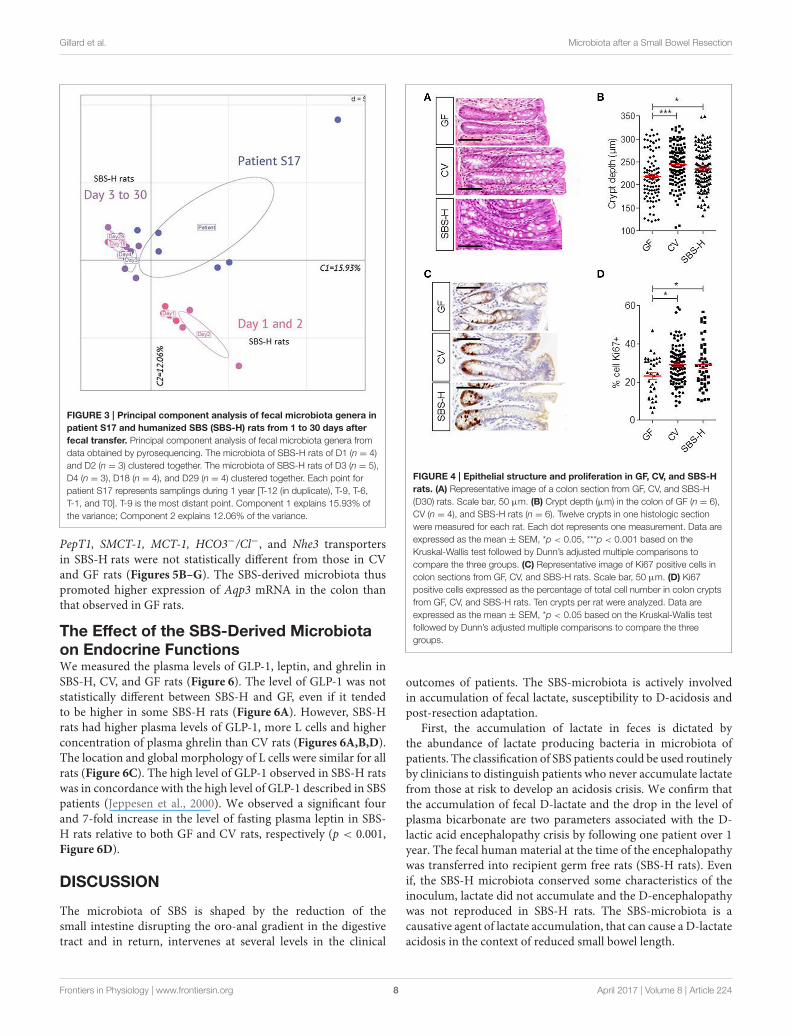
The Figures 2, 3 illustrate the progressive adaptation ofmicrobiota (sampling in S17 patient) after the transfer intorecipient germ free rats. During the 2 first days following thefecal transfer (D1 and D2), the microbiota is adapting to itsnew environment (from a resected gut in human to an intactgut in recipient rats). Thus, the microbiota ecosystem changedprofoundly during the 2 days following its arrival into a newdigestive environment, reflecting an adaptive phase, whereasthe composition of the SBS-H microbiota seemed to stabilizefrom D3 to D30 (Figure 3). All measured parameters (fecallactates, SCFA levels, microbiota composition, pH-value) did notchange between D3 and D30 (data not shown). The Figure 3
also indicates that the microbiota, from D3 to D30 days wascloser to the inoculum than was the microbiota during the2 first days after transfer. The main phylum represented inthe HI was Firmicutes (74%), followed by Bacteroidetes (15%),Actinobacteria (7%), and Proteobacteria (3%) and at D30 after thefecal transfer, the main represented phylum remained Firmicutes
Frontiers in Physiology | www.frontiersin.org 5 April 2017 | Volume 8 | Article 224

Gillard et al. Microbiota after a Small Bowel Resection
FIGURE 1 | Fecal fermentative activity and microbial composition of 16 SBS patients clustered in NLA and LA sub-groups and patient S17. Sixteen
patients, individually described in Mayeur et al. (2013), were clustered depending on the presence (LA) or absence (NLA) of lactates in their feces. One supplementary
patient (S17) was followed during 1 year because it was known to be at risk for D-lactic acidosis. Samplings of S17 were performed 12 (T-12m), 9 (T-9m), 6 (T-6m),
and 1 (T-1m) month before the acidosis crisis (AC at T0). (A) Fecal pH and the level of fecal D and L lactates (mmol/L), (B) the amount of fecal short chain fatty acids:
acetate, propionate, butyrate (mmol/L), (C) fecal bacteria per gram, (D) fecal microbiota diversity (Shannon index), (E) distribution of principal bacterial phyla, (F)
relative abundance of bacterial families in the feces. Lactate-producing bacteria are indicated in red and orange. Lactate-consuming bacteria are indicated by the
different shades of blue.
Frontiers in Physiology | www.frontiersin.org 6 April 2017 | Volume 8 | Article 224

Gillard et al. Microbiota after a Small Bowel Resection
FIGURE 2 | Fecal fermentative activity and microbiota composition of SBS-humanized rats from 1 to 30 days after fecal transfer. Feces of S17 patient
was recovered at the time of acidosis and used as inoculum (HI) for a fecal transfer in recipient germ free rats (SBS-H). The microbiota was analysis in HI and in fecal
samples of SBS-H rats 1 (D1), 2 (D2), and 30 (D30) days after fecal transfer. (A) Fecal pH and concentration of fecal D and L lactates (mmol/L) in HI and SBS-H rats
(n = 6). (B) Amount of acetate, propionate, and butyrate (mmol/L) in the HI and in the feces of SBS-H rats. (C) Principal bacterial phyla in the HI and the feces of
SBS-H rats. (D) Relative abundance of bacterial genera in the HI and the feces of SBS-H rats 1 (D1), 2 (D2), and 30 (D30) days inoculation. Lactate-producing
bacteria are indicated in different shades of pink. Lactate-consuming bacteria are indicated by the different shades of green.
(71%), followed by the Proteobacteria (18%), Bacteroidetes(5%), and Actinobacteria (5%; Figure 2C). Thirty days aftertransfer, the SBS-H microbiota conserved characteristics of theSBS-microbiota, predominated by lactate-producing bacteria(mainly Lactobacillus and Bifidobacterium), a low level oflactate-consuming bacteria (Bacteroides and Veillonella), andundetectable C. leptum (Figure 2D).
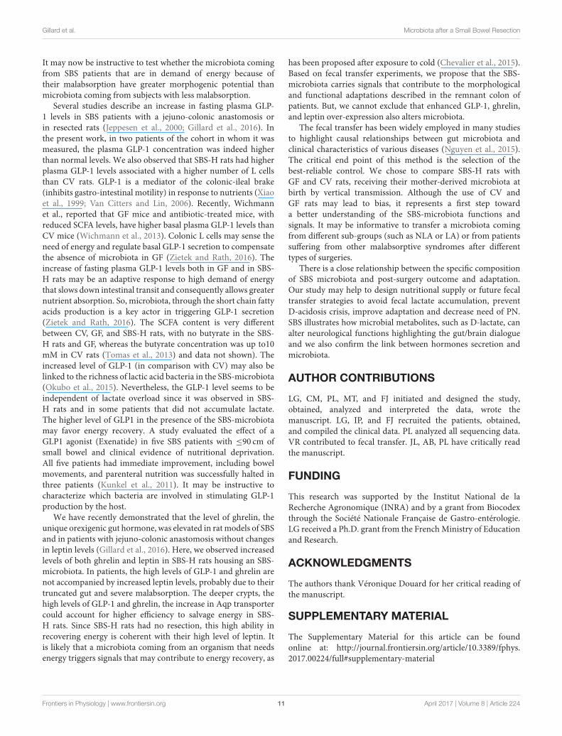
The Trophic Effect of the SBS-Microbiotaon the Colon Epithelium in SBS-H RatsSBS patients usually display deeper crypts than healthy controls(Joly et al., 2009). We thus compared the colonic crypt depth ofSBS-H rats (at D30) to conventional (CV) rats (Figures 4A,B).Histological analysis showed that there was no difference in thecrypt depths between CV (244 ± 32 µm) and SBS-H (235 ± 37µm) rats. Crypt depths were less in GF rats (217 ± 38 µm) than
in CV (P < 0.05) rats, as described previously (Cherbuy et al.,2010). There were a higher number of Ki67-positive cells percrypt both in CV and SBS-H rats than in GF rats (Figures 4C,D,p < 0.05). We thus observed a morphogenic effect triggered bytwo different microbial communities: the SBS-microbiota andthe CV microbiota.
Effect of the SBS-Derived Microbiota onColonic Transporter ExpressionWe explored how SBS-microbiota may change the absorptivecapacities of the colon by examining the expression of differentnutrient and solute transporters that play a key role in nutrientand water salvage. There tended to be a higher level of mRNAencoding Aqp3, an aquaporin water channel, in the colon ofSBS-H rats than CV rats and it was statistically higher (p <
0.05) in GF rats (Figure 5A). The mRNA levels of SGLT-1,
Frontiers in Physiology | www.frontiersin.org 7 April 2017 | Volume 8 | Article 224
Gillard et al. Microbiota after a Small Bowel Resection
FIGURE 3 | Principal component analysis of fecal microbiota genera in
patient S17 and humanized SBS (SBS-H) rats from 1 to 30 days after
fecal transfer. Principal component analysis of fecal microbiota genera from
data obtained by pyrosequencing. The microbiota of SBS-H rats of D1 (n = 4)
and D2 (n = 3) clustered together. The microbiota of SBS-H rats of D3 (n = 5),
D4 (n = 3), D18 (n = 4), and D29 (n = 4) clustered together. Each point for
patient S17 represents samplings during 1 year [T-12 (in duplicate), T-9, T-6,
T-1, and T0]. T-9 is the most distant point. Component 1 explains 15.93% of
the variance; Component 2 explains 12.06% of the variance.
PepT1, SMCT-1, MCT-1, HCO3−/Cl−, and Nhe3 transportersin SBS-H rats were not statistically different from those in CVand GF rats (Figures 5B–G). The SBS-derived microbiota thuspromoted higher expression of Aqp3 mRNA in the colon thanthat observed in GF rats.
The Effect of the SBS-Derived Microbiotaon Endocrine FunctionsWe measured the plasma levels of GLP-1, leptin, and ghrelin inSBS-H, CV, and GF rats (Figure 6). The level of GLP-1 was notstatistically different between SBS-H and GF, even if it tendedto be higher in some SBS-H rats (Figure 6A). However, SBS-Hrats had higher plasma levels of GLP-1, more L cells and higherconcentration of plasma ghrelin than CV rats (Figures 6A,B,D).The location and global morphology of L cells were similar for allrats (Figure 6C). The high level of GLP-1 observed in SBS-H ratswas in concordance with the high level of GLP-1 described in SBSpatients (Jeppesen et al., 2000). We observed a significant fourand 7-fold increase in the level of fasting plasma leptin in SBS-H rats relative to both GF and CV rats, respectively (p < 0.001,Figure 6D).
DISCUSSION
The microbiota of SBS is shaped by the reduction of thesmall intestine disrupting the oro-anal gradient in the digestivetract and in return, intervenes at several levels in the clinical
FIGURE 4 | Epithelial structure and proliferation in GF, CV, and SBS-H
rats. (A) Representative image of a colon section from GF, CV, and SBS-H
(D30) rats. Scale bar, 50 µm. (B) Crypt depth (µm) in the colon of GF (n = 6),
CV (n = 4), and SBS-H rats (n = 6). Twelve crypts in one histologic section
were measured for each rat. Each dot represents one measurement. Data are
expressed as the mean ± SEM, *p < 0.05, ***p < 0.001 based on the
Kruskal-Wallis test followed by Dunn’s adjusted multiple comparisons to
compare the three groups. (C) Representative image of Ki67 positive cells in
colon sections from GF, CV, and SBS-H rats. Scale bar, 50 µm. (D) Ki67
positive cells expressed as the percentage of total cell number in colon crypts
from GF, CV, and SBS-H rats. Ten crypts per rat were analyzed. Data are
expressed as the mean ± SEM, *p < 0.05 based on the Kruskal-Wallis test
followed by Dunn’s adjusted multiple comparisons to compare the three
groups.
outcomes of patients. The SBS-microbiota is actively involvedin accumulation of fecal lactate, susceptibility to D-acidosis andpost-resection adaptation.
First, the accumulation of lactate in feces is dictated bythe abundance of lactate producing bacteria in microbiota ofpatients. The classification of SBS patients could be used routinelyby clinicians to distinguish patients who never accumulate lactatefrom those at risk to develop an acidosis crisis. We confirm thatthe accumulation of fecal D-lactate and the drop in the level ofplasma bicarbonate are two parameters associated with the D-lactic acid encephalopathy crisis by following one patient over 1year. The fecal human material at the time of the encephalopathywas transferred into recipient germ free rats (SBS-H rats). Evenif, the SBS-H microbiota conserved some characteristics of theinoculum, lactate did not accumulate and the D-encephalopathywas not reproduced in SBS-H rats. The SBS-microbiota is acausative agent of lactate accumulation, that can cause a D-lactateacidosis in the context of reduced small bowel length.
Frontiers in Physiology | www.frontiersin.org 8 April 2017 | Volume 8 | Article 224

Gillard et al. Microbiota after a Small Bowel Resection
FIGURE 5 | Transporter expression in colon mucosa of GF, CV, and SBS-H rats. mRNA levels of Aqp3, Sglt-1, PepT-1, SMCT-1, MCT-1, HCO−3 /Cl−, and Nhe3
normalized to the L19 housekeeping gene in colon mucosa from GF (n = 4), CV (n = 4), and SBS-H rats (n = 4) (A–G). Data are expressed as the mean ± SEM,
*P < 0.05 based on the Kruskal-Wallis test followed by Dunn’s adjusted multiple comparisons to compare the three groups.
Second, the SBS-microbiota seems also to be a reservoirof multiple and complex signals that could modify the post-resection adaptation. Our study describes that fecal transfer ofSBS-associated microbiota into recipient germ-free rats triggersmodifications of colon by crypt deepening but does not reducethe level of GLP-1 and ghrelin of GF. Thus, SBS-H and GFrats have higher amount of these hormones than rats housing aconventional microbiota. The SBS-microbiota may favor energyrecovery since its transfer into GF is accompanied by high levelof plasma leptin.
Our cohort of 16 SBS patients is representative of SBS withjejuno-colonic anastomosis associated with severe malabsorptionand hyperphagia (Mayeur et al., 2013). Patients in whom lactateaccumulated (LA), known to be at risk of developing anacidosis crisis, had a gut microbiota enriched in lactate-producing bacteria (Lactobacillaceae, Bifidobacteriaceae,
and Enterobacteriaceae) with a lower proportion of lactate-consuming bacteria (Veillonellaceae, Bacteroidaceae). Thus,the classification of two groups (LA and NLA) reflects therelative abundance of lactate-producing and lactate-consumingbacteria. The specific composition and fermentative activityof the microbiota from SBS patients result from the luminalconstraints due to the extended resection of the small intestine.
The fecal transfer illustrates the adaptability of a specificecosystem in its environment. The inoculum coming from atruncated intestine was transferred into a complete digestivetract. The inoculum adapted progressively to its new gutenvironment in recipient GF rats during the first 2 daysand then stabilized 3 days after fecal transfer. On day 30,the composition of the microbiota established in the gutof SBS-H rats was relatively similar to the human fecalmicrobiota used as inoculum, as has been reported in previous
Frontiers in Physiology | www.frontiersin.org 9 April 2017 | Volume 8 | Article 224

Gillard et al. Microbiota after a Small Bowel Resection
FIGURE 6 | GLP-1, leptin and ghrelin in GF, CV and SBS-H. (A) Fasting plasma levels of GLP-1 in GF (n = 4), CV (n = 5), and SBS-H (n = 6) rats. Plasma GLP-1
levels of all CV rats and 2 SBS-H rats were below the limit of detection. (B) Number of GLP-1 positive cells per mm2 in GF, CV, and SBS-H rats (one section per rat).
(C) Representative image of GLP-1 immunostaining (brown cytoplasm), of colon mucosa sections from GF, CV, and SBS-H rats. Scale bar, 50 µm. (D) Fasting plasma
levels of leptin (pg/mL) in GF (n = 4), CV (n = 5), and SBS-H rats (n = 6). (E) Fasting plasma levels of ghrelin (pg/mL) in GF (n = 3), CV (n = 4), and SBS-H rats (n = 5).
Data are expressed as the mean ± SEM, *p < 0.05, **p < 0.01, ***p < 0.001 based on the Kruskal-Wallis test followed.
studies (Chung et al., 2012; Crouzet et al., 2013). Three maincharacteristics of the SBS-derived microbiota were conserved:the dominance of Lactobacillus and the absence of C. leptumand butyrate. We did not observe an accumulation of lactate inthe feces of SBS-H rats (even after supplementing the drinkingwater with lactose), suggesting that the intact small bowel of therecipient rats protected them from lactate accumulation. Thus,the reduction of the small intestine appears to be required for theaccumulation of lactate but is not sufficient, as fecal lactate is onlyobserved in certain sub-groups of patients.
Intestinal failure includes the malabsorption of vitamins,trace elements, electrolytes, water, and energy. The absorptivefunction of the colon mucosa increases over time in SBS patientswith colon in continuity, despite residual malabsorption, thusdecreasing their need for PN (Nordgaard et al., 1994, 1996).Patients with preserved colonic function gain more energy,especially by fermenting carbohydrates (Nordgaard et al., 1996).Several published studies suggest that the expression of sometransporters may exhibit adaptive flexibility and over expressionin SBS. However, there is no consensus and it is not establishedwhether, which and where precisely in the gut transporters areboosted after resection (Mayeur et al., 2016). The increase in
the level of Aqp3 has been described in a pre-clinical modelin resected rats (Tsujikawa et al., 2003) as we observed inSBS-H rats. It could be speculate that the increased Aqp3may help to save water and exchange metabolites in SBS-Hrats; the mechanisms underlying this over-expression are notknown. We have previously described both in Humans and inpreclinical models a gut epithelium restructuring consequentlyto the resection. With the fecal transfer, we wanted to know if theSBS-microbiota sent specific signals leading to deeper absorptivesurface. The SBS-derived microbiota after fecal transfer in SBS-H rats, triggers crypt deepening and increases proliferation inthe colon but this was also observed in CV rats housing adifferent microbiota. This result is concordant with our previousresults showing that different types of microbe communities(either a microbial population rich in primo-colonizing bacteriaor a population representative of adult microbiota) have similarmorphogenic effect on colon epithelium (Tomas et al., 2013).The trophic effect was not recapitulated when GF rats weremono-colonized with one single commensal, suggesting thatdiversity of the inoculum is required to send signals that shapethe epithelial mucosa (Rul et al., 2011; Turpin et al., 2013;Wrzosek et al., 2013; Tomas et al., 2015; Hoffmann et al., 2016).
Frontiers in Physiology | www.frontiersin.org 10 April 2017 | Volume 8 | Article 224
Gillard et al. Microbiota after a Small Bowel Resection
It may now be instructive to test whether the microbiota comingfrom SBS patients that are in demand of energy because oftheir malabsorption have greater morphogenic potential thanmicrobiota coming from subjects with less malabsorption.
Several studies describe an increase in fasting plasma GLP-1 levels in SBS patients with a jejuno-colonic anastomosis orin resected rats (Jeppesen et al., 2000; Gillard et al., 2016). Inthe present work, in two patients of the cohort in whom it wasmeasured, the plasma GLP-1 concentration was indeed higherthan normal levels. We also observed that SBS-H rats had higherplasma GLP-1 levels associated with a higher number of L cellsthan CV rats. GLP-1 is a mediator of the colonic-ileal brake(inhibits gastro-intestinal motility) in response to nutrients (Xiaoet al., 1999; Van Citters and Lin, 2006). Recently, Wichmannet al., reported that GF mice and antibiotic-treated mice, withreduced SCFA levels, have higher basal plasma GLP-1 levels thanCV mice (Wichmann et al., 2013). Colonic L cells may sense theneed of energy and regulate basal GLP-1 secretion to compensatethe absence of microbiota in GF (Zietek and Rath, 2016). Theincrease of fasting plasma GLP-1 levels both in GF and in SBS-H rats may be an adaptive response to high demand of energythat slows down intestinal transit and consequently allows greaternutrient absorption. So, microbiota, through the short chain fattyacids production is a key actor in triggering GLP-1 secretion(Zietek and Rath, 2016). The SCFA content is very differentbetween CV, GF, and SBS-H rats, with no butyrate in the SBS-H rats and GF, whereas the butyrate concentration was up to10mM in CV rats (Tomas et al., 2013) and data not shown). Theincreased level of GLP-1 (in comparison with CV) may also belinked to the richness of lactic acid bacteria in the SBS-microbiota(Okubo et al., 2015). Nevertheless, the GLP-1 level seems to beindependent of lactate overload since it was observed in SBS-H rats and in some patients that did not accumulate lactate.The higher level of GLP1 in the presence of the SBS-microbiotamay favor energy recovery. A study evaluated the effect of aGLP1 agonist (Exenatide) in five SBS patients with ≤90 cm ofsmall bowel and clinical evidence of nutritional deprivation.All five patients had immediate improvement, including bowelmovements, and parenteral nutrition was successfully halted inthree patients (Kunkel et al., 2011). It may be instructive tocharacterize which bacteria are involved in stimulating GLP-1production by the host.
We have recently demonstrated that the level of ghrelin, theunique orexigenic gut hormone, was elevated in ratmodels of SBSand in patients with jejuno-colonic anastomosis without changesin leptin levels (Gillard et al., 2016). Here, we observed increasedlevels of both ghrelin and leptin in SBS-H rats housing an SBS-microbiota. In patients, the high levels of GLP-1 and ghrelin arenot accompanied by increased leptin levels, probably due to theirtruncated gut and severe malabsorption. The deeper crypts, thehigh levels of GLP-1 and ghrelin, the increase in Aqp transportercould account for higher efficiency to salvage energy in SBS-H rats. Since SBS-H rats had no resection, this high ability inrecovering energy is coherent with their high level of leptin. Itis likely that a microbiota coming from an organism that needsenergy triggers signals that may contribute to energy recovery, as
has been proposed after exposure to cold (Chevalier et al., 2015).Based on fecal transfer experiments, we propose that the SBS-microbiota carries signals that contribute to the morphologicaland functional adaptations described in the remnant colon ofpatients. But, we cannot exclude that enhanced GLP-1, ghrelin,and leptin over-expression also alters microbiota.
The fecal transfer has been widely employed in many studiesto highlight causal relationships between gut microbiota andclinical characteristics of various diseases (Nguyen et al., 2015).The critical end point of this method is the selection of thebest-reliable control. We chose to compare SBS-H rats withGF and CV rats, receiving their mother-derived microbiota atbirth by vertical transmission. Although the use of CV andGF rats may lead to bias, it represents a first step towarda better understanding of the SBS-microbiota functions andsignals. It may be informative to transfer a microbiota comingfrom different sub-groups (such as NLA or LA) or from patientssuffering from other malabsorptive syndromes after differenttypes of surgeries.
There is a close relationship between the specific compositionof SBS microbiota and post-surgery outcome and adaptation.Our study may help to design nutritional supply or future fecaltransfer strategies to avoid fecal lactate accumulation, preventD-acidosis crisis, improve adaptation and decrease need of PN.SBS illustrates how microbial metabolites, such as D-lactate, canalter neurological functions highlighting the gut/brain dialogueand we also confirm the link between hormones secretion andmicrobiota.
AUTHOR CONTRIBUTIONS
LG, CM, PL, MT, and FJ initiated and designed the study,obtained, analyzed and interpreted the data, wrote themanuscript. LG, IP, and FJ recruited the patients, obtained,and compiled the clinical data. PL analyzed all sequencing data.VR contributed to fecal transfer. JL, AB, PL have critically readthe manuscript.
FUNDING
This research was supported by the Institut National de laRecherche Agronomique (INRA) and by a grant from Biocodexthrough the Société Nationale Française de Gastro-entérologie.LG received a Ph.D. grant from the FrenchMinistry of Educationand Research.
ACKNOWLEDGMENTS
The authors thank Véronique Douard for her critical reading ofthe manuscript.
SUPPLEMENTARY MATERIAL
The Supplementary Material for this article can be foundonline at: http://journal.frontiersin.org/article/10.3389/fphys.2017.00224/full#supplementary-material
Frontiers in Physiology | www.frontiersin.org 11 April 2017 | Volume 8 | Article 224

Gillard et al. Microbiota after a Small Bowel Resection
REFERENCES
Briet, F., Flourie, B., Achour, L., Maurel, M., Rambaud, J. C., and
Messing, B. (1995). Bacterial adaptation in patients with short
bowel and colon in continuity. Gastroenterology 109, 1446–1453.
doi: 10.1016/0016-5085(95)90629-0
Caporaso, J. G., Kuczynski, J., Stombaugh, J., Bittinger, K., Bushman,
F. D., Costello, E. K., et al. (2010). QIIME allows analysis of high-
throughput community sequencing data. Nat. Methods 7, 335–336.
doi: 10.1038/nmeth.f.303
Cherbuy, C., Honvo-Houeto, E., Bruneau, A., Bridonneau, C., Mayeur, C.,
Duee, P. H., et al. (2010). Microbiota matures colonic epithelium through a
coordinated induction of cell cycle-related proteins in gnotobiotic rat. Am.
J. Physiol. Gastrointest. Liver Physiol. 299, G348–G357. doi: 10.1152/ajpgi.003
84.2009
Chevalier, C., Stojanovic, O., Colin, D. J., Suarez-Zamorano, N., Tarallo, V., Veyrat-
Durebex, C., et al. (2015). Gut microbiota orchestrates energy homeostasis
during cold. Cell 163, 1360–1374. doi: 10.1016/j.cell.2015.11.004
Chung, H., Pamp, S. J., Hill, J. A., Surana, N. K., Edelman, S. M., Troy, E. B., et al.
(2012). Gut immune maturation depends on colonization with a host-specific
microbiota. Cell 149, 1578–1593. doi: 10.1016/j.cell.2012.04.037
Crenn, P., Morin, M. C., Joly, F., Penven, S., Thuillier, F., and Messing, B.
(2004). Net digestive absorption and adaptive hyperphagia in adult short bowel
patients. Gut 53, 1279–1286. doi: 10.1136/gut.2003.030601
Crouzet, L., Gaultier, E., Del’Homme, C., Cartier, C., Delmas, E., Dapoigny, M.,
et al. (2013). The hypersensitivity to colonic distension of IBS patients can be
transferred to rats through their fecal microbiota.Neurogastroenterol. Motil. 25,
e272–e282. doi: 10.1111/nmo.12103
Deschemin, J. C., Noordine, M. L., Remot, A., Willemetz, A., Afif, C., Canonne-
Hergaux, F., et al. (2016). The microbiota shifts the iron sensing of intestinal
cells. FASEB J. 30, 252–261. doi: 10.1096/fj.15-276840
Drucker, D. J., Erlich, P., Asa, S. L., and Brubaker, P. L. (1996). Induction of
intestinal epithelial proliferation by glucagon-like peptide 2. Proc. Natl. Acad.
Sci. U.S.A. 93, 7911–7916. doi: 10.1073/pnas.93.15.7911
Gillard, L., Billiauws, L., Stan-Iuga, B., Ribeiro-Parenti, L., Jarry, A. C., Cavin, J.
B., et al. (2016). Enhanced ghrelin levels and hypothalamic orexigenic AgRP
and NPY neuropeptide expression in models of jejuno-colonic short bowel
syndrome. Sci. Rep. 6:28345. doi: 10.1038/srep28345
Hoffmann, T. W., Pham, H. P., Bridonneau, C., Aubry, C., Lamas, B., Martin-
Gallausiaux, C., et al. (2016). Microorganisms linked to inflammatory bowel
disease-associated dysbiosis differentially impact host physiology in gnotobiotic
mice. ISME J. 10, 460–477. doi: 10.1038/ismej.2015.127
Jeppesen, P. B. (2012). Teduglutide, a novel glucagon-like peptide 2 analog, in the
treatment of patients with short bowel syndrome. Therap. Adv. Gastroenterol.
5, 159–171. doi: 10.1177/1756283X11436318
Jeppesen, P. B. (2014). Spectrum of short bowel syndrome in adults: intestinal
insufficiency to intestinal failure. JPEN J. Parenter. Enteral. Nutr. 38, 8S–13S.
doi: 10.1177/0148607114520994
Jeppesen, P. B., Hartmann, B., Thulesen, J., Graff, J., Lohmann, J., Hansen, B.
S., et al. (2001). Glucagon-like peptide 2 improves nutrient absorption and
nutritional status in short-bowel patients with no colon. Gastroenterology 120,
806–815. doi: 10.1053/gast.2001.22555
Jeppesen, P. B., Hartmann, B., Thulesen, J., Hansen, B. S., Holst, J. J., Poulsen, S.
S., et al. (2000). Elevated plasma glucagon-like peptide 1 and 2 concentrations
in ileum resected short bowel patients with a preserved colon. Gut 47, 370–376.
doi: 10.1136/gut.47.3.370
Joly, F., Mayeur, C., Bruneau, A., Noordine, M. L., Meylheuc, T., Langella,
P., et al. (2010). Drastic changes in fecal and mucosa-associated microbiota
in adult patients with short bowel syndrome. Biochimie 92, 753–761.
doi: 10.1016/j.biochi.2010.02.015
Joly, F., Mayeur, C., Messing, B., Lavergne-Slove, A., Cazals-Hatem, D.,
Noordine, M. L., et al. (2009). Morphological adaptation with preserved
proliferation/transporter content in the colon of patients with short bowel
syndrome. Am. J. Physiol. Gastrointest. Liver Physiol. 297, G116–G123.
doi: 10.1152/ajpgi.90657.2008
Kaneko, T., Bando, Y., Kurihara, H., Satomi, K., Nonoyama, K., and Matsuura,
N. (1997). Fecal microflora in a patient with short-bowel syndrome and
identification of dominant lactobacilli. J. Clin. Microbiol. 35, 3181–3185.
Kowlgi, N. G., and Chhabra, L. (2015). D-lactic acidosis: an underrecognized
complication of short bowel syndrome. Gastroenterol. Res. Pract. 2015:476215.
doi: 10.1155/2015/476215
Kunkel, D., Basseri, B., Low, K., Lezcano, S., Soffer, E. E., Conklin, J. L.,
et al. (2011). Efficacy of the glucagon-like peptide-1 agonist exenatide in the
treatment of short bowel syndrome. Neurogastroenterol. Motil. 23, e739–e328.
doi: 10.1111/j.1365-2982.2011.01723.x
Lan, A., Bruneau, A., Bensaada, M., Philippe, C., Bellaud, P., Rabot, S.,
et al. (2008). Increased induction of apoptosis by Propionibacterium
freudenreichii TL133 in colonic mucosal crypts of human microbiota-
associated rats treated with 1,2-dimethylhydrazine. Br. J. Nutr. 100, 1251–1259.
doi: 10.1017/S0007114508978284
Le Roy, T., Llopis, M., Lepage, P., Bruneau, A., Rabot, S., Bevilacqua, C., et al.
(2013). Intestinal microbiota determines development of non-alcoholic fatty
liver disease in mice. Gut 62, 1787–1794. doi: 10.1136/gutjnl-2012-303816
Li, W., and Godzik, A. (2006). Cd-hit: a fast program for clustering and comparing
large sets of protein or nucleotide sequences. Bioinformatics 22, 1658–1659.
doi: 10.1093/bioinformatics/btl158
Madsen, K. B., Askov-Hansen, C., Naimi, R. M., Brandt, C. F., Hartmann, B., Holst,
J. J., et al. (2013). Acute effects of continuous infusions of glucagon-like peptide
(GLP)-1, GLP-2 and the combination (GLP-1+GLP-2) on intestinal absorption
in short bowel syndrome (SBS) patients. A placebo-controlled study. Regul.
Pept. 184, 30–39. doi: 10.1016/j.regpep.2013.03.025
Mayeur, C., Gillard, L., Le beyec, J., Bado, A., Joly, F., and Thomas,
M. (2016). Extensive intestinal resection triggers behavioral adaptation,
intestinal remodeling and microbiota transition in short bowel syndrome.
Microorganisms 4:16. doi: 10.3390/microorganisms4010016
Mayeur, C., Gratadoux, J. J., Bridonneau, C., Chegdani, F., Larroque, B., Kapel,
N., et al. (2013). Faecal D/L lactate ratio is a metabolic signature of microbiota
imbalance in patients with short bowel syndrome. PLoS ONE 8:e54335.
doi: 10.1371/journal.pone.0054335
McNeil, N. I. (1984). The contribution of the large intestine to energy supplies in
man. Am. J. Clin. Nutr. 39, 338–342.
Messing, B., Pigot, F., Rongier, M., Morin, M. C., Ndeindoum, U., and
Rambaud, J. C. (1991). Intestinal absorption of free oral hyperalimentation
in the very short bowel syndrome. Gastroenterology 100, 1502–1508.
doi: 10.1016/0016-5085(91)90645-2
Narula, R. K., El Shafei, A., Ramaiah, D., and Schmitz, P. G. (2000). D-
lactic acidosis 23 years after jejuno-ileal bypass. Am. J. Kidney Dis. 36:E9.
doi: 10.1053/ajkd.2000.9005
Nguyen, T. L., Vieira-Silva, S., Liston, A., and Raes, J. (2015). How informative
is the mouse for human gut microbiota research? Dis. Model. Mech. 8, 1–16.
doi: 10.1242/dmm.017400
Nordgaard, I., Hansen, B. S., and Mortensen, P. B. (1994). Colon as
a digestive organ in patients with short bowel. Lancet 343, 373–376.
doi: 10.1016/S0140-6736(94)91220-3
Nordgaard, I., Hansen, B. S., and Mortensen, P. B. (1996). Importance of colonic
support for energy absorption as small-bowel failure proceeds. Am. J. Clin.
Nutr. 64, 222–231.
Okubo, H., Nakatsu, Y., Sakoda, H., Kushiyama, A., Fujishiro, M., Fukushima,
T., et al. (2015). Mosapride citrate improves nonalcoholic steatohepatitis with
increased fecal lactic acid bacteria and plasma glucagon-like peptide-1 level in
a rodent model. Am. J. Physiol. Gastrointest. Liver Physiol. 308, G151–G158.
doi: 10.1152/ajpgi.00198.2014
Pironi, L., Arends, J., Baxter, J., Bozzetti, F., Pelaez, R. B., Cuerda, C., et al. (2015).
ESPEN endorsed recommendations. Definition and classification of intestinal
failure in adults. Clin. Nutr. 34, 171–180. doi: 10.1016/j.clnu.2014.08.017
Rul, F., Ben-Yahia, L., Chegdani, F., Wrzosek, L., Thomas, S., Noordine, M. L.,
et al. (2011). Impact of the metabolic activity of Streptococcus thermophilus
on the colon epithelium of gnotobiotic rats. J. Biol. Chem. 286, 10288–10296.
doi: 10.1074/jbc.M110.168666
Tomas, J., Reygner, J., Mayeur, C., Ducroc, R., Bouet, S., Bridonneau, C., et al.
(2015). Early colonizing Escherichia coli elicits remodeling of rat colonic
epithelium shifting toward a new homeostatic state. ISME J. 9, 46–58.
doi: 10.1038/ismej.2014.111
Tomas, J., Wrzosek, L., Bouznad, N., Bouet, S., Mayeur, C., Noordine, M. L.,
et al. (2013). Primocolonization is associated with colonic epithelial maturation
during conventionalization. FASEB J. 27, 645–655. doi: 10.1096/fj.12-216861
Frontiers in Physiology | www.frontiersin.org 12 April 2017 | Volume 8 | Article 224

Gillard et al. Microbiota after a Small Bowel Resection
Tsujikawa, T., Itoh, A., Fukunaga, T., Satoh, J., Yasuoka, T., and Fujiyama, Y.
(2003). Alteration of aquaporin mRNA expression after small bowel resection
in the rat residual ileum and colon. J. Gastroenterol. Hepatol. 18, 803–808.
doi: 10.1046/j.1440-1746.2003.03033.x
Turpin, W., Humblot, C., Noordine, M. L., Wrzosek, L., Tomas, J., Mayeur, C.,
et al. (2013). Behavior of lactobacilli isolated from fermented slurry (ben-
saalga) in gnotobiotic rats. PLoS ONE 8:e57711. doi: 10.1371/journal.pone.
0057711
Van Citters, G. W., and Lin, H. C. (2006). Ileal brake: neuropeptidergic
control of intestinal transit. Curr. Gastroenterol. Rep. 8, 367–373.
doi: 10.1007/s11894-006-0021-9
Verbeke, K. A., Boobis, A. R., Chiodini, A., Edwards, C. A., Franck, A.,
Kleerebezem, M., et al. (2015). Towards microbial fermentation metabolites
as markers for health benefits of prebiotics. Nutr. Res. Rev. 28, 42–66.
doi: 10.1017/S0954422415000037
Wichmann, A., Allahyar, A., Greiner, T. U., Plovier, H., Lunden, G. O.,
Larsson, T., et al. (2013). Microbial modulation of energy availability
in the colon regulates intestinal transit. Cell Host Microbe 14, 582–590.
doi: 10.1016/j.chom.2013.09.012
Wrzosek, L., Miquel, S., Noordine, M. L., Bouet, S., Joncquel Chevalier-Curt, M.,
Robert, V., et al. (2013). Bacteroides thetaiotaomicron and Faecalibacterium
prausnitzii influence the production of mucus glycans and the development of
goblet cells in the colonic epithelium of a gnotobiotic model rodent. BMC Biol.
11:61. doi: 10.1186/1741-7007-11-61
Xiao, Q., Boushey, R. P., Drucker, D. J., and Brubaker, P. L. (1999).
Secretion of the intestinotropic hormone glucagon-like peptide 2 is
differentially regulated by nutrients in humans. Gastroenterology 117, 99–105.
doi: 10.1016/S0016-5085(99)70555-X
Zietek, T., and Rath, E. (2016). Inflammation meets metabolic disease: gut feeling
mediated by GLP-1. Front. Immunol. 7:154. doi: 10.3389/fimmu.2016.00154
Conflict of Interest Statement: The authors declare that the research was
conducted in the absence of any commercial or financial relationships that could
be construed as a potential conflict of interest.
Copyright © 2017 Gillard, Mayeur, Robert, Pingenot, Le Beyec, Bado, Lepage,
Thomas and Joly. This is an open-access article distributed under the terms of
the Creative Commons Attribution License (CC BY). The use, distribution or
reproduction in other forums is permitted, provided the original author(s) or licensor
are credited and that the original publication in this journal is cited, in accordance
with accepted academic practice. No use, distribution or reproduction is permitted
which does not comply with these terms.
Frontiers in Physiology | www.frontiersin.org 13 April 2017 | Volume 8 | Article 224
View publication statsView publication stats