Embed Size (px)
Citation preview
ORIGINAL RESEARCHpublished: 14 December 2020
doi: 10.3389/fmicb.2020.543260
Frontiers in Microbiology | www.frontiersin.org 1 December 2020 | Volume 11 | Article 543260
Edited by:
Cassandre Sara Lazar,
Université du Québec à
Montréal, Canada
Reviewed by:
William D. Orsi,
Ludwig Maximilian University of
Munich, Germany
Andreas Teske,
University of North Carolina at Chapel
Hill, United States
*Correspondence:
Kai Mangelsdorf
†These authors share first authorship
Specialty section:
This article was submitted to
Extreme Microbiology,
a section of the journal
Frontiers in Microbiology
Received: 16 March 2020
Accepted: 20 November 2020
Published: 14 December 2020
Citation:
Liu Q, Adler K, Lipus D, Kämpf H,
Bussert R, Plessen B, Schulz H-M,
Krauze P, Horn F, Wagner D,
Mangelsdorf K and Alawi M (2020)
Microbial Signatures in Deep
CO2-Saturated Miocene Sediments of
the Active Hartoušov Mofette System
(NW Czech Republic).
Front. Microbiol. 11:543260.
doi: 10.3389/fmicb.2020.543260
Microbial Signatures in DeepCO2-Saturated Miocene Sedimentsof the Active Hartoušov MofetteSystem (NW Czech Republic)Qi Liu 1,2†, Karsten Adler 2,3†, Daniel Lipus 1, Horst Kämpf 3, Robert Bussert 4, Birgit Plessen 5,
Hans-Martin Schulz 3, Patryk Krauze 1,2, Fabian Horn 1, Dirk Wagner 1,2, Kai Mangelsdorf 3*
and Mashal Alawi 1
1 Section Geomicrobiology, GFZ German Research Centre for Geosciences, Potsdam, Germany, 2 Institute of Geosciences,
University of Potsdam, Potsdam, Germany, 3 Section Organic Geochemistry, GFZ German Research Centre for Geosciences,
Potsdam, Germany, 4 Section Applied Geochemistry, Institute of Applied Geosciences, Technische Universität Berlin, Berlin,
Germany, 5 Section Climate Dynamics and Landscape Evolution, GFZ German Research Centre for Geosciences, Potsdam,
Germany
The Hartoušov mofette system is a natural CO2 degassing site in the central Cheb
Basin (Eger Rift, Central Europe). In early 2016 a 108m deep core was obtained from
this system to investigate the impact of ascending mantle-derived CO2 on indigenous
deep microbial communities and their surrounding life habitat. During drilling, a CO2 blow
out occurred at a depth of 78.5 meter below surface (mbs) suggesting a CO2 reservoir
associated with a deep low-permeable CO2-saturated saline aquifer at the transition
from Early Miocene terrestrial to lacustrine sediments. Past microbial communities were
investigated by hopanoids and glycerol dialkyl glycerol tetraethers (GDGTs) reflecting
the environmental conditions during the time of deposition rather than showing a signal
of the current deep biosphere. The composition and distribution of the deep microbial
community potentially stimulated by the upward migration of CO2 starting during Mid
Pleistocene time was investigated by intact polar lipids (IPLs), quantitative polymerase
chain reaction (qPCR), and deoxyribonucleic acid (DNA) analysis. The deep biosphere
is characterized by microorganisms that are linked to the distribution and migration of
the ascending CO2-saturated groundwater and the availability of organic matter instead
of being linked to single lithological units of the investigated rock profile. Our findings
revealed high relative abundances of common soil and water bacteria, in particular
the facultative, anaerobic and potential iron-oxidizing Acidovorax and other members
of the family Comamonadaceae across the whole recovered core. The results also
highlighted the frequent detection of the putative sulfate-oxidizing and CO2-fixating
genus Sulfuricurvum at certain depths. A set of new IPLs are suggested to be indicative
for microorganisms associated to CO2 accumulation in the mofette system.
Keywords: geo-bio interaction, CO2, mofette systems, Eger Rift, microbial lipid biomarker, microbial diversity,
deep biosphere, saline groundwater
Liu et al. Microbial Signatures of Deep Mofette-System
INTRODUCTION
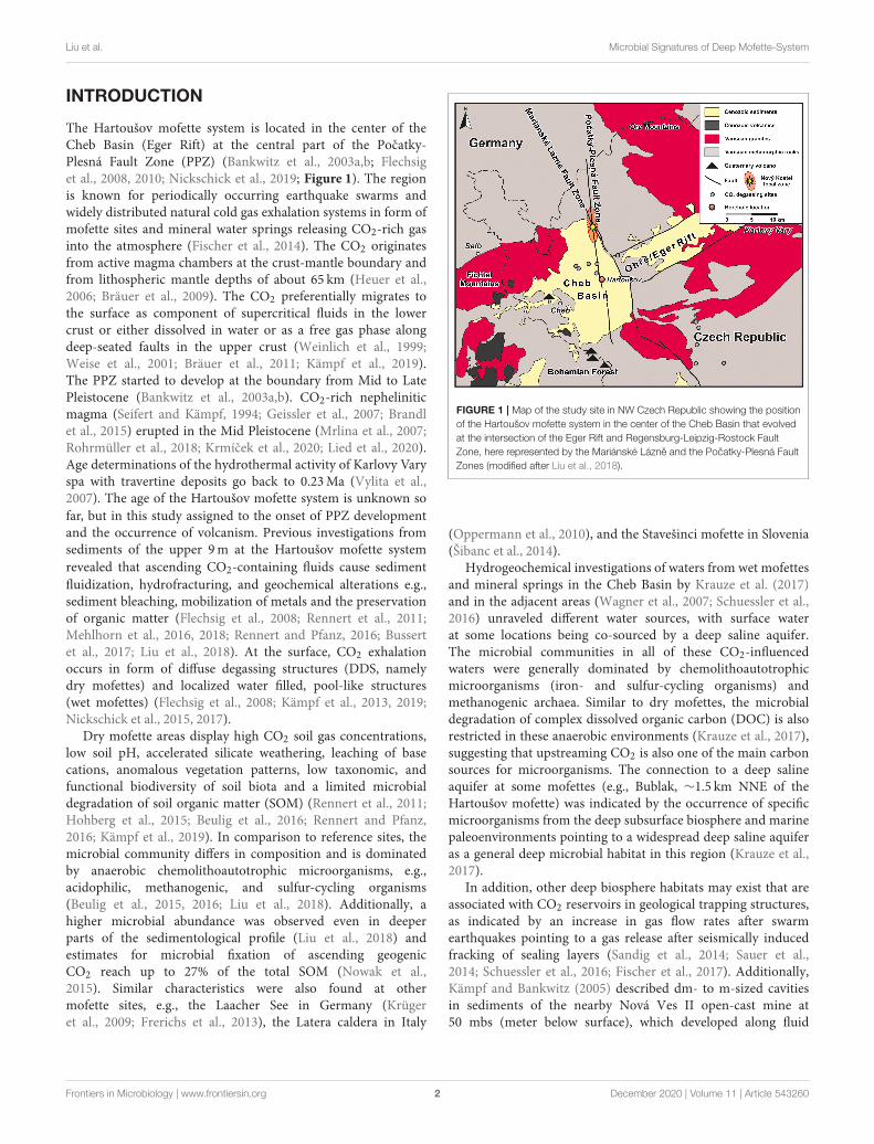
The Hartoušov mofette system is located in the center of theCheb Basin (Eger Rift) at the central part of the Pocatky-Plesná Fault Zone (PPZ) (Bankwitz et al., 2003a,b; Flechsiget al., 2008, 2010; Nickschick et al., 2019; Figure 1). The regionis known for periodically occurring earthquake swarms andwidely distributed natural cold gas exhalation systems in form ofmofette sites and mineral water springs releasing CO2-rich gasinto the atmosphere (Fischer et al., 2014). The CO2 originatesfrom active magma chambers at the crust-mantle boundary andfrom lithospheric mantle depths of about 65 km (Heuer et al.,2006; Bräuer et al., 2009). The CO2 preferentially migrates tothe surface as component of supercritical fluids in the lowercrust or either dissolved in water or as a free gas phase alongdeep-seated faults in the upper crust (Weinlich et al., 1999;Weise et al., 2001; Bräuer et al., 2011; Kämpf et al., 2019).The PPZ started to develop at the boundary from Mid to LatePleistocene (Bankwitz et al., 2003a,b). CO2-rich nepheliniticmagma (Seifert and Kämpf, 1994; Geissler et al., 2007; Brandlet al., 2015) erupted in the Mid Pleistocene (Mrlina et al., 2007;Rohrmüller et al., 2018; Krmícek et al., 2020; Lied et al., 2020).Age determinations of the hydrothermal activity of Karlovy Varyspa with travertine deposits go back to 0.23Ma (Vylita et al.,2007). The age of the Hartoušov mofette system is unknown sofar, but in this study assigned to the onset of PPZ developmentand the occurrence of volcanism. Previous investigations fromsediments of the upper 9m at the Hartoušov mofette systemrevealed that ascending CO2-containing fluids cause sedimentfluidization, hydrofracturing, and geochemical alterations e.g.,sediment bleaching, mobilization of metals and the preservationof organic matter (Flechsig et al., 2008; Rennert et al., 2011;Mehlhorn et al., 2016, 2018; Rennert and Pfanz, 2016; Bussertet al., 2017; Liu et al., 2018). At the surface, CO2 exhalationoccurs in form of diffuse degassing structures (DDS, namelydry mofettes) and localized water filled, pool-like structures(wet mofettes) (Flechsig et al., 2008; Kämpf et al., 2013, 2019;Nickschick et al., 2015, 2017).
Dry mofette areas display high CO2 soil gas concentrations,low soil pH, accelerated silicate weathering, leaching of basecations, anomalous vegetation patterns, low taxonomic, andfunctional biodiversity of soil biota and a limited microbialdegradation of soil organic matter (SOM) (Rennert et al., 2011;Hohberg et al., 2015; Beulig et al., 2016; Rennert and Pfanz,2016; Kämpf et al., 2019). In comparison to reference sites, themicrobial community differs in composition and is dominatedby anaerobic chemolithoautotrophic microorganisms, e.g.,acidophilic, methanogenic, and sulfur-cycling organisms(Beulig et al., 2015, 2016; Liu et al., 2018). Additionally, ahigher microbial abundance was observed even in deeperparts of the sedimentological profile (Liu et al., 2018) andestimates for microbial fixation of ascending geogenicCO2 reach up to 27% of the total SOM (Nowak et al.,2015). Similar characteristics were also found at othermofette sites, e.g., the Laacher See in Germany (Krügeret al., 2009; Frerichs et al., 2013), the Latera caldera in Italy
FIGURE 1 | Map of the study site in NW Czech Republic showing the position
of the Hartoušov mofette system in the center of the Cheb Basin that evolved
at the intersection of the Eger Rift and Regensburg-Leipzig-Rostock Fault
Zone, here represented by the Mariánské Lázne and the Pocatky-Plesná Fault
Zones (modified after Liu et al., 2018).
(Oppermann et al., 2010), and the Stavešinci mofette in Slovenia(Šibanc et al., 2014).
Hydrogeochemical investigations of waters from wet mofettesand mineral springs in the Cheb Basin by Krauze et al. (2017)and in the adjacent areas (Wagner et al., 2007; Schuessler et al.,2016) unraveled different water sources, with surface waterat some locations being co-sourced by a deep saline aquifer.The microbial communities in all of these CO2-influencedwaters were generally dominated by chemolithoautotrophicmicroorganisms (iron- and sulfur-cycling organisms) andmethanogenic archaea. Similar to dry mofettes, the microbialdegradation of complex dissolved organic carbon (DOC) is alsorestricted in these anaerobic environments (Krauze et al., 2017),suggesting that upstreaming CO2 is also one of the main carbonsources for microorganisms. The connection to a deep salineaquifer at some mofettes (e.g., Bublak, ∼1.5 km NNE of theHartoušov mofette) was indicated by the occurrence of specificmicroorganisms from the deep subsurface biosphere and marinepaleoenvironments pointing to a widespread deep saline aquiferas a general deep microbial habitat in this region (Krauze et al.,2017).
In addition, other deep biosphere habitats may exist that areassociated with CO2 reservoirs in geological trapping structures,as indicated by an increase in gas flow rates after swarmearthquakes pointing to a gas release after seismically inducedfracking of sealing layers (Sandig et al., 2014; Sauer et al.,2014; Schuessler et al., 2016; Fischer et al., 2017). Additionally,Kämpf and Bankwitz (2005) described dm- to m-sized cavitiesin sediments of the nearby Nová Ves II open-cast mine at50 mbs (meter below surface), which developed along fluid
Frontiers in Microbiology | www.frontiersin.org 2 December 2020 | Volume 11 | Article 543260
Liu et al. Microbial Signatures of Deep Mofette-System
migration pathways. This suggests the presence of restricted gas-filled cavities, which may function as distinct habitats for thedeep biosphere. An important indication for a CO2-related deepbiosphere was recognized by Bräuer et al. (2005) after a swarmearthquake activity in 2000. They detected an increase inmethaneconcentrations at the Wettin spring (Bad Brambach, Germany)about 20 km north of the Hartoušov mofette system, where asignificant decrease of δ13Cmethane was attributed to microbialmethane production from magmatic CO2 and pre- or co-seismically released hydrogen from the granitic basement. After aswarm earthquake event in 2011, higher methane concentrationswere also detected at the Bublak mofette (Bräuer et al., 2018).
These previous investigations show that ascending geogenicCO2-containing fluids locally alter the sedimentary overburdenand thus change the environmental conditions for microbiallife. Additionally, there is evidence of subsurface structuresthat may host CO2-influenced deep microbial habitats, whichcould function as deep microbial hotspots. However, studiesinvestigating the potential for CO2-related deep microbial lifein the Cheb Basin and the Eger Rift are still missing. Thus,in early 2016 the German Research Centre for Geosciences(GFZ) drilled a 108.5m deep borehole as a test case for theInternational Continental Scientific Drilling Program (ICDP)project “Drilling the Eger Rift” (Dahm et al., 2013). Theborehole was positioned in the Hartoušov mofette system (HJB-1) (50◦07′58′′N, 12◦27′46′′E) and described in detail by Bussertet al. (2017). During drilling, CO2-rich sediments were recoveredbetween 71 and 81 mbs. At a depth of 78.5 mbs a CO2 blowout occurred, suggesting the presence of a subsurface CO2
accumulation. This CO2 reservoir is associated to a basal low-permeable CO2-saturated and saline aquifer (1,892mg L−1 offree dissolved CO2) that occurs between 79 and 85 mbs at thetransition from Early Miocene terrestrial to overlying lacustrinesediments. Hydrogeochemically, the aquifer is characterized bya Na-Ca-HCO3-SO4-type water with a high Fe content ofup to 13.7mg L−1 and a pH of 6.4 (Bussert et al., 2017).Due to the potential of the CO2-saturated aquifer to host avery specialized microbial community we focussed on the coreinterval between 65 and 95 mbs. Our aim was to identify theimpact of mantle-derived CO2 on deep microbial communitiesand to find out whether the low-permeable CO2-saturated andsaline aquifer might act as a hotspot for present deep microbiallife. The methodological approach to characterize the microbialcommunity included lipid biomarker analysis of past andliving microbial biomass (hopanoids, GDGTs and intact polarlipids) as well as DNA analysis such as quantitative PolymeraseChain Reaction (qPCR) and Illumina 16S rRNA gene ampliconsequencing. Furthermore, the microbial signals were comparedto lithological background information and sedimentologicalbulk parameters.
METHODS
Drilling, Coring, and Pump TestA detailed description of the field work including drilling,coring and a pump test was published by Bussert et al.(2017). The drilling was performed with a Drillmec G-25
device installed on a Tatra 815 drilling lorry which discoveredcore material in PVC liners with a length of 3m and adiameter of 0.1m. The drilling mud consisted of homogeneouslyblended pure bentonite. In order to monitor potential drill mudcontamination of the retrieved core material, sodium fluoresceinwas added to the drill mud with a concentration of 5mg L−1
(Supplementary Figure 1D) according to Pellizzari et al. (2013).Subsamples for further analysis were taken about every 0.5m andstored in gasbags flushed with nitrogen at −80◦C directly aftercore recovery in the field. After the drilling campaign a 24 h pumptest within the deep low-permeable CO2-saturated saline aquiferwas performed. The groundwater was filtered, the obtained watersamples geochemically analyzed and the obtained filters stored at−20◦C, respectively.
Sample Processing and ContaminationControlThe initial lithological description of the sample material andthe drill mud contamination control were performed in thelab. The frozen core segments were stored over night at 5◦Cto initiate thawing of the external sample layer and to avoidfluid migration from the rim to the center of the samples. Thethawed rim (∼1 cm) was removed (inner coring), the still frozeninner core described (e.g., Supplementary Figure 1), materialfrom the removed rim (outer rim) tested in triplicates forfluorescein (Pellizzari et al., 2013) and the samples again storedat −80◦C. To ensure that the samples are not contaminatedby external DNA the inner coring technique was repeated in aclean bench (Thermo Scientific, Waltham, USA). The removedmaterial and the outside of the inner core were again testedin triplicates for fluorescein (inner rim). Inner core samples(sample) exceeding the background fluorescence were excludedfrom further analysis (Supplementary Figure 1). The fluoresceinconcentration was measured with a CLARIO star R© platereader (BMG LABTECH GmbH, Ortenberg, Germany). Thebackground fluorescence signal was obtained from samples of ashallow drilling campaign (3m) drilled in 2015 adjacent to ourstudy side without the application of drill mud and fluorescein(Liu et al., 2018).
Bulk Carbon and Nitrogen AnalysesTotal carbon (TC), total organic carbon (TOC), total nitrogen(TN), and the bulk δ13Corg were all analyzed with the sameequipment consisting of a NC2500 Carlo Erba elemental analysercoupled with a ConFlo_III interface on a DELTAplusXL isotoperatio mass spectrometer (IRMS) (Thermo Fischer Scientific).Prior to analysis the sample material was freeze-dried, powderedand homogenized. In order to determine the TC and TN∼25mgof sample material was loaded into tin capsules and the contentwas calibrated against acetanilide. For investigation of TOCand bulk δ13Corg the carbonate content was removed using insitu decalcification. Therefore, depending on the TOC content,3–10mg sample material were loaded into Ag-capsules anddecalcified by drops of 3% HCl followed by 20% HCl and heatedfor 3 h at 75◦C. The calibration was performed using elementalurea and certified isotope standards (USGS24, IAEA-CH-7)and proofed with an internal soil reference sample (Boden3,
Frontiers in Microbiology | www.frontiersin.org 3 December 2020 | Volume 11 | Article 543260
Liu et al. Microbial Signatures of Deep Mofette-System
HEKATECH). All isotope compositions are given relative to theVPDB (Vienna Pee Dee Belemnite) standard in the conventionaldelta notation. The total inorganic carbon (TIC) was calculatedby subtraction of TOC from TC.
Lipid Biomarker Extraction andChromatographic Column SeparationThe freeze-dried, powdered and homogenized sediment samples(about 80 g) were extracted with a modified extraction methodafter Bligh and Dyer (1959) using methanol:dichloromethane(DCM):ammonium acetate buffer (pH 7.5) (2:1:0.8) as initialextraction solvent mixture. The sample material was admixedwith the extraction solvent (4x sample mass in mL, ∼320mL),stirred with a flow-blending rod for 5min and afterwardscentrifuged for 10min with 2,500 rpm. The supernatantwas transferred to a separation funnel and the remainingsample 2 times re-extracted in an ultrasonic bath for 10min,followed by centrifugation and transfer of the supernatantinto the separation funnel. To achieve phase separation, thesolvent ratio in the separation funnel was changed to 1:1:0.9(methanol:DCM:ammonium acetate buffer). Afterwards theorganic phase containing the lipid extract was collected ina turbovap glas and the solvent removed (TurboVap 500).Each fifth sample was a blank. After extraction 5α-Androstaneand deuterium-labeled phosphatidylcholine (PCd54 = 1,2-dimyristoyl-d54-sn-glycero-3-phosphocholine) were added asstandards for compound quantification in the aliphatic and intactpolar lipid fractions, respectively. The obtained total extractswere chromatographically separated into a low polar lipid (20mLchloroform), free fatty acid (50mL methyl formiate with 0.025%glacial acetic acid), glycolipid (20mL acetone), and intact polarlipid (IPLs, 25mL methanol) fraction using two glass syringecolumns filled with dried pure silica (1 g silica gel 63–200µm,dried at 110◦C for 2 h) and Florisil (1 g magnesium silica gel 150–250µm) with the silica column on top of the Florisil column.The IPL fraction was only eluted from the silica column (Zinkand Mangelsdorf, 2004). To improve IPL recovery the silicacolumn was eluted with 25mL methanol:water (60:40) for asecond time. Phase separation was conducted as described above.Finally the IPL fractions were combined and the solvent removed.Afterwards, the IPL fraction was split into two halves: one for thedirect detection of IPLs and one for the detection of polar lipidfatty acids (PLFAs) after saponification (Müller et al., 1993).
After removal of asphaltenes the low polar lipid fraction wasfurther subdivided by Medium Pressure Liquid Chromatography(MPLC) into an aliphatic, aromatic, and Nitrogen-Sulfur-Oxygen-containing compound (NSO) fraction (Radke et al.,1980). The aliphatic fraction was analyzed for hopanoidsand the NSO fraction for glycerol dialkyl glycerol tetraethers(GDGTs). GDGTs have been quantified with regard to an externalarchaeol standard.
Determination of the Lipid BiomarkersAnalysis of IPLs was performed on a Thermo Scientific Ultimate3000 RSUltra high performance liquid chromatograph (UHPLC)coupled to a Q Exactive Plus Orbitrap mass spectrometer(MS) with a heated electrospray (H-ESI II) probe. Samples
were separated with a LiChrospher 100 diol column (2 ×
125mm, 5µm; CS-Chromatographie Service) equipped with apre-column filter. The eluents used for compound separationwere (A) n-hexane:isopropanol:formic acid:ammonia (25% inwater) 79:20:1.2:0.04 v/v and (B) isopropanol:water:formicacid:ammonia (25% in water) 88:10:1.2:0.04 v/v (solventgradients: 1min 100% A, linear increase of B to 65% within20 and 40min for reconditioning). The flow rate was set to0.35 mL/min (modified after Rütters et al., 2001). ESI sourceconditions were as follows: spray voltage −2.2 kV; capillarytemperature 300◦C; nitrogen sheath gas at 49 and auxiliary gasat 12 arbitrary units at a temperature of 419◦C, S-Lens 65V. Theobtained data were acquired in negative and positive ion modewith dependent MS/MS acquisition at ranges of m/z 400–2,000.The full scan and fragment spectra were collected at a resolutionof 280,000 and 70,000 (at m/z 200), respectively.
The aliphatic fraction and PLFAs were determined on aThermo Trace GC Ultra equipped with a Thermo PTV injectionsystem and a SGE BPX5 fused silica capillary column (50mlength, 0.22mm ID, 0.25µmfilm thickness) coupled to a ThermoTrace DSQ Quadrupole MS. Helium was used as carrier gas. Thetemperature of the GC oven was programmed from 50◦C (hold1min) to 310◦C at a rate of 3◦Cmin−1, followed by an isothermalphase of 30min. The injector temperature was programmed from50 to 300◦C at a rate of 10◦C s−1. The MS was operated inelectron impact ionization mode (EI) at 70 eV. Full scan massspectra for compound identification were recorded from m/z 50to 600 at a scan rate of 1.5 scans s−1.
GDGT analysis was conducted on a Shimadzu LC10ADHPLCinstrument coupled to a Finnigan Triple StageQuadrupole (TSQ)7000MS with an atmospheric pressure chemical ionization(APCI) interface. Samples were separated at 30◦C with a PrevailCyano column (2.1 × 150mm, 3µm; Alltech) equipped with apre-column filter. The mobile phase consisted of (A) n-hexaneand (B) isopropanol and compound separation was achievedusing the following solvent gradients: 5min 99% A and 1% B,linear gradient to 1.8% B within 40min, increase to 10% B within1min and holding time for 5min to clean the column, back toinitial solvent conditions within 1min and 16min for columnequilibration (Schouten et al., 2007). The flow rate was set to200 µL min−1. The APCI adjustments were: corona current5 µA giving a voltage of around 5 kV, vaporizer temperature350◦C, capillary temperature 200◦C and nitrogen sheath gas at60 psi (no auxiliary gas). Mass spectra were generated by selectedion monitoring in the positive ion mode for the masses 1295.0,1302.1, 1049.5, 1035.5, 1021.5, and 654.2 each with a width of 7amu (to also obtain neighboring masses) representing major coreGDGTs at a scan rate of 0.33 s.
Compound specific δ13C values of the aliphatic fraction(hopanoids) were determined with a GC-isotope ratiomonitoring (IR)-MS system consisting of an Agilent 7890GC (USA) connected with an open split GC-C/TCIII-Interfacefor compound-specific carbon and hydrogen isotope analysisto a Delta V Plus IRMS (Thermo Fischer Scientific, Germany).The GC-separated organic substances were oxidized to CO2 in acombustion furnace at a temperature of 940◦C on a CuO/Ni/Ptcatalyst. CO2 was transferred to the mass spectrometer to
Frontiers in Microbiology | www.frontiersin.org 4 December 2020 | Volume 11 | Article 543260
Liu et al. Microbial Signatures of Deep Mofette-System
determine carbon isotope ratios. Three microliter of thealiphatic fraction were injected with a split ratio of 1:2 and aninitial temperature of 230◦C to a programmable temperaturevaporization inlet (PTV, Agilent Technology, USA). The injectorwas heated to 300◦C with a heating rate of 12◦C s−1. Theseparation of the aliphatic fractions was attained by a fused silicacapillary column (HP Ultra 1, 50m × 0.2mm ID, 0.33µm FT,Agilent Technology, Germany) with a temperature programstarting from 40 to 300◦C, with a heating rate of 4◦C min−1
and the maximum temperature held for 45min. The carriergas was Helium with a flow rate of 1.0mL min−1. All sampleswere measured in triplicates with a usual standard deviation of≤0.5‰. The quality of the results was checked by measuringn-alkane standards (n-C15, n-C20, and n-C25) with knownisotopic composition (Campro Scientific, Germany). Isotopiccompositions are given in the delta notation relative to theVienna Pee Dee Belemnite (VPDB) standard.
DNA Extraction and PurificationDue to the extremely low amount of biomass in the coresamples, 10 g of powdered sample material was used to extractthe total genomic DNA with the DNeasy R© PowerMax R© Soil Kit(QIAGEN, Venlo, Netherlands). Afterwards, the obtained DNAwas dissolved in 5mL DNA-free water (Carl Roth, Karlsruhe,Germany). For each sampling depth, three independent sampleswere taken from different positions of the core horizon astechnical triplicate. The 5mL DNA solution was concentratedto 100 µL by an Eppendorf Concentrator Plus (EppendorfAG, Hamburg, Germany). The Genomic DNA Clean &ConcentratorTM-10 (Zymo Research, Irvine, CA) was utilized toremove humic acids and other substances that may inhibit thepolymerase chain reaction (PCR). Two DNA extractions weredone from separated sample duplicates. DNA from 1mL DNA-free water (Carl Roth, Karlsruhe, Germany) was extracted as anegative control using the same DNA extraction approach.
In addition to the core material, ∼1 L of the fluid samplesfrom the pump test were filtered (0.2µm) to collect insolubleparticles. The total genomic DNA trapped on the filters wasextracted by the FastDNATM SPIN Kit for Soil and the FastPrep R©
Instrument (MP Biomedicals, Santa Ana, CA) with standardprotocols. The FastPrep R© Instrument homogenizing time andthe homogenizing speed were modified to 30 s and 5.5m s−1
according to Liu et al. (2018).
Quantitative PCRTotal microbial abundance was estimated by determiningthe number of bacterial 16S rRNA gene copies usingquantitative polymerase chain reaction (qPCR) targetingthe V3 region of the 16S gene with the primer pairs341F (5′-CCTACGGGAGGCAGCAG−3′) and 534R (5′-ATTACCGCGGCTGCTGG-3′) (Degelmann et al., 2010). TheqPCR Master Mix consisted of 10 µL SYBR R© FAST qPCRMaster Mix (2X) Universal (KAPA Biosystems, Wilmington,Massachusetts, USA), 5.92 µL PCR water, 0.04 µL forwardprimer (100µM), 0.04 µL reverse primer (100µM), and 4 µLtemplate. The qPCR was programmed as 3min at 95◦C, 40 cyclesof 3 s at 95◦C, 20 s at 60◦C, 30 s at 72◦C, and 3 s at 80◦C for the
plate read. A cloned 16S rRNA gene fragment from Escherichiacoli was used as standard. The qPCR was conducted on a CFX96real-time thermal cycler (Bio-Rad Laboratories Inc., USA) andthe analysis of the quantification data was performed with theCFX ManagerTM software (Bio-Rad Laboratories Inc., USA). Theconcentration range of the standard was optimized and set from103 to 107 16S rRNA gene copies. The R2-value of the standardcurve line was 0.994–0.997.
Illumina MiSeq Amplicon SequencingThe 16S rRNA gene was amplified with OptiTaqTM polymerase(Roboklon, Berlin, Germany) which has a proofreading capabilitydue to the extremely low concentration of extracted total genomicDNA. The PCR reaction solution consisted of 2.5 µL 10x BufferPol C, 0.125 µL OptiTaqTM polymerase, 1 µL dNTP Mix (5mMeach), 1µLMgCl2 (25mM), 17.075µL PCR water, 0.3µL bovineserum albumin, 0.25µL forward primer (20µM), 0.25µL reverseprimer (20µM) and 2.5 µL template. Unique combinationsof barcode-tagged 515F (5′-GTGCCAGCMGCCGCGGTAA-3′)and 806R (5′-GGACTACHVGGGTWTCTAAT-3′) (Caporasoet al., 2011) primers were assigned to each sample. PCRamplifications were performed in volumes of 25 µL ona T100TM thermal cycler (Bio-Rad Laboratories Inc., USA)under the following conditions: 5min at 95◦C, 35 cyclesof 30 s at 95◦C, 45 s at 56◦C, 60 s at 72◦C, and a finalextension step of 7min at 72◦C. A cloned 16S rRNA genefragment from E. coli was used as positive control. Non-template controls were included with each PCR run. The PCRproducts were cleaned up with AMPure XP magnetic beads(Beckman Coulter GmbH, Krefeld, Germany). After measuringthe DNA concentration with a CLARIO star R© plate reader (BMGLABTECH GmbH, Ortenberg, Germany) PCR products werepooled in equimolar amounts. The pooled DNA solution wasconcentrated with Eppendorf Concentrator plus (Eppendorf AG,Hamburg, Germany) to meet the requirement of the IlluminaMiSeq high-throughput sequencing. The final pooled DNAconcentration was 77.05 ng µL−1.
Bioinformatics and Statistical AnalysisSequencing was performed by Eurofins Scientific SE(Luxembourg) on an Illumina MiSeq (2 × 250 bp). Dual-indexed reads were demultiplexed using CutAdapt (Martin,2011) allowing for 10% errors in the primer and no errors inthe barcodes. Individual samples were processed according tothe DADA2 pipeline (Callahan et al., 2016). This includes aninitial sequence truncation (250 bp forward reads; 200 bp reversereads). The quality-filtered reads were used to generate an errormodel that was applied for dereplication, sample inference,and merging of the paired-end reads. All final sequences hada standardized read-orientation and a minimum length of200 bp. The sequence table was created and potential chimerawere filtered using a de novo approach. The resulting ampliconsequence variants (ASVs) were imported into the QIIME2framework (Bolyen et al., 2019) which facilitated the SILVAtaxonomy database (v132) (Quast et al., 2013) and VSEARCH(Rognes et al., 2016) to assign taxonomic units. Singletons andOTUs assigned to chloroplasts and mitochondria were removed
Frontiers in Microbiology | www.frontiersin.org 5 December 2020 | Volume 11 | Article 543260
Liu et al. Microbial Signatures of Deep Mofette-System
from the obtained OTU table. After the filtering processes,11,063,679 sequences were obtained in the 16S rRNA genelibrary in total. The read numbers ranged between 12,999 and243,092 with a mean value of 99,985. The resulting ASV tablewas manually scanned for potential contaminant taxa based onthe negative control, resulting in the removal of ASVs belongingto the taxa Escherichia, Undibacterium, Methylophilaceae,Comamonadaceae, Ralstonia, and Novosphingobium. Due tothe low biomass environment and the high susceptibility ofcontamination introduction via the Power Soil Max kit (Sheiket al., 2018), comparison of drill core and pump test datawas used as an additional screening approach and resultedin the removal of additional contamination ASVs (includingLawsonella and Staphylococcus). The final ASV table was rarifiedto a sequencing depth of 5,541 sequences (lowest availablesequencing count) for alpha diversity estimation. Microbialdiversity in each sample was assessed by calculating ShannonH and Shannon EH indices using the phyloseq package in R(McMurdie and Holmes, 2013). Beta diversity was determinedby the non-metric multidimensional scaling (NMDS) using BrayCurtis distances with PAST3 (Hammer et al., 2001). Sequencingdata was deposited at the European Nucleotide Archive (http://www.ebi.ac.uk/ena) under the accession numbers PRJEB22478(ERS4382097 to ERS4382146 and ERS4382395 to ERS4382400).
Correlation and PCA of Microbial Generaand Lipid BiomarkersThe correlation and Principal Component Analysis (PCA)were performed using the software PAST3 (Hammer et al.,2001). Statistical examinations considered samples investigatedboth DNA and lipid biomarkers. More specifically, correlationsbetween TOC, TIC divided into TIC-Dolomite restricted tothe Cypris Fm. and TIC-Siderite restricted to the Main SeamFm., TN, the TOC/TN-ratio, all microbial genera with arelative abundance >5%, detected functional genera (<5%)related to the methane-, sulfur- and iron-cycles and all detectedlipid biomarkers were investigated. The resulting correlationcoefficients (r) are based on linear regression (Pearson) andstatistical significance was reported as a p-value. Correlationswith a p > 0.05 were considered to be non-significant. ThePCA was carried out for TOC, TIC-Dolomite, TIC-Siderite, TN,TOC/TN-ratio, all genera >5%, additional detected functionalgenera (<5%) and all predominating or relatively constantlydistributed lipid biomarkers. The parameters were normalized to1 with the PAST3 function “row normalized length” and the PCAcalculated on the correlation matrix. Compounds not detectedwere treated as zero.
RESULTS
Stratigraphy and Sample MaterialThe core section between 65 and 95 mbs lithologically consistedof three different units which were from the bottom to the top:(i) a weathered Paleozoic mica schist (95–91.5 mbs, Paleozoicbasement), (ii) sandy to peaty Early Miocene mudstones ofthe Main Seam Formation (Fm.) with lignite fragments androot structures suggesting paleosol horizons in the lower and
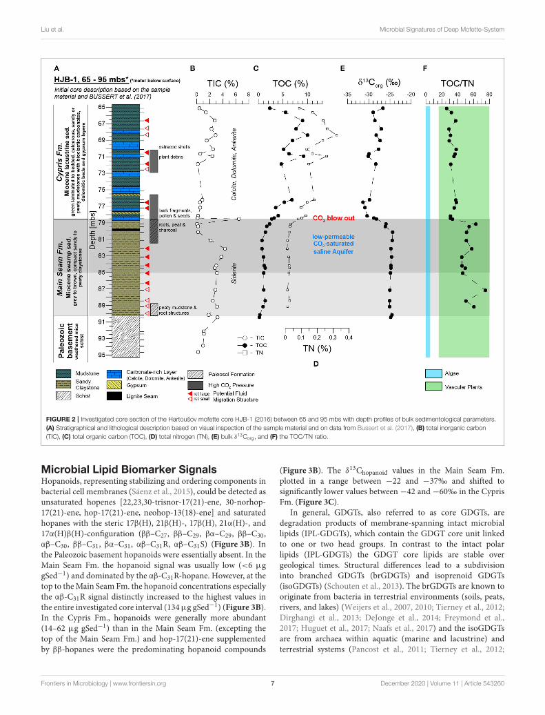
upper sections (91.5–78.5 mbs), and (iii) laminated, calcareous,sandy or peaty Early Miocene mudstones interbedded withbioclastic carbonates, dolomite to ankerite beds, and gypsumlayers of lacustrine origin (78.5–65 mbs) belonging to the CyprisFm. (Bussert et al., 2017; Figure 2A). Identified macrofossilsin the Cypris Fm. were bark fragments, seeds, plant debris,and ostracod shells (Cypris angusta Rss.) (Figure 2A andSupplementary Figure 1). Sediments from the Main Seam andCypris Fm. revealed vein-like structures indicating potentialCO2 ascending pathways with associated mineral alteration andprecipitation. These features were siderite-rich veins and bubblestructures in the Main Seam Fm. and small fractures, dykes andsills with sediment color changes in the Cypris Fm. The CO2 blowout during the drilling campaign occurred at the transition fromthe Cypris Fm. to the underlying Main Seam Fm. This indicatesthat at this transition a dolomite-rich layer (about 30 cm thick)that is widely distributed in the Cheb basin (Smejkal, 1984; Pešek,2014) or the lacustrine sediments themselves act as a sealing layerfor the low-permeable CO2-saturated saline aquifer in the upperMain Seam Fm., resulting in a zone characterized by high CO2
pore pressure (Figure 2A).
Bulk Carbon and NitrogenCarbonates were detected in all three lithological units and areexpressed in total inorganic carbon (TIC) (Figure 2B). In thePaleozoic basement and in the Main Seam Fm. the carbonateswere mainly represented by zoned siderite spheres and veinsthat could have been precipitated from the low-permeable CO2-saturated saline aquifer. At the transition from the Main SeamFm. to the Cypris Fm. carbonates were essentially absent, exceptof the thick (30 cm) dolomite-rich layer at 78.5 mbs. In theCypris Fm., calcite, dolomite, and ankerite predominated. Theiroccurrence together with evaporitic layers indicates a lacustrineorigin (Smejkal, 1984; Pešek, 2014). However, a possible earlydiagenetic alteration cannot be excluded.
Organic matter was not detected in the Paleozoic basement.The TOC contents of theMain Seam Fm. ranged between 0.2 and2.3%. After a small increase the TOC contents remained relativelyconstant at ca. one percent before increasing to 2.3% at the topof the Main Seam Fm. (Figure 2C). In the overlying lacustrineCypris Fm. the TOC contents were significantly higher and showstrong fluctuations between 2.3 and 10%. Bulk δ13Corg data alsochanged with the lithological transition from the Main Seam tothe Cypris Fm. showing relative constant values around −24‰in most parts of the Main Seam Fm. and a strong decrease downto −30‰ at the top (Figure 2E). In the Cypris Fm. the organiccarbon isotope signals fluctuate between−31 and−27‰.
The total nitrogen content (TN, Figure 2D) was mainlypositively correlated with the TOC content (r = 0.99)(Supplementary Table 1). Values were low in theMain Seam Fm.ranging between 0.01 and 0.04% and increase at the top. In theCypris Fm. TN values were significantly higher ranging between0.08 and 0.32%. The TOC/TN ratio ranged between 45 and 75in the Main Seam Fm. and between 26 and 41 in the Cypris Fm.(Figure 2F).
Frontiers in Microbiology | www.frontiersin.org 6 December 2020 | Volume 11 | Article 543260
Liu et al. Microbial Signatures of Deep Mofette-System
FIGURE 2 | Investigated core section of the Hartoušov mofette core HJB-1 (2016) between 65 and 95 mbs with depth profiles of bulk sedimentological parameters.
(A) Stratigraphical and lithological description based on visual inspection of the sample material and on data from Bussert et al. (2017), (B) total inorganic carbon
(TIC), (C) total organic carbon (TOC), (D) total nitrogen (TN), (E) bulk δ13Corg, and (F) the TOC/TN ratio.
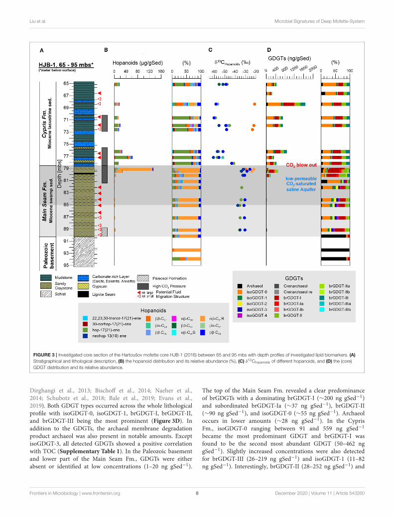
Microbial Lipid Biomarker SignalsHopanoids, representing stabilizing and ordering components inbacterial cell membranes (Sáenz et al., 2015), could be detected asunsaturated hopenes [22,23,30-trisnor-17(21)-ene, 30-norhop-17(21)-ene, hop-17(21)-ene, neohop-13(18)-ene] and saturatedhopanes with the steric 17β(H), 21β(H)-, 17β(H), 21α(H)-, and17α(H)β(H)-configuration (ββ–C27, ββ–C29, βα–C29, ββ–C30,αβ–C30, ββ–C31, βα–C31, αβ–C31R, αβ–C31S) (Figure 3B). Inthe Paleozoic basement hopanoids were essentially absent. In theMain Seam Fm. the hopanoid signal was usually low (<6 µggSed−1) and dominated by the αβ-C31R-hopane. However, at thetop to theMain Seam Fm. the hopanoid concentrations especiallythe αβ-C31R signal distinctly increased to the highest values inthe entire investigated core interval (134µg gSed−1) (Figure 3B).In the Cypris Fm., hopanoids were generally more abundant(14–62 µg gSed−1) than in the Main Seam Fm. (excepting thetop of the Main Seam Fm.) and hop-17(21)-ene supplementedby ββ-hopanes were the predominating hopanoid compounds
(Figure 3B). The δ13Chopanoid values in the Main Seam Fm.plotted in a range between −22 and −37‰ and shifted tosignificantly lower values between −42 and −60‰ in the CyprisFm. (Figure 3C).
In general, GDGTs, also referred to as core GDGTs, aredegradation products of membrane-spanning intact microbiallipids (IPL-GDGTs), which contain the GDGT core unit linkedto one or two head groups. In contrast to the intact polarlipids (IPL-GDGTs) the GDGT core lipids are stable overgeological times. Structural differences lead to a subdivisioninto branched GDGTs (brGDGTs) and isoprenoid GDGTs(isoGDGTs) (Schouten et al., 2013). The brGDGTs are known tooriginate from bacteria in terrestrial environments (soils, peats,rivers, and lakes) (Weijers et al., 2007, 2010; Tierney et al., 2012;Dirghangi et al., 2013; DeJonge et al., 2014; Freymond et al.,2017; Huguet et al., 2017; Naafs et al., 2017) and the isoGDGTsare from archaea within aquatic (marine and lacustrine) andterrestrial systems (Pancost et al., 2011; Tierney et al., 2012;
Frontiers in Microbiology | www.frontiersin.org 7 December 2020 | Volume 11 | Article 543260
Liu et al. Microbial Signatures of Deep Mofette-System
FIGURE 3 | Investigated core section of the Hartoušov mofette core HJB-1 (2016) between 65 and 95 mbs with depth profiles of investigated lipid biomarkers. (A)
Stratigraphical and lithological description, (B) the hopanoid distribution and its relative abundance (%), (C) δ13Chopanoids of different hopanoids, and (D) the (core)
GDGT distribution and its relative abundance.
Dirghangi et al., 2013; Bischoff et al., 2014; Naeher et al.,2014; Schubotz et al., 2018; Bale et al., 2019; Evans et al.,2019). Both GDGT types occurred across the whole lithologicalprofile with isoGDGT-0, isoGDGT-1, brGDGT-I, brGDGT-II,and brGDGT-III being the most prominent (Figure 3D). Inaddition to the GDGTs, the archaeal membrane degradationproduct archaeol was also present in notable amounts. ExceptisoGDGT-3, all detected GDGTs showed a positive correlationwith TOC (Supplementary Table 1). In the Paleozoic basementand lower part of the Main Seam Fm., GDGTs were eitherabsent or identified at low concentrations (1–20 ng gSed−1).
The top of the Main Seam Fm. revealed a clear predominanceof brGDGTs with a dominating brGDGT-I (∼200 ng gSed−1)and subordinated brGDGT-Ia (∼37 ng gSed−1), brGDGT-II(∼90 ng gSed−1), and isoGDGT-0 (∼55 ng gSed−1). Archaeoloccurs in lower amounts (∼28 ng gSed−1). In the CyprisFm., isoGDGT-0 ranging between 91 and 559 ng gSed−1
became the most predominant GDGT and brGDGT-I wasfound to be the second most abundant GDGT (50–462 nggSed−1). Slightly increased concentrations were also detectedfor brGDGT-III (26–219 ng gSed−1) and isoGDGT-1 (11–82ng gSed−1). Interestingly, brGDGT-II (28–252 ng gSed−1) and
Frontiers in Microbiology | www.frontiersin.org 8 December 2020 | Volume 11 | Article 543260
Liu et al. Microbial Signatures of Deep Mofette-System
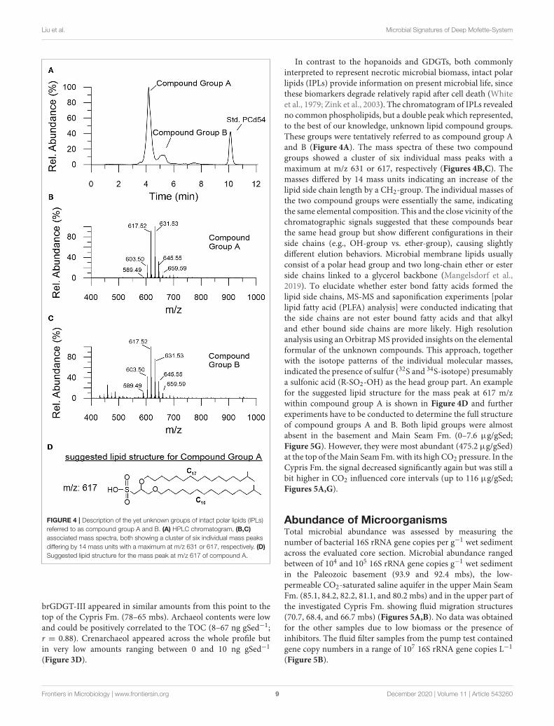
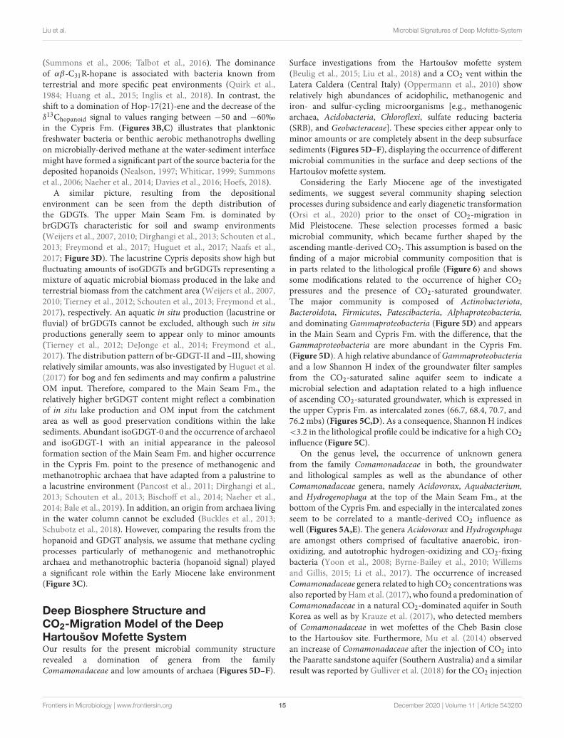
FIGURE 4 | Description of the yet unknown groups of intact polar lipids (IPLs)
referred to as compound group A and B. (A) HPLC chromatogram, (B,C)
associated mass spectra, both showing a cluster of six individual mass peaks
differing by 14 mass units with a maximum at m/z 631 or 617, respectively. (D)
Suggested lipid structure for the mass peak at m/z 617 of compound A.
brGDGT-III appeared in similar amounts from this point to thetop of the Cypris Fm. (78–65 mbs). Archaeol contents were lowand could be positively correlated to the TOC (8–67 ng gSed−1;r = 0.88). Crenarchaeol appeared across the whole profile butin very low amounts ranging between 0 and 10 ng gSed−1
(Figure 3D).
In contrast to the hopanoids and GDGTs, both commonlyinterpreted to represent necrotic microbial biomass, intact polarlipids (IPLs) provide information on present microbial life, sincethese biomarkers degrade relatively rapid after cell death (Whiteet al., 1979; Zink et al., 2003). The chromatogram of IPLs revealedno common phospholipids, but a double peak which represented,to the best of our knowledge, unknown lipid compound groups.These groups were tentatively referred to as compound group Aand B (Figure 4A). The mass spectra of these two compoundgroups showed a cluster of six individual mass peaks with amaximum at m/z 631 or 617, respectively (Figures 4B,C). Themasses differed by 14 mass units indicating an increase of thelipid side chain length by a CH2-group. The individual masses ofthe two compound groups were essentially the same, indicatingthe same elemental composition. This and the close vicinity of thechromatographic signals suggested that these compounds bearthe same head group but show different configurations in theirside chains (e.g., OH-group vs. ether-group), causing slightlydifferent elution behaviors. Microbial membrane lipids usuallyconsist of a polar head group and two long-chain ether or esterside chains linked to a glycerol backbone (Mangelsdorf et al.,2019). To elucidate whether ester bond fatty acids formed thelipid side chains, MS-MS and saponification experiments [polarlipid fatty acid (PLFA) analysis] were conducted indicating thatthe side chains are not ester bound fatty acids and that alkyland ether bound side chains are more likely. High resolutionanalysis using an OrbitrapMS provided insights on the elementalformular of the unknown compounds. This approach, togetherwith the isotope patterns of the individual molecular masses,indicated the presence of sulfur (32S and 34S-isotope) presumablya sulfonic acid (R-SO2-OH) as the head group part. An examplefor the suggested lipid structure for the mass peak at 617 m/zwithin compound group A is shown in Figure 4D and furtherexperiments have to be conducted to determine the full structureof compound groups A and B. Both lipid groups were almostabsent in the basement and Main Seam Fm. (0–7.6 µg/gSed;Figure 5G). However, they were most abundant (475.2 µg/gSed)at the top of theMain Seam Fm. with its high CO2 pressure. In theCypris Fm. the signal decreased significantly again but was still abit higher in CO2 influenced core intervals (up to 116 µg/gSed;Figures 5A,G).
Abundance of MicroorganismsTotal microbial abundance was assessed by measuring thenumber of bacterial 16S rRNA gene copies per g−1 wet sedimentacross the evaluated core section. Microbial abundance rangedbetween of 104 and 105 16S rRNA gene copies g−1 wet sedimentin the Paleozoic basement (93.9 and 92.4 mbs), the low-permeable CO2-saturated saline aquifer in the upper Main SeamFm. (85.1, 84.2, 82.2, 81.1, and 80.2 mbs) and in the upper part ofthe investigated Cypris Fm. showing fluid migration structures(70.7, 68.4, and 66.7 mbs) (Figures 5A,B). No data was obtainedfor the other samples due to low biomass or the presence ofinhibitors. The fluid filter samples from the pump test containedgene copy numbers in a range of 107 16S rRNA gene copies L−1
(Figure 5B).
Frontiers in Microbiology | www.frontiersin.org 9 December 2020 | Volume 11 | Article 543260
Liu et al. Microbial Signatures of Deep Mofette-System
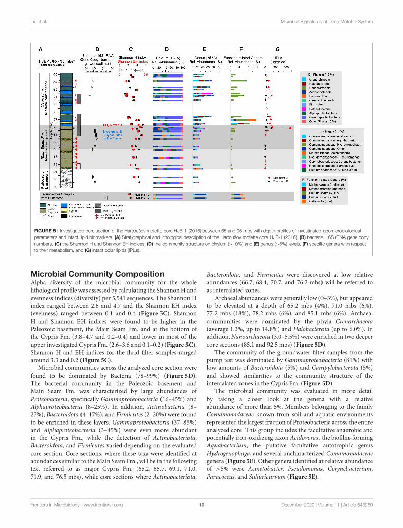
FIGURE 5 | Investigated core section of the Hartoušov mofette core HJB-1 (2016) between 65 and 95 mbs with depth profiles of investigated geomicrobiological
parameters and intact lipid biomarkers. (A) Stratigraphical and lithological description of the Hartoušov mofette core HJB-1 (2016), (B) bacterial 16S rRNA gene copy
numbers, (C) the Shannon H and Shannon EH indices, (D) the community structure on phylum (>10%) and (E) genus (>5%) levels, (F) specific genera with respect
to their metabolism, and (G) intact polar lipids (IPLs).
Microbial Community CompositionAlpha diversity of the microbial community for the wholelithological profile was assessed by calculating the ShannonH andevenness indices (diversity) per 5,541 sequences. The Shannon Hindex ranged between 2.6 and 4.7 and the Shannon EH index(evenness) ranged between 0.1 and 0.4 (Figure 5C). ShannonH and Shannon EH indices were found to be higher in thePaleozoic basement, the Main Seam Fm. and at the bottom ofthe Cypris Fm. (3.8–4.7 and 0.2–0.4) and lower in most of theupper investigated Cypris Fm. (2.6–3.6 and 0.1–0.2) (Figure 5C).Shannon H and EH indices for the fluid filter samples rangedaround 3.3 and 0.2 (Figure 5C).
Microbial communities across the analyzed core section werefound to be dominated by Bacteria (78–99%) (Figure 5D).The bacterial community in the Paleozoic basement andMain Seam Fm. was characterized by large abundances ofProteobacteria, specifically Gammaproteobacteria (16–45%) andAlphaproteobacteria (8–25%). In addition, Actinobacteria (8–27%), Bacteroidota (4–17%), and Firmicutes (2–20%) were foundto be enriched in these layers. Gammaproteobacteria (37–85%)and Alphaproteobacteria (3–45%) were even more abundantin the Cypris Fm., while the detection of Actinobacteriota,Bacteroidota, and Firmicutes varied depending on the evaluatedcore section. Core sections, where these taxa were identified atabundances similar to theMain Seam Fm., will be in the followingtext referred to as major Cypris Fm. (65.2, 65.7, 69.1, 71.0,71.9, and 76.5 mbs), while core sections where Actinobacteriota,
Bacteroidota, and Firmicutes were discovered at low relativeabundances (66.7, 68.4, 70.7, and 76.2 mbs) will be referred toas intercalated zones.
Archaeal abundances were generally low (0–3%), but appearedto be elevated at a depth of 65.2 mbs (4%), 71.0 mbs (6%),77.2 mbs (18%), 78.2 mbs (6%), and 85.1 mbs (6%). Archaealcommunities were dominated by the phyla Crenarchaeota(average 1.3%, up to 14.8%) and Halobacterota (up to 6.0%). Inaddition,Nanoarchaeota (3.0–5.5%) were enriched in two deepercore sections (85.1 and 92.5 mbs) (Figure 5D).
The community of the groundwater filter samples from thepump test was dominated by Gammaproteobacteria (81%) withlow amounts of Bacteroidota (5%) and Campylobacterota (5%)and showed similarities to the community structure of theintercalated zones in the Cypris Fm. (Figure 5D).
The microbial community was evaluated in more detailby taking a closer look at the genera with a relativeabundance of more than 5%. Members belonging to the familyComamonadaceae known from soil and aquatic environmentsrepresented the largest fraction of Proteobacteria across the entireanalyzed core. This group includes the facultative anaerobic andpotentially iron-oxidizing taxon Acidovorax, the biofilm-formingAquabacterium, the putative facultative autotrophic genusHydrogenophaga, and several uncharacterized Comamonadaceaegenera (Figure 5E). Other genera identified at relative abundanceof >5% were Acinetobacter, Pseudomonas, Corynebacterium,Paracoccus, and Sulfuricurvum (Figure 5E).
Frontiers in Microbiology | www.frontiersin.org 10 December 2020 | Volume 11 | Article 543260
Liu et al. Microbial Signatures of Deep Mofette-System
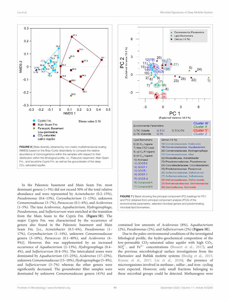
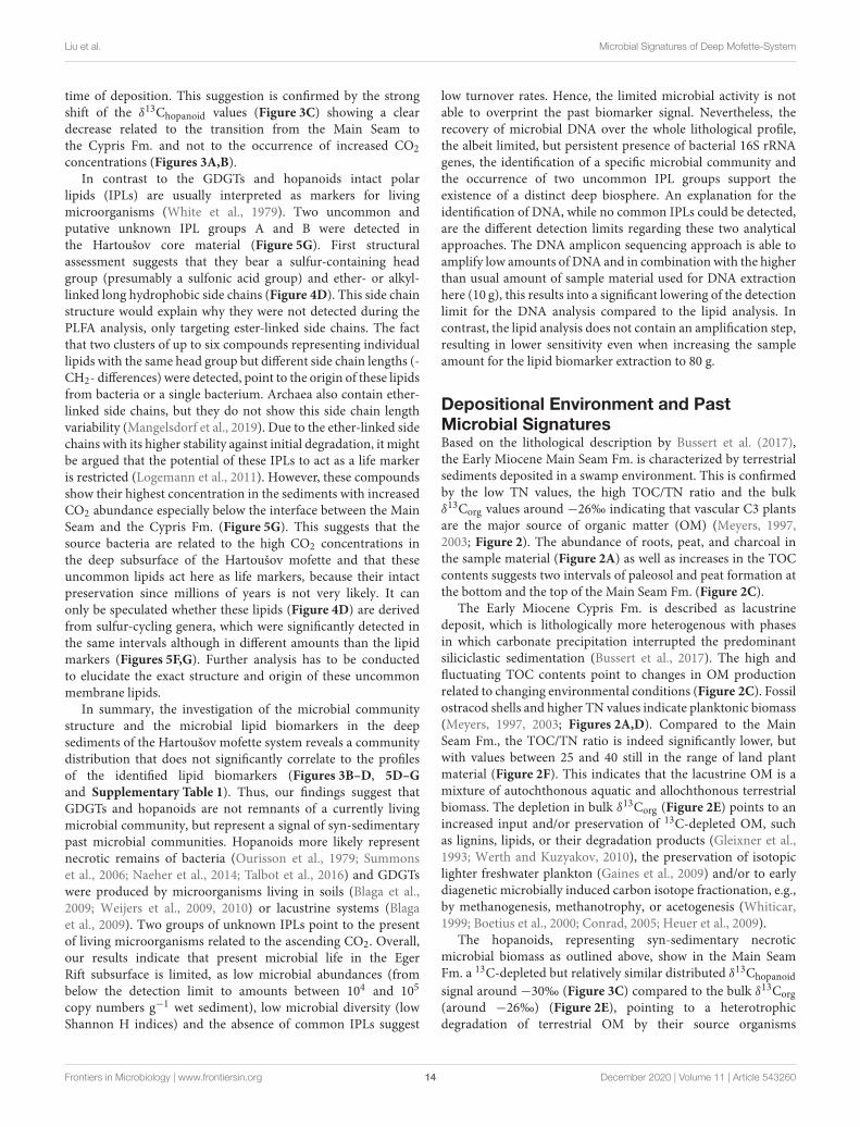
FIGURE 6 | Beta diversity obtained by non-metric multidimensional scaling
(NMDS) based on the Bray-Curtis dissimilarity to compare the relative
abundance of microorganisms within the samples with respect to their
distribution within the lithological profile, i.e., Paleozoic basement, Main Seam
Fm., and lacustrine Cypris Fm. as well as the groundwater of the deep
CO2-saturated aquifer.
In the Paleozoic basement and Main Seam Fm. mostdominant genera (>5%) did not exceed 50% of the total relativeabundance and were represented by Acinetobacter (0.2–15%),Pseudomonas (0.6–13%), Corynebacterium (1–13%), unknownComamonadaceae (3–7%), Paracoccus (0.1–6%), and Acidovorax(1–5%). The taxa Acidovorax, Aquabacterium, Hydrogenophaga,Pseudomonas, and Sulfuricurvum were enriched at the transitionfrom the Main Seam to the Cypris Fm. (Figure 5E). Themajor Cypris Fm. was characterized by the occurrence ofgenera also found in the Paleozoic basement and MainSeam Fm. [i.e., Acinetobacter (0.5–6%), Pseudomonas (1–17%), Corynebacterium (1–14%), unknown Comamonadaceaegenera (3–10%), Paracoccus (0.1–40%), and Acidovorax (4–9%)]. However, this was supplemented by an increasedoccurrence of Aquabacterium (2–13%), Hydrogenophaga (0.4–4%), and Sulfuricurvum (0.4–5%). The intercalated zones weredominated by Aquabacterium (15–25%), Acidovorax (17–23%),unknownComamonadaceae (15–18%),Hydrogenophaga (5–8%),and Sulfuricurvum (3–7%) whereas the other genera weresignificantly decreased. The groundwater filter samples weredominated by unknown Comamonadaceae genera (41%) and
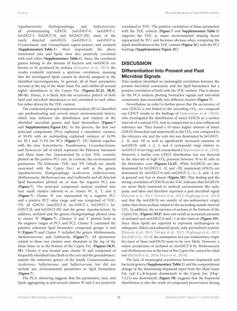
FIGURE 7 | Biplot showing the principal component (PC) loadings for PC1
and PC2 obtained from principal component analysis (PCA) of the
environmental parameters, selected microbial genera and predominating
microbial lipid biomarkers.
contained low amounts of Acidovorax (8%), Aquabacterium(2%), Pseudomonas (2%), and Sulfuricurvum (2%) (Figure 5E).
Due to the paleo-environmental conditions of the investigatedlithological profile, the hydro-geochemical composition of thelow-permeable CO2-saturated saline aquifer with high CO2,SO2−
4 , and Fe2− concentrations (Bussert et al., 2017), andthe previous microbiological surface investigations from theHartoušov and Bublák mofette systems (Beulig et al., 2015;Krauze et al., 2017; Liu et al., 2018), the presence ofmicroorganisms involved in methane-, sulfur-, and iron- cyclingwere expected. However, only small fractions belonging tothese microbial groups could be detected. Methanogens were
Frontiers in Microbiology | www.frontiersin.org 11 December 2020 | Volume 11 | Article 543260
Liu et al. Microbial Signatures of Deep Mofette-System
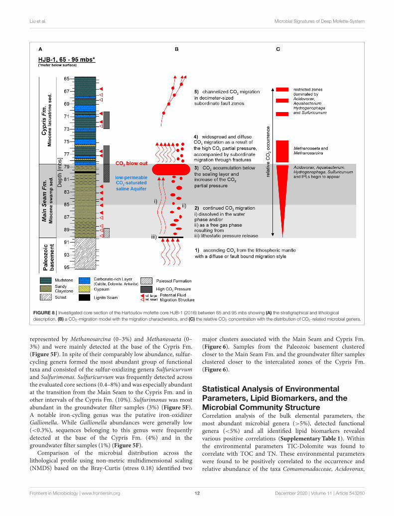
FIGURE 8 | Investigated core section of the Hartoušov mofette core HJB-1 (2016) between 65 and 95 mbs showing (A) the stratigraphical and lithological
description, (B) a CO2-migration model with the migration characteristics, and (C) the relative CO2 concentration with the distribution of CO2-related microbial genera.
represented by Methanosarcina (0–3%) and Methanosaeta (0–3%) and were mainly detected at the base of the Cypris Fm.(Figure 5F). In spite of their comparably low abundance, sulfur-cycling genera formed the most abundant group of functionaltaxa and consisted of the sulfur-oxidizing genera Sulfuricurvumand Sulfurimonas. Sulfuricurvum was frequently detected acrossthe evaluated core sections (0.4–8%) and was especially abundantat the transition from the Main Seam to the Cypris Fm. and inother intervals of the Cypris Fm. (10%). Sulfurimonas was mostabundant in the groundwater filter samples (3%) (Figure 5F).A notable iron-cycling genus was the putative iron-oxidizerGallionella. While Gallionella abundances were generally low(<0.3%), sequences belonging to this genus were frequentlydetected at the base of the Cypris Fm. (4%) and in thegroundwater filter samples (1%) (Figure 5F).
Comparison of the microbial distribution across thelithological profile using non-metric multidimensional scaling(NMDS) based on the Bray-Curtis (stress 0.18) identified two
major clusters associated with the Main Seam and Cypris Fm.(Figure 6). Samples from the Paleozoic basement clusteredcloser to the Main Seam Fm. and the groundwater filter samplesclustered closer to the intercalated zones of the Cypris Fm.(Figure 6).
Statistical Analysis of EnvironmentalParameters, Lipid Biomarkers, and theMicrobial Community StructureCorrelation analysis of the bulk elemental parameters, themost abundant microbial genera (>5%), detected functionalgenera (<5%) and all identified lipid biomarkers revealedvarious positive correlations (Supplementary Table 1). Withinthe environmental parameters TIC-Dolomite was found tocorrelate with TOC and TN. These environmental parameterswere found to be positively correlated to the occurrence andrelative abundance of the taxa Comamonadacceae, Acidovorax,
Frontiers in Microbiology | www.frontiersin.org 12 December 2020 | Volume 11 | Article 543260
Liu et al. Microbial Signatures of Deep Mofette-System
Aquabacterium, Hydrogenophaga, and Sulfuricurvum,all predominating GDGTs (isoGDGT-0, isoGDGT-1,brGDGT-I, brGDGT-II, and brGDGT-III), most of therarely detected isoGDGTs (isoGDGT-2, isoGDGT-4,Crenarchaeol, and Crenarchaeol region-isomer) and archaeol(Supplementary Table 1). Most importantly the abovementioned taxa and lipids were also positively correlatedwith each other (Supplementary Table 1). Since, the correlatedgenera belong to the domain of bacteria and isoGDGTs areknown to be produced by archaea (Schouten et al., 2013) theresults evidently represent a spurious correlation, meaningthat the investigated lipids cannot be directly assigned to theidentified microorganisms. In general, all of these parametersincrease at the top of the Main Seam Fm. and exhibit all aroundhigher abundances in the Cypris Fm. (Figures 2C,D, 3B,D,5D–G). Hence, it is likely that the occurrence pattern of bothlipid and microbial abundances is not correlated to each other,but rather driven by the TOC content.
The conducted principal component analysis (PCA) identifiedone predominating and several minor environmental factors,which may influence the distribution and relation of theidentified microbial genera and lipid biomarkers (Figure 7and Supplementary Figure 2A). In the process, the first twoprincipal components (PCs) explained a cumulative varianceof 50.4% with an outstanding explained variance of 34.9%for PC1 and 15.5% for PC2 (Figure 7). TIC-Siderite togetherwith the taxa Acinetobacter, Pseudomonas, Corynebacterium,and Paracoccus (all of which represent the Paleozoic basementand Main Seam Fm. characterized by a low TOC content)plotted on the positive PC1 axis. In contrast, the environmentalparameters TIC-Dolomite, TOC and TN (which are closelyassociated with the Cypris Fm.) as well as the generaAquabacterium, Hydogenophaga, Acidovorax, Sulfuricurvum,Methanosaeta, Methanosarcina, and Gallionella and all detectedlipid biomarkers plotted on or near the negative PC1 axis(Figure 7). The principal component analysis resulted intofour small clusters referred to as cluster W, X, Y, and Z(Figure 7). Cluster W was represented by a negative PC1and a positive PC2 value range and was comprised of TOC,TN, all GDGTs (isoGDGT-0, iso-GDGT-1, brGDGT-I, br-GDGT-II, and brGDGT-III) and the genus Aquabacterium. Inaddition, archaeol and the genera Hydogenophaga plotted closeto cluster W (Figure 7). Clusters X and Y plotted both inthe negative ranges of PC1 and PC2. Cluster X contained theputative unknown lipid biomarker compound groups A andB (Figure 7) and Cluster Y included the genera Methanosaeta,Methanosarcina, and Gallionella (Figure 7). All parametersrelated to these two clusters were abundant at the top of theMain Seam or at the bottom of the Cypris Fm. (Figures 3B,D,5F). Cluster Z was located near cluster W and comprised offrequently identified taxa (both in the core and the groundwater),namely the unknown genera of the family Comamonadaceae,Acidovorax, Sulfurimonas, and Sulfuricurvum, but did notinclude any environmental parameters or lipid biomarkers(Figure 7).
The PCA clustering suggests that the parameters, taxa, andlipids aggregating in and around clusters W and Z are positively
correlated to TOC. The positive correlation of these parameterswith the TOC content (Figure 7 and Supplementary Table 1)supports the TOC as major environmental shaping factorrepresented by PC1 and becomes obvious when comparing thedepth distributions of the TOC content (Figure 2C) with the PC1loadings (Supplementary Figure 2C).
DISCUSSION
Differentiation Into Present and PastMicrobial SignalsData analysis identified no meaningful correlation between thepresent microbial community and the lipid biomarkers but apositive correlation of both with the TOC content. This is shownby the PCA analysis plotting biomarker signals and microbialcommunity data essentially into different clusters (Figure 7).
Nevertheless, in order to further prove that the occurrence ofcore GDGTs is not linked to the ascending CO2, we comparedour GDGT results to the findings of Oppermann et al. (2010),who investigated the distribution of intact GDGTs in a surfaceinterval of a natural CO2 vent in comparison to a non-influencedreference site. They found a 10 times higher amount of intactGDGTs (branched and isoprenoid) in the CO2 vent compared tothe reference site and the vent site was dominated by brGDGT-I, -II, and -III as well as significantly increased amounts ofisoGDGTs with 1, 2, 3, and 4 cyclopentyl rings relative toisoGDGT-0 (no ring) and crenarchaeol (Oppermann et al., 2010).However, a similar core GDGT distribution was not detectedin the intervals of high CO2 pressure between 70 to 81 mbs inthe Hartoušov core (Figures 3A,D). While brGDGTs are alsodominated by brGDGT-I, -II, and –III, isoprenoid GDGTs aredominated by isoGDGT-0 and isoGDGT-1,−2,−3, and−4 arein general very low or absent (Figure 3D). This finding and thestrong correlation of GDGTs to the TOC indicates that brGDGTsare more likely restricted to surficial environments like soils,peats, and lakes and therefore represent a past microbial signal(Pancost et al., 2011; Tierney et al., 2012; Dirghangi et al., 2013)and that the isoGDGTs are mainly of syn-sedimentary originrather than from archaea related to the ascending mantle-derivedCO2. In addition, the occurrence of archaea at the bottom of theCypris Fm. (Figures 5D,F) does not result in increased amountsof archaeol and isoGDGT-0 and−1 at this interval (Figure 3D).Since these lipids are reported to represent methanogens insubaquatic (lakes) and subaerial (peats, soils, permafrost) systems(Pancost et al., 2011; Tierney et al., 2012; Dirghangi et al., 2013;Bischoff et al., 2014), the assumption of a syn-sedimentary originfor most of these isoGDGTs seem to be very likely. However, aminor production of archaeol or isoGDGT-0 by MethonasaetaandMethanosarcina at the base of the Cypris Fm. cannot be ruledout (Bischoff et al., 2014; Evans et al., 2019).
The lack of meaningful correlations between hopanoids andbacterial genera (Supplementary Table 1) and the compositionalchange of the dominating hopanoid types from the Main SeamFm. (αβ–C31R-hopane dominated) to the Cypris Fm. [Hop-17(21)-ene dominated] (Figure 3B) suggests that the hopanoiddistribution is also the result of compound preservation during
Frontiers in Microbiology | www.frontiersin.org 13 December 2020 | Volume 11 | Article 543260
Liu et al. Microbial Signatures of Deep Mofette-System
time of deposition. This suggestion is confirmed by the strongshift of the δ13Chopanoid values (Figure 3C) showing a cleardecrease related to the transition from the Main Seam tothe Cypris Fm. and not to the occurrence of increased CO2
concentrations (Figures 3A,B).In contrast to the GDGTs and hopanoids intact polar
lipids (IPLs) are usually interpreted as markers for livingmicroorganisms (White et al., 1979). Two uncommon andputative unknown IPL groups A and B were detected inthe Hartoušov core material (Figure 5G). First structuralassessment suggests that they bear a sulfur-containing headgroup (presumably a sulfonic acid group) and ether- or alkyl-linked long hydrophobic side chains (Figure 4D). This side chainstructure would explain why they were not detected during thePLFA analysis, only targeting ester-linked side chains. The factthat two clusters of up to six compounds representing individuallipids with the same head group but different side chain lengths (-CH2- differences) were detected, point to the origin of these lipidsfrom bacteria or a single bacterium. Archaea also contain ether-linked side chains, but they do not show this side chain lengthvariability (Mangelsdorf et al., 2019). Due to the ether-linked sidechains with its higher stability against initial degradation, it mightbe argued that the potential of these IPLs to act as a life markeris restricted (Logemann et al., 2011). However, these compoundsshow their highest concentration in the sediments with increasedCO2 abundance especially below the interface between the MainSeam and the Cypris Fm. (Figure 5G). This suggests that thesource bacteria are related to the high CO2 concentrations inthe deep subsurface of the Hartoušov mofette and that theseuncommon lipids act here as life markers, because their intactpreservation since millions of years is not very likely. It canonly be speculated whether these lipids (Figure 4D) are derivedfrom sulfur-cycling genera, which were significantly detected inthe same intervals although in different amounts than the lipidmarkers (Figures 5F,G). Further analysis has to be conductedto elucidate the exact structure and origin of these uncommonmembrane lipids.
In summary, the investigation of the microbial communitystructure and the microbial lipid biomarkers in the deepsediments of the Hartoušov mofette system reveals a communitydistribution that does not significantly correlate to the profilesof the identified lipid biomarkers (Figures 3B–D, 5D–G
and Supplementary Table 1). Thus, our findings suggest thatGDGTs and hopanoids are not remnants of a currently livingmicrobial community, but represent a signal of syn-sedimentarypast microbial communities. Hopanoids more likely representnecrotic remains of bacteria (Ourisson et al., 1979; Summonset al., 2006; Naeher et al., 2014; Talbot et al., 2016) and GDGTswere produced by microorganisms living in soils (Blaga et al.,2009; Weijers et al., 2009, 2010) or lacustrine systems (Blagaet al., 2009). Two groups of unknown IPLs point to the presentof living microorganisms related to the ascending CO2. Overall,our results indicate that present microbial life in the EgerRift subsurface is limited, as low microbial abundances (frombelow the detection limit to amounts between 104 and 105
copy numbers g−1 wet sediment), low microbial diversity (lowShannon H indices) and the absence of common IPLs suggest
low turnover rates. Hence, the limited microbial activity is notable to overprint the past biomarker signal. Nevertheless, therecovery of microbial DNA over the whole lithological profile,the albeit limited, but persistent presence of bacterial 16S rRNAgenes, the identification of a specific microbial community andthe occurrence of two uncommon IPL groups support theexistence of a distinct deep biosphere. An explanation for theidentification of DNA, while no common IPLs could be detected,are the different detection limits regarding these two analyticalapproaches. The DNA amplicon sequencing approach is able toamplify low amounts of DNA and in combination with the higherthan usual amount of sample material used for DNA extractionhere (10 g), this results into a significant lowering of the detectionlimit for the DNA analysis compared to the lipid analysis. Incontrast, the lipid analysis does not contain an amplification step,resulting in lower sensitivity even when increasing the sampleamount for the lipid biomarker extraction to 80 g.
Depositional Environment and PastMicrobial SignaturesBased on the lithological description by Bussert et al. (2017),the Early Miocene Main Seam Fm. is characterized by terrestrialsediments deposited in a swamp environment. This is confirmedby the low TN values, the high TOC/TN ratio and the bulkδ13Corg values around −26‰ indicating that vascular C3 plantsare the major source of organic matter (OM) (Meyers, 1997,2003; Figure 2). The abundance of roots, peat, and charcoal inthe sample material (Figure 2A) as well as increases in the TOCcontents suggests two intervals of paleosol and peat formation atthe bottom and the top of the Main Seam Fm. (Figure 2C).
The Early Miocene Cypris Fm. is described as lacustrinedeposit, which is lithologically more heterogenous with phasesin which carbonate precipitation interrupted the predominantsiliciclastic sedimentation (Bussert et al., 2017). The high andfluctuating TOC contents point to changes in OM productionrelated to changing environmental conditions (Figure 2C). Fossilostracod shells and higher TN values indicate planktonic biomass(Meyers, 1997, 2003; Figures 2A,D). Compared to the MainSeam Fm., the TOC/TN ratio is indeed significantly lower, butwith values between 25 and 40 still in the range of land plantmaterial (Figure 2F). This indicates that the lacustrine OM is amixture of autochthonous aquatic and allochthonous terrestrialbiomass. The depletion in bulk δ13Corg (Figure 2E) points to anincreased input and/or preservation of 13C-depleted OM, suchas lignins, lipids, or their degradation products (Gleixner et al.,1993; Werth and Kuzyakov, 2010), the preservation of isotopiclighter freshwater plankton (Gaines et al., 2009) and/or to earlydiagenetic microbially induced carbon isotope fractionation, e.g.,by methanogenesis, methanotrophy, or acetogenesis (Whiticar,1999; Boetius et al., 2000; Conrad, 2005; Heuer et al., 2009).
The hopanoids, representing syn-sedimentary necroticmicrobial biomass as outlined above, show in the Main SeamFm. a 13C-depleted but relatively similar distributed δ13Chopanoid
signal around −30‰ (Figure 3C) compared to the bulk δ13Corg
(around −26‰) (Figure 2E), pointing to a heterotrophicdegradation of terrestrial OM by their source organisms
Frontiers in Microbiology | www.frontiersin.org 14 December 2020 | Volume 11 | Article 543260
Liu et al. Microbial Signatures of Deep Mofette-System
(Summons et al., 2006; Talbot et al., 2016). The dominanceof αβ-C31R-hopane is associated with bacteria known fromterrestrial and more specific peat environments (Quirk et al.,1984; Huang et al., 2015; Inglis et al., 2018). In contrast, theshift to a domination of Hop-17(21)-ene and the decrease of theδ13Chopanoid signal to values ranging between −50 and −60‰in the Cypris Fm. (Figures 3B,C) illustrates that planktonicfreshwater bacteria or benthic aerobic methanotrophs dwellingon microbially-derived methane at the water-sediment interfacemight have formed a significant part of the source bacteria for thedeposited hopanoids (Nealson, 1997; Whiticar, 1999; Summonset al., 2006; Naeher et al., 2014; Davies et al., 2016; Hoefs, 2018).
A similar picture, resulting from the depositionalenvironment can be seen from the depth distribution ofthe GDGTs. The upper Main Seam Fm. is dominated bybrGDGTs characteristic for soil and swamp environments(Weijers et al., 2007, 2010; Dirghangi et al., 2013; Schouten et al.,2013; Freymond et al., 2017; Huguet et al., 2017; Naafs et al.,2017; Figure 3D). The lacustrine Cypris deposits show high butfluctuating amounts of isoGDGTs and brGDGTs representing amixture of aquatic microbial biomass produced in the lake andterrestrial biomass from the catchment area (Weijers et al., 2007,2010; Tierney et al., 2012; Schouten et al., 2013; Freymond et al.,2017), respectively. An aquatic in situ production (lacustrine orfluvial) of brGDGTs cannot be excluded, although such in situproductions generally seem to appear only to minor amounts(Tierney et al., 2012; DeJonge et al., 2014; Freymond et al.,2017). The distribution pattern of br-GDGT-II and –III, showingrelatively similar amounts, was also investigated by Huguet et al.(2017) for bog and fen sediments and may confirm a palustrineOM input. Therefore, compared to the Main Seam Fm., therelatively higher brGDGT content might reflect a combinationof in situ lake production and OM input from the catchmentarea as well as good preservation conditions within the lakesediments. Abundant isoGDGT-0 and the occurrence of archaeoland isoGDGT-1 with an initial appearance in the paleosolformation section of the Main Seam Fm. and higher occurrencein the Cypris Fm. point to the presence of methanogenic andmethanotrophic archaea that have adapted from a palustrine toa lacustrine environment (Pancost et al., 2011; Dirghangi et al.,2013; Schouten et al., 2013; Bischoff et al., 2014; Naeher et al.,2014; Bale et al., 2019). In addition, an origin from archaea livingin the water column cannot be excluded (Buckles et al., 2013;Schubotz et al., 2018). However, comparing the results from thehopanoid and GDGT analysis, we assume that methane cyclingprocesses particularly of methanogenic and methanotrophicarchaea and methanotrophic bacteria (hopanoid signal) playeda significant role within the Early Miocene lake environment(Figure 3C).
Deep Biosphere Structure andCO2-Migration Model of the DeepHartoušov Mofette SystemOur results for the present microbial community structurerevealed a domination of genera from the familyComamonadaceae and low amounts of archaea (Figures 5D–F).
Surface investigations from the Hartoušov mofette system(Beulig et al., 2015; Liu et al., 2018) and a CO2 vent within theLatera Caldera (Central Italy) (Oppermann et al., 2010) showrelatively high abundances of acidophilic, methanogenic andiron- and sulfur-cycling microorganisms [e.g., methanogenicarchaea, Acidobacteria, Chloroflexi, sulfate reducing bacteria(SRB), and Geobacteraceae]. These species either appear only tominor amounts or are completely absent in the deep subsurfacesediments (Figures 5D–F), displaying the occurrence of differentmicrobial communities in the surface and deep sections of theHartoušov mofette system.
Considering the Early Miocene age of the investigatedsediments, we suggest several community shaping selectionprocesses during subsidence and early diagenetic transformation(Orsi et al., 2020) prior to the onset of CO2-migration inMid Pleistocene. These selection processes formed a basicmicrobial community, which became further shaped by theascending mantle-derived CO2. This assumption is based on thefinding of a major microbial community composition that isin parts related to the lithological profile (Figure 6) and showssome modifications related to the occurrence of higher CO2
pressures and the presence of CO2-saturated groundwater.The major community is composed of Actinobacteriota,Bacteroidota, Firmicutes, Patescibacteria, Alphaproteobacteria,and dominating Gammaproteobacteria (Figure 5D) and appearsin the Main Seam and Cypris Fm. with the difference, that theGammaproteobacteria are more abundant in the Cypris Fm.(Figure 5D). A high relative abundance of Gammaproteobacteriaand a low Shannon H index of the groundwater filter samplesfrom the CO2-saturated saline aquifer seem to indicate amicrobial selection and adaptation related to a high influenceof ascending CO2-saturated groundwater, which is expressed inthe upper Cypris Fm. as intercalated zones (66.7, 68.4, 70.7, and76.2 mbs) (Figures 5C,D). As a consequence, Shannon H indices<3.2 in the lithological profile could be indicative for a high CO2
influence (Figure 5C).On the genus level, the occurrence of unknown genera
from the family Comamonadaceae in both, the groundwaterand lithological samples as well as the abundance of otherComamonadaceae genera, namely Acidovorax, Aquabacterium,and Hydrogenophaga at the top of the Main Seam Fm., at thebottom of the Cypris Fm. and especially in the intercalated zonesseem to be correlated to a mantle-derived CO2 influence aswell (Figures 5A,E). The genera Acidovorax and Hydrogenphagaare amongst others comprised of facultative anaerobic, iron-oxidizing, and autotrophic hydrogen-oxidizing and CO2-fixingbacteria (Yoon et al., 2008; Byrne-Bailey et al., 2010; Willemsand Gillis, 2015; Li et al., 2017). The occurrence of increasedComamonadaceae genera related to high CO2 concentrations wasalso reported byHam et al. (2017), who found a predomination ofComamonadaceae in a natural CO2-dominated aquifer in SouthKorea as well as by Krauze et al. (2017), who detected membersof Comamonadaceae in wet mofettes of the Cheb Basin closeto the Hartoušov site. Furthermore, Mu et al. (2014) observedan increase of Comamonadaceae after the injection of CO2 intothe Paaratte sandstone aquifer (Southern Australia) and a similarresult was reported by Gulliver et al. (2018) for the CO2 injection
Frontiers in Microbiology | www.frontiersin.org 15 December 2020 | Volume 11 | Article 543260
Liu et al. Microbial Signatures of Deep Mofette-System
into an aquifer at the freshwater Plant Daniel in Escatawpa(Massachusetts, USA). Although, Comamonadaceae have beenfound in non-CO2 influenced subsurface environments suchas the Sanford Underground Research Facility (Jangir et al.,2019) and the Fennoscandian shield (Nyyssönen et al., 2014),the relative abundances in these environments were lowercompared to our results. Thus, the dominance of membersfrom the family Comamonadaceae in the deep sediments of theHartoušov mofette seem to reflect a CO2 influenced microbialcommunity with a good adaptation potential to the prevailingconditions. Hence, our study assumes that some membersof Comamonadaceae, especially the determined sequences ofunknown genera as well as Acidovorax and Aquabacteriumare very adaptive to CO2-dominated ecosystems and can besuggested as indicator for such environments.
The occurrence of Sulfuricurvum and Sulfurimonas is linkedto the high SO2−
4 content of the CO2-saturated saline aquifer(Bussert et al., 2017) and shows an increase of Sulfuricurvumat the transition from the Main Seam to the Cypris Fm. andin the intercalated zones within the Cypris Fm. (Figure 5F).An abundant occurrence of the genus Sulfuricurvum in highlyCO2-influenced subsurface environments was also reported byGulliver et al. (2018). Moreover, the genus Sulfurimonas wasfound in surficial pools of several mofette systems within theCheb Basin (Krauze et al., 2017) and in a CO2-driven geyseron the Colorado Plateau (Utah, USA) (Probst et al., 2018).Thus, Sulfuricurvum and Sulfurimonasmight represent indicatororganisms occurring in CO2 influenced ecosystems with a highSO2−
4 concentration.The relationship of the investigated genera from the family
Comamonadaceae as well as Sulfuricurvum and Sulfurimonas tothe ascending CO2-saturated saline groundwater indicate, thatthe groundwater acts both, as transport mechanism and maincommunity shaping factor for the deep biosphere. As a result,we assume the following CO2 migration model for the deepsediments of the Hartoušov mofette system. The CO2-saturatedgroundwater or the CO2 migrates from the Paleozoic basementinto the low-permeable CO2-saturated saline aquifer and istrapped by the overlaying Cypris Fm. (Figure 8B). Thereby,related to buoyancy and the permanent CO2 supply from themantle, the CO2 pressure increases to the top of the MainSeam Fm. with the highest concentrations occurring between80.5 and 78.5 mbs (Figure 8C), indicated by the CO2 blow outduring the drilling campaign (Figure 8A) and an increase inSulfuricurvum and the uncommon lipid compound groups A andB (Figures 5F,G). This high CO2 pressure causes a widespreaddiffuse groundwater migration into the lower part of the CyprisFm. (between 78.5 and 75 mbs) (Figure 8B). Therein both,potentially produced acetate from OM degradation related toa higher TOC content (Figures 2A,C) and the ascending CO2
itself might act as substrates for methanogenic archaea, namelyMethanosaeta, and Methanosarcina (Zinder et al., 1985; Pateland Sprott, 1990; Figure 5F). Subsequently, in this core intervalpart of the isoGDGT signal representing methanogenic archaealbiomass (Schouten et al., 2013; Naeher et al., 2014) mightalso derive from the current deep biosphere (Figure 3D). Thisdiffuse migration may also be accompanied by a subordinate
migration through small fractures (Figure 8B). Afterwards, thegroundwater migration seem to change into a more channelizedmigration (Figure 8B), indicated by the occurrence of theintercalated zones with higher abundances of Acidovorax,Aquabacterium, Hydrogenophaga, and Sulfuricurvum whichcorrelate with fluid migration structures mentioned in thelithological description from the subsampling (73–65 mbs)(Figures 5A,E and Supplementary Figure 1C) and thus pointto a CO2 migration related to small subordinate fault zones indecimeter-size. A possible syntrophy within this environmentalsetup could be based on an anaerobic, heterotrophic lifestyle ofAcidovorax, Aquabacterium, and other genera from the familyComamonadaceae (Willems et al., 1990; Kalmbach et al., 1999).The fermentation of OM provides hydrogen and sulfur assubstrate for the hydrogen-oxidizing Hydrogenophaga and thesulfur-oxidizer Sulfuricurvum, which both additionally utilizethe ascending CO2 for their metabolism (Willems et al., 1989;Kodama and Watanabe, 2004).
CONCLUSION
The lithological setup of the deep Hartoušov mofette system(65–95 mbs) represents a paleoenvironmental change from anEarly Miocene terrestrial swamp-like (Main Seam Fm.) to alacustrine ecosystem (Cypris Fm.). Since Mid Pleistocene time,this system became overprinted by migration and accumulationof mantle-derived CO2 which forms a potential habitat shapingand stimulating deep microbial life.
The necrotic microbial lipid biomarkers essentially reflectthe environmental conditions during time of deposition andare therefore unsuitable for tracing the deep biosphere at theHartoušov mofette site. This already indicates that the currentbiosphere signal in the deep mofette system is rather smallcompared to the paleo-microbial biomass.
The overall low abundance of microbial signatures from thedeep biosphere in the Hartoušov mofette system suggests that thelow-permeable CO2-saturated aquifer interval does not representa hotspot for deep microbial life as might be expected froma substrate point of view. However, our data indicate that theavailability of organic matter as microbial feedstock and CO2
migration are the main community shaping factors in the deeppart of the mofette system. In the process, CO2 migration andaccumulation occur heterogeneous leading to the formationof niche habitats for CO2-adapted microbial communitiesindependent from the single lithological units of the exploredcore interval. In addition, our results imply that the high relativeabundance of Acidovorax, Aquabacterium,Hydrogenophaga, andunknown genera of the family Comamonadaceae as well as theoccurrence of Sulfuricurvum together with high sulfate contentsin the CO2-saturated groundwater may be indicative for CO2-dominated deep subsurface ecosystems.
A cluster of yet unknown intact polar membrane lipidsdisplays the presence of microbial life associated to higheraccumulations of CO2 in the deep subsurface and show potentialto act as lipid biomarkers for such environmental settings.
Frontiers in Microbiology | www.frontiersin.org 16 December 2020 | Volume 11 | Article 543260
Liu et al. Microbial Signatures of Deep Mofette-System
DATA AVAILABILITY STATEMENT
The datasets generated for this study can be found inthe European Nucleotide Archive (http://www.ebi.ac.uk/ena),accession numbers PRJEB22478 (ERS4382097 to ERS4382146and ERS4382395 to ERS4382400). Lipid data can be found in theSupplementary Material.
AUTHOR CONTRIBUTIONS
QL and KA wrote the manuscript, performed subsampling, andinitial description of the core material as well as PCA analysisin equal manner. QL processed the geomicrobiological analysis,i.e., DNA extractions and purification, gene quantification, andbioinformatical based statistical analyses. KA performed theanalysis of the intact and past lipid membrane biomarkers,and conducted together with BP the bulk elemental and bulkstable isotope analysis. FH and DL were involved in 16S rRNAsequencing data processing. PK and DL were involved instatistical analyses. DW, KM, HK, RB, and H-MS gave essentialtechnical advice and contributed to the interpretation of theresults and valuable discussion. MA and KM supervised thestudy and led the writing of the present manuscript. All authorshave taken part in the manuscript revisions, interpretation ofthe results, writing of the manuscript, and agreed with itsscientific content.
FUNDING
This study was supported by the DeutscheForschungsgemeinschaft (DFG) within the frameworkof the priority program 1006 International Continental
Drilling Program (ICDP) by a grant to MA (AL 1898/1)and KM (MA 2470/5). The drilling of the Hartoušovmofette site and the 16S rRNA gene amplicon (MiSeq)sequencing was financed through the Helmholtz ResearchProgramme Geosystem—the Changing Earth of the GFZ(German Research Centre for Geosciences—HelmholtzCentre Potsdam).
ACKNOWLEDGMENTS
QL gratefully acknowledges financial support from the ChinaScholarship Council. KA and KM are grateful to the DeutscheForschungsgemeinschaft (DFG) for funding the project MA2470/5 and MA for funding AL 1898/1. The authors wouldlike to thank all involved scientists, technicians, and studentresearch assistants of the GFZ—German Research Centre forGeosciences for support and guidance through the samplepreparation, analysis, and interpretation. These importantpeople, who kept the system running are in alphabeticalorder; Oliver Burckhardt, Anke Kaminsky, Cornelia Karger,Axel Kitte, Sebastian Kreutz, Hartmut Liep, Joana MacLean,Doreen Noak, Sylvia Pinkerneil, Anke Saborowski, and AndreaVieth-Hillebrand. Finally, we would like to thank the EditorCassandre Lazar and four reviewers for taking the time tointensely revise our manuscript, which helped us to significantlyimprove it.
SUPPLEMENTARY MATERIAL
The Supplementary Material for this article can be foundonline at: https://www.frontiersin.org/articles/10.3389/fmicb.2020.543260/full#supplementary-material
REFERENCES
Bale, N. J., Sorokin, D. Y., Hopmans, E. C., Koenen, M., Rijpstra, W. I. C.,Villanueva, L., et al. (2019). New insights into the polar lipid compositionof extremely halo(alkali)philic euryarchaea from hypersaline lakes. Front.
Microbiol. 10:377. doi: 10.3389/fmicb.2019.00377Bankwitz, P., Bankwitz, E., Brauer, K., Kampf, H., and Storr, M. (2003a).
Deformation structures in plio-and pleistocene sediments (NWBohemia, central Europe). Spec. Public. Geol. Soc. Lond. 216, 73–93.doi: 10.1144/GSL.SP.2003.216.01.06
Bankwitz, P., Schneider, G., Kämpf, H., and Bankwitz, E. (2003b). Structuralcharacteristics of epicentral areas in Central Europe: study case Cheb Basin(Czech Republic). J. Geodyn. 35, 5–32. doi: 10.1016/S0264-3707(02)00051-0
Beulig, F., Heuer, V. B., Akob, D. M., Viehweger, B., Elvert, M., Herrmann, M.,et al. (2015). Carbon flow from volcanic CO2 into soil microbial communitiesof a wetland mofette. ISME J. 9, 746–759. doi: 10.1038/ismej.2014.148
Beulig, F., Urich, T., Nowak, M., Trumbore, S. E., Gleixner, G., Gilfillan, G.D., et al. (2016). Altered carbon turnover processes and microbiomes insoils under long-term extremely high CO2 exposure. Nat. Microbiol. 1:15025.doi: 10.1038/nmicrobiol.2015.25
Bischoff, J., Mangelsdorf, K., Schwamborn, G., and Wagner, D. (2014). Impactof lake-level and climate changes on microbial communities in a terrestrialpermafrost sequence of the el’gygytgyn crater, far east russian arctic. Permaf.
Periglac. Process. 25, 107–116. doi: 10.1002/ppp.1807Blaga, C. I., Reichart, G.-J., Heiri, O., and Sinninghe Damsté, J. S. (2009).
Tetraether membrane lipid distributions in water-column particulate matter
and sediments: a study of 47 European lakes along a north–south transect. J.Paleolimnol. 41, 523–540. doi: 10.1007/s10933-008-9242-2
Bligh, E. G., and Dyer, W. J. (1959). A rapid method of total lipid extraction andpurification. Can. J. Biochem. Physiol. 37, 911–917. doi: 10.1139/y59-099
Boetius, A., Ravenschlag, K., Schubert, C. J., Rickert, D., Widdel, F., Gieseke, A.,et al. (2000). A marine microbial consortium apparently mediating anaerobicoxidation of methane. Nature 407, 623–626. doi: 10.1038/35036572
Bolyen, E., Rideout, J. R., Dillon, M. R., Bokulich, N. A., Abnet, C. C., Al-Ghalith, G. A., et al. (2019). Reproducible, interactive, scalable and extensiblemicrobiome data science using QIIME 2. Nat.Biotechnol. 37, 852–857.doi: 10.1038/s41587-019-0209-9
Brandl, P. A., Genske, F. S., Beier, C., Haase, K. M., Sprung, P., and Krumm,S. H. (2015). Magmatic evidence for carbonate metasomatism in thelithospheric mantle underneath the Ohre (Eger) rift. J. Petrol. 56, 1743–1774.doi: 10.1093/petrology/egv052
Bräuer, K., Kämpf, H., Faber, E., Koch, U., Nitzsche, H.-M., and Strauch, G. (2005).Seismically triggered microbial methane production relating to the VogtlandNW bohemia earthquake swarm period 2000, Central Europe. Geochem. J. 39,441–450. doi: 10.2343/geochemj.39.441
Bräuer, K., Kämpf, H., Koch, U., and Strauch, G. (2011). Monthly monitoring ofgas and isotope compositions in the free gas phase at degassing locations closeto the Nový Kostel focal zone in the western Eger Rift, Czech Republic. Chem.
Geol. 290, 163–176. doi: 10.1016/j.chemgeo.2011.09.012Bräuer, K., Kämpf, H., Niedermann, S., and Strauch, G. (2018). Monitoring
of helium and carbon isotopes in the western Eger Rift area (CzechRepublic): relationships with the 2014 seismic activity and indications
Frontiers in Microbiology | www.frontiersin.org 17 December 2020 | Volume 11 | Article 543260
Liu et al. Microbial Signatures of Deep Mofette-System
for recent (2000–2016) magmatic unrest. Chem. Geol. 482, 131–145.doi: 10.1016/j.chemgeo.2018.02.017
Bräuer, K., Kämpf, H., and Strauch, G. (2009). Earthquake swarms in non-volcanic regions: what fluids have to say. Geophys. Res. Lett. 36:L17309.doi: 10.1029/2009GL039615
Buckles, L. K., Villanueva, L., Weijers, J. W. H., Verschuren, D., and Damsté,J. S. S. (2013). Linking isoprenoidal GDGT membrane lipid distributionswith gene abundances of ammonia-oxidizing Thaumarchaeota and unculturedcrenarchaeotal groups in the water column of a tropical lake (Lake Challa, EastAfrica). Environ. Microbiol. 15, 2445–2462. doi: 10.1111/1462-2920.12118
Bussert, R., Kämpf, H., Flechsig, C., Hesse, K., Nickschick, T., Liu, Q., et al.(2017). Drilling into an active mofette: pilot-hole study of the impact of CO2-rich mantle-derived fluids on the geo–bio interaction in the western Eger Rift(Czech Republic). Sci. Dril. 23, 13–27. doi: 10.5194/sd-23-13-2017
Byrne-Bailey, K. G., Weber, K. A., Chair, A. H., Bose, S., Knox, T., Spanbauer, T.L., et al. (2010). Completed genome sequence of the anaerobic iron-oxidizingbacterium Acidovorax ebreus strain TPSY. J. Bacteriol. 192, 1475–1476.doi: 10.1128/JB.01449-09
Callahan, B. J., McMurdie, P. J., Rosen, M. J., Han, A. W., Johnson, A. J. A., andHolmes, S. P. (2016). DADA2: High-resolution sample inference from Illuminaamplicon data. Nat. Methods 13, 581–583. doi: 10.1038/nmeth.3869
Caporaso, J. G., Lauber, C. L., Walters, W. A., Berg-Lyons, D., Lozupone, C. A.,Turnbaugh, P. J., et al. (2011). Global patterns of 16S rRNA diversity at a depthof millions of sequences per sample. Proc. Natl. Acad. Sci. U.S.A. 108(Suppl. 1),4516–4522. doi: 10.1073/pnas.1000080107
Conrad, R. (2005). Quantification of methanogenic pathways using stable carbonisotopic signatures: a review and a proposal. Organ. Geochem. 36, 739–752.doi: 10.1016/j.orggeochem.2004.09.006
Dahm, T., Hrubcov,á, P., Fischer, T., Horálek, J., Korn, M., Buske, S., et al.(2013). Eger Rift ICDP: an observatory for study of non-volcanic, mid-crustal earthquake swarms and accompanying phenomena. Sci. Dril. 16, 93–99.doi: 10.5194/sd-16-93-2013
Davies, K. L., Pancost, R. D., Edwards, M. E., Walter-Antony, K. M., Langdon, P.G., and Chaves Torres, L. (2016). Diploptene δ13C values from contemporarythermokarst lake sediments show complex spatial variation. Biogeosciences 13,2611–2621. doi: 10.5194/bg-13-2611-2016
Degelmann, D. M., Borken, W., Drake, H. L., and Kolb, S. (2010).Different atmospheric methane-oxidizing communities in Europeanbeech and Norway spruce soils. Appl. Environ. Microbiol. 76, 3228–3235.doi: 10.1128/AEM.02730-09
DeJonge, C., Stadnitskaia, A., Hopmans, E. C., Cherkashov, G., Fedotov, A.,and Sinninghe Damsté, J. S. (2014). In situ produced branched glyceroldialkyl glycerol tetraethers in suspended particulate matter from theYenisei River, Eastern Siberia. Geochim. Cosmochim. Acta 125, 476–491.doi: 10.1016/j.gca.2013.10.031
Dirghangi, S. S., Pagani, M., Hren, M. T., and Tipple, B. J. (2013). Distribution ofglycerol dialkyl glycerol tetraethers in soils from two environmental transects inthe USA. Organ. Geochem. 59, 49–60. doi: 10.1016/j.orggeochem.2013.03.009
Evans, T. W., Coffinet, S., Könneke, M., Lipp, J. S., Becker, K. W., Elvert, M., et al.(2019). Assessing the carbon assimilation and production of benthic archaeallipid biomarkers using lipid-RIP. Geochim. Cosmochim. Acta 265, 431–442.doi: 10.1016/j.gca.2019.08.030
Fischer, T., Horálek, J., Hrubcová, P., Vavrycuk, V., Bräuer, K., and Kämpf, H.(2014). Intra-continental earthquake swarms in West-Bohemia and Vogtland:a review. Tectonophysics 611, 1–27. doi: 10.1016/j.tecto.2013.11.001
Fischer, T., Matyska, C., and Heinicke, J. (2017). Earthquake-enhancedpermeability – evidence from carbon dioxide release following the ML 3.5 earthquake in West Bohemia. Earth Planet. Sci. Lett. 460, 60–67.doi: 10.1016/j.epsl.2016.12.001
Flechsig, C., Bussert, R., Rechner, J., Schütze, C., and Kämpf, H. (2008). TheHartoušovMofette field in the Cheb Basin,Western Eger Rift (Czech Republic):a comperative geoelectric, sedimentologic and soil gas study of a magmaticdiffuse CO2-degassing structure. Z. Geol. Wiss 36, 177–193.
Flechsig, C., Fabig, T., Rücker, C., and Schütze, C. (2010). Geoelectricalinvestigations in the cheb basin/W-bohemia: an approach to evaluatethe near-surface conductivity structure. Stud. Geophys. Geod. 54, 443–463.doi: 10.1007/s11200-010-0026-6
Frerichs, J., Oppermann, B. I., Gwosdz, S., Möller, I., Herrmann, M., andKrüger, M. (2013). Microbial community changes at a terrestrial volcanicCO2 vent induced by soil acidification and anaerobic microhabitats withinthe soil column. FEMS Microbiol. Ecol. 84, 60–74. doi: 10.1111/1574-6941.12040
Freymond, C. V., Peterse, F., Fischer, L. V., Filip, F., Giosan, L., and Eglinton, T. I.(2017). Branched GDGT signals in fluvial sediments of the Danube River basin:method comparison and longitudinal evolution. Organ. Geochem. 103, 88–96.doi: 10.1016/j.orggeochem.2016.11.002
Gaines, S. M., Eglinton, G., and Rullkötter, J. (2009). Echoes of Life: What Fossil
Molecules Reveal about Earth History. Oxford: Oxford University Press.Geissler, W. H., Kämpf, H., Seifert, W., and Dulski, P. (2007). Petrological
and seismic studies of the lithosphere in the earthquake swarm regionVogtland/NWBohemia, central Europe. J. Volcanol. Geotherm. Res. 159, 33–69.doi: 10.1016/j.jvolgeores.2006.06.011
Gleixner, G., Danier, H. J., Werner, R. A., and Schmidt, H. L. (1993). Correlationsbetween the 13C content of primary and secondary plant products in differentcell compartments and that in decomposing basidiomycetes. Plant Physiol. 102,1287–1290.
Gulliver, D., Lipus, D., Ross, D., and Bibby, K. (2018). Insights into microbialcommunity structure and function from a shallow, simulated CO2-leakageaquifer demonstrate microbial selection and adaptation. Environ. Microbiol.
Rep. 11, 338–351. doi: 10.1111/1758-2229.12675Ham, B., Choi, B.-Y., Chae, G.-T., Kirk, M. F., and Kwon, M. J. (2017).
Geochemical influence on microbial communities at CO2-leakage analog sites.Front. Microbiol. 8:2203. doi: 10.3389/fmicb.2017.02203
Hammer, Ø., Harper, D. A. T., and Ryan, P. D. (2001). PAST-palaeontologicalstatistics, ver. 1.89. Palaeontol. Electron 4, 1–9.
Heuer, B., Geissler, W. H., Kind, R., and Kämpf, H. (2006). Seismic evidencefor asthenospheric updoming beneath the western Bohemian Massif, centralEurope. Geophys. Res. Lett. 33:L05311. doi: 10.1029/2005GL025158
Heuer, V. B., Pohlman, J. W., Torres, M. E., Elvert, M., and Hinrichs, K.-U. (2009).The stable carbon isotope biogeochemistry of acetate and other dissolvedcarbon species in deep subseafloor sediments at the northern Cascadia Margin.Geochim. Cosmochim. Acta 73, 3323–3336. doi: 10.1016/j.gca.2009.03.001UR
Hoefs, J. (2018). Stable Isotope Geochemistry. Cham: Springer.Hohberg, K., Schulz, H.-J., Balkenhol, B., Pilz, M., Thomalla, A., Russell, D.
J., et al. (2015). Soil faunal communities from mofette fields: effects ofhigh geogenic carbon dioxide concentration. Soil Biol. Biochem. 88, 420–429.doi: 10.1016/j.soilbio.2015.06.017
Huang, X., Meyers, P. A., Xue, J., Gong, L., Wang, X., and Xie, S. (2015).Environmental factors affecting the low temperature isomerization ofhomohopanes in acidic peat deposits, central China. Geochim. Cosmochim.
Acta 154, 212–228. doi: 10.1016/j.gca.2015.01.031Huguet, A., Meador, T. B., Laggoun-Défarge, F., Könneke, M., Wu, W., Derenne,
S., et al. (2017). Production rates of bacterial tetraether lipids and fatty acidsin peatland under varying oxygen concentrations. Geochim. Cosmochim. Acta
203, 103–116. doi: 10.1016/j.gca.2017.01.012Inglis, G. N., Naafs, B. D. A., Zheng, Y., McClymont, E. L., Evershed, R. P., and
Pancost, R. D. (2018). Distributions of geohopanoids in peat: implications forthe use of hopanoid-based proxies in natural archives. Geochim. Cosmochim.
Acta 224, 249–261. doi: 10.1016/j.gca.2017.12.029Jangir, Y., Karbelkar, A. A., Beedle, N. M., Zinke, L. A., Wanger, G., Anderson, C.
M., et al. (2019). In situ electrochemical studies of the terrestrial deep subsurfacebiosphere at the sanford underground research facility, South Dakota, USA.Front. Energy Res. 7:121. doi: 10.3389/fenrg.2019.00121
Kalmbach, S., Manz, W., Wecke, J., and Szewzyk, U. (1999). Aquabacterium gen.nov., with description of Aquabacterium citratiphilum sp. nov., Aquabacteriumparvum sp. nov. and Aquabacterium commune sp. nov., three in situ dominantbacterial species from the Berlin drinking water system. Int. J. Syst. Bacteriol.49(Pt. 2), 769–777. doi: 10.1099/00207713-49-2-769
Kämpf, H., and Bankwitz, P. (2005). “Active processes of the western Eger Grabenarea, in the KTB deep crustal laboratory and the western Eger Graben,” inSchriftreihe der Deutschen Gesellschaft für Geowissenschaften, Vol. 40, eds H.Kämpf, A. Petrek, J. Rohrmüller, H. J. Kümpel, W. H. Geissler, 78–82.
Kämpf, H., Bräuer, K., Schumann, J., Hahne, K., and Strauch, G. (2013). CO2
discharge in an active, non-volcanic continental rift area (Czech Republic):
Frontiers in Microbiology | www.frontiersin.org 18 December 2020 | Volume 11 | Article 543260
Liu et al. Microbial Signatures of Deep Mofette-System
characterisation (δ13C, 3He/4He) and quantification of diffuse and vent CO2
emissions. Chem. Geol. 339, 71–83. doi: 10.1016/j.chemgeo.2012.08.005Kämpf, H., Broge, A. S., Marzban, P., Allahbakhshi, M., and Nickschick, T.
(2019). Nonvolcanic carbon dioxide emission at continental rifts: the bublakMofette Area, Western Eger Rift, Czech Republic. Geofluids 2019:4852706.doi: 10.1155/2019/4852706
Kodama, Y., and Watanabe, K. (2004). Sulfuricurvum kujiense gen. nov., sp. nov.,a facultatively anaerobic, chemolithoautotrophic, sulfur-oxidizing bacteriumisolated from an underground crude-oil storage cavity. Int. J. Systemat. Evol.
Microbiol. 54, 2297–2300. doi: 10.1099/ijs.0.63243-0Krauze, P., Kämpf, H., Horn, F., Liu, Q., Voropaev, A., Wagner, D., et al.
(2017). Microbiological and geochemical survey of CO2-dominated mofetteand mineral waters of the Cheb Basin, Czech republic. Front. Microbiol. 8:2446.doi: 10.3389/fmicb.2017.02446
Krmícek, L., Timmerman, M. J., Ziemann, M. A., Sudo, M., and Ulrych, J. (2020).40Ar/39Ar step-heating dating of phlogopite and kaersutite megacrysts fromthe Železná hurka (Eisenbühl) Pleistocene scoria cone, Czech Republic. Geol.Carpathica 71, 382–387. doi: 10.31577/GeolCarp.71.4.6
Krüger, M., West, J., Frerichs, J., Oppermann, B., Dictor, M.-C., Jouliand, C.,et al. (2009). Ecosystem effects of elevated CO2 concentrations on microbialpopulations at a terrestrial CO2 vent at Laacher See, Germany. Energy Procedia1, 1933–1939. doi: 10.1016/j.egypro
Li, C., Zhong, S., Zhang, F., Wang, Z., Jiang, F., and Wan, Y. (2017). Response ofmicrobial communities to supercritical CO2 and biogeochemical influences onmicrobially mediated CO2-saline-sandstone interactions.Chem. Geol. 473, 1–9.doi: 10.1016/j.chemgeo.2017.09.026
Lied, P., Kontny, A., Nowaczyk, N., Mrlina, J., and Kämpf, H. (2020).Cooling rates of pyroclastic deposits inferred from mineral magneticinvestigations: a case study from the Pleistocene Mýtina Maar (CzechRepublic). Int. J. Earth Sci. 109, 1707–1725. doi: 10.1007/s00531-020-01865-1
Liu, Q., Kämpf, H., Bussert, R., Krauze, P., Horn, F., Nickschick, T., et al. (2018).Influence of CO2 degassing on the microbial community in a dry mofettefield in hartoušov, czech republic (Western Eger Rift). Front. Microbiol. 9:2787.doi: 10.3389/fmicb.2018.02787
Logemann, J., Graue, J., Köster, J., Engelen, B., Rullkötter, J., and Cypionka, H.(2011). A laboratory experiment of intact polar lipid degradation in sandysediments. Biogeosciences 8, 2547–2560. doi: 10.5194/bg-8-2547-2011
Mangelsdorf, K., Karger, C., and Zink, K.-G. (2019). “Phospholipids as life markersin geological habitats,” in Hydrocarbons, Oils and Lipids: Diversity, Origin,
Chemistry and Fate, ed H. Wilkes (Cham: Springer), 1–29.Martin, M. (2011). Cutadapt removes adapter sequences from high-throughput
sequencing reads. EMBnet J. 17:10. doi: 10.14806/ej.17.1.200McMurdie, P. J., and Holmes, S. (2013). phyloseq: an R package for reproducible
interactive analysis and graphics of microbiome census data. PLoS ONE
8:e61217. doi: 10.1371/journal.pone.0061217Mehlhorn, J., Besold, J., Lezama Pacheco, J. S., Gustafsson, J. P., Kretzschmar, R.,
and Planer-Friedrich, B. (2018). Coppermobilization and immobilization alongan organic matter and redox gradient-insights from a mofette site. Environ. Sci.Technol. 52, 13698–13707. doi: 10.1021/acs.est.8b02668
Mehlhorn, J., Byrne, J. M., Kappler, A., and Planer-Friedrich, B. (2016). Time andtemperature dependency of carbon dioxide triggered metal(loid) mobilizationin soil. Appl. Geochem. 74, 122–137. doi: 10.1016/j.apgeochem.2016.09.007
Meyers, P. A. (1997). Organic geochemical proxies of paleoceanographic,paleolimnologic, and paleoclimatic processes. Organ. Geochem. 27, 213–250.doi: 10.1016/S0146-6380(97)00049-1
Meyers, P. A. (2003). Applications of organic geochemistry to paleolimnologicalreconstructions: a summary of examples from the Laurentian Great lakes.Organ. Geochem. 34, 261–289. doi: 10.1016/S0146-6380(02)00168-7
Mrlina, J., Kämpf, H., Geissler, W., and van den Bogaard, P. (2007).Assumed quaternary maar structure at the Czech/German border betweenMytina and Neualbenreuth (western Eger Rift, Central Europe): geophysical,petrochemical and geochronological indications. Z. Geol. Wiss. 35, 213–230.doi: 10.3989/mc.2004.v54.i276.257
Mu, A., Boreham, C., Leong, H. X., Haese, R. R., and Moreau, J. W.(2014). Changes in the deep subsurface microbial biosphere resulting froma field-scale CO2 geosequestration experiment. Front. Microbiol. 5:209.doi: 10.3389/fmicb.2014.00209
Müller, K.-D., Nalik, H. P., Schmid, E. N., Husmann, H., and Schomburg, G.(1993). Fast identification of mycobacterium species by GC analysis withtrimethylsulfonium hydroxide (TMSH) for transesterification. J. High Resol.
Chromatogr. 16, 161–165. doi: 10.1002/jhrc.1240160306Naafs, B. D. A., Inglis, G. N., Zheng, Y., Amesbury, M. J., Biester, H., Bindler, R.,
et al. (2017). Introducing global peat-specific temperature and pH calibrationsbased on brGDGT bacterial lipids. Geochim. Cosmochim. Acta 208, 285–301.doi: 10.1016/j.gca.2017.01.038
Naeher, S., Niemann, H., Peterse, F., Smittenberg, R. H., Zigah, P. K.,and Schubert, C. J. (2014). Tracing the methane cycle with lipidbiomarkers in Lake Rotsee (Switzerland). Organ. Geochem. 66, 174–181.doi: 10.1016/j.orggeochem.2013.11.002
Nealson, K. H. (1997). Microorganisms and biogeochemical cycles:what can welearn from layered microbial communities? Rev. Mineral. 35, 3–34.
Nickschick, T., Flechsig, C., Meinel, C., Mrlina, J., and Kämpf, H. (2017).Architecture and temporal variations of a terrestrial CO2 degassing siteusing electric resistivity tomography and self-potential. Int. J. Earth Sci. 106,2915–2926. doi: 10.1007/s00531-017-1470-0
Nickschick, T., Flechsig, C., Mrlina, J., Oppermann, F., and Günther, T. (2019).Large-scale electrical resistivity tomography in the Cheb Basin (Eger Rift) atan ICDP monitoring drill site to image fluid-related structures. Solid Earth 10,1951–1969. doi: 10.5194/se-2019-38
Nickschick, T., Kämpf, H., Flechsig, C., Mrlina, J., and Heinicke, J. (2015). CO2
degassing in the Hartoušov mofette area, western Eger Rift, imaged by CO2
mapping and geoelectrical and gravity surveys. Int. J. Earth Sci. 104, 2107–2129.doi: 10.1007/s00531-014-1140-4
Nowak, M. E., Beulig, F., Fischer, J., von, Muhr, J., Küsel, K., and Trumbore, S. E.(2015). Autotrophic fixation of geogenic CO2 by microorganisms contributesto soil organic matter formation and alters isotope signatures in a wetlandmofette. Biogeosciences 12, 7169–7183. doi: 10.5194/bg-12-7169-2015
Nyyssönen, M., Hultman, J., Ahonen, L., Kukkonen, I., Paulin, L., Laine, P.,et al. (2014). Taxonomically and functionally diverse microbial communitiesin deep crystalline rocks of the Fennoscandian shield. ISME J. 8, 126–138.doi: 10.1038/ismej.2013.125
Oppermann, B. I., Michaelis, W., Blumenberg, M., Frerichs, J., Schulz, H. M.,Schippers, A., et al. (2010). Soil microbial community changes as a result oflong-term exposure to a natural CO2 vent. Geochim. Cosmochim. Acta 74,2697–2716. doi: 10.1016/j.gca.2010.02.006
Orsi, W. D., Schink, B., Buckel, W., and Martin, W. F. (2020). Physiologicallimits to life in anoxic subseafloor sediment. FEMSMicrobiol. Rev. 44, 219–231.doi: 10.1093/femsre/fuaa004
Ourisson, G., Albrecht, P., and Rohmer, M. (1979). Paleochemistry andbiochemistry of a group of natural products: the hopanoids. Pure Appl. Chem.
51, 709–729. doi: 10.1351/pac197951040709Pancost, R. D., McClymont, E. L., Bingham, E. M., Roberts, Z., Charman,
D. J., Hornibrook, E. R. C., et al. (2011). Archaeol as a methanogenbiomarker in ombrotrophic bogs. Organ. Geochem. 42, 1279–1287.doi: 10.1016/j.orggeochem.2011.07.003
Patel, G. B., and Sprott, G. D. (1990). Methanosaeta concilii gen. nov., sp. nov.(“Methanothrix concilii”) andMethanosaeta thermoacetophila nom. rev., comb.nov. Int. J. Syst. Bacteriol. 40, 79–82. doi: 10.1099/00207713-40-1-79
Pellizzari, L., Neumann, D., Alawi, M., Voigt, D., Norden, B., andWürdemann, H.(2013). The use of tracers to assess drill-mud penetration depth into sandstonecores during deep drilling: method development and application. Environ.Earth Sci. 70, 3727–3738. doi: 10.1007/s12665-013-2715-2
Pešek, J. (2014). Tertiary Basins and Lignite Deposits of the Czech Republic. Prague:Czech Geological Survey.
Probst, A. J., Ladd, B., Jarett, J. K., Geller-McGrath, D. E., Sieber, C. M. K.,Emerson, J. B., et al. (2018). Differential depth distribution of microbialfunction and putative symbionts through sediment-hosted aquifers in the deepterrestrial subsurface. Nat. Microbiol. 3, 328–336. doi: 10.1038/s41564-017-0098-y
Quast, C., Pruesse, E., Yilmaz, P., Gerken, J., Schweer, T., Yarza, P., et al. (2013).The SILVA ribosomal RNA gene database project: improved data processingand web-based tools. Nucleic Acids Res. 41, D590–D596. doi: 10.1093/nar/gks1219
Quirk, M. M., Wardroper, A. M. K., Wheatley, R. E.,and Maxwell, J. R. (1984). Extended hopanoids in peat
Frontiers in Microbiology | www.frontiersin.org 19 December 2020 | Volume 11 | Article 543260
Liu et al. Microbial Signatures of Deep Mofette-System
environments. Chem. Geol. 42, 25–43. doi: 10.1016/0009-2541(84)90003-2
Radke, M., Willsch, H., and Welte, D. H. (1980). Preparative hydrocarbon grouptype determination by automated medium pressure liquid chromatography.Anal. Chem. 52, 406–411. doi: 10.1021/ac50053a009
Rennert, T., Eusterhues, K., Pfanz, H., and Totsche, K. U. (2011). Influenceof geogenic CO2 on mineral and organic soil constituents on amofette site in the NW Czech Republic. Eur. J. Soil Sci. 62, 572–580.doi: 10.1111/j.1365-2389.2011.01355.x
Rennert, T., and Pfanz, H. (2016). Hypoxic and acidic — soils on mofette fields.Geoderma 280, 73–81. doi: 10.1016/j.geoderma.2016.06.019
Rognes, T., Flouri, T., Nichols, B., Quince, C., and Mahé, F. (2016).VSEARCH: a versatile open source tool for metagenomics. PeerJ 4:e2584.doi: 10.7717/peerj.2584
Rohrmüller, J., Kämpf, H., Geiß, E., Großmann, J., Grun, I., Mingram, J., et al.(2018). Reconnaissance study of an inferred Quaternary maar structure inthe western part of the Bohemian Massif near Neualbenreuth, NE-Bavaria(Germany). Int. J. Earth Sci. 107, 1381–1405. doi: 10.1007/s00531-017-1543-0
Rütters, H., Sass, H., Cypionka, H., and Rullkötter, J. (2001). Monoalkyletherphospholipids in the sulfate-reducing bacteria Desulfosarcina variabilis
and Desulforhabdus amnigenus. Arch. Microbiol. 176, 435–442.doi: 10.1007/s002030100343
Sáenz, J. P., Grosser, D., Bradley, A. S., Lagny, T. J., Lavrynenko, O.,Broda, M., et al. (2015). Hopanoids as functional analogues of cholesterolin bacterial membranes. Proc. Natl. Acad. Sci. U.S.A. 112, 11971–11976.doi: 10.1073/pnas.1515607112
Sandig, C., Sauer, U., Bräuer, K., Serfling, U., and Schütze, C. (2014). Comparativestudy of geophysical and soil–gas investigations at the Hartoušov (CzechRepublic) natural CO2 degassing site. Environ. Earth Sci. 72, 1421–1434.doi: 10.1007/s12665-014-3242-5
Sauer, U., Watanabe, N., Singh, A., Dietrich, P., Kolditz, O., and Schütze, C. (2014).Joint interpretation of geoelectrical and soil-gas measurements for monitoringCO 2 releases at a natural analogue. Near Surface Geophys. 12, 165–178.doi: 10.3997/1873-0604.2013052
Schouten, S., Hopmans, E. C., and Sinninghe Damsté, J. S. (2013). The organicgeochemistry of glycerol dialkyl glycerol tetraether lipids: a review. Organ.Geochem. 54, 19–61. doi: 10.1016/j.orggeochem.2012.09.006
Schouten, S., Huguet, C., Hopmans, E. C., Kienhuis, M. V. M., and Damsté,J. S. S. (2007). Analytical methodology for TEX86 paleothermometryby high-performance liquid chromatography/atmospheric pressurechemical ionization-mass spectrometry. Anal. Chem. 79, 2940–2944.doi: 10.1021/ac062339v
Schubotz, F., Xie, S., Lipp, J. S., Hinrichs, K.-U., and Wakeham, S. G. (2018).Intact polar lipids in the water column of the eastern tropical North Pacific:abundance and structural variety of non-phosphorus lipids. Biogeosciences 15,6481–6501. doi: 10.5194/bg-15-6481-2018
Schuessler, J. A., Kämpf, H., Koch, U., and Alawi, M. (2016). Earthquake impact oniron isotope signatures recorded in mineral spring water. J. Geophys. Res. SolidEarth 121, 8548–8568. doi: 10.1002/2016JB013408
Seifert, W., and Kämpf, H. (1994). Ba-enrichment in phlogopite of a nephelinitefrom Bohemia. Eur. J. Mineral. 497–502. doi: 10.1127/ejm/6/4/0497
Sheik, C. S., Reese, B. K., Twing, K. I., Sylvan, J. B., Grim, S. L., Schrenk, M. O., et al.(2018). Identification and removal of contaminant sequences from ribosomalgene databases: lessons from the census of deep life. Front. Microbiol. 9:840.doi: 10.3389/fmicb.2018.00840
Šibanc, N., Dumbrell, A. J., Mandic-Mulec, I., and Macek, I. (2014). Impacts ofnaturally elevated soil CO2 concentrations on communities of soil archaea andbacteria. Soil Biol. Biochem. 68, 348–356. doi: 10.1016/j.soilbio.2013.10.018
Smejkal, V. (1984). Isotopic Composition of Carbonates and Differences in
Deposition Environment During the Miocene Lacustrine Sedimentation in the
Krusne Hory Graben. 0323-8776, 372-379UR. Available online at: https://pascal-francis.inist.fr/vibad/index.php?action=getRecordDetail&idt=9271524
Summons, R. E., Bradley, A. S., Jahnke, L. L., andWaldbauer, J. R. (2006). Steroids,triterpenoids and molecular oxygen. Philos. Trans. R. Soc. B Biol. Sci. 361,951–968. doi: 10.1098/rstb.2006.1837
Talbot, H. M., McClymont, E. L., Inglis, G. N., Evershed, R. P., and Pancost,R. D. (2016). Origin and preservation of bacteriohopanepolyol signatures in
Sphagnum peat from Bissendorfer Moor (Germany). Organ. Geochem. 97,95–110. doi: 10.1016/j.orggeochem.2016.04.011
Tierney, J. E., Schouten, S., Pitcher, A., Hopmans, E. C., and Sinninghe Damsté, J.S. (2012). Core and intact polar glycerol dialkyl glycerol tetraethers (GDGTs) inSand Pond,Warwick, Rhode Island (USA): insights into the origin of lacustrineGDGTs.Geochim. Cosmochim. Acta 77, 561–581. doi: 10.1016/j.gca.2011.10.018
Vylita, T., Žák, K., Cílek, V., Hercman, H., and Mikšíková, L. (2007).Evolution of hot-spring travertine accumulation in Karlovy Vary/Carlsbad(Czech Republic) and its significance for the evolution of Teplávalley and Ohre/Eger rift. Zeitschrift für Geomorphologie 51, 427–442.doi: 10.1127/0372-8854/2007/0051-0427
Wagner, C., Mau, M., Schlömann, M., Heinicke, J., and Koch, U. (2007).Characterization of the bacterial flora in mineral waters in upstreamingfluids of deep igneous rock aquifers. J. Geophys. Res. 112:1583.doi: 10.1029/2005JG000105
Weijers, J. W. H., Panoto, E., van Bleijswijk, J., Schouten, S., Rijpstra, W. I.C., Balk, M., et al. (2009). Constraints on the biological source(s) of theOrphan branched tetraether membrane lipids. Geomicrobiol. J. 26, 402–414.doi: 10.1080/01490450902937293
Weijers, J. W. H., Schouten, S., van den Donker, J. C., Hopmans, E. C., andSinninghe Damst,é, J. S. (2007). Environmental controls on bacterial tetraethermembrane lipid distribution in soils. Geochim. Cosmochim. Acta 71, 703–713.doi: 10.1016/j.gca.2006.10.003
Weijers, J. W. H., Wiesenberg, G. L. B., Bol, R., Hopmans, E. C., and Pancost, R. D.(2010). Carbon isotopic composition of branched tetraether membrane lipidsin soils suggest a rapid turnover and a heterotrophic life style of their sourceorganism(s). Biogeosciences 7, 2959–2973. doi: 10.5194/bg-7-2959-2010
Weinlich, F. H., Bräuer, K., Kämpf, H., Strauch, G., Tesar, J., and Weise,S. M. (1999). An active subcontinental mantle volatile system in thewestern Eger rift, Central Europe: gas flux, isotopic (He, C, and N)and compositional fingerprints. Geochim. Cosmochim. Acta 63, 3653–3671.doi: 10.1016/S0016-7037(99)00187-8
Weise, S. M., Bräuer, K., Kämpf, H., Strauch, G., and Koch, U. (2001). Transportof mantle volatiles through the crust traced by seismically released fluids: anatural experiment in the earthquake swarm area Vogtland/NW Bohemia,Central Europe. Tectonophysics 336, 137–150. doi: 10.1016/S0040-1951(01)00098-1
Werth, M., and Kuzyakov, Y. (2010). 13C fractionation at the root–microorganisms–soil interface: a review and outlook for partitioning studies.Soil Biol. Biochem. 42, 1372–1384. doi: 10.1016/j.soilbio.2010.04.009
White, D. C., Davis, W. M., Nickels, J. S., King, J. D., and Bobbie, R. J. (1979).Determination of the sedimentary microbial biomass by extractible lipidphosphate. Oecologia 40, 51–62. doi: 10.1007/BF00388810
Whiticar, M. J. (1999). Carbon and hydrogen isotope systematics ofbacterial formation and oxidation of methane. Chem. Geol. 161, 291–314.doi: 10.1016/S0009-2541(99)00092-3
Willems, A., Falsen, E., Pot, B., Jantzen, E., Hoste, B., Vandamme, P., et al.(1990). Acidovorax, a new genus for Pseudomonas facilis, Pseudomonasdelafieldii, E. Falsen (EF) group 13, EF group 16, and several clinical isolates,with the species Acidovorax facilis comb. nov., Acidovorax delafieldii comb.nov., and Acidovorax temperans sp. nov. Int. J. Syst. Bacteriol. 40, 384–398.doi: 10.1099/00207713-40-4-384
Willems, A., and Gillis, M. (2015). “Hydrogenophaga,” in Bergey’s Manual of
Systematics of Archaea and Bacteria, ed Whitman (Chichester: John Wiley &Sons, Inc.), 1–15.
Willems, A., J., Busse, M., Goor, B., Pot, E., Falsen, E., et al. (1989).Hydrogenophaga, a new genus of hydrogen-oxidizing bacteria thatincludes Hydrogenophaga flava comb. nov. (Formerly Pseudomonasflava), Hydrogenophaga palleronii (Formerly Pseudomonas palleronii),Hydrogenophaga pseudoflava (Formerly Pseudomonas pseudoflava and“Pseudomonas carboxydoflava”), and Hydrogenophaga taeniospiralis(Formerly Pseudomonas taeniospiralis). Int. J. Syst. Evol. Microbiol. 39,319–333. doi: 10.1099/00207713-39-3-319
Yoon, K.-S., Tsukada, N., Sakai, Y., Ishii, M., Igarashi, Y., and Nishihara, H.(2008). Isolation and characterization of a new facultatively autotrophichydrogen-oxidizing Betaproteobacterium, Hydrogenophaga sp. AH-24. FEMS
Microbiol. Lett. 278, 94–100. doi: 10.1111/j.1574-6968.2007.00983.x
Frontiers in Microbiology | www.frontiersin.org 20 December 2020 | Volume 11 | Article 543260
Liu et al. Microbial Signatures of Deep Mofette-System
Zinder, S. H., Sowers, K. R., and Ferry, J. G. (1985). NOTES: Methanosarcina
thermophila sp. nov., a thermophilic, acetotrophic, methane-producingbacterium. Int. J. Syst. Bacteriol. 35, 522–523. doi: 10.1099/00207713-35-4-522
Zink, K.-G., and Mangelsdorf, K. (2004). Efficient and rapid method for extractionof intact phospholipids from sediments combined with molecular structureelucidation using LC–ESI-MS–MS analysis.Anal. Bioanal. Chem. 380, 798–812.doi: 10.1007/s00216-004-2828-2
Zink, K.-G., Wilkes, H., Disko, U., Elvert, M., and Horsfield, B. (2003). Intactphospholipids—microbial “life markers” in marine deep subsurface sediments.Organ. Geochem. 34, 755–769. doi: 10.1016/S0146-6380(03)00041-X
Conflict of Interest: The authors declare that the research was conducted in theabsence of any commercial or financial relationships that could be construed as apotential conflict of interest.
Copyright © 2020 Liu, Adler, Lipus, Kämpf, Bussert, Plessen, Schulz, Krauze, Horn,
Wagner, Mangelsdorf and Alawi. This is an open-access article distributed under the
terms of the Creative Commons Attribution License (CC BY). The use, distribution
or reproduction in other forums is permitted, provided the original author(s) and
the copyright owner(s) are credited and that the original publication in this journal
is cited, in accordance with accepted academic practice. No use, distribution or
reproduction is permitted which does not comply with these terms.
Frontiers in Microbiology | www.frontiersin.org 21 December 2020 | Volume 11 | Article 543260