Embed Size (px)
Citation preview

MICROBIAL PHYSIOLOGY AND BIOCHEMISTRY
Enzymes
Dr. Aditya Mittal Dept. of Biochemical Engineering & Biotechnology
Indian Institute of Technology – Delhi Hauz Khas, New Delhi 110016, India.
Email: [email protected]
(Revised 03-Sep-2007)
CONTENTS Introduction NomenclatureEnzyme-substrate interactionsKinetics
Michaelis-Menten kineticsLineweaver-Burke plotDeviations from hyperbolic kineticsRole of effector moleculesEffect of pHEffect of temperature
RibozymesApplications of Enzymes
Keywords Enzyme activity, enzyme kinetics, Michaelis-Menten kinetics, Lineweaver-Burke plot, Activator, Inhibitor, Reversible Inhibition.

Introduction
What is life? While there are several possible answers to this question, mostly from a philosophical rather than scientific point of view, a common "functionality" that distinguishes a living system from a non-living one is the ability of a living system to reproduce. To maintain their identity of being a living system, living organisms are continuously experiencing very high levels of metabolic activity inside. This metabolic activity comprises of an enormous number of chemical reactions taking place simultaneously and rapidly that change chemical species from one to another thereby resulting in energetic transactions leading to biochemical functionalities. These chemical species do not generally have a disposition to undergo the changes observed in metabolic activity outside the living organism. For example, most of the living cells are able to oxidize glucose resulting in production of carbon dioxide and water along with releasing about 2900 KJ of energy (Garrett & Grisham) that is utilized by the cells. However, if glucose is kept by itself on a shelf, it can be stored for a very long time without getting oxidized immediately. Thus, to ensure timely and specific transformations of chemical species for maintaining metabolic activity, specific bio-catalysts (catalysts in biological systems) are required. These bio-catalysts are called enzymes. So how do enzymes carry out specific chemical transformations? This is illustrated clearly in Fig. 1. One of the initial steps of glycolysis in living cells is conversion of Glucose 1-phosphate (G 1-P) to Glucose 6-Phosphate (G 6-P). This reaction has a negative change in free energy, i.e. thermodynamically it is a spontaneous reaction. However, for the reaction to occur, the G 1-P has to form a transition complex before it is converted to G 6-P. This transition complex can be viewed as an “activated” reactant (an intermediate form of G 1-P). While the final free energy associated with G 6-P is lower than that associated with G 1-P, to reach the former state, G 1-P has to undergo the activated state, activation energy for which is quite high (as seen by the solid curve in Figure 1). However, in presence of an enzyme, this activation energy is lowered substantially (as seen by the dashed curve in Fig. 1), resulting in much “easier” progress of the reaction. Later in the chapter we will discuss the Arrhenius relationship that shows how lower energy indicates a faster reaction also. Summing up the conversion of G 1-P to G 6-P, it is observed that by lowering the activation energy for the reaction, an enzyme makes it possible for reactant molecules with smaller internal energy to react. Several comparisons of enzyme-catalyzed and non-catalytic reaction rates have been studied over a number of years. For example, it has been found by using sophisticated measurements that the rates for spontaneous hydrolysis of ionized phosphate monoesters and diesters are enhanced by ~ 1015- fold by an enzyme called alkaline phosphatase from Escherichia coli (Benkovic and Schiffer, 2003, Science). This means that the enzyme-catalyzed hydrolysis of ionized phosphate mono- and di- esters is 1015 times faster than the non-catalyzed reaction. Therefore, not only enzymes convert specific chemical species into sources of energy for the cell (as in the example of glucose), they also do so in an extremely fast manner (as in the example of phosphate esters) so as to maintain continuity of metabolic activity inside a living system. For now, we will focus on enzymes that are mostly proteins. Exceptional cases, such as RNA molecules as enzymes, will be discussed later as a specific section. Considering enzymes to be proteins, they exhibit the general chemical characteristics common to proteins. From a strictly (bio)chemical point of view, enzymes are macromolecules that are:
2
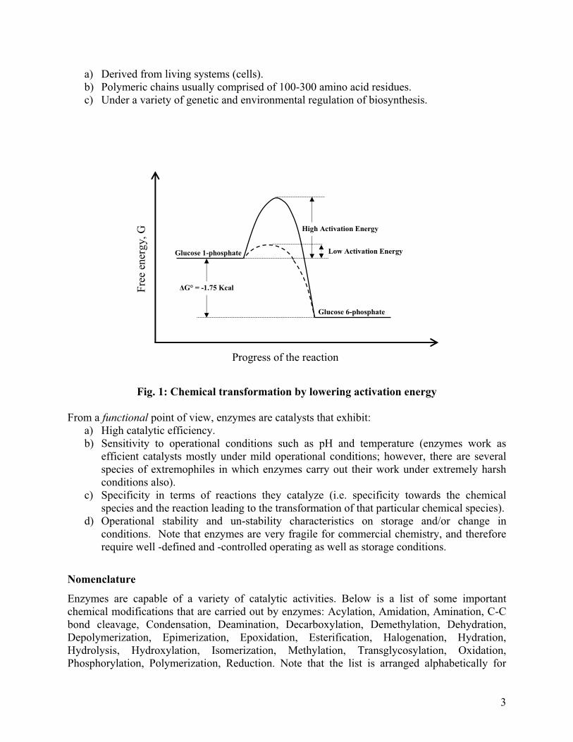
a) Derived from living systems (cells). b) Polymeric chains usually comprised of 100-300 amino acid residues. c) Under a variety of genetic and environmental regulation of biosynthesis.
Glucose 1-phosphate
Glucose 6-phosphate
�G° = -1.75 Kcal
High Activation Energy
Low Activation Energy
Progress of the reaction
Free
ene
rgy,
G
Fig. 1: Chemical transformation by lowering activation energy
From a functional point of view, enzymes are catalysts that exhibit:
a) High catalytic efficiency. b) Sensitivity to operational conditions such as pH and temperature (enzymes work as
efficient catalysts mostly under mild operational conditions; however, there are several species of extremophiles in which enzymes carry out their work under extremely harsh conditions also).
c) Specificity in terms of reactions they catalyze (i.e. specificity towards the chemical species and the reaction leading to the transformation of that particular chemical species).
d) Operational stability and un-stability characteristics on storage and/or change in conditions. Note that enzymes are very fragile for commercial chemistry, and therefore require well -defined and -controlled operating as well as storage conditions.
Nomenclature
Enzymes are capable of a variety of catalytic activities. Below is a list of some important chemical modifications that are carried out by enzymes: Acylation, Amidation, Amination, C-C bond cleavage, Condensation, Deamination, Decarboxylation, Demethylation, Dehydration, Depolymerization, Epimerization, Epoxidation, Esterification, Halogenation, Hydration, Hydrolysis, Hydroxylation, Isomerization, Methylation, Transglycosylation, Oxidation, Phosphorylation, Polymerization, Reduction. Note that the list is arranged alphabetically for
3

convenience. This listing gives a clear indication of the diverse range of chemical reactions that enzymes are capable of carrying out. This not only gives a loaded arsenal of options required for complex biochemical reactions inside living systems, it also provides commercial avenues for carrying out any of the chemical modifications under controlled conditions for commercial applications. Note that using conventional chemical procedures for carrying out these chemical modifications are quite expensive compared to enzymatic procedures due to requirements of high temperatures and pressures as operating conditions in the former. Before proceeding further with understanding enzymes and their functions, it is important to learn about the nomenclature associated with enzymatic processes. Following is the common list of terms and their definitions that are used in discussing enzyme catalyzed reactions:
1. Substrate: This refers to the chemical species that is to be modified or converted by the enzyme into another chemical species. For example, in Figure 1, G 1-P is the substrate. While we study most enzymatic reactions as single substrate reactions, in true biological terms enzymes act on multiple substrates. However, for simplicity purposes, while looking at a single substrate, we assume that the enzymatic reaction kinetics are independent of or insensitive to the other substrates.
2. Product: This refers to the chemical species that is obtained as a result of the enzymatic action on the substrate. For example, in Figure 1, G 6-P is the product. Note that many enzymatic reactions can yield multiple products, however, for simplicity we consider on that subset of reactions in which the desired product is produced.
3. Cofactor & Coenzyme: Cofactor is a small molecule, such as a metal ion, that is required for the enzyme to function. Coenzyme is a specific cofactor and is usually a derivative of some vitamin molecule. Thus, a cofactor can be an inorganic molecule (metal ion) or an organic molecule (vitamin as coenzyme or protein) that is necessary for the catalytic activity of the enzyme.
4. Activator: This is a molecule that participates in the enzymatic reaction helping the binding of the substrate to the enzyme or by stabilization of the enzyme-substrate complex. It modifies the reaction rate and serves as a regulatory device in living cells. Note that an activator actually participates in the specific enzymatic reaction by first “activating” an enzyme. In simplistic terms, an enzyme binds to its activator to become an “activated” enzyme (complex) which then acts on the substrate.
5. Inhibitor: This is a molecule that decreases the enzyme activity or simply abolishes enzyme activity. Inhibitors can affect enzymatic reactions reversibly as well as irreversibly.
6. Inducer & Repressor: It is now clear that enzymes are molecules that are biosynthesized and their activity is required to be regulated (controlled) for a properly functioning living unit. Thus, biosynthesis of enzymes is regulated by molecules that control enzymatic production by induction or repression of genetic and biochemical pathways leading to their production inside the living cells. These molecules are called inducers and repressors respectively. To regulate biochemical pathways, evolution has designed many substrate molecules to act as inducers and product molecules to act as repressors. For example, xylose works as an inducer for gluco-isomerase and cellulose works as an inducer for cellulose for microbial cells. Presence of these molecules in the medium leads to production of this enzyme inside microbial cells. On the other hand, on conversion of xylose or cellulose to glucose (i.e. the product of the chemical modifications), glucose
4

acts as a repressor, shutting down the biochemical production of the enzymes by interfering with the transcription of specific genes.
Functional Nomenclature: The classification of enzymes based on their functions
Enzymes are usually named based on their functions, i.e. specific catalytic activities. This makes classification and understanding of enzymes quite easy. The general rule for enzyme nomenclature is attaching the letters “-ase” to the specific reaction(s) that is(are) catalyzed by that enzyme. Some examples of this are given below:
1. Oxidoreductases: Catalyze oxidation & reduction of the molecules. 2. Transferases: Transfer specific group from one substrate to another. 3. Hydrolases: Break down large molecules by introduction of water. 4. Lyases: Catalyze cleaving of C - C, C – O, C - N and other bonds by elimination,
leaving double bonds or rings or conversely adding groups to double bonds. 5. Isomerases: Catalyze isomerization of substrate molecules. 6. Ligases: Catalyzes condensation of two molecules (coupled with cleavage of ATP,
GTP etc.). In addition to the above, there is a special “form” of proteins which are inactive precursors and need to be themselves chemically modified (usually by cleavage of one or more peptide bonds), before they are able to work as enzymes. This form of proteins is called the Zymogen form of proteins. Thus zymogens are the inactive precursor protein molecules, which are activated by some chemical modification (usually carried out by another enzyme) resulting in their biochemical activity. Just like proteins, enzymes can be obtained from a variety of sources, depending on which they can be sub-classified in terms of nomenclature as well as specificity of their activities. It is important to note that activity of enzymes carrying out the same chemical modifications can vary depending on the source of the enzyme. This is due to several possible differences, for example, proteases (that cleave the peptide bonds) from bacterial sources do not have glycosylation (sugars added on to proteins), where as plant derived proteases like papain have glycosylations. Similarly, lipases found in animal sources (e.g. porcine pancreatic lipase) have different activity compared to lipases from microbial sources for the same chemical modification. The differences will become clearer in the next sections of this chapter when different parameters governing enzymatic activities will be discussed in detail.
Units of Enzyme Activity
Historically, purified enzymes have not been used often for assessing activity in terms of product formation (see the section on “Kinetics of Enzyme Catalyzed Reactions” for details). Therefore, the classically, enzyme activity (of an enzymatic preparation, that may or may not contain purified form of enzyme) is expressed in terms of “international units” abbreviated as IU, which is the quantity of enzyme that converts 1 �mol of substrate into products per minute. The S. I. Unit for enzyme activity is “katal”, symbolized as kat, that is defined as the quantity of enzyme that produces 1 mole of product per second. Note that 1 kat = 6 x 10⁷ IU.
5

Enzyme-substrate interactions
An enzyme molecule “E” has to bind with a substrate molecule “S” first for any enzymatic reaction to proceed. Formation of this enzyme-substrate complex, “ES”, is the first step in enzymatic catalysis. Binding of substrates to enzymes take place at specific “binding” sites that contain particular amino acid residues that facilitate not only binding affinities but also specificity towards substrate molecules. In other words, binding site amino acids on enzymes are responsible for (a) desired binding affinities for substrates and (b) ensuring specificity for substrates. Once the substrates are bound, a subset of the amino acids involved in binding, or a different set of amino acids that are not exposed in free enzyme but become capable of directly interacting with the substrate molecules subsequent to binding, form the ES transition state complex. These amino acids are called active site residues. Note that while binding site amino acid residues are responsible for specificity towards substrates, it is the active site residues that determine the catalytic efficiency of the enzyme molecules. The active site residues are directly involved in breaking and formation of new chemical bonds. While enzymes come in a variety of structures, sizes and have different catalytic functions, the binding and active sites of enzymes share certain characteristics. These can be briefly stated as below:
1. Substrates bind to enzymes by multiple weak interactions (i.e. non-covalent interactions). These are reversible interactions, with well defined equilibrium constants usually in the ranges varying from mM to M values. These weak interactions are quite similar to those that are involved in protein folding, i.e. electrostatic and hydrogen bonding, hydrophobic interactions and van der Waal forces. Note that van der Waal forces are significant only when multiple substrate atoms come in very close vicinity to multiple enzyme atoms.
2. Usually binding sites on enzymes are in form of “gaps” or “clefts” or “crevices”, i.e. some kind of three dimensional structures which have vacancies for a particular shape to fit into them. These particular shapes are exactly complementary to the shapes of substrate molecules that specifically fit into these three dimensional structures. The first visualization of such shape dependent specificity of substrate-enzyme reactions was provided by Fischer in 1890 using the “Lock-and-key” model of interactions. Fig. 2 shows a cartoon representation of this model. The substrate S has a perfect complementary shape to a particular site in the enzyme E. This way, S fits into E just like a key fits into a lock, giving the ES complex. Note that in Figure 2, reactions subsequent to ES formation are shaded, since these will be discussed later. However, after several years of research, it was found that the lock-and-key model is not the most accurate representation of enzyme-substrate interactions. Koshland Jr., in 1958, postulated that shapes of binding sites of enzyme molecules can change significantly on the approach of substrate molecules due to the weak interactions taking place between the particular amino acid residues on the enzyme and the substrate molecules. Here, the enzymes take complementary shape of the substrate only after binding of the substrate has been initiated. This is shown by the cartoon in Fig. 3. This model, which has now been proven for several ES complexes by structural studies, is called the “Induced-fit” model for enzyme substrate interactions. Note that in Figure 3 also, reactions subsequent to ES formation are shaded, since these will be discussed later.
6

3. The active site of an enzyme takes up a very small fraction of the total enzyme volume. For an enzyme made of about 100 amino acid residues, the active site is usually comprised of only a few residues that can vary in number from 2 to 5. It is believed that the need for the rest of the amino acids, which have no apparent role in the enzyme activity, is to stabilize the enzyme structure.
4. Keeping the overall structure of the enzyme same, changing of one or more residues on the active and/or binding sites can greatly affect not only the catalytic efficiency but also specificity of the enzymes. This feature is largely utilized in the field of molecular enzyme engineering, where specific enzymes with desired catalytic activities for specialized substrates are produced using genetic modification of the protein sequences.
SE
Fig. 2:“Lock and key” model of enzyme-substrate interaction Summarizing enzyme-substrate interactions, it is important to remember that substrates are bound to enzymes by multiple weak interactions and only a very small number of amino acid residues are actually involved in making and breaking of bonds. These amino acid residues form a three dimensional entity called the active site of the enzyme.
kSk-S
E
P
E
P
ES
7

Fig. 3: “Induced fit” model for enzyme-substrate interaction
Kinetics of Enzyme Catalyzed Reactions
Enzyme kinetics is useful for quantitative analysis of the effect of various parameters that influence the rate of an enzyme catalyzed reaction. These parameters are concentrations of various interacting species like the enzyme, substrate, inhibitors, activators, pH, water activity and temperature. Essentially, after obtaining experimental data for an enzyme catalyzed reaction (e.g. product formation as a function of time), a mechanistic hypothesis is tested in which rate expressions are written that provide one or more variable parameters. If the rate expressions, and the equations derived from them, fit the data well then these can form the basis of understanding the actual mechanism of the enzymatic reaction. More importantly, inability to fit the experimental data with a particular set of equations helps rule out the “incorrect” mechanistic hypothesis that resulted in the equations in the first place. Further, after getting the “correct” mechanistic hypothesis by finding the set of equations which actually fit the data, it becomes possible to vary the operational conditions to control the enzymatic activity.
Before going further into the details of enzyme kinetics, it is useful to consider the historical perspective through which enzyme kinetics has evolved. The rates of reactions catalyzed by enzymes were studied in late 19th century, when (a) enzymes were mostly in form of “crude
kSk-S
S
E
P
E
P
E
ES
8

extracts” from microbial sources (i.e. impure preparations) and (b) methods to determine product formation (i.e. enzymatic activity measurements) were quite primitive. This was due to lack of detection of product formation at the early stages of the reaction when the product concentration is very low, leading to measurements being done after letting the reaction run for a period of time. The first kinetic studies were reported by O’Sullivan and Tompson in 1890 on the enzyme invertase that converts sucrose to glucose and fructose. Note that in the present day, invertase is called �-fructofurnosidase. They essentially found that the enzyme (invertase) was capable of withstanding higher temperatures in presence of its substrate (cane sugar: the main component of which is sucrose) without getting “spoiled”. This led them to conclude that the enzyme must be forming a complex with its substrate. In 1892 Brown reported that that rate of hydrolysis of sucrose by living yeast cells was independent of sucrose concentration, and more importantly, the kinetics deviated from second-order kinetics. Subsequently in 1902, Brown confirmed the findings with purified invertase (rather than living microbial cells which were producing the enzyme). The results led Brown to suggest the involvement of an enzyme-substrate complex that limited the achievable rate of the enzymatic reaction. However, this meant that the enzyme-substrate complex had to exist for a brief instant of time before breaking down into products and a maximum rate would be reached only when the substrate concentration would be high enough for all enzyme molecules to be present as the enzyme-substrate complex. Clearly, the requirement of a fixed lifetime for the enzyme-substrate complex required questioning the simple chemical equilibrium fundamentals, where for two reacting species in a closed system, neither of the species is ever in the completely complex form. Therefore, Victor Henri proposed a mechanism in 1903 that was conceptually similar to that of Brown, but was expressed more precisely by assuming an equilibrium between the free enzyme and the enzyme-substrate complex. All of the above studies were done without buffered solutions and the experimental data supporting either of the above proposals was not particularly reproducible, leading to a general non-acceptance of the mechanisms.
Michaelis-Menten Kinetics
In 1913, Leonor Michaelis and Maud Leonora Menten carried out experiments with invertase at a fixed pH using buffered solutions. Their experimental findings seemed to agree with the suggestions of Henri and they proposed a mechanism as follows:
E + S � ES � E + P
The main assumption in this mechanism was equilibrium between E and ES. They also assumed total enzyme concentration [E]0 << [S]. Note that bar brackets, i.e. [ ], denote concentration of a given species and the units are “M” (Molar = mol/liter).
By considering only the “initial” reaction rate, when [P] ~ 0, Michaelis and Menten applied enzyme conservation to derive an expression for the velocity of an enzyme catalyzed reaction. The derviation goes as follows:
E + S � ES � E + P k1 k2
k-1
9

Now, at equilibrium,
[S]1k
ES][1-k [E]
ES][1-k E][S][1k
��
�
The enzyme conservation equation gives [E]0 = [E] + [ES]
}[S]1k
ES][1-k {1 [ES] [ES]
[S]1k
ES][1-k [E] 0 �����
Therefore, (initial) velocity of the reaction “v0” is given by
[S] K[S]V
[S] 1k1-k
[S][E]2k [ES]2k v
S
Max00 �
�
�
���
[S] K[S]V v
S
max0 ��� (1)
where, Vmax = k2[E]0 is the maximum possible velocity of the reaction for product formation, and KS is the equilibrium constant for the equilibrium between free and complexed (with substrate) enzyme species. The strongest assumption in Eq. 1 is equilibrium. This implicitly puts a constraint on the rate constants for the forward and backward reactions for the enzyme-substrate complex formation. More importantly, if the enzyme-substrate complex is very short lived, i.e. it cannot be observed experimentally then assuming equilibrium does not seem valid. The limitations of Eq. 1 were overcome by Briggs and Haldane in 1925. They took the same reaction scheme:
E + S � ES � E + P k1 k2
k -1
However, instead of assuming equilibrium, they assumed a steady state for the enzyme-substrate complex. This essentially meant that even if the ES complex is very short lived, the number of
ES complexes remains the same at any given point. Mathematically, this means 0. dt
d[ES]�
Therefore, writing the rate equation for the ES complex, Briggs and Haldane got,
1k2k 1-k
K where,[S][ES]K [E]
)[ES]2k 1-(k [E][S]1k 0 dt
d[ES]
MM �
���
����
10

Following the enzyme conservation and exactly the same treatment as done by Michaelis-Menten in 1913, they got the final expression for the initial velocity of an enzymatic reaction as:
[S] K[S]V v
M
max0 �� (2)
Owing to the similarity in the expression derived by Briggs and Haldane to that derived by Michaelis-Menten, Eq. 2 is the generally accepted Michaelis-Menten equation in the current scenario, with KM being called the Michaelis constant. Note that Eq. 2 has the same maximum velocity of the reaction as Eq. 1, i.e. Vmax = k2[E]0. k2 is also referred to as the “catalytic constant” and more generally symbolized by kcat. It is also sometimes called the “turnover number” since its reciprocal defines the number of catalytic cycles per unit time. Figure 4 shows the graphical representation of Eq. 2 (referred to as the Michaelis-Menten equation from now on). From Eq. 2 and Figure 4, it is clear that KM can be defined as that substrate concentration [S] at which the reaction velocity is half the maximum velocity of the enzymatic reaction. For understanding purposes, KM is quite often considered a good indicator for affinity of the enzyme for a substrate. Lower the KM, higher would be the affinity since the enzymatic reaction would proceed faster (half the maximum velocity is reached at much lower substrate concentration). The following features also need to be noted from Eq. 2 and Fig. 4:
1. As [S] � � (i.e. [S] >> KM), v0 = Vmax. This represents zero-order kinetics (initial velocity of the reaction is independent of substrate concentration).
2. When [S] << KM, v0 = Vmax[S]/KM. This represents first-order kinetics (initial velocity of the reaction has a first order dependence on the substrate concentration).
Summarizing the kinetic parameters of the Michaelis-Menten equation, the following salient points need to be remembered:
1. Vmax is not a fundamental kinetic constant, as it depends on [E]0. 2. Both Vmax and KM are “phenomenological” parameters and not “real” kinetic parameters.
The real kinetic parameters are the rate constants k1, k-1 and k2, that are not determinable using the Michaelis-Menten kinetics.
3. v0 does not reach Vmax at finite value of [S]. Even at [S] = 100 KM, v0 = 100Vmax /101. 4. KM denotes affinity of the substrate to the enzyme. 5. k2 = kcat = turn over number is 1 - 108 /sec for most enzymes. 6. kcat / KM is also defined as the “Specificity Constant” for an enzyme towards a substrate.
It is important to note that kcat / KM is also often utilized as a measure of “Catalytic Efficiency” of enzymes, especially while comparing activities of more than one enzyme on a particular substrate. However, there are several limitations of utilizing this parameter as a measure of catalytic efficiency. In fact, Eisenberg et al. (2007) have demonstrated very elegantly that utilizing kcat / KM as a measure of catalytic effectiveness of an enzyme is not only incorrect, but also misleading in many cases.
11

Vmax
Vmax /2
Fig. 4: Graphical representation of the Michaelis-Menten equation Having understood Michaelis-Menten kinetics and their origin, the assumptions that are intrinsic to the kinetic treatment of enzyme catalyzed data need to be re-emphasized as follows:
1. Michaelis-Menten kinetics is applicable to only single substrate enzymatic reactions. In view of this, how does one apply this to enzymes working inside cellular systems, where most of the enzyme species are involved with multiple substrates? The simplest explanation towards this is the assumption that except for the substrate in question, all the other substrates are “infinite” or present in much larger quantities than the substrate in question.
2. The Michaelis-Menten kinetics assumes formation of a single E-S complex. 3. Only initial reaction rates are of interest in analyzing kinetic data, i.e. it is assumed that
the measurement of the enzymatic reaction is done for v0 � [P] ~ 0. 4. A steady state is assumed with respect to the E-S complex. 5. Kinetic analysis assumes constant pH, temperature, ionic strength and water activity for
the enzymatic reaction.
[S]
[vo]
KM
12

6. The turnover number is often a “lumped” parameter, i.e. it may involve several kinetic steps lumped into one.
The significance of Michaelis-Menten kinetics, in spite of the above assumptions, is highlighted by the fact that even after about a 100 years of its conceptualization, it has withstood the test of time and serves as one of the most important quantitative tool to understand enzyme kinetics. It provides profound insight into processes taking place in living cells. For example, during glycolysis, the first step involves formation of glucose-1-phosphate (G-1-P) from glucose. Now, G-1-P can either be directed by an enzyme towards glycogen synthesis for storage or can be converted to Fructose-1,6-biP for progressing into glycolysis using another enzyme. The KM for G-1-P of the former enzyme is at least a 1000 fold larger than the KM for G-1-P of the latter enzyme. This automatically ensures that when glucose is in low concentrations, the cells easily break down glucose to derive energy using the latter pathway and the moment glucose is in large excess, only then is glycogen storage pathway initiated. Apart from providing an understanding of intracellular enzymatic processes, Michaelis-Menten kinetics is very important in assay procedures for enzymes and for design of large scale enzymatic processes for commercially relevant products.
Lineweaver-Burke Plot
Data analysis using the Michaelis-Menten kinetics can sometimes be a challenging task. For example, it is clear from Fig. 4 that unless the experimental data can be obtained for substrate concentrations at which the initial velocity of the enzymatic reaction starts reaching an asymptotic value, non-linear fitting of the data using Eq. 2 may not yield very accurate kinetic parameters. Moreover, traditionally, non-linear fitting of data to obtain mechanistic or kinetic parameters is not considered very elegant. Therefore, a linearized form of Michaelis-Menten kinetics was developed that results in a “Lineweaver-Burke Plot”. Essentially, consider the Michaelis-Menten equation:
[S] K[S]V v
M
max0 ��
Now, if a reciprocal is taken on both sides, we get:
maxmax
M
0 V1
[S]1
VK
v1 �� (3)
Eq. 3 is a straight line of the form y = mx + c between the reciprocals of initial velocity and substrate concentrations respectively, with the slope “m” and the Y-intercept as “c”. The slope and the intercepts on the X- and Y- axes provide direct values for the Michaelis-Menten kinetic parameters. Fig. 5 shows the Lineweaver-Burke plot and its elegant simplicity in deducing the kinetic parameters of an enzyme catalyzed reaction. Here, it is important to note that some key quantitative limitations are associated with the Lineweaver-Burke plot. The main limitation comes when one is working with a large range of substrate concentrations. Since the plot involves reciprocal of substrate concentrations, this leads to “data clustering” with smaller substrate concentrations forming extreme and segregated data
13

points towards the right in Fig. 5 and higher substrate concentrations clustering near the intersection of the X and Y axes. To overcome these limitations, other linear methods using different arithmetic rearrangements of Eq. 2 have been developed (Eadie-Hofstee, Wolf-Hanes and Cornish-Bowden). However, they are beyond the scope of this chapter and can be found in the references provided. Moreover, as a diagnostic and simple analytical method, the Lineweaver-Burke plot serves the purpose quite well for determining the Michaelis-Menten kinetic parameters.
Slope = KM /Vmax
Fig. 5: The Lineweaver-Burke plot
Deviations from Hyperbolic Enzyme Kinetics
All enzymatic reactions do not follow hyperbolic Michaelis-Menten kinetics as shown in Fig. 4. There are certain cases, where just in the presence of the enzyme molecules and single substrate molecules, the kinetics observed deviate from the hyperbolic relationship between the initial velocity of the reaction and substrate concentration. These can be broadly categorized in two categories:
1. Substrate inhibition: An enzymatic reaction is “inhibited” in presence of the substrate itself. This might sound unrealistic initially, but a slightly deeper look into the enzyme-substrate interaction can actually show why this would happen. Consider an enzyme called succinate dehydrogenase that converts succinate to fumerate in the TCA cycle.
1/vo
1/[S]
1/Vmax-1/KM
14

Now, the chemical structure of succinate is –OOC-CH2-CH2-COO–. Both the carboxyl groups are required to be bound to the enzyme. At very high succinate concentrations, however, two succinate molecules bind to the enzyme with one carboxyl group from each succinate. This forms a dead end complex with the enzyme not able to act on either of the two succinate molecules. In fact, substrate inhibition, at high substrate concentrations, is also a feature of invertase that converts sucrose into glucose and fructose. Recollecting from our previous discussions, this was the first enzyme that was studied for developing theoretical knowledge on the kinetics of enzyme catalyzed reactions. One of the reasons why hyperbolic kinetics of Michaelis-Menten were developed only as late as 1913 was that invertase did not show hyperbolic kinetics. The kinetic scheme for substrate inhibition can be written as:
E + S � ES � E + P k1 k2
k-1+ S kS k-S
ESS
The species ESS is formed by a substrate molecule binding to the ES complex and is a dead end for product formation. Clearly, at very high [S], ES converts to ESS rather than producing P. Using the mass action law and total enzyme balance, it can be shown that
S
S-SS
1
1-S
SS
S
020
kk K and
kk K where
K[S]
[S]K 1
[E]k v
��
���
(4)
A graphical representation for Eq. 4 is shown in Fig. 6. Note that the actual maximum velocity of the reaction that is reached in case of substrate inhibition is lower than the theoretical Vmax as per the Michaelis-Menten equation. Differentiating initial velocity with respect to substrate concentration in Eq. 4, and finding the maxima by the second derivative, yields the maximum possible velocity of the reaction with substrate inhibition (shown by the horizontal dashed arrow in Fig. 6) when [S] = {KS.KSS}1/2. This is shown in Fig. 6 by a dashed vertical arrow and denoted by [S]m.
2. Allosteric Enzyme Kinetics: Involvement of “cooperative” binding of the substrate leads to deviation from hyperbolic kinetics. In this case also, as in the above case, multiple binding sites for substrate exist. Binding of a substrate molecule at one site induces conformational changes that result in altered affinities for the binding sites. Fig. 7 shows that cooperative binding of the substrate molecules yields a sigmoidal dependence (solid curve) of initial velocity on the substrate concentration in contrast to hyperbolic kinetics (dashed curve). Note that these results are very similar to oxygen binding to hemoglobin, which also is a cooperative binding process. The sigmoidal velocity curve is very important for regulation of enzymatic reactions inside living cells (prokaryotic and
15

eukaryotic alike). Sigmoidal initial velocity implies that over a certain concentration range, the reaction velocity of the enzyme can respond to changes in [S] more readily, leading to a better control of cell metabolism. The Hill equation for cooperative interactions is often utilized to kinetically analyze the sigmoidal initial velocity dependence on substrate concentration for allosteric enzymes. The analytical expression represented by the sigmoid in Fig. 7 is:
n
nmax
0 [S] K [S]V v
�� (5)
where, K is the Hill’s constant (different from Michaelis constant) and “n” is the “Hill’s coefficient” giving the approximate number of cooperative units in the enzyme. Note that Eq. 5 is derived by explicitly assuming that the concentration of all enzyme-substrate complexes containing less than “n” substrate molecules will be negligible. This assumption often results in the value of “n” coming out to be a non-integer (i.e. a rational number), which is then rounded off to represent the cooperative units = the minimum number of binding sites on the enzyme.
Vmax
Fig. 6: Substrate inhibition
v0
[S]
Maximum v0
[S]m = {KS.KSS}1/2
16

Vmax
v0
[S]
Figure 7: Cooperative binding in allosteric enzyme kinetics
The above discussion clearly demonstrates deviations from the hyperbolic Michaelis-Menten enzyme kinetics occurs when there are multiple sites for the substrate molecules on enzymes. However, there is one case, when there is no deviation from hyperbolic kinetics is observed, in spite of multiple substrate binding sites on the enzyme molecules. This is when the substrate binding sites on the enzyme are identical and independent of each other. In this case, suppose there are three binding sites on the enzyme for substrate molecules, then hyperbolic kinetics is still observed, with the “effective” enzyme concentration being three times more than the actual enzyme concentration.
Role of Effector Molecules in Enzyme Kinetics
Effector molecules affect rates of enzymatic reactions. They can be broadly classified into activators and inhibitors. Activators are regulatory devices for enzymes in living cells. They modify the reaction rates by participating in the binding of substrate molecules to the enzyme or by stabilization of the ES complex. Ca2+ is one of the common examples of an activator for conversion of starch to dextrins by the action of -amylase. Similarly Co2+ acts as an activator for the enzyme glucose isomerase which converts glucose to fructose. The kinetic expressions for activator molecules can be written based on the following reaction schemes:
17

E + A � EA ka
k-a
EA + S � EAS � EA + P k1 k2
k-1
As done previously, applying steady state on EAS and following the total enzyme conservation, the initial velocity of the reaction is calculated as:
a
a-a
aM
max0
kk K where
[S] }[A]K{1K
[S]V v
�
���
(6)
Notice that compared to Eq. 2, the effective KM value in Eq. 6 has changed, i.e. }[A]K{1K a
M � , with
a clear dependence on the dissociation constant of the activator and the activator concentration itself.
The other class of effector molecules are called inhibitors. As the name clearly suggest, they can inhibit enzymatic reactions. “Irreversible” inhibitors decrease the enzyme activity in a course of time to zero. These essentially bind to the enzyme molecules resulting in permanent conformational changes in the enzyme structure leading to an inactive state. Examples of irreversible inihibitors are heavy metal ions. Mercury is poisonous in nature since it acts as an irreversible inhibitor. In contrast, “reversible” inhibitors are “milder” and are used for regulatory as well as commercially important therapeutic purposes for enzymes implicated in metabolic pathways and diseases. A generalized scheme for reversible inhibitions is shown below, with I as the inhibitor. For simplicity, equilibrium constants (dissociation constants) for various reactions are shown, instead of rate constants. Remember that dissociation constants are the ratio of backward to forward rate constants for a reaction.
E + S � ES � E + P KES k2
+ + I I
S + EI ESI (Inactive)
KEI
KES
KESI
Notice that in the above generalized reaction scheme, the inhibitor is capable of interacting with various enzyme species (E, ES), with different dissociation constants. However, the substrate has the same dissociation constant for reacting with different enzyme species (E, EI). What is clear
18

from the reaction scheme is that the reaction can be driven in one direction or the other by modulating the concentrations of different reacting species (more specifically substrate and inhibitor), thereby tilting the reaction balance in a particular direction. A critical study on reversible inhibition of enzymes should include: (a) pattern of binding of inhibitor I to E or ES, (b) rate expressions of reactions in which I is involved, (c) effect(s) of [I] on the Michaelis-Menten kinetic parameters of the reaction, (d) effect(s) of I on the Lineweaver-Burke plot and (e) the degree of inhibition by I. The degree of inhibition is defined by i = 1 – (vI/v0), where vI is the initial velocity of the reaction in presence of the inhibitor. The above parameters provide general features for classifying reversible inhibitors into competitive (KESI � �), non-competitive (KESI = KEI ), and un-competitive inhibitors (KEI � �). We look at each of these in detail below. Competitive Inhibition
In this form of inhibition, “I” is a substrate analog and competes with S for the same active site on E, i.e. S and I are mutually exclusive with respect to the active site of the enzyme. Here, neither the ES complex can combine with I, nor can the EI complex bind S. EI by itself is a dead end species. In the generalized reaction scheme shown above, competitive inhibition is represented by putting KESI = �. Applying the Michaelis-Menten methodology for deriving initial velocity of the reaction, using the steady state assumption, we get
[S] }K[I]{1K
[S]V v
EIM
max0
��� (7)
Notice that the maximum velocity of the enzymatic reaction is independent of the inhibitor, but the Michaelis constant has effectively changed by a factor that includes the inhibitor concentration and the dissociation constant of the inhibitor. Since it is being multiplied by a value greater than 1, there is an increase in the value of the Michaelis constant. This essentially means that the amount of substrate required for reaching half the maximum velocity of the reaction now increases due to the presence of a competitive inhibitor. It is also interesting to note that the degree of inhibition for a competitive inhibitor is given by:
[S] }K[I]{1K
K[I]K
i
EIM
EIM
��� .
Taking reciprocal on both sides of Eq. 7 for looking at the Lineweaver-Burke form, we get
maxEImax
M
0 V1
[S]1}
K[I]{1
VK
v1 ��� (8)
Fig. 8 shows the graphical representation for Eq. 8. Since the maximum velocity of the reaction is not affected by the competitive inhibitor, the straight lines in presence or absence of the inhibitor intersect on the Y-axis at the reciprocal value of the maximum velocity. From the slope of the lines without and with inhibitor, as well as from the X-intercepts, one can then calculate the dissociation constant of the inhibitor with the enzyme.
19

[I]
1/v0
Fig. 8: Competitive inhibition
Non-Competitive Inhibition
In this form of inhibition, “I” does not influence S binding to E. S and I bind reversibly, randomly and independently at different sites on E. The ESI complex formed is inactive, i.e. ESI is a dead end species. In the generalized reaction scheme shown above, competitive inhibition is represented by putting KESI = KEI. For simplicity, let KI = KESI = KEI. Applying the Michaelis-Menten methodology for deriving initial velocity of the reaction, using the steady state assumption, we get
[S] K
[S]
K[I]1
V
vM
I
max
0 �
�� (9)
Notice that the Michaelis constant of the enzymatic reaction is independent of the inhibitor, but the maximum velocity of the reaction has effectively changed by a factor that includes the inhibitor concentration and the dissociation constant of the inhibitor. Since it is being divided by a value greater than 1, there is a reduction in the maximum velocity of the reaction in this case. This essentially means that no amount of substrate can increase the maximum velocity of the reaction to the value obtained without inhibitor, and, the amount of substrate required to reach half the maximum velocity remains the same. It is also interesting to note that the degree of
1 KM ( 1 + I ) KEI
1/[S]
-1/ KM
[I] = 0
1/ Vmax
20

inhibition for a non-competitive inhibitor is given by: IK [I]
[I] i �
� , which is independent of
substrate concentration. Taking reciprocal on both sides of Eq. 9 for looking at the Lineweaver-Burke form, we get
}K[I]{1
V1
[S]1}
K[I]{1
VK
v1
ImaxImax
M
0
���� (10)
Fig. 9 shows the graphical representation for Eq. 10. Since the Michaelis constant of the reaction is not affected by the non-competitive inhibitor, the straight lines in presence or absence of the inhibitor intersect on the X-axis at the negative reciprocal value of the Michaelis constant. From the slope of the lines without and with inhibitor, as well as from the Y-intercepts, one can then calculate the dissociation constant of the inhibitor with the enzyme. Notice in Fig. 9 that the lines start becoming steeper as the inhibitor concentration is increased, resulting in an increase in the Y-intercept indicating a decrease in the value of maximum velocity.
[I]1/v0
Fig. 9: Non-competitive inhibition
Un-Competitive Inhibition
In this form of inhibition, “I” binds reversibly to ES yielding an inactive ESI complex, but is incapable of binding to E alone. This kind of inhibition is rare for uni-substrate reactions, but quite useful for bisubstrate reactions. Since I cannot bind to E and only to ES, in the generalized reaction scheme shown above, competitive inhibition is represented by putting KEI = �. Applying the Michaelis-Menten methodology for deriving initial velocity of the reaction, using the steady state assumption, we get
1/[S]
[I] = 0
21
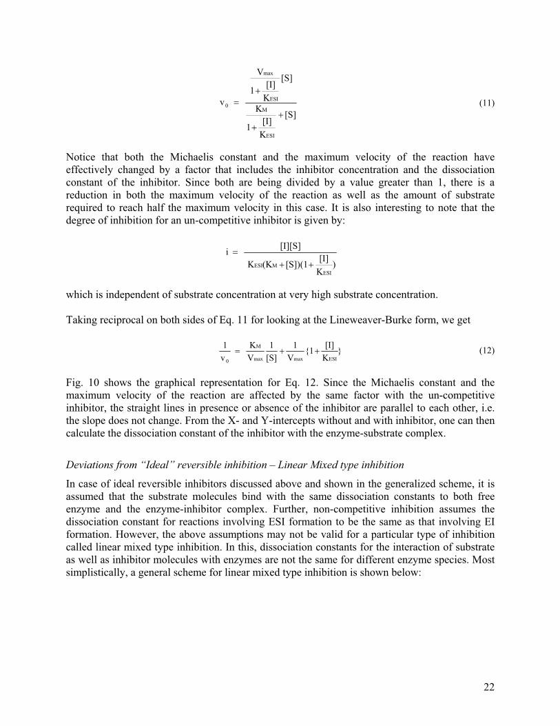
[S]
K[I]1
K
[S]
K[I]1
V
v
ESI
M
ESI
max
0
��
�� (11)
Notice that both the Michaelis constant and the maximum velocity of the reaction have effectively changed by a factor that includes the inhibitor concentration and the dissociation constant of the inhibitor. Since both are being divided by a value greater than 1, there is a reduction in both the maximum velocity of the reaction as well as the amount of substrate required to reach half the maximum velocity in this case. It is also interesting to note that the degree of inhibition for an un-competitive inhibitor is given by:
)K[I][S])(1 (KK
[I][S] i
ESIMESI ��
�
which is independent of substrate concentration at very high substrate concentration. Taking reciprocal on both sides of Eq. 11 for looking at the Lineweaver-Burke form, we get
}K[I]{1
V1
[S]1
VK
v1
ESImaxmax
M
0
��� (12)
Fig. 10 shows the graphical representation for Eq. 12. Since the Michaelis constant and the maximum velocity of the reaction are affected by the same factor with the un-competitive inhibitor, the straight lines in presence or absence of the inhibitor are parallel to each other, i.e. the slope does not change. From the X- and Y-intercepts without and with inhibitor, one can then calculate the dissociation constant of the inhibitor with the enzyme-substrate complex.
Deviations from “Ideal” reversible inhibition – Linear Mixed type inhibition
In case of ideal reversible inhibitors discussed above and shown in the generalized scheme, it is assumed that the substrate molecules bind with the same dissociation constants to both free enzyme and the enzyme-inhibitor complex. Further, non-competitive inhibition assumes the dissociation constant for reactions involving ESI formation to be the same as that involving EI formation. However, the above assumptions may not be valid for a particular type of inhibition called linear mixed type inhibition. In this, dissociation constants for the interaction of substrate as well as inhibitor molecules with enzymes are not the same for different enzyme species. Most simplistically, a general scheme for linear mixed type inhibition is shown below:
22

E + S � ES � E + P KES k2
+ + I I
S + EI ESI (Inactive)
KEI
�KES
�KEI
[I] = 0
1 [ I ] ax
Fig. 10: Un-competitive inhibition Notice that in the above simplified scheme, the inhibitor is capable of interacting with various enzyme species (E, ES), with different dissociation constants such that the interaction of the inhibitor with ES has a dissociation constant different from the interaction of the inhibitor with free enzyme by a factor of �. Similarly, the substrate has a different dissociation constant for reacting with different enzyme species (E, EI), with the difference represented by the same factor �. Note that the same � is used here just for simplicity. In reality, the difference factor for the dissociation constants may be different. Applying the Michaelis-Menten methodology for deriving initial velocity of the reaction, using the steady state assumption, we get
KM
Vmax
1/Vmax
1/[S]
1/v0 Vm KESI
1+{ } Slope =
-1/KM
23

[S] K
[S]
}
K[I][S]{1 }
K[I]{1K
[S]V v
}KEI[I]{1
}KEI[I]{1
M
}KEI[I]
{1
Vmax
EIEIM
max0
�
����
�
�
�
�
(13)
Notice that both the maximum velocity and the Michaelis constant have effectively changed by a factor that includes the inhibitor concentration, the dissociation constants of the inhibitor and the factor . Clearly he degree of inhibition also now depends on . Taking reciprocal on both sides of Eq. 13 for looking at the Lineweaver-Burke form, we get
max
EI
EImax
M
0 V
}K[I]{1
[S]1}
K[I]{1
VK
v1
���� (14)
Fig. 11 shows the graphical representation for Eq. 14 for ��. Since both the maximum velocity and the Michaelis constant are affected in linear mixed type inhibition, the straight lines in presence or absence of the inhibitor intersect at a point which is neither on the X-axis nor on the Y-axis. From the slope of the lines without and with inhibitor, as well as from the X- and Y-intercepts, one can then calculate the dissociation constants of the inhibitor with the enzyme species.
Fig. 11: Linear Mixed type inhibition
1/[S]
1/v0
[I] for
� > 1
24

Effect of pH on enzyme kinetics
Our discussion on enzymatic reactions so far has clearly pointed out those weak interactions between substrate and enzyme molecules that lead to the catalytic activity of enzymes resulting in product formation. Electrostatic interactions are the key to enzymatic modification of substrates. Most active sites have one or more ionizable residues whose ionization state is defined by pH of the medium in which the reaction is being carried out. Thus, while pH may affect the overall stability of protein conformation that is responsible for enzymatic activity, it also can lead to a shift in equilibrium of the enzymatic reaction. For instance, alcohol dehydrogenase produces acetaldehyde from ethanol in presence of NAD+. This reaction results in formation of NADH along with acetaldehyde and also release of a free proton. Now at a low pH, when there is an abundance of free protons to begin with, the reaction equilibrium rapidly shifts in the reverse direction where the acetaldehyde is converted back into ethanol in presence of NADH. Further, if the enzyme is somewhat insensitive to pH (which is very rarely the case) and/or the substrate is very susceptible to ionization (which is many times the case, especially with protein substrates), then a change in pH leads to ionization of the substrate leading to a change in the fraction of the substrate binding to the enzyme. Changes in pH can also affect enzymatic reactions in the way effector molecules affect them. This is indeed the case when a proton can behave as an activator or an inhibitor for the enzyme. The effects of pH on the kinetics of enzyme catalyzed reactions can very well be studied using the equations already described above for different effector molecules by replacing [A] or [I] by [H+] = 10-pH. The dissociation constants for such analyses are provided by pKa’s of the specific interacting residues on the enzyme. Figure 12 shows a typical graph of kinetics of an enzyme catalyzed reaction as a function of pH. Notice that there is a pH optimum of the reaction that is clearly shown by a peak. At different pH values, both the initial velocity and the maximum velocity of the reaction are affected. Experimentally, pH titrations for enzymatic activities may not yield as sharp peaks for pH optimum as shown in Fig. 12. Instead, one would observe either a broader peak or, in extreme cases, a plateau. This serves as a strong diagnostic to predict the number active site residues since a single peak would signify an overwhelming dependence on a single ionization site or a plateau would indicate presence of two possible ionization sites. On the same lines, the pH titrations of enzymatic activity can also help in generating hypothesis about the identity of active site residues, since pKa values of individual amino acid residues are well known. However, this needs to be carefully considered since it is now well established that ionization properties of amino acids actually change when they are a part of a folded protein compared to their presence in free form in solutions. Effect of Temperature
Just as for proteins in general, temperature is also a very important parameter for enzymatic reactions. While thermal deactivation of enzyme itself can provide important information about enzyme substrate interactions, the temperature dependence of ES complex formation and the catalytic step leading to product formation are important in understanding the working mechanism of the enzyme. A classical experiment used to find dissociation constants for ES complex formation involves measurements done on E and S interactions at 4 ºC, at which negligible product formation takes place leading to an accumulation of the ES complex. Therefore, by varying substrate concentrations and by measuring the fraction of substrate or
25

enzyme involved in ES complex formation, one can determine the dissociation constant of an enzyme with its substrate. The temperature dependence of the catalytic step provides a useful tool to study and control enzymatic reactions. The rate constant, k2, representing the catalytic step of the enzyme follows the Arrhenius relationship with temperature i.e. k2 = Aexp(-EA/RT), where EA represents the activation energy, A is the Arrhenius coefficient, R is the molar gas constant and T is the absolute temperature.
v0 or
Vmaxopt. pH
pH
Figure 12: Effect of pH on enzyme kinetics
Ribozymes
All our discussion on enzymes above is essentially based on proteins as enzymes. However, it is important to introduce another class of molecules called ribozymes (ribozyme = ribonucleic acid enzyme) which are RNA molecules that catalyze biochemical reactions. These are also called RNA enzymes. Sidney Altman and Thomas Cech discovered that some RNA molecules have catalytic activity which is shown by their ability to cleave themselves (self-splicing RNA molecules). They were awarded the Nobel prize for this discovery in 1989. Many natural ribozymes catalyze cleavage of RNAs (either themselves or others) by transesterification or peptidyl transfer activity, but now it is well established that ribozymes also catalyze the
26

aminotransferase activity of the ribosome. Other ribozymes have also been discovered which carry out the general acid/base catalysis of reactions (for example, the hepatitis delta virus ribozyme). Considering the fact that ribozymes are much simpler structures than many protein enzymes, and RNA comes before proteins in the “central dogma” of biology, they have generated a great interest in evolution of life on earth. In fact, many people consider ribozymes to be living fossils of a life based primarily on nucleic acids before assembling of complex structures into uni- and multi- cellular organisms took place.
Applications of Enzymes
As mentioned in the “Nomenclature” section, enzymes provide commercial avenues for carrying out a variety of chemical modifications under controlled (and in-expensive as well as much less harsh compared to chemical methods) conditions for commercial applications. Wide commercial applications range from production of alcohol from starchy sources (by using amylases to convert the starch source into fermentable sugars like glucose and fructose) to production of drugs from different chemical or biochemical compounds. Antibiotics are the most common examples of the latter. Production of pharmaceutically relevant forms of penicillins (by utilizing an acylase), cyclosporins, cephalosporins, and many modern day antibiotics requires enzymatic treatments of native compounds that are chemically modified by specific enzymes to achieve biocompatible and bioactive compounds. Enzymes can themselves also act as therapeutic agents in a variety of ways ranging from wound healing to cosmetic applications to applications in internal medicine. Along with the above, enzymes are extremely important in the field of medical diagnostics, where enzymatic reactions provide relatively simple and user-friendly technologies for detecting presence of compounds. If the substrate of an enzyme is present in a biological sample, the enzyme can detect its presence by converting the substrate into a product that can then be measured (most often by utilizing optical properties of the substrate or product). Similar strategies for non-diagnostic applications render enzymes as extremely useful analytical agents due to the simplicity of the assays based on enzymatic activity. One of the breakthrough developments of modern day, that has revolutionized biological research in this genomic era, is the technology behind Polymerase Chain Reaction (PCR), which has now evolved into very rigorous reverse transcriptase PCR and real time PCR. This technology is based on the enzymatic activity of DNA/RNA polymerases and enables one to amplify both DNA as well as RNA sequences. While most of our discussion in this chapter is predominantly applicable to enzymes in solutions, it is important to note that the above applications often utilize “immobilized” enzymes. Enzyme molecules are often anchored to a support via simple adsorption mechanism or through covalent binding and a continuous stream of substrate is maintained over the anchored enzyme. This process, when coupled with proper recycling of the continuous stream, leads to efficient conversion of substrates into products, without losing any enzyme. Enzyme molecules can also be immobilized by entrapment within membranes or small particles, such that the pores of these entrapping structures are small enough to allow substrates and products to permeate in and out (e.g. glucose) but do not allow relatively large enzyme molecules to pass through. While basic kinetic mechanisms of enzymes remain the same for immobilized enzymes (compared to those in solutions), some modifications of kinetic equations occurs due to immobilization coupled with mass-transfer parameters that need to be accounted for during such reactions. Introduction to
27
these modifications and parameters is beyond the scope of this chapter and can be found in the suggested reading mentioned in different degree of detail. Finally, enzymes have played the single largest role in basic biological research for over a century in terms of understanding the ubiquitous process of protein folding. This has been made possible due to the fact that enzymatic activity provides a rigorous quantitative measure for a protein folded in an “active” or “native” conformation under specific conditions.
Suggested Reading 1. Garrett & Grisham. Biochemistry, 2nd Ed.. Saunders College Publishing, 1999. 2. Athel Cornish-Bowden, Fundamentals of Enzyme Kinetics (3rd Ed.), Portland Press, 2004. 3. Stryer, Biochemistry (4th Ed.), W. H. Freeman and Co., NY, 1995. 4. Madigan, Martinko & Parker, Brock Biology of Microorganisms (9th Ed.), Prentice Hall Intl. Inc., 2000. 5. Glazer & Nikaido, Microbial Biotechnology: Fundamentals of Applied Microbiology, W. H. Freeman and
Company, 1995. 6. Benkovic and Schiffer, A Perspective on Enzyme Catalysis, Science, Vol. 301: pp. 1196-1202, 2003. 7. Eisenthal, Danson & Hough, Catalytic efficiency and kcat/KM: a useful comparator?, Trends in Biotechnology,
Vol. 25: pp. 247-249, 2007.
28