Embed Size (px)
Citation preview

ORIGINAL RESEARCHpublished: 25 August 2016
doi: 10.3389/fmicb.2016.01311
Frontiers in Microbiology | www.frontiersin.org 1 August 2016 | Volume 7 | Article 1311
Edited by:
Jyoti Prakash Tamang,
Sikkim University, India
Reviewed by:
Catarina Prista,
Instituto Superior de Agronomia,
Portugal
Braulio Esteve-Zarzoso,
Rovira i Virgili University, Spain
Jose Antonio Curiel,
Instituto de Ciencias de la Vid y del
Vino, Spain
*Correspondence:
Zhijun Qiao
Hongjiang Yang
Specialty section:
This article was submitted to
Food Microbiology,
a section of the journal
Frontiers in Microbiology
Received: 29 April 2016
Accepted: 08 August 2016
Published: 25 August 2016
Citation:
Qin H, Sun Q, Pan X, Qiao Z and
Yang H (2016) Microbial Diversity and
Biochemical Analysis of Suanzhou: A
Traditional Chinese Fermented Cereal
Gruel. Front. Microbiol. 7:1311.
doi: 10.3389/fmicb.2016.01311
Microbial Diversity and BiochemicalAnalysis of Suanzhou: A TraditionalChinese Fermented Cereal GruelHuibin Qin 1, 2, Qinghui Sun 1, Xuewei Pan 1, Zhijun Qiao 2* and Hongjiang Yang 1*
1 Key Laboratory of Industrial Microbiology, Ministry of Education, Tianjin Key Laboratory of Industrial Microbiology, College of
Biotechnology, Tianjin University of Science and Technology, Tianjin, China, 2 Key Laboratory of Crop Gene Resources and
Germplasm Enhancement on Loess Plateau, Ministry of Agriculture, Shanxi Key Laboratory of Genetic Resources and
Genetic Improvement of Minor Crops, Institute of Crop Germplasm Resources of Shanxi Academy of Agricultural Sciences,
Taiyuan, China
Suanzhou as a traditional Chinese gruel is fermented from proso millet and millet. The
biochemical analysis showed Suanzhou had relatively high concentrations of lactic acid,
acetic acid, and free amino acids. The metagenomics of Suanzhou were studied, with the
analysis of the V4 region of 16S rRNA gene, the genera Lactobacillus and Acetobacter
were found dominant with the average abundance of 58.2 and 24.4%, respectively; and
with the analysis of the ITS1 region between 18S and 5.8S rRNA genes, 97.3% of the
fungal community was found belonging to the genus Pichia and 2.7% belonging to five
other genera. Moreover, the isolates recovered from 59 Suanzhou samples with various
media were identified with the 16S rRNA or 18S rRNA gene analyses. Lactobacillus
fermentum (26.9%), L. pentosus (19.4%), L. casei (17.9%), and L. brevis (16.4%) were
the four dominant Lactobacillus species; Acetobacter lovaniensis (38.1%), A. syzygii
(16.7%), A. okinawensis (16.7%), and A. indonesiensis (11.9%) were the four dominant
Acetobacter species; and Pichia kudriavzevii (55.8%) and Galactomyces geotrichum
(23.1%) were the two dominant fungal species. Additionally, L. pentosus p28-c and
L. casei h28-c1 were selected for the fermentations mimicking the natural process.
Collectively, our data demonstrate that Suanzhou is a nutritional food high in free amino
acids and organic acids. Diverse Lactobacillus, Acetobacter, and yeast species are
identified as the dominant microorganisms in Suanzhou. The isolated strains can be
further characterized and used as starters for the industrial production of Suanzhou
safely.
Keywords: Suanzhou, metagenomic analysis, lactic acid bacteria, acetic acid bacteria, yeast, free amino acid
INTRODUCTION
Many types of ethnic fermented cereal foods are widely consumed across the world. Compared withfoods cooked directly from raw materials, fermented cereal foods are generally more tasteful, easilydigested, and richer in various nutrients, such as vitamins, organic acids, and free amino acids(Blandino et al., 2003). Almost all types of cereals have been prepared into many kinds of foodsin various natural fermented processes. Diverse microorganisms, mainly comprised of a numberof bacteria and yeast species originated from the cereal grains and local environments, have beenidentified with diverse techniques (Tamang et al., 2016a,b).
Qin et al. Characterization of the Fermented Cereal Food Suanzhou
A number of indigenous fermented foods have been madeof rice or rice as the main material, such as Idli, dominantwith Leuconostoc lactis (Saravanan and Shetty, 2016); Ang-kakalso named Chinese red rice, dominant with Monascus strains(Lotong and Suwanarit, 1990); Selroti, dominant with multipleLAB and yeast species (Das et al., 2012); and Jiuniang or Laozao,dominant with Rhizopus, Mucor, Monilia, Aspergillus, and yeastspecies (Li and Hsieh, 2004). Wheat is an important sourceof diet proteins. However, wheat-based foods may contain acertain level of gluten which may cause allergic reactions in someindividuals. Fermented wheat flour foods can greatly reducethe gluten content to safe levels, such as Sourdough, dominantwith Lactobacillus species, and Saccharomyces cerevisiae (Settanniet al., 2005); Bhatooru, dominant with S. cerevisiae, Lactobacillusplantarum, and Bacillus sp. (Savitri and Bhalla, 2013); and Miso,dominant with Pediococcus acidilactici (Asahara et al., 1992).Maize based fermented foods mainly include doklu, dominantwith Lactobacillus fermentum, L. plantarum, and Pediococcuspentosaceus (Assohoun-Djeni et al., 2016); and Ogi, dominantwith P. acidilactici and Lactobacillus paraplantarum (Okeke et al.,2015). Sorghum based fermented foods are consumed in anumber of African countries, such as Injera (Fischer et al., 2014),Kisra (Mohammed et al., 1991), and Hussuwa (Yousif et al.,2010), and all of them are rich in lactic acid bacteria (LAB). Milletis another important cereal grain and consumed as a staple foodthroughout the world (Saleh et al., 2013). Dosa (Palanisamy et al.,2012) and Ben-saalga (Tou et al., 2006) are two types of fermentedmillet foods which are also rich in LAB.
Prosomillet andmillet are highly drought-resistant crops withlow demanding to environments. In northwestern China, prosomillet and millet are commonly fermented to make Suanzhou, asour gruel food easily prepared in local individual households.Until now, no studies of Suanzhou have been conducted interms of its nutrients and microbial populations. In this work,totally 59 Suanzhou samples were collected for metagenomicDNA analysis and detection of free amino acids and organic acidsconcentration. Additionally, the dominant microorganisms inSuanzhou were isolated, identified, and characterized for possibleapplications in industrial production of Suanzhou.
MATERIALS AND METHODS
Preparation of SuanzhouSuanzhou is a gruel made of fermented cereals prepared inindividual households. Briefly, four types of raw materials wereused in fermentations, group A containing samples fermentedfrom millet with a small amount of rice (<10%), group B frommillet, group C from white proso millet, and group D fromred proso millet (Table S1). About 100 g grains were soakedin the fermentation soup or supernatant from the previousfermentation and kept at room temperature for 24 h in a jarsealed with a lid. Fermented grains are taken away for cooking.Raw materials are again added and soaked in the acidic soupfor future fermentation and consumption. The water loss issupplemented with boiled water. Thirty samples (h1-30) werefrom Hequ county, in which different proso millet were used(Table S1). Twenty-nine samples (p1-30, the sample p25 was
contaminated and removed from the analyses) from Pianguancounty, in whichmillet was used as main rawmaterial (Table S1).Both counties are located in Shanxi Province, China. All theSuanzhou samples were collected after 24 h incubation. Two 50-ml sour soup samples were obtained from each jar-fermentor andstored at 4◦C for assays.
Metagenomic Analysis of SuanzhouSamplesThe samples for metagenomic analysis were randomly selectedon the basis of raw materials used for fermentation from tworegions, Pianguan County and Hequ County. Suanzhou sampleswere centrifuged and the pellets were subjected to the extractionof genomic DNA by using Qiagen DNA blood and tissue kit(Qiagen, Dutch). To investigate the bacterial communities, thehypervariable V4 region (∼207 bp) of the 16S rRNA genewas analyzed with the primers 520-F (5′ AYTGGGYDTAAAGNG 3′) and 802-R (5′ TACNVGGGTATCTAATCC 3′) (Coleet al., 2005). To investigate the fungal communities, the ITS1region between 18S and 5.8S rRNA genes was analyzed withthe primers ITS1-F (5′ CTTGGTCATTTAGAGGAAGTAA 3′)and ITS2 (5′ GCTGCGTTCTTCATCGATGC 3′) (Schnabel et al.,1999). Metagenomic sequencing was performed on an IlluminaMiSeq system by Shanghai Personal Biotechnology Co., Ltd.,China (http://www.personalbio.cn). The obtained sequenceswere assigned to the operational taxonomic units (OTUs)with a threshold of 97% pairwise identity using the BLASTNtool in the NCBI (http://blast.ncbi.nlm.nih.gov/Blast.cgi). Thespecies diversity, richness, and abundance were estimated bythe Shannon, Chao1, and ACE indices (http://www.mothur.org/wiki/). Totally, 1,046,828 clean sequencing reads with lengtharound 225 bp were obtained from the libraries of the 24Suanzhou samples. Venn diagram was used to group the sampleson basis of the genus level (http://bioinformatics.psb.ugent.be/webtools/Venn/). The possible correlations of the microbialcommunities were analyzed by the online software Cytoscape(www.mothur.org/wiki/Otu.association).
Determination of pH, Lactic Acid, AceticAcid, and Free Amino AcidsThe pH was measured by using a Sartorius pH indicator. Lacticacid, acetic acid, and free amino acids were determined byusing ACQUITY UPLC M-Class System with BEH C18 Column(2.1× 50mm× 1.7µm) and PDA detector (Waters Corporation,Milford, MA, USA). The supernatants of Suanzhou sampleswere subjected to filtration by the syringe filter (0.2µm poresize). The filtrate was directly used for the lactic acid and aceticacid analysis. The mobile phase used was prepared by mixing0.01mol/l KH2PO4 (pH 3.0) and CH3CN in a ratio of 98:2.Free amino acids in the supernatant samples were determinedaccording to the manual from Waters and the method describedpreviously (Fiechter et al., 2011). The reagent 6-aminoquinolyl-N-hydroxysuccinimidyl carbamate (AQC, Waters) was used toderivatize amino acids. Amino acids AAS18 and A9906 (Sigma-Aldrich) were used as analytical standards. All experimentswere repeated three times. The values for pH, organic acids,
Frontiers in Microbiology | www.frontiersin.org 2 August 2016 | Volume 7 | Article 1311

Qin et al. Characterization of the Fermented Cereal Food Suanzhou
and free amino acids were subjected to one-way analysis ofvariance (ANOVA) by Tukey’s method of the statistical softwareStatistica 7.0.
Isolation and Identification of theMicroorganisms from SuanzhouSerial dilutions of Suanzhou samples with 0.9% NaCl (normalsaline) were prepared. The dilutions were spread on the plateswith the selective media, including Lactobacilli MRS agar(pH 5.0) for the isolation of LAB (De Man et al., 1960), GYC agarwith CaCO3 for the isolation of acetic acid bacteria (AAB; Rasporand Goranovic, 2008), and YEPD agar for the isolation of yeastspecies (Treco and Lundblad, 2001). The plates were incubatedat 30◦C for 48 h to enumerate the colonies. All experiments wererepeated three times. Average and standard deviation (STDEV)were calculated using Excel.
Bacterial genomic DNA was extracted with the E.Z.N.A. R©
Bacterial DNA Kit (Omega Bio-tek Inc., USA) from a widevariety of gram positive and negative bacterial species. Primers27f (5′ AGAGTTTGATCCTGGCTCAG 3′) and 1492r (5′ GGTTACCTTGTTACGACTT 3′) were used for amplification ofthe 16S rRNA gene (Lane, 1991). Fungal genomic DNA wasextracted with the method as described previously (Löffleret al., 1997). Primers NS1 (5′ GTAGTCATATGCTTGTCTC3′) and NS4 (5′ CTTCCGTCAATTCCTTTAAG 3′) were usedto amplify the 18S rRNA gene (White et al., 1990). Theamplicons were sequenced directly and the obtained sequenceswere deposited in GenBank. The accession number of 16SrRNA gene sequences was KX150543 through KX150609 for theLAB isolates and KX150610 through KX150652 for the AABisolates. The accession number of 18S rRNA gene sequences wasKX150653 through KX150704 for the yeast isolates. Sequencesimilarity was analyzed by using the online tool BLAST in theNCBI (http://blast.ncbi.nlm.nih.gov/Blast.cgi). Sequences wereassigned to species level when similarities were at 97% or higher.The phylogenetic tree was constructed with the software MEGA6(Tamura et al., 2013).
Growth Curve of LAB StrainsThe growth curve of the isolated LAB isolates was determinedin MRS medium (pH6.0). The single colony was inoculated in3ml fresh MRS medium for static cultivation at 30◦C for 12 h.The overnight precultures were diluted in fresh MRS mediumto OD600 <0.2. The mixtures were dispensed into the 96-wellplates with 250µl per well. The culture was then grown for staticcultivation at 30◦C for 48 h. The OD600 was recorded at the 10-min intervals by the Synergy H1 Multi-Mode Reader (BioTekInstruments, Inc., Winooski, VT, USA). All experiments wererepeated three times. Average and STDEV were calculated usingExcel.
In-lab Fermentation of SuanzhouProso millet (100 g) was weighed and cleaned with water. Afterdrying, proso millet was put in a 1-l bottle and filled with 900mldistilled water. The mixture was pasteurized at 65◦C for 30minand ready for in-lab fermentation of Suanzhou. Two of theisolated LAB strains were selected and used as starter separately.
Overnight culture of each strain was transferred in the sterilizedproso millet suspension with 5% inoculation for static cultivationat 30◦C. The fermented grains were replaced with fresh rawproso millet daily. As aforementioned, Suanzhou samples wereanalyzed in terms of pH, lactic acid, and free amino acids. LABcells were enumerated by the standard plating method. Theexperiment was repeated three times. Average and STDEV werecalculated using Excel.
RESULTS
Biochemical Characteristics of SuanzhouSamplesA collection of 59 Suanzhou samples were subjected to theanalysis of acidity, lactic acid, and acetic acid. The pH valueranged from 3.22± 0.01 to 5.15± 0.02 in all samples (Table S1).The lactic acid concentration was from 0.74 ± 0.02 to 6.20 ±
0.04mg/ml in the samples fermented from proso millet and2.93 ± 0.00 to 17.00 ± 0.00mg/ml in the samples from millet(Table S1). The acetic acid concentration was from 0.33 ± 0.00to 7.66± 0.05mg/ml in the samples fermented from proso milletand 0.28 ± 0.04 to 3.80 ± 0.00mg/ml in the samples from millet(Table S1). With statistical analysis, the significant differenceswere observed in the comparison pairs, including groups A andC, A and D, B and C, and B and D, suggesting the raw materialswere associated with the organic acid levels in Suanzhou.
Analysis of Free Amino Acids ContentThe content of free amino acids was measured, ranging from81.93 ± 5.01 to 665.47 ± 2.19µ g/ml in the samples fermentedfrom proso millet and 67.91 ± 0.41 to 1257.30 ± 0.93µ g/mlin the samples from millet (Table S1). Of the total amino acids,essential amino acids accounted for 13.27 ± 0.29 to 48.02 ±
0.48% in the samples fermented from proso millet and 27.50 ±
0.09 to 51.81± 0.02% in the samples frommillet (Table S1).Withstatistical analysis, Suanzhou fermented from the different cerealsdisplayed the significant difference in the content of free aminoacids among the comparison pairs, including groups A and C, Aand D, B and C, and B and D. However, rice had no effects on theamino acids levels, possibly due to the low ratio (<10%) in theraw material.
Analysis of the V4 Region of 16S rRNAGeneThe species diversity, richness, and evenness in the 24 Suanzhousamples were estimated by the collector rarefaction curves ofthe observed species, chao1, and Shannon indices (Figure S1).The results showed that the libraries were relatively well-sampledand constructed. The bacterial diversity was mainly analyzedat the genus level. In the 24 Suanzhou samples, the top 4dominant species groups were the genus Lactobacillus, thegenus Acetobacter, the family Acetobacteraceae, and the orderLactobacillales, with an average abundance of 58.20± 0.28, 24.40± 0.23, 9.00 ± 0.15, and 2.00 ± 0.04%, respectively (Figure 1).The sample h25 had 55 OTUs, the maximum of all testedsamples; while only 21 OTUs were found in the sample h19,
Frontiers in Microbiology | www.frontiersin.org 3 August 2016 | Volume 7 | Article 1311

Qin et al. Characterization of the Fermented Cereal Food Suanzhou
FIGURE 1 | Abundance of the top 12 abundant operational taxonomic units (OTUs) among the Suanzhou samples. OTUs with the abundance <0.10%
were not included in this diagram. G: OTUs at the genus level. F: OTUs at the family level. O: OTUs at the order level. Others: all OTUs with the abundance ≥0.10%
were included. In summary, h10 with 0.10% Streptophyta (order); h12 with 0.10% Psychrobacter and 0.10% Clostridium; h15 with 0.10% Psychrobacter and 0.3%
Streptophyta (order); h17 with 0.30% Psychrobacter, 0.10% Micrococcaceae, 0.10% Phenylobacterium, and 0.10% Methylobacteriaceae (family); h21 with 0.20%
Psychrobacter, 0.10% Micrococcaceae (family), 0.10% Phenylobacterium, 0.10% Bacteroides, and 0.30% Bacteroidales (order); h22 with 0.10% Psychrobacter, and
2.60% Gluconacetobacter; h24 with 1.30% Psychrobacter, 0.20% Micrococcaceae (family); 0.5% Phenylobacterium, 0.40% Methylobacteriaceae (family), 0.10%
Bacteroides, 0.10% Bacteroidales (order), 0.10% Brochothrix, 0.10% Caulobacteraceae (family), 0.10% Balneimonas, 0.90% Methylobacterium, 0.10% Halomonas,
0.30% Acinetobacter, 0.10% Enhydrobacter, and 0.10% Stenotrophomonas; h25 with 0.40% Psychrobacter, 0.30% Streptophyta (order), 0.20% Phenylobacterium,
0.10% Methylobacteriaceae (family); 0.10% Bacteroides, 0.20% Myroides, 0.20% Sphingobacterium, 0.10% Bacteroidales (order), 0.10% Brochothrix, 0.20%
Staphylococcus, 0.10% Gemellales (order), 0.10% Kuenenia, 0.20% Enterobacteriaceae (family), 0.10% Klebsiella, 0.20% Acinetobacter, 0.10%
Xanthomonadaceae (family); h26 with 0.10% Streptophyta (order); h29 with 0.10% Psychrobacter, and 0.50% Pediococcus; p1 with 0.40% Psychrobacter, 0.10%
Micrococcaceae (family), 0.40% Streptophyta (order), 0.10% Phenylobacterium, and 0.10% Methylobacteriaceae (family); p2 with 0.10% Comamonas, 0.20%
Enterobacteriaceae (family), 0.10% Klebsiella, 0.10% Moraxellaceae (family), 0.10% Acinetobacter, and 0.10% Xanthomonadaceae (family); p3 with 0.20%
Psychrobacter, 0.10% Streptophyta, 0.10% Phenylobacterium, 0.10% Methylobacteriaceae (family), 0.60% Bacteroides, 0.20% Bacteroidales (order), 0.30%
Bacteroidia (class), 0.30% Barnesiellaceae (family), 0.10% Veillonellaceae (family), 0.10% Desulfovibri, and 0.10% Akkermansia; p4 with 0.20% Streptophyta (order);
p6 with 0.20% Psychrobacter, 0.10% Micrococcaceae (family), 0.10% Streptophyta (order), 0.10% Phenylobacterium, 0.10% Methylobacteriaceae (family), and
0.10% Bifidobacteriaceae (family); p9 with 0.20% Psychrobacter, 0.10% Phenylobacterium, and 0.10% Methylobacteriaceae (family); p10 with 0.20%
Psychrobacter, 0.10% Micrococcaceae (family), 0.10% Phenylobacterium, 0.10% Actinomyces, 1.10% Chryseobacterium, 0.20% Klebsiella, and 0.30%
Acinetobacter; p12 with 0.20% Psychrobacter, 0.10% Micrococcaceae (family), 0.10% Streptophyta (order), 0.10% Phenylobacterium, 0.20% Methylobacteriaceae
(family), 0.10% Bacteroidales (order); p16 with 0.80% Psychrobacter, 0.30% Micrococcaceae (family), 0.30% Streptophyta (order), 0.30% Phenylobacterium, 0.30%
Methylobacteriaceae (family), 0.10% Nocardioidaceae (family), 0.10% Myroides, 0.10% Brochothrix, 0.10% Paenibacillus; p18 with 0.30% Psychrobacter, 0.10%
Micrococcaceae (family), 0.10% Phenylobacterium, 0.10% Methylobacteriaceae (family), 0.10% Halomonas; and p20 with 0.10% Psychrobacter, 0.10% Streptophyta
(order), 0.40% Halomonas, and 0.20% Thermus.
mainly including Lactobacillus (53.80%) and Acetobacter (44.7%)(Figure S2).
There were many microorganisms commonly present in theSuanzhou samples with low abundance. Psychrobacter specieswere detected in 17 Suanzhou samples with the averageabundance from 0.10 to 1.30%. Phenylobacterium species weredetected in 12 Suanzhou samples with the average abundancefrom 0.10 to 0.50%. Streptophyta (order) species were detectedin 11 Suanzhou samples with the average abundance from 0.10to 0.40%. Methylobacteriaceae (family) species were detected in11 Suanzhou samples with the average abundance from 0.10to 0.40%. Micrococcaceae (family) species were detected in 9Suanzhou samples with the average abundance from 0.10 to0.30% (Figure 1).
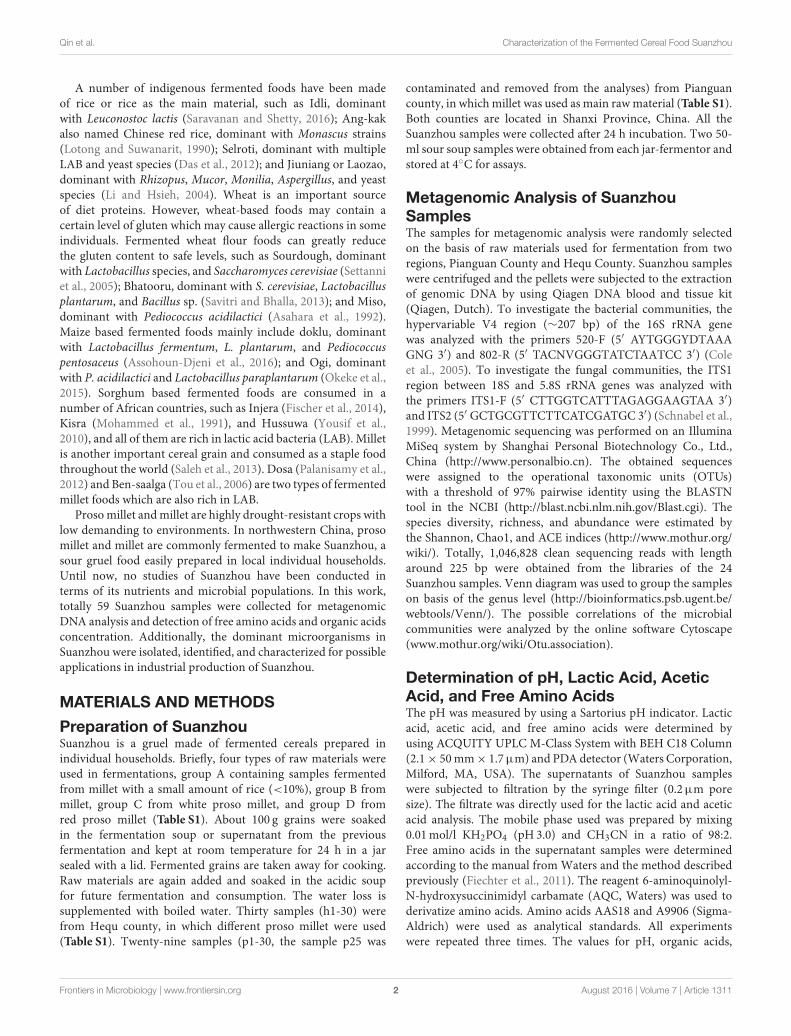
Analysis of the ITS1 RegionThe sample h21 was analyzed for its fungal communitiesby the Miseq system. In the obtained sequences, 29.40%showed no blast hits. Of the remaining sequences, at thegenus level, the abundance of Pichia, Xeromyces, Candida,Issatchenkia, Cryptococcus, and Trichosporon accountedfor 97.30, 1.50, 0.81, 0.35, 0.02, and 0.01%, respectively(Figure 2). Pichia was the prominent OTUs in the sampleh21.
In contrast to the fungal population analysis, the bacterialdiversity was also determined in the sample h21. The dominantOTUs included Acetobacteraceae (family) 35.0%, Lactobacillales(order) 15.0%, Acetobacter 22.8%, Lactobacillus 20.3%, andLactobacillaceae (family) 4.1% (Figure 1).
Frontiers in Microbiology | www.frontiersin.org 4 August 2016 | Volume 7 | Article 1311
Qin et al. Characterization of the Fermented Cereal Food Suanzhou
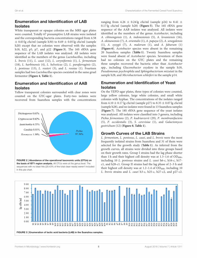
Enumeration and Identification of LABIsolatesWhite transparent or opaque colonies on the MRS agar plateswere counted. Totally 67 presumptive LAB strains were isolatedand the corresponding bacteria concentrations ranged from 4.58± 0.03 lg cfu/ml (sample h30) to 8.69 ± 0.05 lg cfu/ml (sampleh20) except that no colonies were observed with the samplesh18, h22, p5, p7, and p22 (Figure 3). The 16S rRNA genesequence of the LAB isolates was analyzed. All isolates wereidentified as the members of the genus Lactobacillus, includingL. brevis (11), L. casei (12), L. coryniformis (1), L. fermentum(18), L. harbinensis (4), L. helveticus (2), L. parafarraginis (2),L. pentosus (13), L. reuteri (3), and L. rossiae (1). Fourteensamples had two Lactobacillus species coexisted in the same gruelfermentor (Figure 4, Table 1).
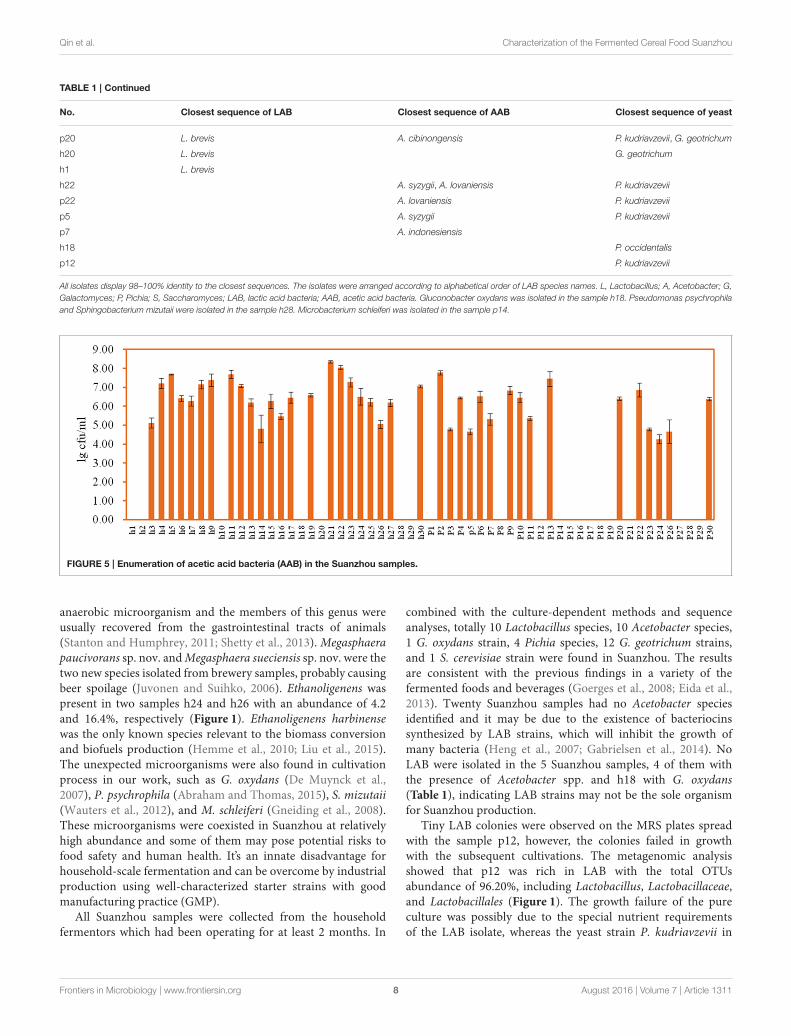
Enumeration and Identification of AABIsolatesYellow transparent colonies surrounded with clear zones werecounted on the GYC-agar plates. Forty-two isolates wererecovered from Suanzhou samples with the concentrations
FIGURE 2 | Abundance of the operational taxonomic units (OTUs) on
the basis of IST1 region analysis. All OTUs were at the genus level. The
sequences with no blast hits (29.43% of the total clean reads) weren’t included
in this pie-chart.
ranging from 4.26 ± 0.24 lg cfu/ml (sample p24) to 8.41 ±
0.17 lg cfu/ml (sample h28) (Figure 5). The 16S rRNA genesequence of the AAB isolates was analyzed. All isolates wereidentified as the members of the genus Acetobacter, includingA. cibinongensis (1), A. indonesiensis (5), A. lovaniensis (16),A. okinawensis (7), A. orientalis (1), A. papaya (2), A. senegalensis(1), A. syzygii (7), A. malorum (1), and A. fabarum (1)(Figure 6). Acetobacter species were absent in the remaining20 Suanzhou samples (Table 1). Twenty Suanzhou sampleswere found absent of Acetobacter species. Seventeen of themhad no colonies on the GYC plates and the remainingthree samples recovered the bacteria other than Acetobacterspp., including Gluconobacter oxydans in the sample h18,Pseudomonas psychrophila and Sphingobacterium mizutaii in thesample h28, andMicrobacterium schleiferi in the sample p14.
Enumeration and Identification of YeastIsolatesOn the YEPD-agar plates, three types of colonies were counted,large yellow colonies, large white colonies, and small whitecolonies with hyphae. The concentrations of the isolates rangedfrom 4.10 ± 0.17 lg cfu/ml (sample p27) to 8.35 ± 0.07 lg cfu/ml(sample h28), and no isolates were found in 13 Suanzhou samples(Figure 7). The 18S rRNA gene sequence of the yeast isolateswas analyzed. All isolates were classified into 3 genera, includingPichia fermentans (2), P. kudriavzevii (29), P. membranifaciens(5), P. occidentalis (3), S. cerevisiae (1), and Galactomycesgeotrichum (12) (Figure 8, Table 1).
Growth Curves of the LAB StrainsL. fermentum, L. pentosus, L. casei, and L. brevis were the mostfrequently isolated strains from Suanzhou and 31 of them wereselected for the growth study (Table 1). As inferred from thegrowth curves, all strains were divided into three groups basedon their growth rates. Group I strains had the lag phase shorterthan 1 h and their highest cell density was at 1.3–1.6 of OD600,including 10 L. pentosus strains and L. casei h6-c, h16-c, h17-c1, and h28-c1. Group II strains had the lag phase of 2–3 h andtheir highest cell density was at 1.2–1.4 of OD600, including 10L. brevis strains and L. casei h3-c, h25-c, h27-c2, and p27-c2.
FIGURE 3 | Enumeration of lactic acid bacteria (LAB) in the Suanzhou samples.
Frontiers in Microbiology | www.frontiersin.org 5 August 2016 | Volume 7 | Article 1311

Qin et al. Characterization of the Fermented Cereal Food Suanzhou
FIGURE 4 | The cladogram tree of the lactic acid bacteria (LAB). The partial 16S rRNA gene sequences (921–925 bp) were analyzed using the Neighbor-Joining
method. L, Lactobacillus.
Group III strains grew very slowly without perceptible lag phasesand their highest cell density was at 0.2–0.4 of OD600, includingL. fermentum p17-c1, L. fermentum p27-c1, and L. casei h12-c2(Figure S3).
In-lab Fermentation of SuanzhouTwo fast growing strains L. pentosus p28-c and L. casei h28-c1 were selected for a 5 d-fermentation mimicking the naturalprocess for Suanzhou preparation. Proso millet was used as rawmaterial and the relevant parameters were analyzed daily. With5% innoculum, the population of L. casei h28-c1 decreased onelog unit after 5 days of fermentation, while the population ofL. pentosus p28-c increased from 7.16 ± 0.16 to 8.44 ± 0.11 lgcfu/ml after 2 d cultivation (Figure 9). The pH value of the twocultures decreased from about 6.6 to 3.5 after 1-day fermentationand remained constant, consistent with the elevated lactic acidcontent in the cultures (Figure 9). The amount of the total aminoacids, essential amino acids, and alanine increased significantlyduring the fermentation. Strain L. casei h28-c1 produced higheramount of free amino acids and essential amino acids than strainL. pentosus p28-c (Figure 9).
DISCUSSION
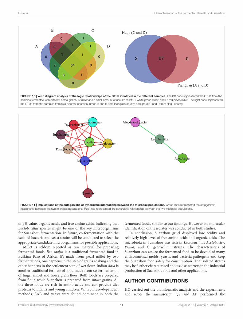
In our work, totally 69 OTUs at the genus level were detected inSuanzhou samples with the metagenomic analysis. The possiblelogic relationship of the OTUs identified among the samples wasanalyzed with the Venn diagram (Figure 10). The samples weredivided into group A, B, C, and D based on the cereals, which
were made of millet with a small amount of rice, millet, whiteproso millet, and red proso millet, respectively (Table S1). Fifty-four of them were found in all groups and 67 OTUs found in thesamples from both counties, indicating no significant differenceexisted. The associations among the microbial populations in thesamples were predicted. Of the top 20 abundant OTUs found inthe metagenomic analysis, the genera Lactobacillus, Acetobacter,and Gluconacetobacter had no correlations with the remaining17 OTUs. However, the abundance of Lactobacillus was inverselycorrelated with Acetobacter and Gluconacetobacter, respectively(Figure 11). Could the antagonistic effect between the generaLactobacillus, Acetobacter, and Gluconacetobacter lead to thedying off of any strains was still a question remained be answerby in-lab fermentations using the isolated microbes.
The majority of environmental microorganisms areinculturable with the available methods. In Suanzhou samples,only a few species including LAB, AAB, and yeasts were foundwith the culture-dependent methods, much less than the OTUsidentified with the metagenomic analysis. Furthermore, with themetagenomic analysis, microbial structure was found not uniqueamong Suanzhou samples and some special OTUs were detectedin the individual samples (Figure 1). Gluconacetobacter wasdetected in the sample h22 with the abundance of 2.6%, whichwas commonly the dominant bacterium found in the traditionalvinegar production (Hommel, 2014). Akkermansia was detectedin the sample p3 with a low abundance 0.10%. A. muciniphila isthe type species of this genus and related with the diet-inducedobesity (Everard et al., 2013).Megasphaera species was dominantin the sample h24 with an abundance of 31.00%. It’s a strictly
Frontiers in Microbiology | www.frontiersin.org 6 August 2016 | Volume 7 | Article 1311
Qin et al. Characterization of the Fermented Cereal Food Suanzhou
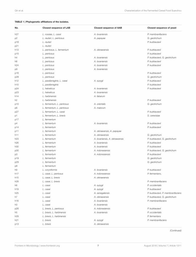
TABLE 1 | Phylogenetic affiliations of the isolates.
No. Closest sequence of LAB Closest sequence of AAB Closest sequence of yeast
h27 L. rossiae, L. casei A. lovaniensis P. membranifaciens
p2 L. reuteri, L. pentosus A. papayae G. geotrichum
p18 L. reuteri P. kudriavzevii
p21 L. reuteri
h13 L. pentosus, L. fermentum A. okinawensis P. kudriavzevii
p15 L. pentosus P. kudriavzevii
h4 L. pentosus A. lovaniensis P. kudriavzevii, G. geotrichum
h8 L. pentosus A. lovaniensis P. kudriavzevii
h24 L. pentosus A. lovaniensis P. kudriavzevii
p9 L. pentosus A. lovaniensis
p16 L. pentosus P. kudriavzevii
p28 L. pentosus G. geotrichum
h12 L. parafarraginis, L. casei A. syzygii P. kudriavzevii
h10 L. parafarraginis P. kudriavzevii
p24 L. helveticus A. lovaniensis P. kudriavzevii
p23 L. helveticus A. lovaniensis
h14 L. harbinensis A. fabarum
h2 L. harbinensis P. kudriavzevii
p10 L. fermentum, L. pentosus A. orientalis G. geotrichum
p6 L. fermentum, L. pentosus A. malorum
p27 L. fermentum, L. casei P. kudriavzevii
p1 L. fermentum, L. brevis S. cerevisiae
p17 L. fermentum
p4 L. fermentum A. lovaniensis P. kudriavzevii
p14 L. fermentum P. kudriavzevii
p11 L. fermentum A. okinawensis, A. papayae
h11 L. fermentum A. okinawensis G. geotrichum
h23 L. fermentum A. lovaniensis, A. okinawensis P. kudriavzevii, G. geotrichum
h26 L. fermentum A. lovaniensis P. kudriavzevii
h30 L. fermentum A. lovaniensis P. kudriavzevii
p30 L. fermentum A. indonesiensis P. kudriavzevii, G. geotrichum
p3 L. fermentum A. indonesiensis P. kudriavzevii
p19 L. fermentum G. geotrichum
p29 L. fermentum G. geotrichum
p8 L. fermentum
h9 L. coryniformis A. lovaniensis P. kudriavzevii
h17 L. casei, L. pentosus A. indonesiensis P. fermentans,
h15 L. casei, L. brevis A. okinawensis
h28 L. casei, L. brevis P. membranifaciens
h6 L. casei A. syzygii P. occidentalis
h19 L. casei A. syzygii P. kudriavzevii
h25 L. casei A. senegalensis P. kudriavzevii, P. membranifaciens
h7 L. casei A. okinawensis P. kudriavzevii, G. geotrichum
h16 L. casei A. lovaniensis P. membranifaciens
h3 L. casei A. lovaniensis
p26 L. brevis, L. pentosus A. indonesiensis P. kudriavzevii
h5 L. brevis, L. harbinensis A. lovaniensis P. occidentalis
h29 L. brevis, L. harbinensis P. fermentans
h21 L. brevis A. syzygii P. membranifaciens
p13 L. brevis A. okinawensis
(Continued)
Frontiers in Microbiology | www.frontiersin.org 7 August 2016 | Volume 7 | Article 1311
Qin et al. Characterization of the Fermented Cereal Food Suanzhou
TABLE 1 | Continued
No. Closest sequence of LAB Closest sequence of AAB Closest sequence of yeast
p20 L. brevis A. cibinongensis P. kudriavzevii, G. geotrichum
h20 L. brevis G. geotrichum
h1 L. brevis
h22 A. syzygii, A. lovaniensis P. kudriavzevii
p22 A. lovaniensis P. kudriavzevii
p5 A. syzygii P. kudriavzevii
p7 A. indonesiensis
h18 P. occidentalis
p12 P. kudriavzevii
All isolates display 98–100% identity to the closest sequences. The isolates were arranged according to alphabetical order of LAB species names. L, Lactobacillus; A, Acetobacter; G,
Galactomyces; P, Pichia; S, Saccharomyces; LAB, lactic acid bacteria; AAB, acetic acid bacteria. Gluconobacter oxydans was isolated in the sample h18. Pseudomonas psychrophila
and Sphingobacterium mizutaii were isolated in the sample h28. Microbacterium schleiferi was isolated in the sample p14.
FIGURE 5 | Enumeration of acetic acid bacteria (AAB) in the Suanzhou samples.
anaerobic microorganism and the members of this genus wereusually recovered from the gastrointestinal tracts of animals(Stanton and Humphrey, 2011; Shetty et al., 2013). Megasphaerapaucivorans sp. nov. andMegasphaera sueciensis sp. nov. were thetwo new species isolated from brewery samples, probably causingbeer spoilage (Juvonen and Suihko, 2006). Ethanoligenens waspresent in two samples h24 and h26 with an abundance of 4.2and 16.4%, respectively (Figure 1). Ethanoligenens harbinensewas the only known species relevant to the biomass conversionand biofuels production (Hemme et al., 2010; Liu et al., 2015).The unexpected microorganisms were also found in cultivationprocess in our work, such as G. oxydans (De Muynck et al.,2007), P. psychrophila (Abraham and Thomas, 2015), S. mizutaii(Wauters et al., 2012), and M. schleiferi (Gneiding et al., 2008).These microorganisms were coexisted in Suanzhou at relativelyhigh abundance and some of them may pose potential risks tofood safety and human health. It’s an innate disadvantage forhousehold-scale fermentation and can be overcome by industrialproduction using well-characterized starter strains with goodmanufacturing practice (GMP).
All Suanzhou samples were collected from the householdfermentors which had been operating for at least 2 months. In
combined with the culture-dependent methods and sequenceanalyses, totally 10 Lactobacillus species, 10 Acetobacter species,1 G. oxydans strain, 4 Pichia species, 12 G. geotrichum strains,and 1 S. cerevisiae strain were found in Suanzhou. The resultsare consistent with the previous findings in a variety of thefermented foods and beverages (Goerges et al., 2008; Eida et al.,2013). Twenty Suanzhou samples had no Acetobacter speciesidentified and it may be due to the existence of bacteriocinssynthesized by LAB strains, which will inhibit the growth ofmany bacteria (Heng et al., 2007; Gabrielsen et al., 2014). NoLAB were isolated in the 5 Suanzhou samples, 4 of them withthe presence of Acetobacter spp. and h18 with G. oxydans(Table 1), indicating LAB strains may not be the sole organismfor Suanzhou production.
Tiny LAB colonies were observed on the MRS plates spreadwith the sample p12, however, the colonies failed in growthwith the subsequent cultivations. The metagenomic analysisshowed that p12 was rich in LAB with the total OTUsabundance of 96.20%, including Lactobacillus, Lactobacillaceae,and Lactobacillales (Figure 1). The growth failure of the pureculture was possibly due to the special nutrient requirementsof the LAB isolate, whereas the yeast strain P. kudriavzevii in
Frontiers in Microbiology | www.frontiersin.org 8 August 2016 | Volume 7 | Article 1311

Qin et al. Characterization of the Fermented Cereal Food Suanzhou
FIGURE 6 | The cladogram tree of the acetic acid bacteria (AAB). The partial 16S rRNA gene sequences (1005–1026 bp) were analyzed using the
Neighbor-Joining method. A, Acetobacter.
FIGURE 7 | Enumeration of yeasts in the Suanzhou samples.
Suanzhou may provide the nutrients essential for the growth ofthe isolate (Assohoun-Djeni et al., 2016). Strain L. fermentump17-c1, L. fermentum p27-c1, and L. casei h12-c2 also exhibitedlow growth rates in the pure cultures, significantly lowerthan the relevant cell numbers in the corresponding Suanzhousamples (Figure 3 and Figure S3). The results indicated thatthe Suanzhou micro-ecosystems possibly provided necessarynutrients for the growth of microorganisms either by degradationof the cereals or by the biosynthesis.
In our work, several yeasts were identified. Pichia species werecommonly isolated in the most samples of Suanzhou withoutdetecting other fungi. The result is consistent with the previousfindings that Pichia species can antagonize and decrease the
abundance of a number of yeast and mold pathogens in variousniches (Golubev, 2006;Mukherjee et al., 2014).G. geotrichumwascoexisted with Pichia species in 5 Suanzhou samples, showingthat its growth wasn’t affected by Pichia, also consistent withthe previous findings (Viljoen, 2006). G. geotrichum or itsanamorph Geotrichum candidum was widely present in the earlystages of ripening on soft cheeses (Marcellino et al., 2001) andsome strains were the starter organisms for fermented foodsand beverages (Goerges et al., 2008; Tamang et al., 2016a).Only one S. cerevisiae strain was isolated from the sample p1which was absent of Acetobacter strains (Table 1). The data isconsistent with the fact that acetic acid can suppress the growthof S. cerevisiae in sour dough (Suihko and Mäkinen, 1984).
Frontiers in Microbiology | www.frontiersin.org 9 August 2016 | Volume 7 | Article 1311

Qin et al. Characterization of the Fermented Cereal Food Suanzhou
FIGURE 8 | The cladogram tree of the yeasts. The partial 18S rRNA gene sequences (928–944 bp) were analyzed using the Neighbor-Joining method. P, Pichia.
S, Saccharomyces. G, Galactomyces.
FIGURE 9 | Preparation of Suanzhou with in-lab fermentations. Strain L. casei h28-c1 and L. pentosus p28-c were used as starters. Bacterial cell density
(lg cfu/ml), pH, lactic acid, total free amino acid, essential amino acid, and alanine content were evaluated, respectively.
Taken together, the yeast strains could play an inhibitory roleagainst food-borne pathogens and also provide some nutrientsfor the growth of other microbes (Goerges et al., 2006; Viljoen,2006).
LAB strains have been widely used in fermented food industryas starters (Giraffa et al., 2010). In our wok, 2 lactobacillus strainswere used for in-lab fermentations separately. The results showedthat the in-lab products were similar to traditional gruel in terms
Frontiers in Microbiology | www.frontiersin.org 10 August 2016 | Volume 7 | Article 1311
Qin et al. Characterization of the Fermented Cereal Food Suanzhou
FIGURE 10 | Venn diagram analysis of the logic relationships of the OTUs identified in the different samples. The left panel represented the OTUs from the
samples fermented with different cereal grains, A: millet and a small amount of rice; B: millet; C: white proso millet; and D: red proso millet. The right panel represented
the OTUs from the samples from two different counties: group A and B from Pianguan county, and group C and D from Hequ county.
FIGURE 11 | Implications of the antagonistic or synergistic interactions between the microbial populations. Green lines represented the antagonistic
relationship between the two microbial populations. Red lines represented the synergistic relationship between the two microbial populations.
of pH value, organic acids, and free amino acids, indicating thatLactobacillus species might be one of the key microorganismsfor Suanzhou fermentation. In future, co-fermentation with theisolated bacteria and yeast strains will be conducted to select theappropriate candidate microorganisms for possible applications.
Millet is seldom reported as raw material for preparingfermented foods. Ben-saalga is a traditional fermented food inBurkina Faso of Africa. It’s made from pearl millet by twofermentations, one happens in the step of grains soaking and theother happens in the settlement step of wet flour. Indian dosa isanother traditional fermented food made from co-fermentationof finger millet and horse gram flour. Both foods are preparedfrom flour, while Suanzhou is prepared from intact grains. Allthe three foods are rich in amino acids and can provide dietproteins to infants and young children. With culture-dependentmethods, LAB and yeasts were found dominant in both the
fermented foods, similar to our findings. However, no molecularidentification of the isolates was conducted in both studies.
In conclusion, Suanzhou gruel displayed low acidity andrelatively high-level of free amino acids and organic acids. Themicrobiota in Suanzhou was rich in Lactobacillus, Acetobacter,Pichia, and G. geotrichum strains. The characteristics ofSuanzhou can assure the fermented food to be devoid of manyenvironmental molds, yeasts, and bacteria pathogens and keepthe Suanzhou food safely for consumption. The isolated strainsmay be further characterized and used as starters in the industrialproduction of Suanzhou food and other applications.
AUTHOR CONTRIBUTIONS
HQ carried out the bioinformatic analysis and the experimentsand wrote the manuscript. QS and XP performed the
Frontiers in Microbiology | www.frontiersin.org 11 August 2016 | Volume 7 | Article 1311

Qin et al. Characterization of the Fermented Cereal Food Suanzhou
bioinformatic analysis. ZQ designed the experiments. HYdesigned the experiments and wrote the manuscript.
ACKNOWLEDGMENTS
This work is supported by the China Agriculture ResearchSystem (CARS-07-13.5-A12), National Natural ScienceFoundation of China (31370205), Shanxi ProvinceYouth Science and Technology Research Foundation(2015021151), and Shanxi Academy of Agricultural Sciences(ygg-1413).
SUPPLEMENTARY MATERIAL
The Supplementary Material for this article can be foundonline at: http://journal.frontiersin.org/article/10.3389/fmicb.2016.01311
Table S1 | Biochemical analysis of the acid-gruel samples.
Figure S1 | The collector retraction curves of the observed species,
Chao1, and Shannon indice of the metagenomic libraries of the 24
Suanzhou samples.
Figure S2 | The OTUs distributions in the 24 Suanzhou samples.
Figure S3 | Growth curves of the isolated lactic acid bacteria.
REFERENCES
Abraham, W. P., and Thomas, S. (2015). Draft genome sequence of Pseudomonas
psychrophila MTCC 12324, isolated from the arctic at 79 degrees N. Genome
Announc. 3:e00578-15. doi: 10.1128/genomeA.00578-15
Asahara, N., Zhang, X. B., and Ohta, Y. (1992). Antimutagenicity and mutagen-
binding activation of mutagenic pyrolyzates by microorganisms isolated from
japanese miso. J. Sci. Food Agric. 58, 395–401. doi: 10.1002/jsfa.2740580314
Assohoun-Djeni, N. M. C., Djeni, N. T., Messaoudi, S., Lhomme, E., Koussemon-
Camara, M., Ouassa, T., et al. (2016). Biodiversity, dynamics and antimicrobial
activity of lactic acid bacteria involved in the fermentation of maize flour
for doklu production in Côte d’Ivoire. Food Control 62, 397–404. doi:
10.1016/j.foodcont.2015.09.037
Blandino, A., Al-Aseeri, M. E., Pandiella, S. S., Cantero, D., and Webb, C. (2003).
Cereal-based fermented foods and beverages. Food Res. Int. 36, 527–543. doi:
10.1016/S0963-9969(03)00009-7
Cole, J. R., Chai, B., Farris, R. J., Wang, Q., Kulam, S. A., Mcgarrell, D. M.,
et al. (2005). The ribosomal database project (RDP-II): sequences and tools
for high-throughput rRNA analysis. Nucleic Acids Res. 33, D294–D296. doi:
10.1093/nar/gki038
Das, A., Raychaudhuri, U., and Chakraborty, R. (2012). Cereal based functional
food of Indian subcontinent: a review. J. Food Sci. Technol. 49, 665–672. doi:
10.1007/s13197-011-0474-1
De Man, J. C., Rogosa, M., and Sharpe, M. E. (1960). A medium for the
cultivation of lactobacilli. J. Appl. Bacteriol. 23, 130–135. doi: 10.1111/j.1365-
2672.1960.tb00188.x
De Muynck, C., Pereira, C. S., Naessens, M., Parmentier, S., Soetaert, W., and
Vandamme, E. J. (2007). The genus Gluconobacter oxydans: comprehensive
overview of biochemistry and biotechnological applications. Crit. Rev.
Biotechnol. 27, 147–171. doi: 10.1080/07388550701503584
Eida, M. F., Nagaoka, T., Wasaki, J., and Kouno, K. (2013). Phytate degradation by
fungi and bacteria that inhabit sawdust and coffee residue composts. Microbes
Environ. 28, 71–80. doi: 10.1264/jsme2.ME12083
Everard, A., Belzer, C., Geurts, L., Ouwerkerk, J. P., Druart, C., Bindels, L. B., et al.
(2013). Cross-talk between Akkermansia muciniphila and intestinal epithelium
controls diet-induced obesity. Proc. Natl. Acad. Sci. U.S.A. 110, 9066–9071. doi:
10.1073/pnas.1219451110
Fiechter, G., Pavelescu, D., and Mayer, H. K. (2011). UPLC analysis of free amino
acids in wines: profiling of on-lees aged wines. J. Chromatogr. B Analyt. Technol.
Biomed. Life Sci. 879, 1361–1366. doi: 10.1016/j.jchromb.2011.02.005
Fischer,M.M., Egli, I. M., Aeberli, I., Hurrell, R. F., andMeile, L. (2014). Phytic acid
degrading lactic acid bacteria in tef-injera fermentation. Int. J. Food Microbiol.
190, 54–60. doi: 10.1016/j.ijfoodmicro.2014.08.018
Gabrielsen, C., Brede, D. A., Nes, I. F., andDiep, D. B. (2014). Circular bacteriocins:
biosynthesis and mode of action. Appl. Environ. Microbiol. 80, 6854–6862. doi:
10.1128/AEM.02284-14
Giraffa, G., Chanishvili, N., and Widyastuti, Y. (2010). Importance of lactobacilli
in food and feed biotechnology. Res. Microbiol. 161, 480–487. doi:
10.1016/j.resmic.2010.03.001
Gneiding, K., Frodl, R., and Funke, G. (2008). Identities of Microbacterium spp.
encountered in human clinical specimens. J. Clin. Microbiol. 46, 3646–3652.
doi: 10.1128/JCM.01202-08
Goerges, S., Aigner, U., Silakowski, B., and Scherer, S. (2006). Inhibition of Listeria
monocytogenes by food-borne yeasts. Appl. Environ. Microbiol. 72, 313–318.
doi: 10.1128/AEM.72.1.313-318.2006
Goerges, S., Mounier, J., Rea, M. C., Gelsomino, R., Heise, V., Beduhn, R.,
et al. (2008). Commercial ripening starter microorganisms inoculated
into cheese milk do not successfully establish themselves in the
resident microbial ripening consortia of a South German red smear
cheese. Appl. Environ. Microbiol. 74, 2210–2217. doi: 10.1128/AEM.
01663-07
Golubev,W. I. (2006). “Antagonistic interactions among yeasts,” in Biodiversity and
Ecophysiology of Yeasts, eds G. Péter and C. Rosa (Berlin; Heidelberg: Springer),
197–219. doi: 10.1007/3-540-30985-3_10
Hemme, C. L., Mouttaki, H., Lee, Y.-J., Zhang, G., Goodwin, L., Lucas,
S., et al. (2010). Sequencing of multiple clostridial genomes related to
biomass conversion and biofuel production. J. Bacteriol. 192, 6494–6496. doi:
10.1128/JB.01064-10
Heng, N. C. K., Wescombe, P. A., Burton, J. P., Jack, R. W., and Tagg, J. R.
(2007). “The diversity of bacteriocins in gram-positive bacteria,” in Bacteriocins:
Ecology and Evolution, eds M. A. Riley and M. A. Chavan (Berlin; Heidelberg:
Springer), 45–92.
Hommel, R. K. (2014). “Acetobacter A2 - Batt, Carl A,” in Encyclopedia of Food
Microbiology, 2nd Edn., ed M. L. Tortorello (Oxford: Academic Press), 3–10.
doi: 10.1016/B978-0-12-384730-0.00001-X
Juvonen, R., and Suihko, M.-L. (2006). Megasphaera paucivorans sp. nov.,
Megasphaera sueciensis sp. nov. and Pectinatus haikarae sp. nov., isolated from
brewery samples, and emended description of the genus Pectinatus. Int. J. Syst.
Evol. Microbiol. 56, 695–702. doi: 10.1099/ijs.0.63699-0
Lane, D. J. (1991). “16S/23S rRNA sequencing,” in Nucleic Acid Techniques in
Bacterial Systematics, ed E. Stackebrandt and M. Goodfellow (New York, NY:
John Wiley and Sons), 115–175.
Li, J. R., and Hsieh, Y. H. (2004). Traditional Chinese food technology and cuisine.
Asia Pac. J. Clin. Nutr. 13, 147–155.
Liu, B. F., Xie, G. J., Wang, R. Q., Xing, D. F., Ding, J., Zhou, X., et al. (2015).
Simultaneous hydrogen and ethanol production from cascade utilization of
mono-substrate in integrated dark and photo-fermentative reactor. Biotechnol.
Biofuels 8, 8. doi: 10.1186/s13068-014-0191-x
Löffler, J., Hebart, H., Schumacher, U., Reitze, H., and Einsele, H. (1997).
Comparison of different methods for extraction of DNA of fungal pathogens
from cultures and blood. J. Clin. Microbiol. 35, 3311–3312.
Lotong, N., and Suwanarit, P. (1990). Fermentation of ang-kak in plastic bags and
regulation of pigmentation by initial moisture content. J. Appl. Bacteriol. 68,
565–570. doi: 10.1111/j.1365-2672.1990.tb05221.x
Marcellino, N., Beuvier, E., Grappin, R., Guéguen, M., and Benson, D. R.
(2001). Diversity of Geotrichum candidum strains isolated from traditional
cheesemaking fabrications in France. Appl. Environ. Microbiol. 67, 4752–4759.
doi: 10.1128/AEM.67.10.4752-4759.2001
Mohammed, S. I., Steenson, L. R., and Kirleis, A. W. (1991). Isolation and
characterization of microorganisms associated with the traditional sorghum
fermentation for production of sudanese kisra. Appl. Environ. Microbiol. 57,
2529–2533.
Mukherjee, P. K., Chandra, J., Retuerto,M., Sikaroodi, M., Brown, R. E., Jurevic, R.,
et al. (2014). Oral mycobiome analysis of HIV-infected patients: identification
Frontiers in Microbiology | www.frontiersin.org 12 August 2016 | Volume 7 | Article 1311
Qin et al. Characterization of the Fermented Cereal Food Suanzhou
of pichia as an antagonist of opportunistic fungi. PLoS Pathog. 10:e1003996. doi:
10.1371/journal.ppat.1003996
Okeke, C. A., Ezekiel, C. N., Nwangburuka, C. C., Sulyok, M., Ezeamagu, C. O.,
Adeleke, R. A., et al. (2015). Bacterial diversity andmycotoxin reduction during
maize fermentation (steeping) for ogi production. Front. Microbiol. 6:1402. doi:
10.3389/fmicb.2015.01402
Palanisamy, B. D., Rajendran, V., Sathyaseelan, S., Bhat, R., and Venkatesan, B. P.
(2012). Enhancement of nutritional value of finger millet-based food (Indian
dosa) by co-fermentation with horse gram flour. Int. J. Food Sci. Nutr. 63, 5–15.
doi: 10.3109/09637486.2011.591367
Raspor, P., and Goranovic, D. (2008). Biotechnological applications of acetic acid
bacteria. Crit. Rev. Biotechnol. 28, 101–124. doi: 10.1080/07388550802046749
Saleh, A. S. M., Zhang, Q., Chen, J., and Shen, Q. (2013). Millet grains: nutritional
quality, processing, and potential health benefits. Compr. Rev. Food Sci. Food
Safety 12, 281–295. doi: 10.1111/1541-4337.12012
Saravanan, C., and Shetty, P. K. (2016). Isolation and characterization of
exopolysaccharide from Leuconostoc lactis KC117496 isolated from idli batter.
Int. J. Biol. Macromol. 90, 100–106. doi: 10.1016/j.ijbiomac.2015.02.007
Savitri, and Bhalla, T. C. (2013). Characterization of bhatooru, a traditional
fermented food of Himachal Pradesh: microbiological and biochemical aspects.
3 Biotech. 3, 247–254. doi: 10.1007/s13205-012-0092-2
Schnabel, G., Schnabel, E. L., and Jones, A. L. (1999). Characterization of
ribosomal DNA from venturia inaequalis and its phylogenetic relationship to
rDNA from other tree-fruit venturia species. Phytopathology 89, 100–108. doi:
10.1094/PHYTO.1999.89.1.100
Settanni, L., Van Sinderen, D., Rossi, J., and Corsetti, A. (2005). Rapid
differentiation and in situ detection of 16 sourdough lactobacillus
species by multiplex PCR. Appl. Environ. Microbiol. 71, 3049–3059. doi:
10.1128/AEM.71.6.3049-3059.2005
Shetty, S. A., Marathe, N. P., Lanjekar, V., Ranade, D., and Shouche, Y.
S. (2013). Comparative genome analysis of Megasphaera sp. reveals niche
specialization and its potential role in the human gut. PLoS ONE 8:e79353. doi:
10.1371/journal.pone.0079353
Stanton, T. B., and Humphrey, S. B. (2011). Persistence of antibiotic resistance:
evaluation of a probiotic approach using antibiotic-sensitive Megasphaera
elsdenii strains to prevent colonization of swine by antibiotic-resistant strains.
Appl. Environ. Microbiol. 77, 7158–7166. doi: 10.1128/AEM.00647-11
Suihko, M. L., and Mäkinen, V. (1984). Tolerance of acetate, propionate and
sorbate by Saccharomyces cerevisiae and Torulopsis holmii. Food Microbiol. 1,
105–110. doi: 10.1016/0740-0020(84)90019-4
Tamang, J. P., Watanabe K., and Holzapfel, W. H. (2016a). Review: diversity of
microorganisms in global fermented foods and beverages. Front. Microbiol.
7:377. doi: 10.3389/fmicb.2016.00377
Tamang, J. P., Shin, D. H., Jung, S.-J., and Chae, S. W. (2016b). Functional
properties of microorganisms in fermented foods. Front. Microbiol. 7:578. doi:
10.3389/fmicb.2016.00578
Tamura, K., Stecher, G., Peterson, D., Filipski, A., and Kumar, S. (2013). MEGA6:
molecular evolutionary genetics analysis version 6.0. Mol. Biol. Evol. 30,
2725–2729. doi: 10.1093/molbev/mst197
Tou, E. H., Guyot, J. P., Mouquet-Rivier, C., Rochette, I., Counil, E., Traoré,
A. S., et al. (2006). Study through surveys and fermentation kinetics of the
traditional processing of pearl millet (Pennisetum glaucum) into ben-saalga,
a fermented gruel from Burkina Faso. Int. J. Food Microbiol. 106, 52–60. doi:
10.1016/j.ijfoodmicro.2005.05.010
Treco, D. A., and Lundblad, V. (2001). “Preparation of yeast media,” in Current
Protocols in Molecular Biology, eds F. M. Ausubel, R. Brent, R. E. Kingston, D.
D. Moore, J. G. Seidman, J. A. Smith, and K. Struhl (New York, NY: JohnWiley
& Sons, Inc.), 13.1.1–13.1.7.
Viljoen, B. C. (2006). “Yeast ecological interactions. Yeast’Yeast, Yeast’Bacteria,
Yeast’Fungi interactions and yeasts as biocontrol agents,” in Yeasts in Food
and Beverages, eds A. Querol and G. Fleet (Berlin; Heidelberg: Springer),
83–110.
Wauters, G., Janssens, M., De Baere, T., Vaneechoutte, M., and Deschaght,
P. (2012). Isolates belonging to CDC group II-i belong predominantly to
Sphingobacterium mizutaii Yabuuchi et al. 1983: emended descriptions of S.
mizutaii and of the genus Sphingobacterium. Int. J. Syst. Evol. Microbiol. 62,
2598–2601. doi: 10.1099/ijs.0.037325-0
White, T. J., Bruns, T., Lee, S., and Taylor, J. (1990). “Amplification and direct
sequencing of fungal ribosomal RNA genes for phylogenetics,” in PCR Protocols:
A Guide to Methods and Applications, eds M. A. Innis, D. H. Gelfand, J. J.
Sninsky, and T. J. White (San Diego, CA: Academic Press), 315–322.
Yousif, N. M. K., Huch, M., Schuster, T., Cho, G.-S., Dirar, H. A., Holzapfel,
W. H., et al. (2010). Diversity of lactic acid bacteria from Hussuwa, a
traditional African fermented sorghum food. Food Microbiol. 27, 757–768. doi:
10.1016/j.fm.2010.03.012
Conflict of Interest Statement: The authors declare that the research was
conducted in the absence of any commercial or financial relationships that could
be construed as a potential conflict of interest.
Copyright © 2016 Qin, Sun, Pan, Qiao and Yang. This is an open-access article
distributed under the terms of the Creative Commons Attribution License (CC BY).
The use, distribution or reproduction in other forums is permitted, provided the
original author(s) or licensor are credited and that the original publication in this
journal is cited, in accordance with accepted academic practice. No use, distribution
or reproduction is permitted which does not comply with these terms.
Frontiers in Microbiology | www.frontiersin.org 13 August 2016 | Volume 7 | Article 1311