Embed Size (px)
Citation preview

Microbes and Health Kit:"What Causes Yogurtness?"™
Catalog #166-5030EDU
explorer.bio-rad.com
Store components of this kit at room temperature.
For technical service, call your local Bio-Rad office or, in the U.S., call 1-800-4BIORAD (1-800-424-6723)
Duplication of any part of this document permitted for classroom use only.Please visit explorer.bio-rad.com to access our selection of language
translations for Biotechnology Explorer kit curricula.
Biotechnology Explorer™

Dear Educator,
The chemistry of the bacterial cell is brought into focus as students examine bacteriaand their interaction with the environment. Enzyme catalyzed chemical reactions in bacteria provide energy for the bacteria as they change food into secreted waste products.In some cases, bacterial waste products can be the cause of disease symptoms and inother cases they may create foods and nutrients for people. Thus bacteria can sometimesbe our friends and other times our foes. For a long time, biotechnology has utilizedfriendly bacteria in the production of foods such as cheese, sauerkraut, kimchi, coffee,sour cream, vinegar, sausage, and yogurt. Other bacteria cause cholera, typhus, leprosy,tuberculosis, and anthrax. In this lab students will examine both the risks and benefits ofbacteria to better understand their role in disease and food production.
Discover the cause of disease. In the 18th century bacterial diseases were still adeadly mystery. Bacteria were sometimes found in diseased humans and animals —but did the bacteria cause the disease or did the bacteria merely follow a diseasecaused by another unknown agent? To know the cause is the first step toward cureor prevention. Join Robert Koch, Louis Pasteur, and the founders of modern microbiology in a thrilling search to find the bacterial culprit behind a new disease.The new disease examined in this lab is "yogurtness" — an affliction of "healthy" milkthat causes it to become acidic and thick. What is the cause of yogurtness? Can youuse Koch’s postulates, the standard of proof in the identification of microbial diseaseagents, to identify the guilty microbe in this inquiry based activity?
Students will use microscopes, agar plates, and their powers of observation toidentify the bacteria used to produce yogurt and to provide proof for their hypothesizedidentification. Use this kit to examine metabolism, cellular chemistry, and the role ofbacteria in both disease and food microbiology.
This curriculum was developed in collaboration with Peggy Skinner of the BushSchool in Seattle Washington. We would like to thank her for her invaluable guidanceand contribution to this curriculum.
Ron MardigianFounderBiotechnology Explorer Program
Create context. Reinforce learning. Stay current.New scientific discoveries and technologiescreate more content for you to teach,but not more time. BiotechnologyExplorer kits help you teach moreeffectively by integrating multiplecore content subjects into a single lab. Connect conceptswith techniques and putthem into context withreal-world scenarios.


Table of Contents
PageIntroduction ......................................................................................................................1
Timeline............................................................................................................................1
Curriculum Fit ..................................................................................................................2
Storage Instructions..........................................................................................................2
Safety Issues....................................................................................................................2
Kit Inventory Checklist ......................................................................................................3
Instructor’s Manual ..........................................................................................................5
Background................................................................................................................5
Instructor’s Advanced Preparation ..................................................................................10
Lesson 1: ................................................................................................................10
Lesson 2: ................................................................................................................12
Lesson 3: ................................................................................................................13
Quick Guide ..................................................................................................................14
Student Manual ..............................................................................................................19
Background..............................................................................................................19
Lesson 1: Postulate one ..........................................................................................21
Lesson 1: Postulate two ..........................................................................................24
Lesson 2: Postulate two ..........................................................................................26
Lesson 2: Postulate three ........................................................................................29
Lesson 3: Postulate four ..........................................................................................32
Appendix A: Glossary ................................................................................................36
Appendix B: Instructor’s Answer Guide ....................................................................40
Appendix C: Additional Information ..........................................................................44


IntroductionThis lab activity uses simple techniques and common substances to demonstrate the discoveryprocess of a disease-causing organism by following Koch’s postulates. The lab also showshow bacterial cells take up food and use enzymatic reactions to gain energy by convertingfood into lactic acid.
Objectives• To utilize Koch’s postulates to find a causative agent for disease
• To practice microbial techniques
• To design a controlled experiment
• To reach a scientific conclusion from data and defend that conclusion
Robert Koch , a German physician who lived from 1843–1910, developed four basic principlesto identify the causative agent for a particular disease. The postulates are:
1. The microorganism must be found in all organisms suffering from the disease, but notin healthy organisms.
2. The microorganism must be isolated from a diseased organism and grown in pure culture.
3. The cultured microorganism should cause disease when introduced into a healthy organism.
4. The microorganism must be again isolated from the inoculated, diseased experimentalhost and identified as identical to the original specific causative agent.
In this lab students will compare milk and yogurt physically and microscopically and identifybacteria in the yogurt as a possible cause of a condition called "yogurtness". Yogurtness isthe condition of being like yogurt. They will then isolate and culture the bacteria from yogurton agar plates and reintroduce the pure cultured bacteria into milk to see if they can induceyorgurtness. If the milk turns into yogurt they can then identify the bacteria and determine ifit is the same bacteria as in their pure culture. Koch’s postulates are used to conduct anexperiment to determine the cause of yorgurtness. Milk will model the "healthy" individualand yogurt will model the "diseased" individual. Yogurtness is caused by one or morestrains of yogurt producing bacteria: Streptococcus thermophilus (Streptococcus salivarius subsp. thermophilus), Lactobacillus bulgaricus (Lactobacillus delbruecki subsp.bulgaricus), Lactobacillus acidophilus, Lactobacillus casei, or Bifidobacterium bifidum. Ofcourse, it is important to remember that yogurt producing bacteria do not cause any realdisease and that yogurt itself is a very healthy and beneficial food.
TimelinePre-Lesson: Prepare agar plates 3–7 days ahead (30–60 minutes instructor preparation).Purchase milk and yogurt.
Lesson 1: Comparison of milk and yogurt. Inoculation of agar plates with yogurt followed by incubation at 37°C for 1–2 days (50 minute class, 30–60 minute instructor preparation).
Lesson 2: Identification of bacteria and inoculation of milk followed by incubation at 37°C for 1–2 days (50 minute class, 30–60 minute instructor preparation).
Lesson 3: Identification of bacteria from newly made yogurt (50 minute class, 15 minute instructorpreparation).
1

Curriculum Fit• Students develop abilities to conduct inquiry-based experiments
• Students formulate scientific hypothesis and conclusions using data, logic, and evidence
• Students develop an understanding of the role of microbes in disease and health
• Students learn microbiology skills commonly used in research
• Students gain knowledge of how cells break down food to form other products
Storage InstructionsAll components of this kit may be stored at room temperature.
Safety IssuesThe Escherichia coli bacteria HB101 K-12 strain contained in this kit is not a pathogenicorganism like the E. coli strain O157:H7 that has sometimes been implicated in food poisoning.HB101 K-12 has been genetically crippled to prevent its growth unless grown on anenrichedmedium. However, handling of the E. coli K-12 strain requires the use of standard microbiological practices. These practices include, but are not limited to, the following: worksurfaces are decontaminated once a day and after any spill of viable material; all contaminated liquid or solid wastes are decontaminated before disposal; all persons mustwash their hands: (i) after they handle material containing bacteria, and (ii) before exitingthe laboratory; all procedures are performed carefully to minimize the creation of aerosols;mechanical pipeting devices are used, mouth pipetting is prohibited; eating, drinking, smoking, and applying cosmetics are not permitted in the work area; wearing protectiveeyewear and gloves is strongly recommended. The E. coli bacteria are used in this kit onlyas a control and thus their use may be eliminated if there are concerns.
If an autoclave is not available, all solutions and components (loops and pipets) that havecome in contact with bacteria can be placed in a fresh 10% bleach solution for at least 20 min for sterilization. A shallow pan of this solution should be placed at every lab station.No matter what you choose, all used loops and pipets should be collected for sterilization.Sterilize Petri dishes by covering the agar with 10% bleach solution. Let the plate stand for1 hr or more and then pour excess plate liquid down the drain. Once sterilized, the agarplates can be double bagged and treated as normal trash. Safety glasses are recommendedwhen using bleach solutions.
Ampicillin may cause allergic reactions or irritation to the eyes, respiratory system, andskin. In case of contact with eyes, rinse immediately with plenty of water and seek medicaladvice. Wear suitable protective clothing. Ampicillin is a member of the penicillin family ofantibiotics. Those with allergies to penicillin or any other member of the penicillin family ofantibiotics should avoid contact with ampicillin.
Please refer to the Material Safety Data Sheets (MSDS) available from Bio-Rad by calling (800)-4BIORAD in the United States, or see www.bio-rad.com for further information onreagents in this kit. Please consult your local environmental health and safety regulationsfor proper disposal.
Do not eat the yogurt created in the laboratory. There are several websites in the additionalinformation section with protocols for making yogurt if you would like to make yogurt forconsumption.
2
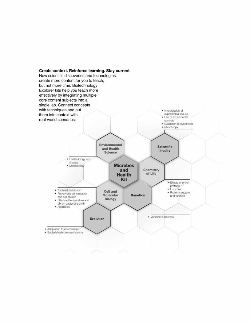
Kit Inventory ChecklistThis section lists the equipment and reagents necessary to conduct the microbes andhealth experiment in your classroom or teaching laboratory. We recommend student teamsof 2–4 students per workstation. The kit contains reagents for 32 students working at 8workstations made up of 4 students each.
Kit Components Number/Kit (✔✔)
Ampicillin 2 vials ❒
LB nutrient agar powder (to make 500 ml) 1 pouch ❒
Petri dishes, sterile, bags of 20 2 bags ❒
Culture tubes, sterile, bags of 25 3 pks ❒
Inoculation loops, sterile, 10 µl, packs of 10 loops 8 pks ❒
E. coli HB101 K-12, lyophilized 1 vial ❒
LB broth capsules, bags of 12 (to make 50 ml each)* 1 bag ❒
Disposable plastic transfer pipets, packs of 10 pipets 1 pack ❒
* LB broth capsules are included to extend the activity by using liquid cultures, if desired.
Required Accessories Number/Kit (✔✔)
Microwave or autoclave 1 ❒
Incubator at 37°C (Catalog #166-0501EDU) 1 ❒
500 ml graduated cylinder 1 ❒
1 L flask or bottle 1 ❒
Microscopes 1/workstation ❒
Microscope slides and cover slips 40 slides/80 cover slips ❒
pH paper (pH range 4–7 or wider) 48 pieces ❒
Permanent marker pens 8 ❒
Table sugar (sucrose) 10 grams ❒
Toothpicks or micropipet tips box ❒
Distilled water 1 L ❒
Milk 400 ml ❒
Plain cow’s milk yogurt (2–4 brands, must be 100 ml ❒labeled as containing live and active cultures – the latest available expiration date is preferred)
Optional Accessories
Magnifying glasses (to view bacterial colony morphology)
3

Refills Available SeparatelyMicrobes and Health kit refill package, (Catalog #166-5021EDU) includes Ampicillin, 12 LBbroth capsules, LB nutrient agar powder, and HB101 K-12 E.coli
LB nutrient agar powder (Catalog #166-0600EDU) 1 package
Ampicillin (Catalog #166-0407EDU) 1 vial
E. coli strain HB101 K-12 (Catalog #166-0408EDU) 1 vial
4
Background for Instructors
Bacteria Are EverywhereBacteria are the single most successful form of life on the Earth. There are probably morebacteria, more species, and more total biomass of bacteria than any other lifeforms.Bacteria are found in soil, water, in and on animals, in and on plants, in and on humans,and even miles below the ground. There is speculation that bacteria or similar forms of lifemay exist on Mars or other planets.
Characteristics of BacteriaBacteria are one of the three great domains of life along with Eukarya (Animals, Plants, andFungi) and Archaea (ancient bacteria-like organisms classified as a separate domain of lifeby Carl Woese in 1977). Bacteria and Archaea are classified as prokaryotes, single-celledcreatures usually too small to be seen by the unaided eye. Bacteria are so small that itwould take 5,000 to 50,000 in a row to stretch for an inch. Bacteria have no separate compartment (nucleus) to hold their DNA as eukaryotes do. Bacteria can sometimes, butnot always, move by tiny tails called flagella. Bacteria sometimes grow connected to otherbacteria forming chains. Some types of bacteria that may been seen in this lab grow connected in chains.
Bacteria as PathogensWhen we think of bacteria we usually think of disease. In fact, only a tiny minority of bacteria arecapable of causing disease. Bacteria that do cause disease have played an enormous role inthe history of humanity—cholera, typhus, the bubonic plague, tuberculosis, and other bacteria have sickened and killed millions. The development of antibiotics has greatly reducedthe dangers of bacterial diseases. However, due to the overuse of antibiotics some bacterialstrains (such as methicillin-resistant Staphylococcus aureus or MRSA) have developed antibioticresistance leaving humanity exposed to the reemergence of old bacterial threats.
Bacteria can also spoil food such as milk. Milk is an ideal growth medium for bacteria and maycontain both spoilage bacteria capable of souring milk, and pathogenic bacteria which mightcause disease in humans, such as brucellosis, bovine tuberculosis, and scarlet fever. Milk ispasteurized by heating to 62.9°C for 30 min, or 71.6°C for 15 sec, and then cooling rapidly.Pasteurization destroys all pathogenic bacteria, and most but not all, spoilage bacteria. Thusmilk still needs to be kept cool when stored. Grade A milk should contain less than 30,000 bacteria per milliliter.
History of BacteriologyAnton van Leeuwenhoek of the Netherlands first saw bacteria through a microscope in 1676and called them animalcules (tiny animals). Later Christian Gottfried Ehrenberg coined the term“bacterium” (meaning “small staff” in Greek) in 1828. In 1835 Agostino Bassi proposed the“germ theory of disease” which connected the spread of disease to unseen microorganisms, asprevously bacteria were thought to arise spontaneously in suitable environments. Louis Pasteurand John Tyndall showed that boiled broth grew bacteria only when exposed to the air thus disproving the theory of spontaneous generation. In 1875 Robert Koch was able to offer convincing proof of the germ theory by proving that anthrax was caused by bacteria. Koch’s setof rules (Koch’s postulates) for proving the cause of anthrax are the basis for assigning thecause of disease to a particular microbe. The postulates are also the basis for the experimentsin this lab.
5
INS
TR
UC
TOR
'S M
AN
UA
LB
AC
KG
RO
UN
D
Instructor’s Manual
6
Types of Bacteria and Bacterial ColoniesThere are several distinct morphologies or shapes of bacteria. The three major shapes are coccus (spherical), bacillus (rod-shaped), and spirillum (spiral). Cocci and bacilli can exist singly,in pairs (diplococci or diplobacilli), attached in long strings (streptococci or streptobacilli), or connected in other arrangements (staphylococci or staphylobacilli). There are various forms ofspiral bacteria too, such as comma-shaped (Bdellovibrio), helical (Helicobacter pylori), or longtwisted spirochete forms. It is best to examine fresh cultures as older bacteria are occasionallyoddly shaped and may have lost motility.
Bacteria increase in number by binary fission (splitting in half). Some bacteria can divide every15–20 minutes! A single bacterium on a solid medium, such as an agar plate, increases logarithmically so that overnight a single bacterium becomes millions or billions. These millionsor billions of bacteria form a visible "colony" on an agar plate. A colony of bacteria can itself havea distinct form and be large or small. Some bacterial colonies are so small that they cannot beseen with the unaided eye. Colonies may be circular, irregular, or branching. The edge of thecolony may be smooth, wavy or serrated. The colony may be flat, raised or raised only in thecenter.
Bacteria are also differentiated by their cell walls. Some have thick cell walls made of peptidoglycan molecules. The cell walls of these bacteria take up a dye called Gram stain andthus are called Gram-positive bacteria. Other bacteria have thinner cell walls that do not absorbGram stain and thus are called Gram-negative bacteria. The lactic acid bacteria found in yogurtare Gram-positive bacteria. The HB101 K-12 E. coli bacteria provided in this kit are Gram-negative bacteria.
Bacterial MetabolismLike all living things bacteria require food, often in the form of sugars, to gain energy. Bacteriabreak down sugars chemically into other molecules using enzymes. Enzymes are large proteinsthat speed up chemical reactions. This process of bacterial metabolism is often called fermentation.
Some bacteria require oxygen from the air to grow and are called aerobes. Other bacteria growonly in the absence of oxygen and are called anaerobes. Some bacteria can grow either with orwithout oxygen and are referred to as facultative anaerobes. Aerobic bacteria use oxygen tobreak sugar into intermediate products and then finally into carbon dioxide and water. Lackingoxygen, anaerobic or facultative anaerobic bacteria usually do not reduce sugars completely tocarbon dioxide and water. Often these bacteria convert sugar into pyruvic acid and then convertthe pyruvic acid into other by-products.
Yogurt forming bacteria are anaerobes and break down milk sugar (lactose) into pyruvic acidand then into lactic acid using enzymes. Lactic acid is the by-product or waste product made bylactic acid bacteria. Lactic acid also lowers the pH of milk making it acidic. The acidic conditionscause casein (a common protein in milk) to denature (or curdle) and become more solid. Inaddition the acidic conditions inhibit the growth of other microorganisms that might spoil theyogurt. Thus lactic acid causes the yogurt to stay fresh, while at the same time remainingdigestible by people who can break lactic acid down for additional energy. Other bacteria canbreak down sugars and pyruvic acid and make other by-products. The E. coli bacteria breaksugar down into succinic acid, ethanol, acetic acid, formic acid, and lactic acid.
INS
TR
UC
TOR
'S M
AN
UA
LB
AC
KG
RO
UN
D
Instructor’s Manual
7
Koch’s PostulatesBy the mid-19th century, the famous French scientist Louis Pasteur had conducted extensivestudies on the role of bacteria in fermentation, and had shown conclusively that germs did notspontaneously appear in susceptible hosts (spontaneous generation), but rather needed tocome in contact with the host first. There was already a prevailing assumption at the time thatmicrobes were in some way connected with disease, but whether their presence was the causeor just a result of disease was unclear. Furthermore, many infected tissues contained more thanone type of microorganism. This made it difficult to define with certainty the role any particulartype of bacterium played in disease. The work of Pasteur and others, along with improved techniques in microscopy and the discovery of semi-solid culture media, all paved the way forthe work of Robert Koch.
Koch had been studying anthrax, a deadly disease that infects both humans and animals, andhe noticed that certain rod-shaped bacteria and their spores were characteristically found in thetissues of sick sheep. He meticulously isolated these bacteria, which he named Bacillusanthracis, and grew pure cultures in a medium consisting of the aqueous humor of cattle or rabbiteyeballs. Next, he introduced the bacteria from the cultures into healthy rabbits. When the rabbitssubsequently developed symptoms of anthrax, Koch again isolated the bacteria from the rabbittissue and observed them under the microscope to confirm that they were indeed the sameones he had seen in his original culture.
The steps he used are now known as "Koch’s postulates." Meeting the criteria laid down byKoch is referred to as "satisfying Koch’s postulates" and is considered the standard evidencerequired to show that a microorganism causes a particular disease.
To demonstrate Koch’s postulates, students must do the following:
• Describe and record the symptoms shown
• Isolate and identify the suspected pathogen from the infected material and establish a pureculture
• Use the pure culture to infect new material. Describe and record the symptoms shown bythe material. Check that these are the same as their original observations
• Again isolate and identify the organism
Beneficial Bacteria and YogurtDespite our longstanding association of bacteria with disease, most bacteria are essentiallyharmless. In fact, many bacteria are beneficial. Bacteria break down waste organic material.Rhizobium bacteria take nitrogen from the air and convert it into a usable form (fixation).Intestinal bacteria break down indigestible material and synthesize nutrients. Some types of bacteria are necessary for the manufacture of certain food products, such as cheese, kimchi,sour cream, pickles, and yogurt.
Yogurt is made by adding specific strains of bacteria into milk, which is then fermented undercontrolled temperatures and environmental conditions. The bacteria ingest natural milk sugarsand release lactic acid as a waste product thus making the milk acidic. The increased aciditycauses casein (the most common milk protein) to tangle into a solid mass (called curd) in a process called denaturation. The increased acidity (the usual pH of yogurt is 4–5) also inhibitsthe growth of other dangerous bacteria. To be classified and sold as yogurt in the United Statesit is required that yogurt must contain the bacteria strains Streptococcus thermophilus(Streptococcus salivarius subsp. thermophilus) and Lactobacillus bulgaricus (Lactobacillus delbruecki subsp. bulgaricus). Often these two are cocultured with other lactic acid bacteria fortaste or health effects including Lactobacillus acidophilus, Lactobacillus casei, or Bifidobacterium
INS
TR
UC
TOR
'S M
AN
UA
LB
AC
KG
RO
UN
D
Instructor’s Manual

bifidum. In most countries, a product may be called yogurt only if live bacteria are present in thefinal product. A small amount of live yogurt can be used to inoculate a new batch of yogurt, asthe bacteria reproduce and multiply during fermentation. Pasteurized products, which have noliving bacteria, are called fermented milk. In the United States yogurt must contain at least a bil-lion viable bacteria per gram at the time of manufacture and at least a million viable bacteria pergram at the expiration date.
Yogurt has nutritional benefits beyond those of milk—people who are lactose intolerant oftenenjoy yogurt without ill effects, apparently because live yogurt cultures contain enzymes whichhelp break down lactose inside the intestine. Yogurt also has medical uses, in particular for avariety of gastrointestinal conditions, such as preventing antibiotic-associated diarrhea.
In this lab students will isolate the bacterial strains found in a yogurt sample on agar in a petridish, then use those same strains to inoculate fresh milk to find out if they can reproduce thesame yogurt. Students should be able to conclude that the acidic, solidified nature of yogurt iscaused by bacteria acting upon milk.
AntibioticsEarly attempts to treat bacterial infections sometimes employed substances, such as arsenic orstrychnine, that were nearly as toxic to humans as to bacteria. In 1928 Alexander Fleming discovered penicillin, a compound produced by mold, that inhibited the growth of bacteria withoutserious harmful effects upon humans. Many different types of antibacterial antibiotics have beendiscovered since that time. These antibiotics have vastly reduced the incidence of bacterial disease. Modern society has almost forgotten how great the dangers of bacterial disease oncewere. Careless misuse of antibiotics now threatens a return of potent bacterial diseases.Massive amounts of antibiotics are used in animal feed inadvertently selecting for the growth ofbacteria resistant to many classes of antibiotics. People often needlessly take antibiotics for viralinfections – again selecting for the growth of antibiotic resistant bacteria. In addition patients oftendiscontinue use of antibiotics as soon as they feel better leaving the most resistant bacteria inplace to start a new round of infection.
Antibacterial antibiotics are either bactericidal (kill bacteria) or bacteriostatic (prevent bacteriafrom dividing). There are many different modes of action for antibiotics. For instance, someinhibit the function of important enzymes present only in bacteria and not in mammals. Othersdestroy components of bacterial cell walls that are not used in mammalian cells.
The antibiotic ampicillin is included in this kit both as an additional control and as a tool to allowfurther experimentation. Ampicillin is a beta-lactam antibiotic similar to penicillin and amoxicillin. Itinhibits Gram-positive bacteria and some Gram-negative bacteria, such as E. coli, and it acts by preventing the synthesis of bacterial cell walls eventually leading to the death of the bacteria.Ampicillin is widely used in molecular biology as a selective agent since the gene for resistanceto ampicillin (encoding for the beta-lactamase enzyme) can be inserted into bacteria on plasmidsthat may also carry genes of interest to scientists. Those bacteria that survive on ampicillin containing media will also have the gene of interest.
Sterile TechniqueWhen culturing bacteria it is important to avoid contamination. Contaminating bacteria andmolds are found everywhere, including on hands and lab benchtops, so it is important to avoidthese surfaces. The round circle at the end of inoculating loops and the surfaces of agar platesshould not be touched or placed onto potential contaminating surfaces. Wipe down lab bencheswith 70% alcohol or a 10% bleach solution wearing appropriate safety equipment.
8
INS
TR
UC
TOR
'S M
AN
UA
LB
AC
KG
RO
UN
D
Instructor’s Manual

Lactobacillus bulgaricus(rod-shaped)
Streptococcus thermophilus
(spherical-shaped)
9
INS
TR
UC
TOR
'S M
AN
UA
LB
AC
KG
RO
UN
D
Instructor’s Manual

10
Instructor’s Advance PreparationThese instructions are designed for 8 workstations with up to 4 students per workstation.
Lesson 1 Advanced Preparation Step 1: 3–7 days prior to laboratory1. Prepare LB sugar agar plates as below.
2. To prepare one package of LB nutrient agar, add 500 ml of distilled water to a 1 L or largerErlenmeyer flask. This should make enough agar for 30–40 plates.
3. Add the entire contents of one LB nutrient agar packet. Add 5 g of table sugar (sucrose).Swirl the flask to dissolve the agar and sugar. Heat the agar to boiling in a microwave.
4. Repeat the heating and swirling cycle about three times until all the agar is dissolved. Thereshould be no more clear specks swirling around. Be careful to allow the flask to cool a littlebefore swirling so that the hot medium does not boil over onto your hand.
5. Once all the agar has been dissolved, allow the agar to cool so that the outside of the flaskis just comfortable to hold (50°C). Be careful not to let the agar cool so much that it begins tosolidify.
Pouring Plates 1. Label the outside of the bottom plate with date and "LBS".
2. Stack the empty plates 4-8 high. Starting with the bottom plate lift the lid and the platesabove straight up and to the side with one hand and pour the agar with the other.
3. Fill the plate about one-third to one-half (~10 ml) with agar, replace the lid and continue upthe stack. Once the agar cools label the bottom plate "LBS" and record the date.
4. After the plates have dried for two days at room temperature they can be used or stackedand stored in the original plastic sleeve to prevent dehydration. The stack is inverted, thesleeve taped shut, and the plates stored upside down in the refrigerator until used.
LBN
UTR
IENT
AGAR
INS
TR
UC
TOR
'S M
AN
UA
LA
DVA
NC
E P
RE
PAR
AT
ION
Instructor’s Manual
Add water Add agarpacket
and sugar
Swirl
Microwaveto boiling

Lesson 1 Advanced Preparation Step 2: 24 hr or less prior to lesson 1 laboratory1. Prepare sterile water
Add approximately 100 ml of distilled water to a loosely capped bottle. Heat until boilingusing a microwave or hot plate. Simmer for 10 minutes to sterilize using a low power settingon the microwave or hot plate. Alternatively use an autoclave. Seal container and allow tocool.
2. Rehydrate E. coli HB101 K-12 bacteria
Using a sterile pipet, rehydrate the lyophilized E. coli HB101 K-12 by adding 250 µl of sterilewater directly to the vial. Recap the vial and shake to mix. Store the rehydrated bacteria inthe refrigerator until used (preferably within 24 hours).
3. Obtain samples of milk and yogurt
a. Purchase milk and 2–4 brands of fresh plain cow’s milk yogurt labeled as containing liveand active cultures and having the freshest best before date. Store in refrigerator.
b. Label 8 culture tubes "milk" and aliquot approximately 5 ml of milk into each milk tube.Store in refrigerator until use.
c. Optional: Aliquot the yogurt into culture tubes or small labeled cups or beakers to distribute between groups. The exact volume is not important. Alternatively have studentsuse yogurt directly from the yogurt container. Store in refrigerator until use.
Student WorkstationsMaterials Needed for Each Workstation Provided by Quantity/workstaion
Microscope Instructor 1
Microscope slides Instructor 2
Cover slips Instructor 4
pH paper (pH range 4-7 or wider) Instructor 3 pieces
Cup with 5 ml yogurt* Prepared by instructor 1
Culture tube with 5 ml milk Prepared by instructor 1
LB sugar agar plates Prepared by instructor 3
Sterile inoculation loops Kit 3
Toothpicks or micropipet tips Instructor 3
Marker pen Instructor 1
Sterile water Instructor 1 ml
Common Workstation Quantity
Vial with E. coli rehydrated on ice 1
* Distribute the different brands of yogurt between the groups and have groups examine the properties of different yogurt brands. Each group should label the tube with initials so it can besaved for subsequent lessons.
11
INS
TR
UC
TOR
'S M
AN
UA
LA
DVA
NC
E P
RE
PAR
AT
ION
Instructor’s Manual
Lesson 2 Advanced Preparation: 24 hr or less prior to lesson 2 laboratory1. Prepare scalded milk
a. Add approximately 300 ml of milk to a beaker or pan and heat to just boiling/bubbling(86–93°C) and then turn off heat and allow to cool—store in the refrigerator for periodsover two hours. This can be done with care in a microwave using a low power setting.Take care that the milk does not boil over onto your hand.
b. Aliquot approximately 5 ml into 48 culture tubes (6 per workstation). Store in a refrigerator.
2. Rehydrate Ampicillin
Ampicillin is shipped freeze-dried in a small vial. Add 1 ml of sterile water directly to one vial tomake a 30 mg/ml ampicillin solution. Recap the vial and shake to mix. Label the vial with theconcentration of ampicillin. Rehydrated ampicillin can be stored in the refrigerator for up to 1 month and in the freezer for up to 1 year.
Student WorkstationsMaterials Needed for Each Workstation Provided by Quantity/workstation
Microscope Instructor 1
Microscope slides Instructor 3–6
Cover slips Instructor 3–6
pH paper (pH range 4-7 or wider) Instructor 2 pieces
Cup with 5 ml yogurt Instructor 1
(from lesson 1)
Culture tube with 5 ml scalded milk Prepared by instructor 6
Inoculated LB sugar agar plates From lesson 1 3
Sterile inoculation loops Kit 3
Magnifying glass (if available) Instructor 1
Sterile loops Instructor 4
Sterile water Instructor 1 ml
Marker pen Instructor 1
Toothpicks or micropipet tips Instructor 6
Common Workstation Provided by Quantity
Vial with rehydrated ampicillin (on ice) Instructor 1
12
INS
TR
UC
TOR
'S M
AN
UA
LA
DVA
NC
E P
RE
PAR
AT
ION
Instructor’s Manual

INS
TR
UC
TOR
'S M
AN
UA
LA
DVA
NC
E P
RE
PAR
AT
ION
Lesson 3 Advanced Preparation
Student WorkstationsMaterials Needed for Each Workstation Provided by Quantity/workstation
Cultured milk tubes From lesson 2 6 tubes
Microscope Instructor 1
Microscope slides Instructor 4
Cover slips Instructor 8
Yogurt plates From lesson 2 1
pH paper (pH range 4-7 or wider) Instructor 6 pieces
Toothpicks or micropipet tips Instructor box
13
Instructor’s Manual

Quick Guide
Lesson 1
Postulate 1: Identify possible pathogens
1. Compare yogurt and milk with respect toappearance, smell, and pH. Record observations.
2. Label left hand edge of slide "yogurt" andright hand edge “milk”.
3. Dip toothpick in yogurt, mix with a drop ofwater on left hand side of slide, and coverwith cover slip.
4. Add drop of milk to right hand side of slideand cover with cover slip.
5. Observe yogurt and milk under the micro-scope. Describe and draw what you see.
6. Repeat steps 1–5 with a different brand ofyogurt.
Postulate 2: Isolate and culture suspectedpathogens
7. Label 3 LB sugar agar plates on the bottom(not the lid) with your initials and one as"milk", one as "yogurt", and the third as "E. coli".
14
QU
ICK
GU
IDE
milk yogurt E. coli
Quick Guide
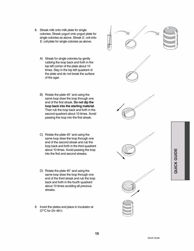
8. Streak milk onto milk plate for singlecolonies. Streak yogurt onto yogurt plate forsingle colonies as above. Streak E. coli ontoE. coli plate for single colonies as above.
A) Streak for single colonies by gently rubbing the loop back and forth in thetop left corner of the plate about 10times. Stay in the top left quadrant ofthe plate and do not break the surfaceof the agar.
B) Rotate the plate 45° and using thesame loop draw the loop through oneend of the first streak. Do not dip theloop back into the starting material.Then rub the loop back and forth in thesecond quadrant about 10 times. Avoidpassing the loop into the first streak.
C) Rotate the plate 45° and using thesame loop draw the loop through oneend of the second streak and rub theloop back and forth in the third quadrantabout 10 times. Avoid passing the loopinto the first and second streaks.
D) Rotate the plate 45° and using thesame loop draw the loop through oneend of the third streak and rub the loopback and forth in the fourth quadrantabout 10 times avoiding all previousstreaks.
9. Invert the plates and place in incubator at37°C for 24–48 h.
15
Quick Guide
QU
ICK
GU
IDE
Lesson 2
Postulate 2 continued: Isolate and culturesuspected pathogens
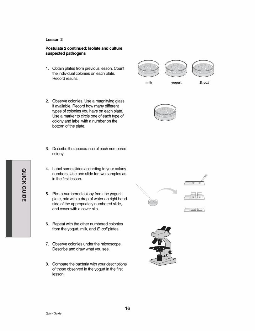
1. Obtain plates from previous lesson. Countthe individual colonies on each plate.Record results.
2. Observe colonies. Use a magnifying glassif available. Record how many differenttypes of colonies you have on each plate.Use a marker to circle one of each type ofcolony and label with a number on the bottom of the plate.
3. Describe the appearance of each numberedcolony.
4. Label some slides according to your colonynumbers. Use one slide for two samples asin the first lesson.
5. Pick a numbered colony from the yogurtplate, mix with a drop of water on right handside of the appropriately numbered slide,and cover with a cover slip.
6. Repeat with the other numbered coloniesfrom the yogurt, milk, and E. coli plates.
7. Observe colonies under the microscope.Describe and draw what you see.
8. Compare the bacteria with your descriptionsof those observed in the yogurt in the firstlesson.
16
QU
ICK
GU
IDE
Quick Guide
milk yogurt E. coli
17
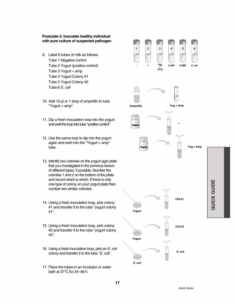
Postulate 3: Inoculate healthy individualwith pure culture of suspected pathogen
9. Label 6 tubes of milk as follows:Tube 1 Negative controlTube 2 Yogurt (positive control)Tube 3 Yogurt + ampTube 4 Yogurt Colony #1Tube 5 Yogurt Colony #2Tube 6 E. coli
10. Add 10 µl or 1 drop of ampicillin to tube"Yogurt + amp".
11. Dip a fresh inoculation loop into the yogurtand swirl the loop into tube "positive control".
12. Use the same loop to dip into the yogurtagain and swirl into the "Yogurt + amp"tube.
13. Identify two colonies on the yogurt agar platethat you investigated in the previous lessonof different types, if possible. Number thecolonies 1 and 2 on the bottom of the plateand record which is which. If there is onlyone type of colony on your yogurt plate thennumber two similar colonies.
14. Using a fresh inoculation loop, pick colony#1 and transfer it to the tube "yogurt colony#1".
15. Using a fresh inoculation loop, pick colony#2 and transfer it to the tube "yogurt colony#2".
16. Using a fresh inoculation loop, pick an E. colicolony and transfer it to the tube "E. coli".
17. Place the tubes in an incubator or waterbath at 37°C for 24–48 h.
Quick Guide
QU
ICK
GU
IDE
Ampicillin Yog + Amp
+
Yog + Amp
COL#1
COL#2
Yogurt
Yogurt
E. coli
E. coli

Lesson 3
Postulate 4: Isolate and identify suspectedpathogen from newly diseased individual
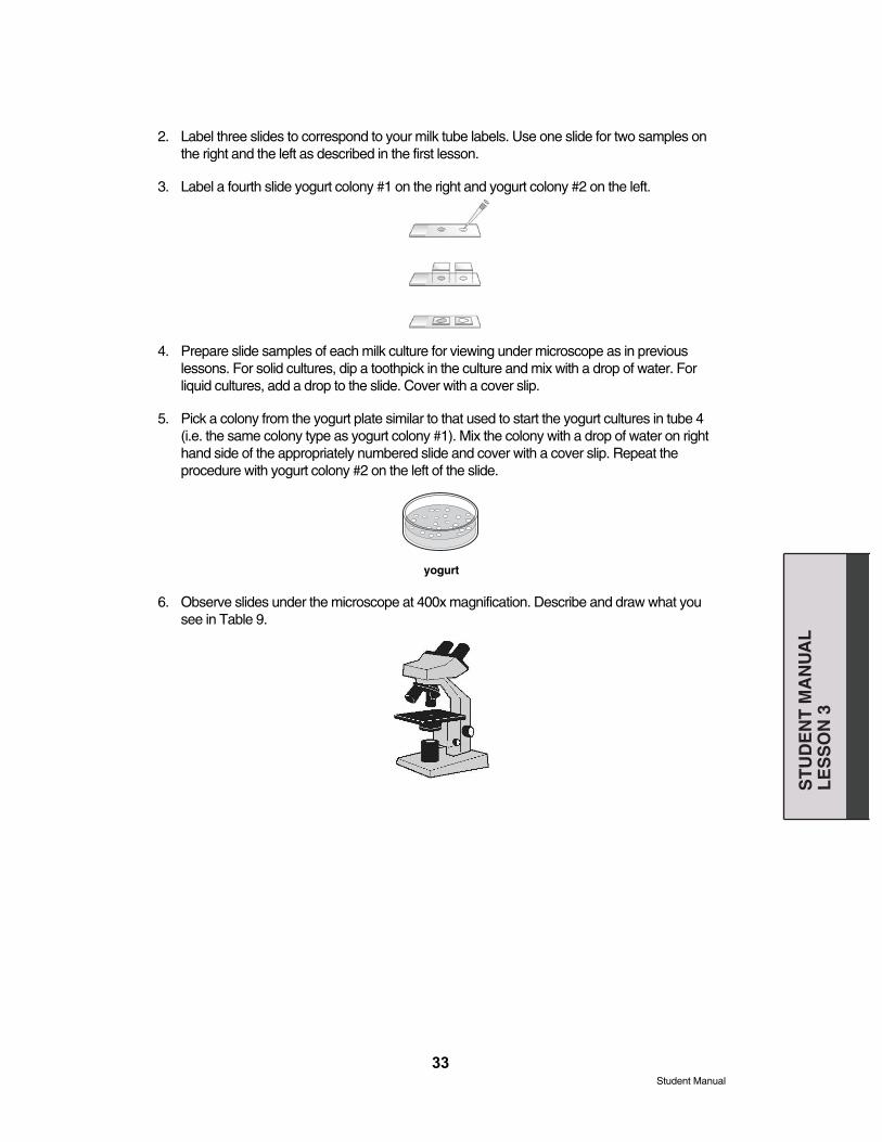
1. Obtain milk tubes and yogurt agar platefrom previous lesson. Describe each milkculture with respect to appearance, smell,and pH.
2. Label 3 slides according your milk tubelabels. Use one slide for two samples onthe right and the left as in the first lesson.
3. Label a fourth slide yogurt colony #1 onthe right and yogurt colony #2 on the left.
4. Prepare slide samples of each milk culturefor viewing under microscope as in previous lessons. For solid cultures, diptoothpick in culture and mix with a drop ofwater. For liquid cultures, add a drop tothe slide. Cover with cover slip.
5. Pick a colony from the yogurt plate similarto that used to start the yogurt cultures intube 4 (i.e. the same colony type asyogurt colony #1). Mix colony with a dropof water on right hand side of the appropriately numbered slide and coverwith cover slip. Repeat with yogurt colony#2 on the left of the slide.
6. Observe slides under the microscope.Describe and draw what you see.
7. Using the microscope compare any bacteria in the newly infected cultures inmilk tubes 4 and 5 with the pure bacteriaused to inoculate these cultures. Are theythe same?
18
Quick Guide
QU
ICK
GU
IDE
Student Manual
Background
In the 1800's microbial diseases were a terrifying mystery. People sickened and died withoutapparent cause. It had long been suspected that contact with an infected individual was necessaryfor the transmission of disease, but this was not true for all diseases. Early microbiologists acted asdetectives on the trail of a multitude of microbial killers. They were able to view bacteria from diseased individuals with microscopes, but how could they prove that the bacteria actuallycaused the disease? We will use Koch’s postulates, a series of tests devised by Robert Koch, aGerman physician of the 1800s. Koch’s postulates are widely used to prove that a particularmicrobe causes a particular disease.
Koch's postulates:
1. The microorganism must be found in all organisms suffering from the disease, but not inhealthy organisms.
2. The microorganism must be isolated from a diseased organism and grown in pure culture.
3. The cultured microorganism should cause disease when introduced into a healthy organism.
4. The microorganism must be again isolated from the inoculated, diseased experimental hostand identified as identical to the original specific causative agent.
Since it is dangerous and often unethical to experiment on humans, scientists often use modelsystems to simulate diseases in humans. Frequently, medical researchers will examine diseasesin animals so that they can learn more about similar diseases in humans. You will use a modelto test Koch’s postulates. In this model, milk will represent a healthy individual. At times milk willdevelop a condition that causes it to thicken and turn into yogurt. This is the "yogurtness disease."You will play the role of a medical investigator from a time over a hundred years ago. You suspectthat the yogurtness disease may be caused by something that is found in yogurt. You will useKoch’s postulates to prove or disprove the hypothesis that microbes found in yogurt are thecause of yogurtness disease. Of course it is important to remember that real yogurt is a veryhealthy food and that any microbes found in yogurt are harmless and do not cause disease inhealthy humans. Only a small minority of any bacteria cause disease in humans. In fact the "probiotic" (beneficial) bacteria found in yogurt may be helpful for digestion and may promotegood health.
Ampicillin may cause allergic reactions or irritation to the eyes, respiratory system, and skin. Incase of contact with eyes, rinse immediately with plenty of water and seek medical advice. Wearsuitable protective clothing. Ampicillin is a member of the penicillin family of antibiotics. Thosewith allergies to penicillin or any other member of the penicillin family of antibiotics should avoidcontact with ampicillin.
19
ST
UD
EN
T M
AN
UA
LB
AC
KG
RO
UN
D
Student Manual

20
Pre-lab Focus Questions1. What diseases do you know of that are caused by bacteria?
2. What diseases do you know of that are not caused by bacteria?
3. What characteristics allow bacteria to cause diseases?
4. How are bacterial diseases treated?
5. How can the spread of bacterial diseases be prevented?
6. Are all bacteria harmful? If not, describe the benefits of some bacteria.S
TU
DE
NT
MA
NU
AL
BA
CK
GR
OU
ND
Student Manual

Lesson 1: Postulate 1
Identify Possible Pathogen
Compare the healthy individual (milk) and the diseased individual (yogurt) for differentproperties.
1. Describe the differences between the milk and yogurt in texture, smell, color, and any otherobservable characteristics. Also test the pH of the milk and each type of yogurt. Yourinstructor will have directions for use of the pH indicator.
Record the differences between yogurt and milk in Table 1 below. Different groups will have different types of yogurt – share your samples if possible. Examine more than one type ofyogurt. Are there any attributes that are common to both types of yogurt but not milk?
Table 1. Milk and yogurt characteristics.
Characteristics Milk Yogurt 1 Yogurt 2
Texture
Color
Smell
pH
OtherObservations
Examine the milk and yogurt under the microscope.
2. Label the left side of a microscope slide "yogurt" and the right side "milk."
3. Use a toothpick or micropipette tip to place a small amount of yogurt on the left side of theslide. The yogurt should come from below the surface if possible. Add one drop of sterile waterand mix with a toothpick or micropipette tip. Place a cover slip over the top of the mixture.
21
ST
UD
EN
T M
AN
UA
LL
ES
SO
N 1
Student Manual
22
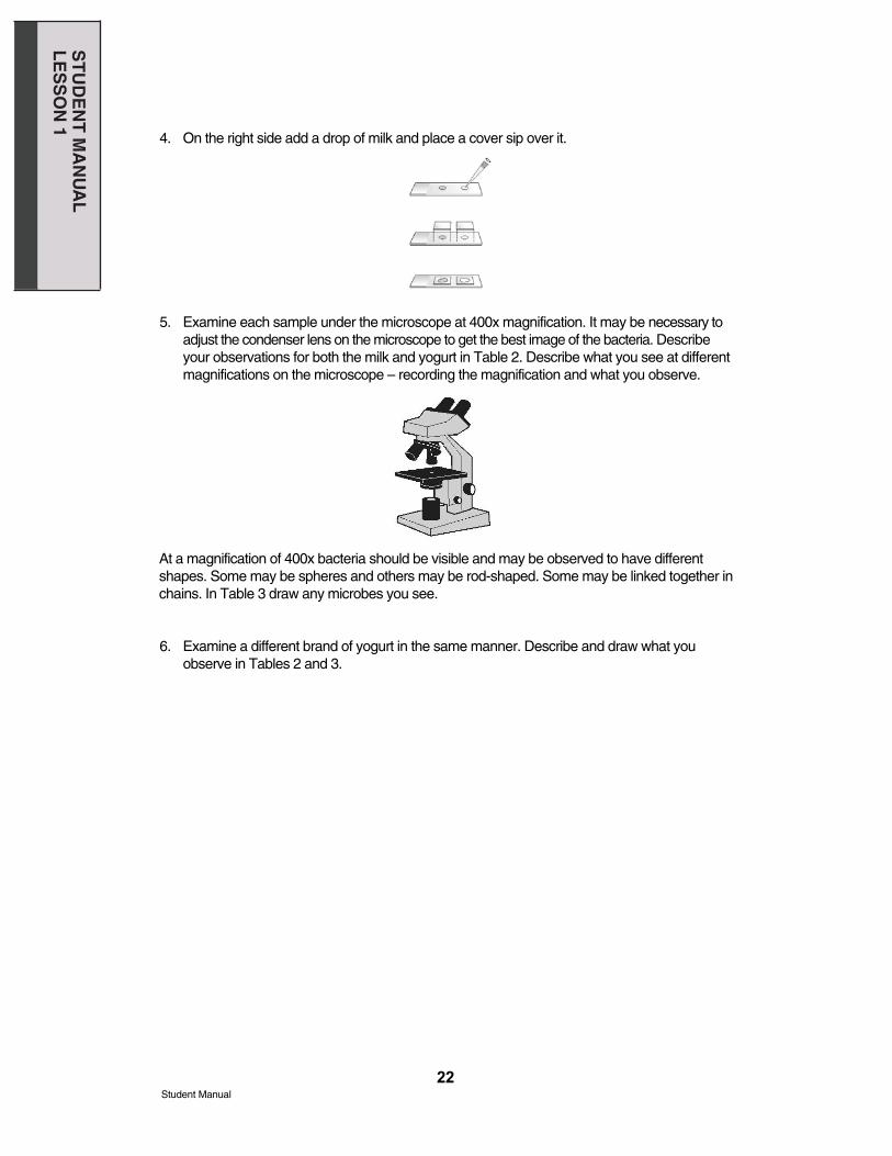
4. On the right side add a drop of milk and place a cover sip over it.
5. Examine each sample under the microscope at 400x magnification. It may be necessary toadjust the condenser lens on the microscope to get the best image of the bacteria. Describeyour observations for both the milk and yogurt in Table 2. Describe what you see at differentmagnifications on the microscope – recording the magnification and what you observe.
At a magnification of 400x bacteria should be visible and may be observed to have differentshapes. Some may be spheres and others may be rod-shaped. Some may be linked together inchains. In Table 3 draw any microbes you see.
6. Examine a different brand of yogurt in the same manner. Describe and draw what youobserve in Tables 2 and 3.
ST
UD
EN
T M
AN
UA
LL
ES
SO
N 1
Student Manual

Table 2: Descriptions of milk and yogurt under the microscope.
Milk Yogurt 1 Yogurt 2
Table 3: Drawings of microbes seen under the microscope.
Milk Yogurt 1 Yogurt 2
23
ST
UD
EN
T M
AN
UA
LL
ES
SO
N 1
Student Manual
24
Lesson 1 continued: Postulate 2
Isolate and culture the suspected pathogen
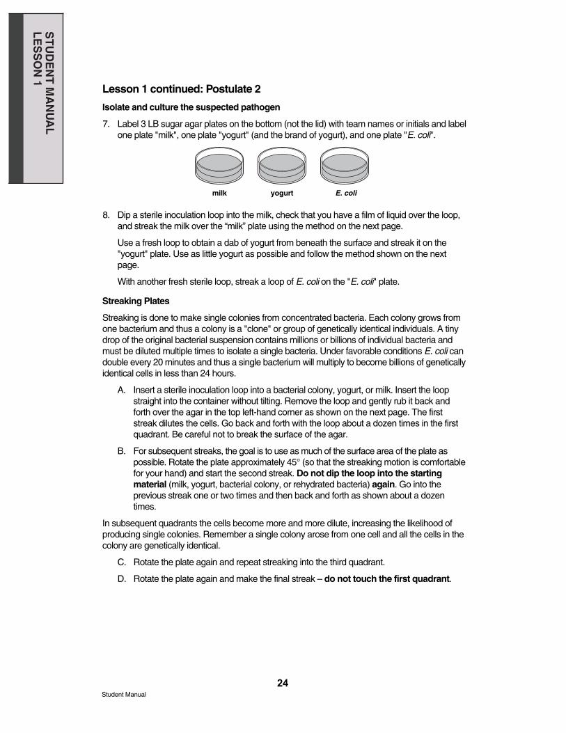
7. Label 3 LB sugar agar plates on the bottom (not the lid) with team names or initials and labelone plate "milk", one plate "yogurt" (and the brand of yogurt), and one plate "E. coli".
8. Dip a sterile inoculation loop into the milk, check that you have a film of liquid over the loop,and streak the milk over the “milk” plate using the method on the next page.
Use a fresh loop to obtain a dab of yogurt from beneath the surface and streak it on the"yogurt" plate. Use as little yogurt as possible and follow the method shown on the nextpage.
With another fresh sterile loop, streak a loop of E. coli on the "E. coli" plate.
Streaking Plates
Streaking is done to make single colonies from concentrated bacteria. Each colony grows fromone bacterium and thus a colony is a "clone" or group of genetically identical individuals. A tinydrop of the original bacterial suspension contains millions or billions of individual bacteria andmust be diluted multiple times to isolate a single bacteria. Under favorable conditions E. coli candouble every 20 minutes and thus a single bacterium will multiply to become billions of geneticallyidentical cells in less than 24 hours.
A. Insert a sterile inoculation loop into a bacterial colony, yogurt, or milk. Insert the loopstraight into the container without tilting. Remove the loop and gently rub it back andforth over the agar in the top left-hand corner as shown on the next page. The firststreak dilutes the cells. Go back and forth with the loop about a dozen times in the first quadrant. Be careful not to break the surface of the agar.
B. For subsequent streaks, the goal is to use as much of the surface area of the plate as possible. Rotate the plate approximately 45° (so that the streaking motion is comfortablefor your hand) and start the second streak. Do not dip the loop into the startingmaterial (milk, yogurt, bacterial colony, or rehydrated bacteria) again. Go into the previous streak one or two times and then back and forth as shown about a dozentimes.
In subsequent quadrants the cells become more and more dilute, increasing the likelihood ofproducing single colonies. Remember a single colony arose from one cell and all the cells in thecolony are genetically identical.
C. Rotate the plate again and repeat streaking into the third quadrant.
D. Rotate the plate again and make the final streak – do not touch the first quadrant.
milk yogurt E. coli
ST
UD
EN
T M
AN
UA
LL
ES
SO
N 1
Student Manual
25
9. Stack up your plates and tape them together. Put your group name and class period on thebottom of the stack and place upside down in a 37°C incubator for 24–48 hours.
It is extremely important to follow the streak protocol to thin out the bacteria in order to have individual colonies on the plate. Otherwise the colonies may be too close together to count orpick individually.
B
rotate 45°
rotate 45°rotate 45°
A
C D
ST
UD
EN
T M
AN
UA
LL
ES
SO
N 1
Student Manual

Lesson 2: Postulate 2Isolate Pathogen and Grow in Pure Culture
Analyze results from the previous lesson
1. Examine each of your three plates and describe what you see. Use a magnifying glass ifone is available.
Are there colonies on each of the plates? Count the number of separate individual colonies andrecord the numbers in Table 4. The first and second quadrants may be completely covered witha lawn of bacteria.
Table 4: The number of separate individual colonies on each plate.
Yogurt Milk E. coli
2. Do all your colonies look the same? Find a colony of each type that is isolated from othercolonies and circle it. Label each circle with a number on the bottom of the agar plate – notthe lid. Count and record the number of different types of colonies on each plate in Table 5.
Table 5: The number of different types of colonies on each plate.
Yogurt Milk E. coli
3. Describe the appearance (morphology) of at least two of the circled colonies. Use a magnifying glass if one is available. Are the colonies large or small? Are the colonies circularor irregular? Is the edge of the colony even or irregular? Is the colony flat or raised? What isthe color of the colony? Record the number of the colony, which plate it is on, and describeand draw the morphology of the colony in Table 6.
26
milk yogurt E. coli
ST
UD
EN
T M
AN
UA
LL
ES
SO
N 2
Student Manual

Table 6: Morphology of the bacterial colonies.
Yogurt colony #1 Yogurt colony #2
Milk colony E. coli colony
4. Label some slides to correspond with your circled colonies. Use one slide for two samplesas in the first lesson.
5. Using a new toothpick take the circled colony and put it on the slide. Add a drop of water,mix, and put a cover slip on top.
6. Repeat this procedure with the other circled colonies from the yogurt, milk, and E. coli plates.
27
ST
UD
EN
T M
AN
UA
LL
ES
SO
N 2
Student Manual
milk yogurt E. coli
milk yogurt E. coli

7. Examine the colony slides under the microscope at 400x magnification. Observe and drawthe bacteria, their shape, and if they are linked together. Record your results in Table 7.
Table 7: Description and drawing of bacteria under a microscope.
Yogurt 1 Yogurt 2
Milk E. coli
8. Compare these bacteria with your description of those from the yogurt in the first lesson. Doany of the bacteria appear to be the same? If so which ones?
28
ST
UD
EN
T M
AN
UA
LL
ES
SO
N 2
Student Manual
Lesson 2 continued: Postulate 3
Inoculate a healthy individual with the pure culture of suspected pathogen
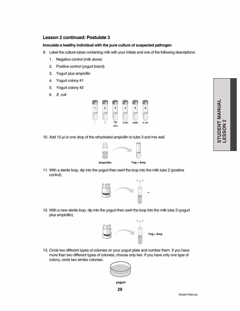
9. Label the culture tubes containing milk with your initials and one of the following descriptions:
1. Negative control (milk alone)
2. Positive control (yogurt brand)
3. Yogurt plus ampicillin
4. Yogurt colony #1
5. Yogurt colony #2
6. E. coli
10. Add 10 µl or one drop of the rehydrated ampicillin to tube 3 and mix well.
11. With a sterile loop, dip into the yogurt then swirl the loop into the milk tube 2 (positive control).
12. With a new sterile loop, dip into the yogurt then swirl the loop into the milk tube 3 (yogurtplus ampicillin).
13. Circle two different types of colonies on your yogurt plate and number them. If you havemore than two different types of colonies, choose only two. If you have only one type ofcolony, circle two similar colonies.
29
ST
UD
EN
T M
AN
UA
LL
ES
SO
N 2
Student Manual
yogurt
Ampicillin Yog + Amp
+
Yog + Amp
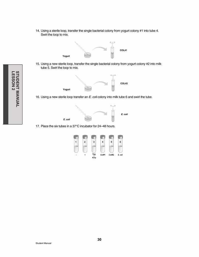
14. Using a sterile loop, transfer the single bacterial colony from yogurt colony #1 into tube 4.Swirl the loop to mix.
15. Using a new sterile loop, transfer the single bacterial colony from yogurt colony #2 into milktube 5. Swirl the loop to mix.
16. Using a new sterile loop transfer an E. coli colony into milk tube 6 and swirl the tube.
17. Place the six tubes in a 37°C incubator for 24–48 hours.
30
COL#1
Yogurt
Yogurt
E. coli
COL#2
E. coli
ST
UD
EN
T M
AN
UA
LL
ES
SO
N 2
Student Manual

Lesson 2 Post Lab Focus Questions
1. What could we conclude if we see more than one type of bacteria growing on an agar platestreaked with yogurt?
2. If there is more than one type of bacteria how could this affect our investigation into the"yogurtness" disease?
3. Which of Koch’s postulates are tested by adding bacteria from yogurt to milk?
4. What do you expect to see in the 6 tubes of milk after incubation?
Tube 1 – Negative control (milk alone)
Tube 2 – Positive control (yogurt brand)
Tube 3 – Yogurt plus ampicillin
Tube 4 – Yogurt colony #1
Tube 5 – Yogurt colony #2
Tube 6 – E. coli
5) Do all bacteria cause milk to turn into yogurt? Which of the controls tests for this?
6) Why add an antibiotic (ampicillin) in one of the tubes?
31
ST
UD
EN
T M
AN
UA
LL
ES
SO
N 2
Student Manual

Lesson 3: Postulate 4
Isolate and identify suspected pathogen from newly diseased individual
1. After the 24–48 hour incubation examine each of the 6 tubes and the original yogurt andrecord observations.
Table 8: Characteristics of milk cultures
Tube Texture Color Smell pHTube 1 Negative Control
Tube 2Positive Control
Tube 3Yogurt + Ampicillin
Tube 4First Colony
Tube 5Second Colony
Tube 6E. coli
Control Yogurt(cup)
32
ST
UD
EN
T M
AN
UA
LL
ES
SO
N 3
Student Manual
2. Label three slides to correspond to your milk tube labels. Use one slide for two samples onthe right and the left as described in the first lesson.
3. Label a fourth slide yogurt colony #1 on the right and yogurt colony #2 on the left.
4. Prepare slide samples of each milk culture for viewing under microscope as in previouslessons. For solid cultures, dip a toothpick in the culture and mix with a drop of water. For liquid cultures, add a drop to the slide. Cover with a cover slip.
5. Pick a colony from the yogurt plate similar to that used to start the yogurt cultures in tube 4(i.e. the same colony type as yogurt colony #1). Mix the colony with a drop of water on righthand side of the appropriately numbered slide and cover with a cover slip. Repeat the procedure with yogurt colony #2 on the left of the slide.
6. Observe slides under the microscope at 400x magnification. Describe and draw what yousee in Table 9.
33
ST
UD
EN
T M
AN
UA
LL
ES
SO
N 3
Student Manual
yogurt

Table 9: Description of milk cultures under microscope
Tube Description under microscope Drawing under microscope
Tube 1Negative control
Tube 2Positive control
Tube 3Yogurt + Ampicillin
Tube 4First Colony
Tube 5Second Colony
Tube 6E. coli
Controlyogurt(cup)
7. Compare the newly infected cultures in tubes 4 and 5 with the pure bacteria – are they thesame?
34
ST
UD
EN
T M
AN
UA
LL
ES
SO
N 3
Student Manual
Lesson 3 Focus Questions1. From your results, what can you conclude about what causes milk to turn into yogurt?
2. What evidence do you have to support your conclusions?
3. Can any bacteria turn milk into yogurt? What evidence do you have to support your answer?
4. Can yogurt-making bacteria be prevented from making yogurt? What evidence do youhave to support your answer?
5. If you had just added yogurt to the milk and found that it made yogurt, what would that showand what would that fail to show?
6. Why is it important to inoculate milk with bacteria from a single colony rather than from multiplebacterial colonies?
7. Some bacteria will only grow when they have access to specific types of nutrients. If somebacteria in the yogurt would only grow in milk, and would not grow on agar, how would thishave affected your investigation?
35
ST
UD
EN
T M
AN
UA
LL
ES
SO
N 3
Student Manual
36
Appendix AGlossaryAerobe — Aerobes are bacteria that require oxygen for survival.
Agar — Agar is a jelly-like substance obtained from seaweed. It is made of linked sugars(a polysaccharide) and is used to make media for growing bacteria.
Ampicillin — Ampicillin is a penicillin-like bactericidal antibiotic that inhibits the synthesis ofthe peptidoglycan component of bacterial cells walls especially in Gram-positive bacteriabut also in some Gram-negative bacteria, such as E. coli.
Anaerobe — Anaerobes are bacteria that do not require oxygen for survival. Anaerobesmay die in the presence of oxygen.
Antibiotic — An antibiotic is a chemical that prevents or reduces the growth of bacteria orother microbes.
Anthrax — Anthrax is an often fatal disease that affects both animals and humans.Bacillus anthracis bacteria were identified as the cause of anthrax by Robert Koch in 1876.Anthrax is a possible agent of biowarfare or bioterrorism.
Archaea — Archaea are bacteria-like, single-celled micro organisms with no nucleus.Archaea were once thought to be a type of bacteria but are now known to be an entirelyseparate domain of life along with Bacteria and Eukaryota (animals, plants, fungi, and protozoans).
Bacillus — A bacillus is a rod-shaped bacteria.
Bacteria — Bacteria are single-celled, microorganisms with no nucleus. Bacteria are oneof the three domains of life along with Archaea and Eukaryota (animals, plants, and fungi).
Bactericidal — An antibiotic or other agent that kills bacteria is said to be bactericidal.
Bacteriostatic — An antibiotic or other agent that prevents the growth of bacteria is said tobe bacteriostatic.
Bifidobacterium bifidum — Bifidobacterium bifidum is a Gram-positive bacteria present inthe guts of animals and humans. Bifidobacterium bifidum aids in digestion and thus is a"probiotic" bacteria. It is added to some varieties of yogurt.
Binary fission — Most bacteria reproduce asexually by duplicating their DNA and dividinginto two equal halves.
Casein — Casein is the major protein in milk. When denatured it causes milk to solidify orcurdle into yogurt or cheese. Casein is denatured by enzymes or acidic conditions but notby heat.
Clone — A clone is a group of genetically identical organisms.
Coccus — A coccus is a spherical bacteria.
Colony — A clump of genetically identical bacterial cells growing on an agar plate.Because all the cells in a single colony are genetically identical, they are called clones.
Curd — Curd is denatured milk protein (casein) which becomes solid.
Denaturation — Denaturation is the process of altering the structure of proteins by someexternal stress such as heat, acid, or a change in salt concentration. Casein proteins in milkare denatured to form curd.
AP
PE
ND
IX A

37
E. coli — Escherichia coli is a Gram-negative facultative anaerobic bacillus bacterium. Itinhabits the intestines of animals and humans and may benefit them by producing vitaminK and preventing the spread of harmful bacteria. Harmless genetically weakened forms ofE. coli, such as the HB101 K-12 strain used in this kit, are used in many scientific applications.Normally E. coli is harmless but a few strains such as O157:H7 can cause disease.
Enzyme — An enzyme is a protein that catalyzes a chemical reaction. Rennin is anenzyme which causes milk proteins (casein) to coagulate (solidify) into curd.
Eukaryotes — Eukaryotes are one of the three domains of life along with Bacteria andArchaea. DNA in eukaryotic cells is contained in a special compartment called a nucleus.Eukaryotes include all animals, plants, fungi, and single-celled protozoans.
Facultative anaerobe — Facultative anaerobes are bacteria that can use oxygen but donot need it and thus can grow either in the presence or absence of oxygen.
Germ theory — Germ theory proposes that disease is caused by microbes. Disease couldbe transmitted from a diseased individual to a healthy individual by passage of thesemicrobes. Prior to the acceptance of germ theory it was thought that diseases arose spontaneously. Agostino Bassi first formally stated germ theory based on his observation ofdisease in silkworms. Robert Koch devised Koch’s postulates as a test of germ theory anddemonstrated that anthrax was caused by the bacteria Bacillus anthracis.
Gram-positive — Bacteria whose cell walls contain only one lipid membrane surroundedby a thick layer of peptidoglycans are Gram-positive because they take up Gram stain.Lactobacillus acidophilus, Lactobacillus bulgaricus, and Streptococcus thermophillis areGram-positive bacteria.
Gram-negative — Bacteria whose cell walls contain a second lipid membrane on the outside of a thin layer of peptidoglycans and interior lipid membrane are gram negativebecause they do not take up Gram stain. E. coli are Gram-negative bacteria.
Gram stain — Gram stain contains two dyes: crystal violet and safranin. The crystal violetis taken up by Gram-positive bacteria and will appear purple or blue. Only the safranin istaken up by Gram-negative bacteria and will appear pink or red.
Koch’s postulates — A series of tests devised by Robert Koch used to assign the causeof a disease to a particular microbe.
1. The microorganism must be found in all organisms suffering from the disease, but notin healthy organisms.
2. The microorganism must be isolated from a diseased organism and grown in pure culture.
3. The cultured microorganism should cause disease when introduced into a healthy organism.
4. The microorganism must be again isolated from the inoculated, diseased experimentalhost and identified as identical to the original specific causative agent.
Koch, Robert — Robert Koch was a German doctor who lived from 1843 to 1910. He discovered the bacteria that caused anthrax, tuberculosis, and cholera. He also developedKoch’s postulates, a series of tests used to assign the cause of disease to a particularmicrobe.
Lactic acid — Lactic acid (C3H6O3) is sometimes known as milk acid. It is produced by thefermentation of lactose or other carbohydrates by lactic acid bacteria. Lactic acid causesthe denaturation (or curdling) of casein into solid form. Lactic acid is a normal byproduct ofmetabolism and may accumulate during exercise causing temporary side pains.
AP
PE
ND
IX A

Lactic acid bacteria — Bacteria that break down (ferment) sugars, such as lactose intolactic acid, are termed lactic acid bacteria. Lactobacillus acidophilus, Lactobacillus bulgaricus, and Streptococcus thermophillis are all lactic acid bacteria. Lactic acid bacteriaare tolerant to acidic low pH conditions but many other bacteria are not. Thus lactic acidbacteria preserve food, such as yogurt or sauerkraut from the effects of spoilage bacteria.
LB — Luria Bertani broth (sometimes called lysogeny broth) is composed of yeast extract,tryptone, and sodium chloride and is commonly used to culture bacteria.
LBS — Luria Bertani Sugar is LB with sugar added to promote the growth of particular bacteria that would not grow as well on plain LB media.
Lactobacillus acidophilus — Lactobacillus acidophilus is a beneficial probiotic Gram-positive,rod-shaped lactic acid bacterium. It tolerates warm acidic conditions. It is added to sometypes of yogurt.
Lactobacillus bulgaricus — Lactobacillus delbrueckii subsp. bulgaricus is a beneficial probiotic Gram-positive, rod-shaped lactic acid bacterium. It tolerates warm acidic conditions.It is present in most types of yogurt.
Lactobacillus casei — Lactobacillus casei is a beneficial probiotic Gram-positive, rod-shaped lacticacid bacterium. It is added to some types of yogurt.
Lactose — Lactose (C12H22O11) is often referred to as milk sugar. It is the main type ofsugar in milk. Adults in many parts of the world cannot digest lactose without the aid of probiotic bacteria as are found in yogurt.
Media — Media is a substance such as an agar plate or nutrient broth that will support thegrowth of microbes.
Microbe — Microbes or microorganisms are single-celled organisms such as bacteria,archaea, yeast, or protozoans, usually visible only under a microscope. The term may alsoinclude viruses although they are not strictly alive.
Microbiology — Microbiology is the study of microbes.
Pasteur, Louis — Louis Pasteur was a French chemist who lived from 1822–1895. Alongwith Robert Koch he was one of the founders of the science of microbiology. Pasteur isfamous for creating the first vaccine for rabies, a viral disease, and for devising the processof pasteurization.
Pasteurization — Pasteurization is a process used to destroy nearly all pathogenic bacteria and, most but not all of, spoilage bacteria in milk or other liquid foods. Milk is pasteurized by heating to 62.9°C for 30 min, or 71.6°C for 15 sec, and is then cooled rapidly.Pasteurization was invented in 1862 by Louis Pasteur.
Penicillin — Penicillin is a bactericidal antibiotic that inhibits the synthesis of the peptidoglycan component of bacterial cells walls especially in Gram-positive bacteria.Penicillin was discovered by Alexander Fleming in 1928 and was the first antibiotic to beused medically.
Peptidoglycan — Peptidoglycans are sugar-peptide molecules that make up part of thecell walls of bacteria. Gram-positive bacteria have a thick layer of peptidoglycan on the outside of their lipid membrane. Gram-negative bacteria have a thin layer of peptidoglycansbetween their two lipid membranes. Some antibiotics such as penicillin or ampicillin preventthe production of peptidoglycans. Archaea and eukaryotes do not have peptidoglycans.
38
AP
PE
ND
IX A
Petri dish — Petri dishes are small round flat containers made of glass or plastic. They arecommonly used to hold media used to culture microbes. Petri dishes were invented bymicrobiologist Julius Petri, an assistant to Robert Koch.
Probiotic — Probiotic bacteria aid in the digestion of foods, such as lactose in milk, andthus are added to some foods for their health benefits. The most common type of probioticsare lactic acid bacteria, such as those found in yogurt.
Pyruvic acid — Pyruvic acid (C3H4O3) is created by the fermentation of carbohydrates.Pyruvic acid can be further fermented by some bacteria to make lactic acid.
Spontaneous generation — At one time microbes and other pests were believed to arisespontaneously from decaying matter. For instance, maggots were thought to appear spontaneously in meat. Louis Pasteur and others showed that microbes could not appearwithout being introduced from an external source. The idea of spontaneous generation hassince been replaced by the germ theory of disease.
Streptococcus thermophillus — Streptococcus salivarius subsp. thermophillis is a beneficialprobiotic Gram-positive spherical lactic acid bacterium. It tolerates warm acidic conditions.It is present in most types of yogurt.
Yogurt — Yogurt is a healthy food made from milk fermented by lactic acid bacteria, suchas Lactobacillus delbrueckii subsp. bulgaricus and Streptococcus salivarius subsp. thermophillis. The bacteria make lactic acid as a byproduct which lowers the pH and curdlescasein proteins in the milk. The acidic conditions help prevent growth of spoilage bacteria.Yogurt is also more digestible than milk for many people because the lactose milk sugarhas been fermented into more digestible lactic acid.
Yogurtness — Yogurtness is a made up term meaning the "condition of being like yogurt".
39
AP
PE
ND
IX A
Appendix BInstructors Answer Guide
Pre-Lab Focus Questions1. What diseases do you know of that are caused by bacteria?
Answers may vary but some bacterial diseases are listed below:Anthrax, bacterial meningitis, bacterial pneumonia, botulism, brucellosis, bubonicplague, chlamydia, cholera, dental caries (cavities), diphtheria, dysentery, gonorrhea,Legionnaires’ disease, leprosy, Lyme disease, pertussis (whooping cough),rocky mountain spotted fever, salmonella (food poisoning), scarlet fever, shigel-losis, strep throat, syphilis, toxic shock syndrome, tuberculosis, typhus
2. What diseases do you know of that are not caused by bacteria?
Answers may vary.Some viral diseases are: AIDS, common cold, ebola hemorrhagic fever, equine encephalitis, dengue, fifthdisease, foot and mouth disease, Hanta virus hemorrhagic fever, Hepatitis A,Hepatitis B, Hepatitis C, herpes, influenza, measles (rubeola), mononucleosis,mumps, polio, rabies, roseola, rubella (german measles), SARS, smallpox, varicella (chicken pox), viral meningitis, viral pneumonia, warts, west nile virus,yellow fever
Some environmental and autoimmune diseases are:Alzheimer’s disease, arthritis, asthma, cancer, emphysema, Parkinson’s disease,lupus
Some fungal and parasitic disease are: Athlete’s foot, candidasis, giardiasis, histoplasmosis, leismaniasis, malaria, ringworm, river blindness, thrush, toxoplasmosis, trichinosis
Some prionic (protein caused) diseases are:Creutzfeld- Jacob disease, fatal insomnia, mad cow’s disease,
Some genetic diseases are:Celiac disease, down syndrome, hemophilia, muscular dystrophy, phenylke-tonuria, sickle-cell disease, Tay-Sachs disease
3. What characteristics allow bacteria to cause diseases?
They are small and thus can invade host organisms. They can survive outside thehost at least for a brief period and thus can be transmitted. They can grow without being eliminated by the hosts immune system and create toxic wasteproducts which harms the host.
4. How are bacterial diseases treated?
Bacterial diseases are often treated with antibiotics. Antibiotics are not effectiveagainst viruses and other non-bacterial diseases. So a common cold cannot betreated with antibiotics. Of course an individual with a bacterial disease shouldconsult a doctor to discuss other treatment options.
5. How can the spread of bacterial diseases be prevented?
40
AP
PE
ND
IX B
Good hygiene such as washing hands is important. It is also important to ensurethat sources of water and food are clean and free of bacteria. Diseased individualsmight be quarantined and prevented from having contact with healthy individuals.
6. Are all bacteria harmful? If not, describe the benefits of some bacteria.
No, most bacteria are harmless to humans. Some bacteria perform useful tasks.Bacteria in our digestive system break down indigestible foods into forms usefulfor humans and may even synthesize nutrients. Bacteria living in plants covertatmospheric nitrogen into a useable form (fixation). Bacteria clean up our environment by degrading toxins and dead organic matter. Bacteria are alsohelpful in the creation of many food stuffs, such as cheese, sauerkraut, andyogurt.
41
AP
PE
ND
IX B
Lesson 2 Post Lab Focus Questions1. What could we conclude if we see more than one type of bacteria growing on an agar
plate streaked with yogurt?
We might conclude that one of the types of bacteria caused the disease and theother was coincidental or perhaps and “opportunistic infection.” We might alsoconclude that both types of bacteria are necessary to cause yogurtness. Wecould even conclude that either type of bacteria could cause yogurtness.
2. If there is more than one type of bacteria how could this affect our investigation into the"yogurtness" disease?
We would not know for sure which type of bacteria caused the disease.
3. Which of Koch's postulates are tested by adding bacteria from yogurt to milk?
Postulate three.
4. What do you expect to see in the 6 tubes of milk after incubation?
Tube 1 – Negative control (milk alone)
The milk is probably unchanged.
Tube 2 – Positive control (yogurt, brand)
The milk probably turns into yogurt.
Tube 3 – Yogurt plus ampicillin
The milk is unchanged if the antibiotic inhibits the growth of the yogurt forming bacteria. Otherwise it will turn into yogurt.
Tube 4 – Yogurt colony #1
The milk might turn into yogurt if the first type of bacteria causes yogurt to form.
Tube 5 – Yogurt colony #2
The milk might turn into yogurt if the second type of bacteria causes yogurt to form.
Tube 6 – E. coli
The milk might turn into yogurt if all bacteria cause the creation of yogurt but nototherwise.
5. Do all bacteria cause milk to turn into yogurt? Which of the controls tests for this?
Tube number 6 with the E. coli tests if all bacteria cause yogurt to form.
6. Why are we using an antibiotic (ampicillin) in one of the tubes?
Ampicillin is an antibiotic which inhibits the growth of bacteria. If the antibioticprevents the creation of yogurt that is additional proof that bacteria cause theproduction of yogurt. The addition of the antibiotic is also a test of a possiblepreventative measure. We did not test the antibiotic as a cure. To test a cure wewould have to add ampicillin to a tube of yogurt and see if it turns back into milk.(It does not since the ampicillin cannot renature the curdled milk proteins).
42
AP
PE
ND
IX B

Lesson 3 Focus Questions1. From your results, what can you conclude about what causes milk to turn into yogurt?
One or more types of bacteria from yogurt causes “yogurtness.”
2. What evidence do you have to support your conclusions?
If we have successfully followed Koch’s postulates we have: 1) isolated a possiblecausative agent from yogurt (the diseased individual); 2) grown that microbe in apure culture on an agar plate; 3) reintroduced that agent into a healthy individual(milk) and seen the symptoms of the disease (the milk turns into yogurt) and; 4) again isolated the same bacteria from the newly infected individual.
3. Can any bacteria turn milk into yogurt? What evidence do you have to support youranswer?
No, the E. coli in tube 6 should not have turned the milk into yogurt.
4. Can yogurt-making bacteria be prevented from making yogurt? What evidence do youhave to support your answer?
Yes, the ampicillin should have prevented the milk in tube 6 from turning intoyogurt. However we can only say this if the positive control (tube 2) does turninto yogurt.
5. If you had just added yogurt to the milk and found that it made yogurt, what would thatshow and what would that fail to show?
It would show that something in yogurt causes yogurt to form. It would not showwhat the causative agent was. It could be a bacteria, virus, fungus, prion, parasite,or even something else.
6. Why is it important to inoculate milk with bacteria from a single colony rather than frommultiple bacterial colonies?
Yogurt and diseased individuals may harbor more than one kind of bacteria. Wewould not know what type of bacteria causes yogurt to form.
7. Some bacteria will only grow when they have access to specific types of nutrients. Ifsome bacteria in the yogurt would only grow in milk, and would not grow on agar, howwould this have affected your investigation?
The causative bacteria would not grow on the plate and thus no bacterial colonythat we found would cause the disease to appear in a healthy individual. Wemight try the experiment again with a different, perhaps more general nutrientmedia in the hope of being able to grow the right bacteria. Some disease -causing bacteria, such as tuberculosis, grow poorly if at all on any artificial mediumand thus it is difficult to work with them.
43
AP
PE
ND
IX B

Appendix CAdditional Information
MicrobiologyThe Microbe Zoo:http://commtechlab.msu.edu/sites/dlc-me/zoo/
Introduction to the Bacteria:http://www.ucmp.berkeley.edu/bacteria/bacteria.html
Microbiology for the Public:http://www.bact.wisc.edu/GenInfo.html
The Microbiology Network:http://www.microbiol.org/
Making YogurtMaking Yogurt Illustrated:http://biology.clc.uc.edu/Fankhauser/Cheese/yogurt_making/YOGURT2000.htm
Making Yogurt at home:http://chetday.com/howtomakeyogurt.htm
Yogurt Production:http://www.waksmanfoundation.org/labs/lsu/yogurt.html
Probiotic BacteriaHuffnagle, Gary and Wernick, Sarah, The Probiotic Revolution, Bantam (2008)
Trademarks"What Causes Yogurtness?" is a trademark of Bio-Rad Laboratories
44
AP
PE
ND
IX C


Life ScienceGroup
07-1013 0108 Sig 1207Bulletin 5694 US/EG Rev A
Bio-Rad Laboratories, Inc.
Web site www.bio-rad.com USA 800 4BIORAD Australia 61 02 9914 2800 Austria 01 877 89 01 Belgium 09 385 55 11 Brazil 55 21 3237 9400 Canada 905 364 3435 China 86 21 6426 0808 Czech Republic 420 241 430 532 Denmark 44 52 10 00 Finland 09 804 22 00 France 01 47 95 69 65 Germany 089 318 84 0 Greece 30 210 777 4396 Hong Kong 852 2789 3300 Hungary 36 1 455 8800 India 91 124 4029300 Israel 03 963 6050 Italy 39 02 216091 Japan 03 6361 7000 Korea 82 2 3473 4460 Mexico 52 555 488 7670 The Netherlands 0318 540666 New Zealand 0508 805 500 Norway 23 38 41 30 Poland 48 22 331 99 99 Portugal 351 21 472 7700 Russia 7 495 721 14 04 Singapore 65 6415 3188 South Africa 27 861 246 723 Spain 34 91 590 5200 Sweden 08 555 12700 Switzerland 061 717 95 55 Taiwan 886 2 2578 7189 United Kingdom 020 8328 2000
1665031 Rev A