Embed Size (px)
Citation preview
An alternative surgical approach to treatment of obstructive urolithiasis in a ram
Michel José Sales Abdalla Helayel1, Andrigo Barboza De Nardi2, Deborah Penteado Martins Dias2, Márcio Gianordoli Teixeira Gomes1,
Sandro Estevan Moron1, Adriano Cardoso Bomfim1
1Tocantins Federal University, UFT, Araguaína, TO, Brazil2São Paulo State University, Unesp, Jaboticabal, SP, Brazil
Received December 5, 2014Accepted June 24, 2015
Abstract
Urolithiasis is defined as calculi formation into the urinary tract. The aetiology is related to mineral imbalance in the diet and in most cases, successful treatment is achieved only through surgical procedures. Furthermore, the breeding ability could be impaired. This report describes the technique of cranial penile urethrostomy associated with partial penectomy performed for the treatment of urolithiasis in a ram. Although limited to cranial obstructions, this technique is low-costs, only the penile tissues are accessed, general anaesthesia is not required and breeding ability may be preserved.
Breeder, calculi, small ruminant, surgery, urinary tract
Urolithiasis is characterized by urinary calculi, or uroliths, in the urinary tract. Calculi are aggregates of minerals and mucoprotein that may be single or multiple (Van Metre 2004). Dietary imbalance is crucial in the development of the disorder and calculi composition is directly related to the mineral concentration in high-grain diets. The composition of uroliths varies according to geographic location, but they are commonly formed by calcium salts and phosphatic complexes. High-grain diets overwhelm the salivary excretion mechanism and cause excessive urinary excretion of phosphorus (Belknap and Pugh 2002).
The obstruction site is usually in areas of the narrowed urethral diameter, more commonly at the urethral process or vermiform appendage of sheep and goats (Van Metre 2004; Ewoldt et al. 2008). Clinical signs are associated with mucosa damage and obstruction of the urinary flow, including anorexia, dysuria and stranguria, abdominal bilateral distention, tenesmus, colic, weight shifting, bruxism, urethral pulsation ventral to the anal sphincter and a tendency to rectal prolapse. Blood clots, crystals or small stones could be identified on preputial hairs. (Belknap and Pugh 2002; Ewoldt et al. 2008).
Medical treatment of obstructive urolithiasis in ruminants is usually unsuccessful and a surgical intervention to remove the urolith or bypass the obstruction is indicated. However, surgical methods could result in loss of urinary continence, inability of males to breed, and other complications associated with urethral stricture (Ewoldt et al. 2008). This report describes the technique of cranial penile urethrostomy associated with partial penectomy as an approach for the treatment of obstructive urolithiasis in a ram.
Case descriptionAn 11-month-old (80 kg) Santa Inês ram was admitted to the Veterinary Teaching Hospital
with the complaint of acute onset of hyporexia, dysuria, and haematuria of 2-day duration. The owners reported that the ram was raised in an intensive system, and consistently fed concentrated pelleted feed (1.5 kg per day) and hay. Water and mineral salt containing
ACTA VET. BRNO 2015, 84: 393-396; doi:10.2754/avb201584040393
Address for correspondence:Dr. Deborah DiasDepartment of Veterinary Medicine and Surgery College of Agricultural and Veterinary SciencesSão Paulo State University, Unesp Via de acesso Prof. Paulo Donato Castellane, s/n Jaboticabal – SP, 14884-900, Brazil
Phone: +55 16 32092626E-mail: [email protected]; [email protected]://actavet.vfu.cz/
phosphorus was provided ad libitum. No history of dietary or management change was reported.
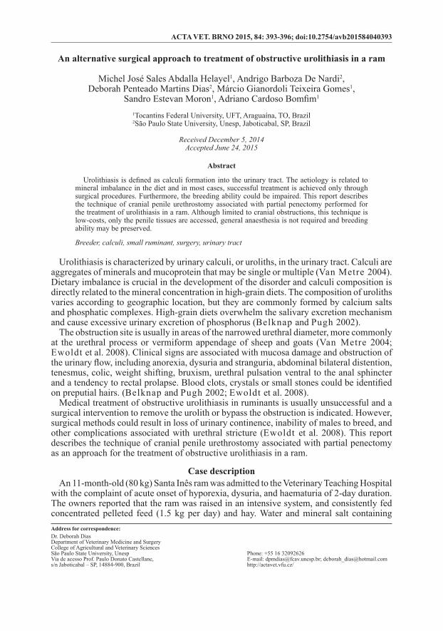
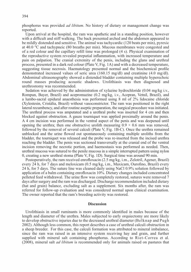
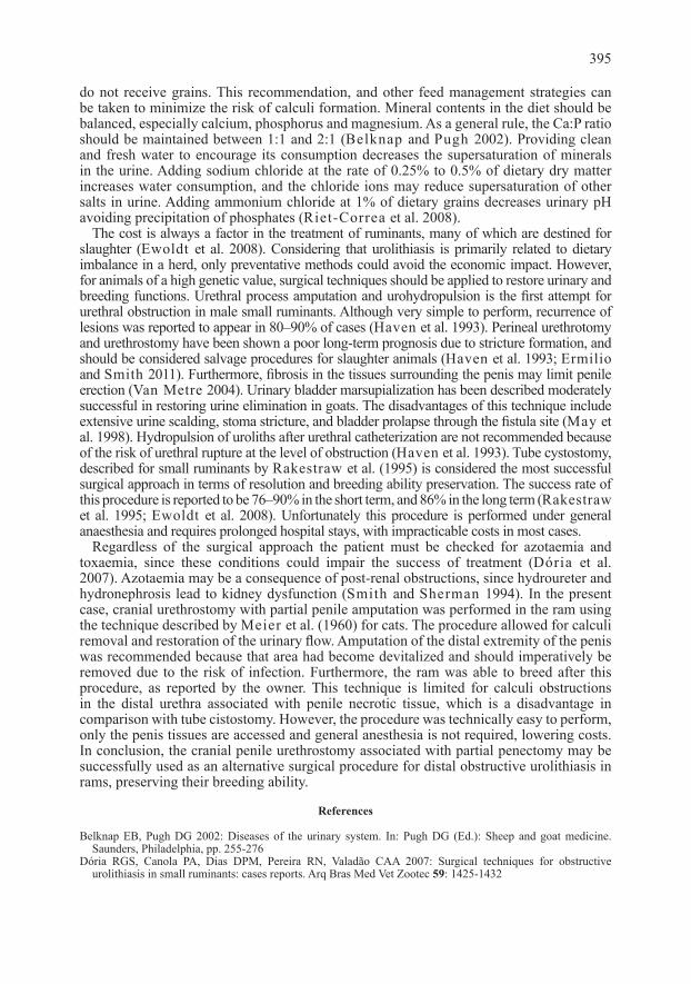
Upon arrival at the hospital, the ram was apathetic and in a standing position, however with a difficult and stiff walking. The back presented arched and the abdomen appeared to be mildly distended and sensitive. The animal was tachycardic (120 beats per min), pyrexic at 40.0 °C and tachypneic (80 breaths per min). Mucous membranes were congested and of a red colour and the capillary refill time was prolonged (4 s). Physical examination of the reproductive system revealed preputial inflammation, with increased temperature and pain on palpation. The cranial extremity of the penis, including the glans and urethral process, presented in a dark red colour (Plate V, Fig. 1A) and with a decreased temperature, suggesting tissue necrosis. Haematology presented normal and the biochemical profile demonstrated increased values of seric urea (160.15 mg/dl) and creatinine (4.0 mg/dl). Abdominal ultrasonography showed a distended bladder containing multiple hyperechoic round masses producing acoustic shadows. Urolithiasis was then diagnosed and urethrostomy was recommended.
Sedation was achieved by the administration of xylazine hydrochloride (0.04 mg/kg i.v., Rompun, Bayer, Brazil) and acepromazine (0.2 mg/kg, i.v., Acepran, Vetnil, Brazil), and a lumbo-sacral epidural anaesthesia was performed using 8 ml of 2% lidocaine chloride (Xylestesin, Cristália, Brazil) without vasoconstrictor. The ram was positioned in the right lateral recumbency, and after routine aseptic preparation, the surgical procedure was initiated. The urethral process was amputated and a urethral probe was inserted for 4 cm and then blocked against obstruction. A gauze tourniquet was applied proximally around the penis. A 4 cm incision was performed in the ventral aspect of the penis and was deepened until opening the urethra; then one obstructive urolith measuring 0.9 × 0.6 mm was removed, followed by the removal of several calculi (Plate V, Fig. 1B-C). Once the urethra remained unblocked and the urine flowed out spontaneously containing multiple uroliths from the bladder, the tourniquet was released and the probe was re-inserted from the incised urethra reaching the bladder. The penis was sectioned transversally at the cranial end of the ventral incision removing the necrotic portion, and haemostasis was performed as needed. Then, urethral mucosa was sutured to the penile mucosa in a simple interrupted pattern using nylon 0, creating a new urethral stoma (Plate V, Fig. 1D). Surgical time was around 90 min.
Postoperatively, the ram received enrofloxacin (2.5 mg/kg, i.m., Zelotril, Agener, Brazil) every 24 h, for 7 days and meloxicam (0.5 mg/kg, i.m., Maxicam, Ourofino, Brazil) every 24 h, for 5 days. The suture line was cleaned daily using NaCl 0.9% solution followed by application of a balm containing enrofloxacin 10%. Dietary changes included concentrated pelleted feed withdrawal. The urine flow was completely restored, sutures were removed 7 days after surgery and the ram was discharged. Discharge recommendation included dietary (hat and grain) balance, excluding salt as a supplement. Six months after, the ram was referred for follow-up evaluation and was considered normal upon clinical examination. The owner reported that the ram’s breeding activity was restored.
Discussion
Urolithiasis in small ruminants is more commonly identified in males because of the length and diameter of the urethra. Males subjected to early orquiectomy are more likely to develop obstructive injuries due to the decreased urethral diameter (Belknap and Pugh 2002). Although less common, this report describes a case of urethral calculi obstruction in a sheep breeder. For this case, the calculi formation was attributed to mineral imbalance, since the ram was raised in an intensive system receiving hay and grain, and further supplied with mineral salt containing phosphorus. According to Riet-Correa et al. (2008), mineral salt ad libitum is recommended only for animals raised on pastures that
394
do not receive grains. This recommendation, and other feed management strategies can be taken to minimize the risk of calculi formation. Mineral contents in the diet should be balanced, especially calcium, phosphorus and magnesium. As a general rule, the Ca:P ratio should be maintained between 1:1 and 2:1 (Belknap and Pugh 2002). Providing clean and fresh water to encourage its consumption decreases the supersaturation of minerals in the urine. Adding sodium chloride at the rate of 0.25% to 0.5% of dietary dry matter increases water consumption, and the chloride ions may reduce supersaturation of other salts in urine. Adding ammonium chloride at 1% of dietary grains decreases urinary pH avoiding precipitation of phosphates (Riet-Correa et al. 2008).
The cost is always a factor in the treatment of ruminants, many of which are destined for slaughter (Ewoldt et al. 2008). Considering that urolithiasis is primarily related to dietary imbalance in a herd, only preventative methods could avoid the economic impact. However, for animals of a high genetic value, surgical techniques should be applied to restore urinary and breeding functions. Urethral process amputation and urohydropulsion is the first attempt for urethral obstruction in male small ruminants. Although very simple to perform, recurrence of lesions was reported to appear in 80–90% of cases (Haven et al. 1993). Perineal urethrotomy and urethrostomy have been shown a poor long-term prognosis due to stricture formation, and should be considered salvage procedures for slaughter animals (Haven et al. 1993; Ermilio and Smith 2011). Furthermore, fibrosis in the tissues surrounding the penis may limit penile erection (Van Metre 2004). Urinary bladder marsupialization has been described moderately successful in restoring urine elimination in goats. The disadvantages of this technique include extensive urine scalding, stoma stricture, and bladder prolapse through the fistula site (May et al. 1998). Hydropulsion of uroliths after urethral catheterization are not recommended because of the risk of urethral rupture at the level of obstruction (Haven et al. 1993). Tube cystostomy, described for small ruminants by Rakestraw et al. (1995) is considered the most successful surgical approach in terms of resolution and breeding ability preservation. The success rate of this procedure is reported to be 76–90% in the short term, and 86% in the long term (Rakestraw et al. 1995; Ewoldt et al. 2008). Unfortunately this procedure is performed under general anaesthesia and requires prolonged hospital stays, with impracticable costs in most cases.
Regardless of the surgical approach the patient must be checked for azotaemia and toxaemia, since these conditions could impair the success of treatment (Dória et al. 2007). Azotaemia may be a consequence of post-renal obstructions, since hydroureter and hydronephrosis lead to kidney dysfunction (Smith and Sherman 1994). In the present case, cranial urethrostomy with partial penile amputation was performed in the ram using the technique described by Meier et al. (1960) for cats. The procedure allowed for calculi removal and restoration of the urinary flow. Amputation of the distal extremity of the penis was recommended because that area had become devitalized and should imperatively be removed due to the risk of infection. Furthermore, the ram was able to breed after this procedure, as reported by the owner. This technique is limited for calculi obstructions in the distal urethra associated with penile necrotic tissue, which is a disadvantage in comparison with tube cistostomy. However, the procedure was technically easy to perform, only the penis tissues are accessed and general anesthesia is not required, lowering costs. In conclusion, the cranial penile urethrostomy associated with partial penectomy may be successfully used as an alternative surgical procedure for distal obstructive urolithiasis in rams, preserving their breeding ability.
References
Belknap EB, Pugh DG 2002: Diseases of the urinary system. In: Pugh DG (Ed.): Sheep and goat medicine. Saunders, Philadelphia, pp. 255-276
Dória RGS, Canola PA, Dias DPM, Pereira RN, Valadão CAA 2007: Surgical techniques for obstructive urolithiasis in small ruminants: cases reports. Arq Bras Med Vet Zootec 59: 1425-1432
395

Ermilio EM, Smith MC 2011: Treatment of emergency conditions in sheep and goats. Vet Clin N Am-Food A 27: 33-45
Ewoldt JM, Jones ML, Miesner MD 2008: Surgery of obstructive urolithiasis in ruminants. Vet Clin N Am-Food A 24: 455-465
Haven ML, Bowman KF, Engelbert TA, Blikslager AT 1993: Surgical management of urolithiasis in small ruminants. Cornell Vet 83: 47-55
May KA, Moll HD, Wallace LM, Pleasant RS, Howard RD 1998: Urinary bladder marsupialization for treatment of obstructive urolithiasis in male goats. Vet Surg 27: 583-588
Meier FW 1960: Management of urethral obstruction and stenosis in the male cat. J Am Vet Med Assoc 137: 67-70
Rakestraw PC, Fubini SL, Gilbert RO, Ward JO 1995: Tube cystostomy for treatment of obstructive urolithiasis in small ruminants. Vet Surg 24: 498-505
Riet-Correa F, Simões SVD, Vasconcelos JS 2008: Urolithiasis in sheep and goats. Pesq Vet Bras 28: 319-322Smith MC, Sherman DM 1994: Goat medicine. Lippincott Williams & Wilkins, Baltimore, 620 p.Van Metre DC 2004: Urolithiasis. In: Fubini S, Ducharme N (Eds): Farm animal surgery. Saunders, St. Louis,
pp. 534-547
396
Plate VHelayel M. et al.: An alternative ... pp. 393-396
Fig. 1. Cranial extremity of the penis of a ram showing devitalized tissue due to obstructive urolithiasis (A). Calculi removal through a ventral urethral incision (B). Calculi removed from a ventral urethral incision. Bar = 1 cm (C). Penis of a ram after cranial penile urethrostomy (D).
![Springer-Verlag, LNCS 9879, pages 61{79 doi:10.1007/978 … · Robust Password-Protected Secret Sharing Michel Abdalla, ... and Blakey [Bla79]. This ... Camenisch et al. [CLLN14]](https://img.pdfslide.us/doc/110x75/5b3d4ff47f8b9a213f8dc66d/springer-verlag-lncs-9879-pages-6179-doi101007978-robust-password-protected.jpg)