Embed Size (px)
Citation preview
ECHOCARDIOGRAPHY FOR RADIOLOGISTS
Michael A. Bolen, M.D. Cardiovascular and Thoracic Imaging Imaging and Heart and Vascular Institutes Cleveland Clinic

� Brief overview of transthoracic echocardiography
modes and acquisition
� Discuss why knowledge of this modality can be
beneficial to the radiologist
Objectives

� Leading causes of death in US: Heart disease, cancer
(Nearly equivalent, CDC 2009).
� Daily cardiac imaging at our institution– cardiovascular
CT: 30-45, MRI: 10-15, echocardiography: 300
(excludes vascular studies…..)
� Technology development: combined efforts of multiple
physicists, engineers and clinicians over past 50 years
Echocardiography

Acquisition Methods
� Motion mode (M mode)
� Two-dimensional (2D) echo
� Doppler – continuous wave,
pulsed wave, color flow
� Newer developments – speckle
tracking, 3D
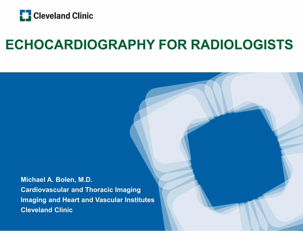
� Similar to earliest ultrasound images – single line of
interrogation reflected from structures
� Location and strength of these interfaces recorded over time,
the motion is recorded
� Limited anatomic depiction, but temporal resolution is fantastic
(sub ms, compared with industry best 75 ms CT)
M- Mode Echocardiography

� Time of transit and signal intensity of returning signal again measured, in this
mode spanning a 90 degree arc; anatomy is much better demonstrated
� Color flow and Doppler can be simultaneously acquired and superimposed
� Temporal resolution is good (5-50 ms)
Two dimensional (2D) echo

� Doppler is dependent upon change in frequency of the
transmitted ultrasound
� This shift of frequency can be converted to velocity
Doppler echocardiography

� Pulsed wave – discrete ultrasound waves sent at predefined rate (PRF);
location can be determined but peak velocity is limited
� Continuous wave – continuous receive and transmit US signal, higher
velocities can be measured but there is range ambiguity
� Color flow or color Doppler imaging – variation of pulsed Doppler, shares its
strengths/weaknesses – colorized image showing velocity and direction of
blood flow superimposed on 2D image
Doppler echocardiography
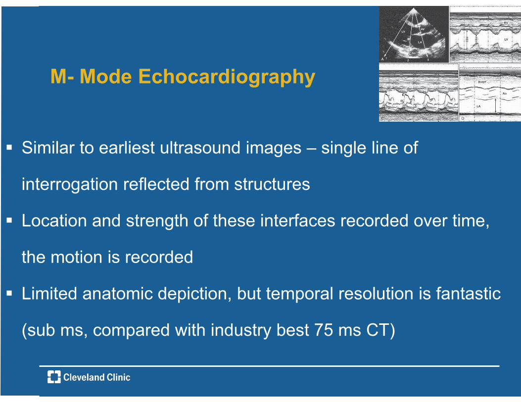
Echocardiography imaging windows


73 year old male with prior MVR with annuloplasty ring, now with symptoms of heart failure

M Mode images at level of aortic valve and mitral leaflets

Apical views: 5ch color flow and CW doppler

Findings
Systolic anterior motion of mitral valve associated with prior
repair with dynamic outflow tract obstruction* Findings corroborated on TEE later same week, patient
scheduled for redo MVR
* Risk factors for SAM with LVOT obstruction post MVR: myxomatous redundant leaflets, non dilated hyperdynamic LV, short distance between MV coaptation point and IVS after repair Sternik L et al Tex Heart Inst J 2005

60 year old male with prior STEMI, concern for PE

TTE: Apical 4ch and 2ch with and without contrast

Imaging windows and echocardiography
Images will only be as good as the sonographic window In previous case, good apical windows were challenging to
acquire due to shape and location of LV apex

� Combined efforts of members of CT/MRI and echo sections can be
productive – below are a few examples
� Cardiac MR assessment of aortic regurgitation: holodiastolic flow reversal in
the descending aorta helps stratify severity Radiology 2011; Development of
a Consensus Document to Improve Multireader Concordance and Accuracy
of Aortic Regurgitation Severity Grading by Echocardiography Versus CMR
Am J Cardiol 2012
Teamwork

� Important, widely used tool in the array of cardiovascular imaging options
� Anatomy and principles of ultrasound imaging may already be familiar to
radiologist with interest in cardiac imaging
� Increasing knowledge of this modality can improve effectiveness of work in
other areas of cardiac imaging, and may lead to collaborative investigations
or involvement with imaging service
Echocardiography

� Thank you for your attention
� TTE training opportunity, ongoing education, and
slides: Dr.’s Borkowski, Flamm, Marwick, Grimm,
Popovic, Al Soleiman
Acknowledgements