Embed Size (px)
Citation preview
HAL Id: hal-03331901https://hal.archives-ouvertes.fr/hal-03331901
Submitted on 2 Sep 2021
HAL is a multi-disciplinary open accessarchive for the deposit and dissemination of sci-entific research documents, whether they are pub-lished or not. The documents may come fromteaching and research institutions in France orabroad, or from public or private research centers.
L’archive ouverte pluridisciplinaire HAL, estdestinée au dépôt et à la diffusion de documentsscientifiques de niveau recherche, publiés ou non,émanant des établissements d’enseignement et derecherche français ou étrangers, des laboratoirespublics ou privés.
MICAL-L1 is required for cargo protein delivery to thecell surface
R. Sikora, P. Bun, L. Danglot, M. Alqabandi, P. Bassereau, F. Niedergang, T.Galli, A. Zahraoui
To cite this version:R. Sikora, P. Bun, L. Danglot, M. Alqabandi, P. Bassereau, et al.. MICAL-L1 is required forcargo protein delivery to the cell surface. Biology Open, Royal Society, 2021, 10 (6), pp.bio058008.�10.1242/bio.058008�. �hal-03331901�
RESEARCH ARTICLE
MICAL-L1 is required for cargo protein delivery to the cell surfaceR. Sikora1,*, P. Bun2,3,*, L. Danglot2,3, M. Alqabandi4, P. Bassereau4, F. Niedergang1, T. Galli2,5 andA. Zahraoui1,2,‡
ABSTRACTSecreted proteins are transported along intracellular route from theendoplasmic reticulum through the Golgi before reaching the plasmamembrane. Small GTPase Rab and their effectors play a key role inmembrane trafficking. Using confocal microscopy, we showed thatMICAL-L1 was associated with tubulo-vesicular structures andexhibited a significant colocalization with markers of the Golgiapparatus and recycling endosomes. Super resolution STORMmicroscopy suggested at the molecular level, a very closeassociation of MICAL-L1 and microdomains in the Golgi cisternae.Using a synchronized secretion assay, we report that the shRNA-mediated depletion of MICAL-L1 impaired the delivery of a subset ofcargo proteins to the cell surface. The process of membranetubulation was monitored in vitro, and we observe that recombinantMICAL-L1-RBD domain may contribute to promote PACSINs-mediated membrane tubulation. Interestingly, two hydrophobicresidues at the C-terminus of MICAL-L1 appeared to be importantfor phosphatidic acid binding, and for association with membranetubules. Our results reveal a new role for MICAL-L1 in cargo deliveryto the plasma membrane.
KEYWORDS: Rab effectors, MICAL-L1, Membrane traffic, Membranetubules
INTRODUCTIONMembrane trafficking is a tightly regulated process required forplasma membrane (PM) homeostasis, adhesion, and cell polarity.Secreted cargoes are transported from the endoplasmic reticulumto the Golgi apparatus (GA) and are then addressed to the plasmamembrane. Small GTPase Rab proteins and their effectors regulatemembrane trafficking between different intracellular compartments(Stenmark, 2009; Wandinger-Ness and Zerial, 2014). Rab8 plays arole in the exocytosis of AP-1B-dependent cargo (Ang et al., 2003).It is also required for the outgrowth of the primary cilium, andregulates apical protein localization in intestinal cells (Nachuryet al., 2007; Sato et al., 2007; Yamamura et al., 2008). In addition,
Rab8 and molecule interacting with CasL 3 (MICAL3) cooperate incontrolling the docking and fusion of exocytotic carriers (Grigorievet al., 2011). Rab13 regulates tight junction assembly by controllingthe delivery of tight junction proteins and downregulation of PKAactivity (Köhler et al., 2004; Marzesco and Zahraoui, 2005;Morimoto et al., 2005). It also regulates membrane traffickingbetween GA and PM (Nokes et al., 2008).
Molecule interacting with CasL-like1 (MICAL-L1), wasidentified as an effector of several Rabs including Rab 8, 11, 13and 35 (Abou-Zeid et al., 2011; Giridharan et al., 2012; Kobayashiet al., 2014; Sharma et al., 2009; Zahraoui, 2014). MICAL-L1 has acalponin homology (CH), a LIM (Lin-1l, Isl-1 andMec-3), a prolinerich (PRD) and a Rab binding domains (RBD) that binds GTP-bound Rab proteins. MICAL proteins contain an N-terminalflavoprotein monooxygenase domain (FAD) involved in F-actinoxidation and disassembly (Grigoriev et al., 2011; Hung et al.,2013). However, MICAL-L1 and 2 sequences do not contain theFAD. MICAL-L1 regulates endocytic recycling and plays a rolein ciliogenesis (Sharma et al., 2010, 2009; Xie et al., 2019). Inthis study, we found that MICAL-L1 is partially associated withmarkers of the Golgi apparatus and recycling endosomes. We theninvestigated whether MICAL-L1 regulates the biosynthetic deliveryof membrane proteins. To avoid non physiological temperatureconditions and to monitor trafficking of different cargo proteins, theRUSH (retention using selective hooks) system was used(Boncompain et al., 2012). The RUSH assay allows the retentionof a tagged cargo of interest in the endoplasmic reticulum (ER) andthen the cargo is released following the addition of biotin in the cellmedia. This method proved to be very powerful to quantitativelyassess the secretion of several cargoes (Boncompain et al., 2012;Boncompain and Weigel, 2018; Chen et al., 2017). We used theRUSH assay to characterize MICAL-L1 function in transport ofmembrane proteins to the PM. Depletion of MICAL-L1 experimentssuggest that the protein is required for surface delivery of a subset ofcargo proteins. Our data further indicate that two amino acids atMICAL-L1 C-terminus are important for phosphatidic acid (PA)binding and association with membrane tubules.
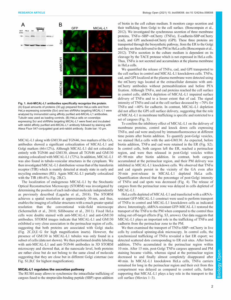
RESULTSMICAL-L1 is associated with Golgi like structure and withcytoplasmic tubulo-vesiclesWe used an affinity-purified polyclonal antibodies raised againstthe C-terminal domain (amino acid 520 to 863) to probe animmunoblot of cell extracts prepared from untransfected, Scramble(scr) and shRNAs targetingMICAL-L1 transfected HeLa cells. Anti-MICAL-L1 antibodies recognizes specifically a band of 130 kDacorresponding to the endogenous MICAL-L1 protein. The intensityof this band significantly decreased in MICAL-L1 silenced cells(∼70% depletion, Fig. 1A). In addition, the endogenous MICAL-L1staining was significantly reduced in MICAL-L1-shRNA-treatedcells as compared with mock-treated cells (Fig. 1B), thus furthervalidating our antibody.We then determinedMICAL-L1 intracellularlocalization in HeLa cells. Double labeling experiments withReceived 17 November 2020; Accepted 22 April 2021
1Universite de Paris, InsermU1016-CNRSUMR 8104, Institut Cochin, Paris, France.2Universite de Paris, Institute of Psychiatry and Neuroscience of Paris (IPNP),INSERM U1266, Membrane Traffic in Healthy & Diseased Brain, Paris, France.3Universite de Paris, Institute of Psychiatry and Neuroscience of Paris (IPNP),INSERM U1266, NeurImag Imaging facility, 75014 Paris, France. 4LaboratoirePhysico Chimie Curie, Institut Curie, PSL Research University, CNRS, UMR168,75005, Paris, France. 5GHU PARIS psychiatrie & neurosciences, F-75014 Paris,France.*These authors contributed equally to this work
‡Author for correspondence ([email protected])
L.D., 0000-0001-6190-6605; P.B., 0000-0002-8544-6778; T.G., 0000-0001-8514-7455; A.Z., 0000-0001-7424-8322
This is an Open Access article distributed under the terms of the Creative Commons AttributionLicense (https://creativecommons.org/licenses/by/4.0), which permits unrestricted use,distribution and reproduction in any medium provided that the original work is properly attributed.
1
© 2021. Published by The Company of Biologists Ltd | Biology Open (2021) 10, bio058008. doi:10.1242/bio.058008
BiologyOpen
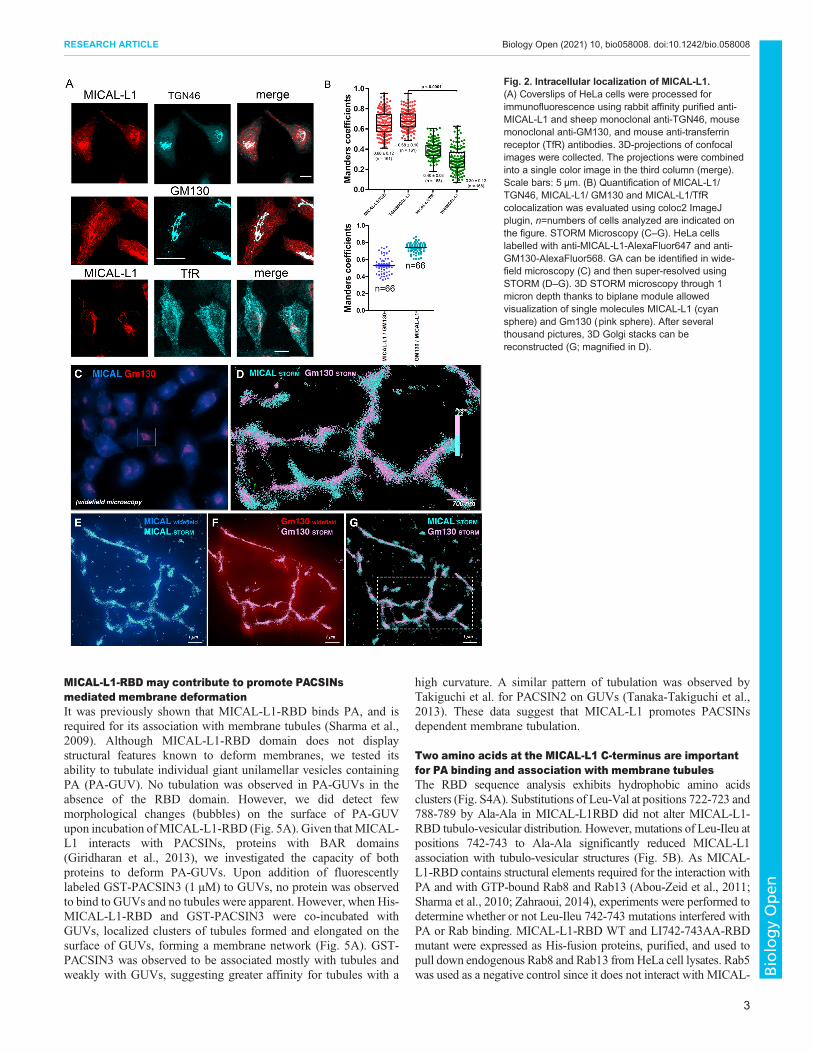
MICAL-L1 along with GM130 and TGN46, two markers of the GA,antibodies showed a significant colocalization of MICAL-L1 andGolgi markers (66±12%). Although MICAL-L1 did not colocalizeentirely with TGN46 and GM130, almost all TGN46 and GM130staining colocalized withMICAL-L1 (72%). In addition, MICAL-L1was also found in tubulo-vesicular structures in the cytoplasm. Wethen investigatedMICAL-L1 distribution versus that of the transferrinreceptor (TfR) which is mainly detected at steady state in early andrecycling endosomes (RE). Again MICAL-L1 partially colocalizedwith the TfR (40±8%; Fig. 2B,C).The localization of endogenous MICAL-L1 by the Stochastic
Optical Reconstruction Microscopy (STORM) was investigated bydetermining the position of each individual molecule independentlyas previously described (Lagache et al., 2018). The STORMachieves a spatial resolution at approximately 30 nm, and thus,enables the imaging of cellular structures with a much greater spatialresolution than the conventional wide-field microscope(Schermelleh et al., 2010; Sillibourne et al., 2011). Fixed HeLacells were double stained with anti-MICAL-L1 and anti-GM130antibodies. STORM images indicate that MICAL-L1 and GM130exhibited a very close association in the perinuclear region of cells,suggesting that both proteins are associated with Golgi stacks(Fig. 2C,D,E–G for high magnification insets). However, thepresence of GM130 in MICAL-L1 tubules was only found in asubset of cells (data not shown). We then performed double labelingwith anti-MICAL-L1 and anti-TGN46 antibodies in 3D STORMmicroscopy and showed that, at the nanoscale level, both proteinsare rather close but do not belong to the same cloud of moleculesuggesting that they are close but in different Golgi cisternae (seeFig. S1,B,C for highest magnification).
MICAL-L1 regulates the secretion pathwayThe RUSH assay allows to synchronize the intracellular trafficking ofcargoes fused to the streptavidin-binding peptide (SBP) upon addition
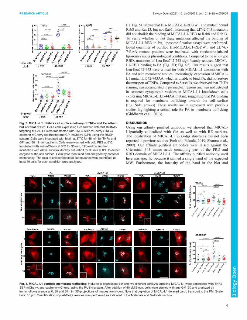
of biotin in the cell culture medium. It monitors cargo secretion andtheir trafficking from Golgi to the cell surface. (Boncompain et al.,2012). We investigated the synchronous secretion of three membraneproteins, TNFα−SBP−mCherry (TNFα), E-cadherin-SBP-mCherry(cad), and GPI anchored-mCherry (GPI). These three proteins aretransported through the biosynthetic pathway, from the ER to the Golgiand they are then delivered to the PM inHeLa cells (Boncompain et al.,2012). TNFα secretion in the culture medium is dependent on itscleavage by the TACE protease which is not expressed in HeLa cells.Thus, TNFα is not secreted and accumulates at the plasma membranein HeLa cells.
We quantified the release of TNFα, cad, and GPI transported tothe cell surface in control and MICAL-L1 knockdown cells. TNFα,cad, and GPI localized at the plasma membrane were detected usingthe mCherry tags located at the extracellular domains with anti-mCherry antibodies without permeabilization and before PFAfixation. Although TNFα, and cad proteins reached the cell surfacein control cells, shRNA depletion of MICAL-L1 impaired surfacedelivery of TNFα and to a lesser extent that of cad. The signalintensity of TNFα and cad at the cell surface deceased by ∼70% forTNFα and ∼40% for cadherin. In contrast, MICAL-L1 depletiondid not affect the GPI cell surface delivery, suggesting that the roleof MICAL-L1 in membrane trafficking is specific and restricted to aset of cargoes (Fig. 3).
To confirm the inhibitory effect of MICAL-L1 on the delivery ofmembrane proteins, control and knock down cells expressingTNFα, and cad were analyzed by immunofluorescence at differenttime points after biotin addition. To quantify post-Golgi vesicles,we stained HeLa cells with the anti-GM130. As expected, beforebiotin addition, TNFα and cad were retained in the ER (Fig. S2).In control cells, both cargoes left the ER, reached a perinuclearregion, and were then released in post-Golgi vesicles within45–90 min after biotin addition. In contrast, both cargoesaccumulated at the perinuclear region, and their PM delivery wasinhibited in MICAL-L1 knockdown cells. We observed that TNFαand cad signals persist in the vicinity of the GA even after30 min post-release in MICAL-L1 depleted HeLa cells.Quantification showed that the percentage of post-Golgi intensityof TNFα and cad spots was decreased (Fig. 4). The release ofcargoes from the perinuclear zone was delayed in cells depleted ofMICAL-L1.
HeLa cells depleted of MICAL-L1 and transfected with a siRNA-resistant GFP-MICAL-L1 construct were used to perform transportof TNFα in control and MICAL-L1 knockdown cells as indicatedabove. Interestingly, shRNA-resistant GFP-MICAL-L1 restored thetransport of the TNFα to the PM when compared to the control thusruling out off-target effects (Fig. S3, arrows). Our data suggests thatMICAL-L1 plays an important role in the trafficking of TNFα andcadherin from the perinuclear zone to the PM.
We then examined the transport of TNFα-SBP−mCherry in livecells by confocal spinning-disk microscopy. In control cells, thesynchronized trafficking of TNFα revealed a fast ER export; wedetected scattered dots corresponding to ER exit sites. After biotinaddition, TNFα accumulated in the perinuclear region within10 min. After 15 min, post-Golgi TNFα cargoes appeared and PMstaining became visible, whereas signal at the perinuclear regiondecreased to and finally almost completely disappeared after40 min. In MICAL-L1 knockdown HeLa cells, TNFα carriersremained for long in the perinuclear region and their exit from thiscompartment was delayed as compared to control cells, furthersupporting that MICAL-L1 plays a key role in the transport to thecell surface (Movies 1–3).
Fig. 1. Anti-MICAL-L1 antibodies specifically recognize the protein.(A) Equal amounts of proteins (30 µg) prepared from HeLa cells and fromHeLa expressing scramble (Scr) and two shRNAs targeting MICAL-L1 wereanalyzed by immunoblot using affinity purified anti-MICAL-L1 antibodies.Tubulin was used as loading controls. (B) HeLa cells on coverslipsexpressing Scr and shRNAs targeting MICAL-L1 were fixed and incubatedwith rabbit affinity purified anti-MICAL-L1 antibody followed by staining withAlexa Fluor 547-conjugated goat anti-rabbit antibody. Scale bar: 10 µm.
2
RESEARCH ARTICLE Biology Open (2021) 10, bio058008. doi:10.1242/bio.058008
BiologyOpen
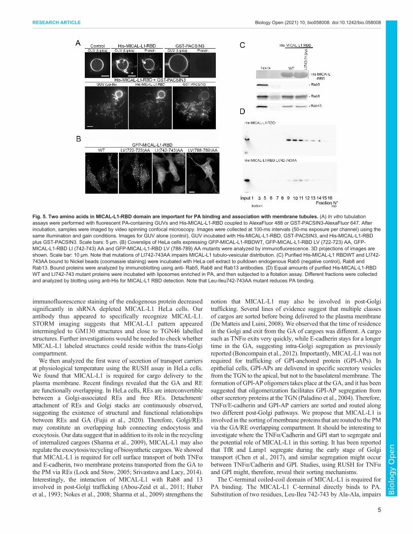
MICAL-L1-RBD may contribute to promote PACSINsmediated membrane deformationIt was previously shown that MICAL-L1-RBD binds PA, and isrequired for its association with membrane tubules (Sharma et al.,2009). Although MICAL-L1-RBD domain does not displaystructural features known to deform membranes, we tested itsability to tubulate individual giant unilamellar vesicles containingPA (PA-GUV). No tubulation was observed in PA-GUVs in theabsence of the RBD domain. However, we did detect fewmorphological changes (bubbles) on the surface of PA-GUVupon incubation ofMICAL-L1-RBD (Fig. 5A). Given thatMICAL-L1 interacts with PACSINs, proteins with BAR domains(Giridharan et al., 2013), we investigated the capacity of bothproteins to deform PA-GUVs. Upon addition of fluorescentlylabeled GST-PACSIN3 (1 µM) to GUVs, no protein was observedto bind to GUVs and no tubules were apparent. However, when His-MICAL-L1-RBD and GST-PACSIN3 were co-incubated withGUVs, localized clusters of tubules formed and elongated on thesurface of GUVs, forming a membrane network (Fig. 5A). GST-PACSIN3 was observed to be associated mostly with tubules andweakly with GUVs, suggesting greater affinity for tubules with a
high curvature. A similar pattern of tubulation was observed byTakiguchi et al. for PACSIN2 on GUVs (Tanaka-Takiguchi et al.,2013). These data suggest that MICAL-L1 promotes PACSINsdependent membrane tubulation.
Two amino acids at the MICAL-L1 C-terminus are importantfor PA binding and association with membrane tubulesThe RBD sequence analysis exhibits hydrophobic amino acidsclusters (Fig. S4A). Substitutions of Leu-Val at positions 722-723 and788-789 by Ala-Ala in MICAL-L1RBD did not alter MICAL-L1-RBD tubulo-vesicular distribution. However, mutations of Leu-Ileu atpositions 742-743 to Ala-Ala significantly reduced MICAL-L1association with tubulo-vesicular structures (Fig. 5B). As MICAL-L1-RBD contains structural elements required for the interaction withPA and with GTP-bound Rab8 and Rab13 (Abou-Zeid et al., 2011;Sharma et al., 2010; Zahraoui, 2014), experiments were performed todetermine whether or not Leu-Ileu 742-743 mutations interfered withPA or Rab binding. MICAL-L1-RBD WT and LI742-743AA-RBDmutant were expressed as His-fusion proteins, purified, and used topull down endogenous Rab8 and Rab13 fromHeLa cell lysates. Rab5was used as a negative control since it does not interact with MICAL-
Fig. 2. Intracellular localization of MICAL-L1.(A) Coverslips of HeLa cells were processed forimmunofluorescence using rabbit affinity purified anti-MICAL-L1 and sheep monoclonal anti-TGN46, mousemonoclonal anti-GM130, and mouse anti-transferrinreceptor (TfR) antibodies. 3D-projections of confocalimages were collected. The projections were combinedinto a single color image in the third column (merge).Scale bars: 5 µm. (B) Quantification of MICAL-L1/TGN46, MICAL-L1/ GM130 and MICAL-L1/TfRcolocalization was evaluated using coloc2 ImageJplugin, n=numbers of cells analyzed are indicated onthe figure. STORM Microscopy (C–G). HeLa cellslabelled with anti-MICAL-L1-AlexaFluor647 and anti-GM130-AlexaFluor568. GA can be identified in wide-field microscopy (C) and then super-resolved usingSTORM (D–G). 3D STORM microscopy through 1micron depth thanks to biplane module allowedvisualization of single molecules MICAL-L1 (cyansphere) and Gm130 (pink sphere). After severalthousand pictures, 3D Golgi stacks can bereconstructed (G; magnified in D).
3
RESEARCH ARTICLE Biology Open (2021) 10, bio058008. doi:10.1242/bio.058008
BiologyOpen
L1. Fig. 5C shows that His- MICAL-L1-RBDWT and mutant boundRab8 and Rab13, but not Rab5, indicating that LI742-743 mutationsdid not abolish the binding of MICAL-L1-RBD to Rab8 and Rab13.To verify whether or not these mutations affected the binding ofMICAL-L1-RBD to PA, liposome flotation assays were performed.Equal quantities of purified His-MICAL-L1-RBDWT and LL742-743AA mutant proteins were incubated with rhodamine-labeledliposomes under physiological conditions. Compared to thewild-typeRBD, mutations of Leu-Ileu742-743 significantly reduced MICAL-L1-RBD binding to PA (Fig. 5D; Fig. S5). Our results suggest thatLeu-Ileu742-743 were critical for both MICAL-L1 association withPA and with membrane tubules. Interestingly, expression of MICAL-L1 mutant LI742-743AA, which is unable to bind PA, did not restorethe transport of TNFα. Compared to Scr cells, we observed that TNFαstaining was accumulated in perinuclear regions and was not detectedin scattered cytoplasmic vesicles in MICAL-L1 knockdown cellsexpressing MICAL-L1LI744AA mutant, suggesting that PA bindingis required for membrane trafficking towards the cell surface(Fig. S4B, arrows). These results are in agreement with previousstudies highlighting a critical role for PA in membrane trafficking(Giridharan et al., 2013).
DISCUSSIONUsing our affinity purified antibody, we showed that MICAL-L1partially colocalized with GA as well as with RE markers.The localization of MICAL-L1 in Golgi structures has not beenreported in previous studies (Etoh and Fukuda, 2019; Sharma et al.,2009). Our affinity purified antibodies were raised against theC-terminal 343 amino acids containing part of the PRD andRBD domain of MICAL-L1. The affinity purified antibody usedhere was specific because it stained a single band of the expectedMW. Furthermore, the intensity of the band in the blot and
Fig. 4. MICAL-L1 controls membrane trafficking. HeLa cells expressing Scr and two different shRNAs targeting MICAL-L1 were transfected with TNFα-SBP-mCherry, and cadherin-mCherry, using the RUSH system. After addition of 40 µM Biotin, cells were stained with anti-GM130 and analyzed byimmunofluorescence at 0, 30 and 60 min. 3D-projections of images are shown. Note that depletion of MICAL-L1 delayed cargo transport to the PM. Scalebars: 10 µm. Quantification of post-Golgi vesicles was performed as indicated in the Materials and Methods section.
Fig. 3. MICAL-L1 inhibits cell surface delivery of TNFα and E-cadherinbut not that of GPI. HeLa cells expressing Scr and two different shRNAstargeting MICAL-L1 were transfected with TNFα-SBP-mCherry (TNFα),cadherin-mCherry (cadherin)t and GPI-mCherry (GPI) using the RUSHsystem. Cells were incubated with biotin at 37°C for 45 min for TNFα andGPI and 90 min for cadherin. Cells were washed with cold PBS at 0°C,Incubated with anti-mCherry at 0°C for 30 min, followed by anotherincubation with AlexaFluor647 donkey anti-rabbit for 30 min at 0°C to detectcargoes at the cell surface. Cells were then fixed and analyzed by confocalmicroscopy. The ratio of cell surface/total fluorescence was quantified, atleast 40 cells for each condition were analyzed.
4
RESEARCH ARTICLE Biology Open (2021) 10, bio058008. doi:10.1242/bio.058008
BiologyOpen
immunofluorescence staining of the endogenous protein decreasedsignificantly in shRNA depleted MICAL-L1 HeLa cells. Ourantibody thus appeared to specifically recognize MICAL-L1.STORM imaging suggests that MICAL-L1 pattern appearedintermingled to GM130 structures and close to TGN46 labelledstructures. Further investigations would be needed to check whetherMICAL-L1 labeled structures could reside within the trans-Golgicompartment.We then analyzed the first wave of secretion of transport carriers
at physiological temperature using the RUSH assay in HeLa cells.We found that MICAL-L1 is required for cargo delivery to theplasma membrane. Recent findings revealed that the GA and REare functionally overlapping. In HeLa cells, REs are interconvertiblebetween a Golgi-associated REs and free REs. Detachment/attachment of REs and Golgi stacks are continuously observed,suggesting the existence of structural and functional relationshipsbetween REs and GA (Fujii et al., 2020). Therefore, Golgi/REsmay constitute an overlapping hub connecting endocytosis andexocytosis. Our data suggest that in addition to its role in the recyclingof internalized cargoes (Sharma et al., 2009), MICAL-L1 may alsoregulate the exocytosis/recycling of biosynthetic cargoes. We showedthat MICAL-L1 is required for cell surface transport of both TNFαand E-cadherin, two membrane proteins transported from the GA tothe PM via REs (Lock and Stow, 2005; Srivastava and Lacy, 2014).Interestingly, the interaction of MICAL-L1 with Rab8 and 13involved in post-Golgi trafficking (Abou-Zeid et al., 2011; Huberet al., 1993; Nokes et al., 2008; Sharma et al., 2009) strengthens the
notion that MICAL-L1 may also be involved in post-Golgitrafficking. Several lines of evidence suggest that multiple classesof cargos are sorted before being delivered to the plasma membrane(DeMatteis and Luini, 2008). We observed that the time of residencein the Golgi and exit from the GA of cargoes was different. A cargosuch as TNFα exits very quickly, while E-cadherin stays for a longertime in the GA, suggesting intra-Golgi segregation as previouslyreported (Boncompain et al., 2012). Importantly, MICAL-L1 was notrequired for trafficking of GPI-anchored protein (GPI-APs). Inepithelial cells, GPI-APs are delivered in specific secretory vesiclesfrom the TGN to the apical, but not to the basolateral membrane. Theformation of GPI-AP oligomers takes place at the GA, and it has beensuggested that oligomerization facilitates GPI-AP segregation fromother secretory proteins at the TGN (Paladino et al., 2004). Therefore,TNFα/E-cadherin and GPI-AP carriers are sorted and routed alongtwo different post-Golgi pathways. We propose that MICAL-L1 isinvolved in the sorting ofmembrane proteins that are routed to the PMvia the GA/RE overlapping compartment. It should be interesting toinvestigate where the TNFα/Cadherin and GPI start to segregate andthe potential role of MICAL-L1 in this sorting. It has been reportedthat TfR and Lamp1 segregate during the early stage of Golgitransport (Chen et al., 2017), and similar segregation might occurbetween TNFα/Cadherin and GPI. Studies, using RUSH for TNFαand GPI might, therefore, reveal their sorting mechanisms.
The C-terminal coiled-coil domain of MICAL-L1 is required forPA binding. The MICAL-L1 C-terminal directly binds to PA.Substitution of two residues, Leu-Ileu 742-743 by Ala-Ala, impairs
Fig. 5. Two amino acids in MICAL-L1-RBD domain are important for PA binding and association with membrane tubules. (A) In vitro tubulationassays were performed with fluorescent PA-containing GUVs and His-MICAL-L1-RBD coupled to AlexaFluor 488 or GST-PACSIN3-AlexaFluor 647. Afterincubation, samples were imaged by video spinning confocal microscopy. Images were collected at 100-ms intervals (50-ms exposure per channel) using thesame illumination and gain conditions. Images for GUV alone (control), GUV incubated with His-MICAL-L1-RBD, GST-PACSIN3, and His-MICAL-L1-RBDplus GST-PACSIN3. Scale bars: 5 µm. (B) Coverslips of HeLa cells expressing GFP-MICAL-L1-RBDWT, GFP-MICAL-L1-RBD LV (722-723) AA, GFP-MICAL-L1-RBD LI (742-743) AA and GFP-MICAL-L1-RBD LV (788-789) AA mutants were analyzed by immunofluorescence. 3D projections of images areshown. Scale bar: 10 µm. Note that mutations of LI742-743AA impairs MICAL-L1 tubulo-vesicular distribution. (C) Purified His-MICAL-L1 RBDWT and LI742-743AA bound to Nickel beads (coomassie staining) were incubated with HeLa cell extract to pulldown endogenous Rab5 (negative control), Rab8 andRab13. Bound proteins were analyzed by immunoblotting using anti- Rab5, Rab8 and Rab13 antibodies. (D) Equal amounts of purified His-MICAL-L1-RBDWT and LI742-743 mutant proteins were incubated with liposomes enriched in PA, and then subjected to a flotation assay. Different fractions were collectedand analyzed by blotting using anti-His for MICAL-L1 RBD detection. Note that Leu-Ileu742-743AA mutant reduces PA binding.
5
RESEARCH ARTICLE Biology Open (2021) 10, bio058008. doi:10.1242/bio.058008
BiologyOpen
both the interaction ofMICAL-L1-Cter with PA and the targeting totubulo-vesicular structures. Compared to the wild-type RBD,mutations of Leu-Ileu742-743 significantly reduced MICAL-L1-RBD binding to PA, suggesting that additional residues were alsoimplicated. Indeed, two KR residues at positions 851-852 arerequired for optimal membrane association (Sharma et al., 2009). Ithas been previously shown that a stretch of hydrophobic residues atpositions 721-726 were critical for MICAL-L1 tubule association. Itis possible that LV722-723 mutations alone are not sufficient, butlarger region (721-726) is necessary for MICAL-L1 associationwith tubules (Sharma et al., 2009).Although MICAL-L1-Cter domain does not encompass any
obvious amphipathic helical sequence or displays homology toBAR domains, we show that it is able to promote PACSIN mediateddeformation of GUVs in vitro. Surprisingly, PACSIN3 alone (atleast under our experimental conditions) is not able to inducetubulation of GUVs, suggesting a high energy barrier for itsoligomerization and induction of membrane tubulation, alsoreported for PACSIN2 (Tanaka-Takiguchi et al., 2013).Our data and others (Sharma et al., 2009; Giridharan et al.,
2013) suggest that PA is a key component for the recruitment ofMICAL-L1 to membrane tubules. PA has been implicated inmembrane fission, suggesting that it may promote the release oftubulo-vesicular carriers (Jovanovic et al., 2006). We speculatethat MICAL-L1 scaffold couples membrane shaping componentssuch as PACSINs, Rabs and cargo sorting to PA microdomains,which may facilitate membrane sorting/targeting during cargodelivery to the plasma membrane. Interestingly, Rab10, a partner ofMICAL-L1, has been involved in the regulation of tubularendosome formation through KIF13A and KIF13B motors (Etohand Fukuda, 2019). Thus, MICAL-L1 in a complex with itsinteracting proteins might facilitate membrane deformation andcontribute to the formation of post-GA/RE carriers in the secretorypathway.
MATERIALS AND METHODSConstructsCloning of MICAL-L1 into pEGFP-C1 and mCherry vectors werepreviously described (Abou-Zeid et al., 2011). All the RUSH plasmidsused in this study, use streptavidin-KDEL as a hook. Briefly, the hook(streptavidin-tagged protein) allows retention of the SBP-tagged cargo in theER in the absence of biotin thanks to streptavidin–SBP interaction(Boncompain et al., 2012). The release of the RUSH cargoes was inducedby addition of 40 µM of D-biotin (Sigma-Aldrich).
Lipids reagentsAll reagents and β-casein from bovine milk (>99%) were purchased fromSigma-Aldrich (Sigma-Aldrich, France). DOPC (1,2-dioleoyl-sn-glycero-3-phosphatidylcholine), DOPS (1,2-dioleoyl-sn-glycero-3-phospho-L-serine), DOPE (1,2-dioleoyl-sn-glycero-3-phosphatidylethanol- amine),Egg PA (L-α-phosphatidic acid) and Egg Rhod PE (L-α-phosphatidylethanolamine-N-(lissamine rhodamine B sulfonyl) werepurchased from Avanti Polar Lipids, Inc. (Avanti Polar Lipids, USA).Stock solutions of lipids in chloroform (10 mg/ml) were stored at −20°C inamber vials (Sigma-Aldrich, France). Lipid stock solutions were mixed toachieve the desired molar ratio of DOPC/DOPE/DOPS/Egg PA/Egg RhodPE (33/33/13/20/0.8) at a total concentration of 1 mg/ml in chloroform.After use, argon was added to vials before sealing with paraffin film(Parafilm, USA) to prevent lipid oxidation.
MutagenesisFor mutagenesis of hydrophobic amino acids at the Cter of MICAL-L1,QuickChange site-directed mutagenesis kit (Agilent Technologies Inc.,Santa Clara, CA, USA) was used. Substitutions of amino acids were
performed according to the manufacturer’s instructions. Oligonucleotidesfor mutagenesis of LV722-723 to AA, oligo sens, 5′-GAGCTTGAA-CCAGTCCGCCGCCATGTCATCCTCACGG-3′; oligo anti-sens, 5′-CCG-TGAGGATGACATGGCGGCGGCTGGTTCAAGCTC-3′. Formutagenesis of LI722-723 to AA, oligo sens, 5′-TGCTGCTTG-AAGACATAGGCG-GCCTCGGACTCTCGCCGCAC-3′, oligo anti-sens,5′-GTGCGGCGA-GAGTCCGAGGCCGCCTATGTCTTCAAGCAGCA-3′. For mutagenesis of LV788-789 to AA, oligo sens, 5′-CTGCTCAATGAGGGTCGCAGCCTCCTGCATCAGCACC-3′, oligoanti-sens, 5′-GGTGCTGATGCAGGAGGCTGCGACCCTCATTGAGC-AG-3′. All mutations were verified by sequencing.
AntibodiesPurified His-MICAL-L1-RBD protein (amino acids 520-863 of MICAL-L1) was injected into rabbits to generate polyclonal antibodies (Covalab.Villeurbanne, France). The resulting antiserum was affinity purified againstHis-MICAL-L1RBD protein. anti-mCherry and anti-Histidine (His) rabbitpolyclonal antibodies were from Roche (Basel, Switzerland), anti-GM130monoclonal antibody (BD Transduction laboratory, CA, USA), anti-humanTGN46 (Bio-Rad, CA, USA),anti-trasferrin receptor (Thermo FisherScientific, Rockford, IL, USA), anti Rab5, Rab8 from Transductionlaboratories (USA) and Rab13 from Sigma-Aldrich (USA) and donkey-affinity purified secondary antibodies conjugated to AlexaFluor 488, 568,and 647 were from Jackson ImmunoResearch Laboratories (West Grove,PA, USA). The protein disulphide-isomerase (PDI) mouse monoclonalantibody from Enzo Life Sciences (France).
Cell culture and transfectionHeLa cells (ATCC CCL-2) were grown in DMEM containing 10% fetal calfserum (FCS) (Gibco,Watham,MA, USA) supplemented with 10% fetal calfserum, 2 mM glutamine, 100 U/ml penicillin, and 10 mg/ml streptomycin.The cells were incubated at 37°C under a 5% CO2 atmosphere. Stable HeLacells expressing shRNA ofMICAL-L1 were generated. Positive clones wereselected in the same medium supplemented with 0.3 µg/ml zeocin (Gibco,Waltham, MA, USA). Stably transfected clones were maintained underselection in 0.1 µg/ml of zeocin. The shRNA sequences that efficientlyinhibited proteins expression were as follow: MICAL-L1 shRNA oligo-sens, 5′-ACCTCGTGGAGCCTAGAGTGGAACAATC AAGAGTTGTTCCACTCTAGGCTCCACTT-3′; MICAL-L1 shRNA oligo-antisens, 5′-CAAAAAGTGGAGCCTAGAGTGGAACAACTCTTGATTGTTCCACTCTAGGCTCCACG-3′; PACSIN3 shRNA oligo-sens, 5′-ACCTCGGCTTGTTCTAGCGTGTATTATCA AGAGTAATACACGCTAGAACAAGCCTT-3′; PACSIN3 shRNA oligo-antisens, 5′-CAAAAAGGCTTGTTCTAGCGTGTATTACTCTTGATAATACACGCTAGAACAAGCCG-3′. HeLacells were transfected with Lipofectamine 3000 according to themanufacturer’s protocol (Invitrogen, Grand Island, NY, USA).
ImmunoblotHeLa cells were lysed in buffer, 20 mM Tris HCl pH 7.5, 150 mM NaCl,0.5% NP-40 with a protease cocktail inhibitor (Sigma-Aldrich). Solubilizedmaterial was recovered by centrifugation at 18.000 g for 15 min at 4°Cand supernatants were collected. Protein amounts were determinedusing the Pierce BCA assay (Life Technologies, PA, USA) and equalquantities of proteins were separated by SDS-PAGE and transferredelectrophoretically to nitrocellulose filters. Immunoblots were performedusing anti-MICAL-L1 antibodies and enhanced chemiluminescenceaccording to the manufacturer’s protocol (Thermo Fisher Scientific,Rockford, IL, USA).
GST pull-down assayThe cDNA encoding His-MICAL-L1-RBD (amino acids 520-863) WT orLI742-743AAwas inserted in a pET15b expression vector using NdeI-XhoIrestriction sites. The His-MICAL-L1-RBD fusion proteins were produced inE. coli and purified on Ni2-beads It were then incubated with HeLa cellextract for 4 h at 4°C, washed and bound material was analyzed by SDS-PAGE and immunoblotting using anti-Rab5, Rab83 and Rab13 antibodies.
6
RESEARCH ARTICLE Biology Open (2021) 10, bio058008. doi:10.1242/bio.058008
BiologyOpen
Liposome flotation assayLiposomes were prepared with a mass ratio composition of 87% POPC, 3%Lissamine rhodamine phosphatidyl ethanolamine and 10% of POPC, POPAor POPS in Hepes/NaCl buffer (25 mM Hepes pH 7.3, 150 mM NaCl).800 nM of GST-PACSIN3 or His-MICAL-L1-Cter purified proteins wereincubated with 500 µM of each liposome preparation for 30 min at roomtemperature. Samples were adjusted to 55% sucrose and loaded at thebottom of a Beckman SW55 Ti centrifugation tube. Samples were thenoverlaid by a discontinuous sucrose gradient (50%, 40%, 30%, 20%) andHepes/NaCl buffer was added on the top of the tube. Liposomes werecentrifuged at 150,000× g for 4 h at 4°C. Fractions were collected from thetop and separated by SDS-PAGE and analyzed by immunoblot using rabbitpolyclonal anti-His antibodies to detect MICAL-L1 RBD.
Preparation of giant unilamellar vesicles (GUVs)GUVs were prepared by the eletroformation method using conductingIndium Tin Oxide coated glass slides (ITO, Präzisions glas & optik GmBH,Germany) (Mathivet et al., 1996;Meleard et al., 2009; Morales-Penningstonet al., 2010). A lipid solution of 10 µl was deposited on ITO slides by using a5 µl Hamilton syringe to make a dry lipid film as thin as possible. The lipidcoated ITO slides, assembled with sigillum wax (Vitrex, Denmark), weredried under vacuum for 45 min at room temperature. The lipid films werethen hydrated with a sucrose/Tris growth buffer (100 mM of sucrose and10 mM of Tris, at pH 7.4) and sinusoidale AC current at 1V (peak to peak)with a frequency of 10 Hz was applied for 45–90 min at room temperature.GUVs were extracted by pipetting directly from GUV-rich regions from theformation chamber. Collected GUVs were then transferred in observationbuffer (70 mM of NaCl and 10 mM of Tris, at pH 7.4) with an osmolarity of10–20 mOsm higher than that of the growth buffer.
Protein-membrane binding assayObservation chambers were prepared using 60×24 mm coverslips (Menzel-Gläser, Germany) and 40×22 mm coverslips (VWR International, France).Before use, the chambers were passivated with a 5 mg/ml β-casein solution(100 mM NaCl and 10 mM Tris, at pH7.4) for 15–30 min to preventGUVs from adhering to the glass surface. Chambers were then rinsedseveral times and filled with observation buffer. GUVs were incubated witheither MICAL-L1-RBD at 1 µM, or GST-PACSIN3 at 1 µM in theobservation buffer, or with both of them simultaneously. The protein wasallowed to bind to GUVs for 20–40 min on ice before observation. GUVswere observed with a spinning disk confocal microscope inverted NikonEclipse Ti-E microscope with 100x oil objective. Images were recordedwith an EM-CCD Evolve camera. The exposure time for all imageswas 50 ms
Immunofluorescence stainingHeLa Cells on coverslips were fixed with 4% paraformaldehyde for 15 minat room temperature and permeabilized using 2% BSA and 0.1% TritonX100 for 15 min at room temperature in PBS. Cells were then incubatedwith primary antibody in 2% BSA, 0.1% triton X100 in PBS for 1 h at roomtemperature and then incubated with secondary conjugated antibody for30 min at room temperature in the same buffer. After washing, samples weremounted in prolong (eBioscience). Alexa Fluor 488-transferrin pulse-chaseassays were done as described previously (Jovic et al., 2009).
Fixed cell confocal imagingImage acquisition was performed on an inverted confocal microscope(Leica DMI6000) with a 63x (1.4 NA) objective and a MicroMAX camera(Princeton Instruments) or ORCA Flash4.0 (Hamamatsu). Z stack of 7-10plans (0.4 microns step) were acquired using Metamorph software(Molecular Devices, Sunnyvale, CA, USA). Images were then generatedby compiling three-dimensional maximum intensity projections of plansusing ImageJ software.
To evaluate protein colocalization we used Coloc 2, a pre-installedplugin on FIJI. We employed a bisection threshold regression on region ofinterests corresponding to single cell masks. Numerical correlation
parameters such as Mander’s coefficients are recorded as well as the 2Dintensity histogram.
For transport of TNFα-mCherry, cadherin-mCherry, and GPI-mCherry tothe cell surface, cells were washed at 0°C with PBS. Proteins at the cellsurface were stained with anti-Cherry antibodies at 0°C for 60 min followedby incubation at 0°C with Alexa 746 donkey anti-rabbit secondaryantibodies. Cells were then fixed with 4% PFA and analyzed. Cherryfluorescence gave the total amount of proteins expressed in the cell. Theratio of cell surface over total fluorescence was calculated. Experiment wereperformed in triplicate and more than 50 cells per experiment werequantified.
Image quantificationTo quantify the ratio of cargo transported to the surface, we first segmentedcell morphology using the GFP channel. Background removal wasperformed by globally subtracting the mean fluorescent intensity of aregion outside cells. The generated cell mask was then used to measure themean fluorescence intensity of cargoes at the surface and within the cell.n=number of cells quantified is indicated in the figure.
To quantify the amount of cargoes exiting the Golgi (post-Golgi vesicles)we both segmented endogenously GFP-expressing cell and GM130-labelled Golgi morphology. We performed a rolling ball of 50 pixels toremove background signal. We then measured the cargo fluorescenceintensity in the entire cell without taking into account the Golgi contribution
Statistical significance testStatistical tests used were unpaired nonparametric tests such as t-test andKruskal–Wallis followed by a post hoc Dunn’s test.
Live cell imaging setup – Spinning diskHeLa cells were seeded onto 18 mm-diameter glass coverslips, 1 day beforetransfection. Twenty hours after transfection with the TNFα-SBPEGFPRUSH plasmid (Boncompain et al., 2012), coverslips were transferred into aChamlide chamber, filled with pre-warmed DMEM medium (Invitrogen).At time 2 min, medium was removed and D-biotin (Sigma-Aldrich) at40 μM final was introduced in the chamber. Time-lapse acquisitions weredone at 37°C in a thermostat-controlled chamber. Fluorescent images weresequentially acquired every 40 s for 60 min using a HCX APO 1.3 glycerol63 X objective and MetaMorph software (Molecular Device). We used aLEICA DMI8 microscope (LEICA MICROSYSTEMS) equipped with aCSU-X1 spinning-disk confocal unit (Yokogawa Electric Corporation) andan ORCA -Flash4.0 V3 Digital sCMOS camera (Hamamatsu Photonics) ina controlled environment box (37°C and 5% CO2, PECON). Themicroscopy system was equipped with a laser combiner (Errol) comprisesof a 488 nm (KVANT) and a 561 nm (Oxxius LBX) laser line. GFP (resp.mCherry/mRFP) emission light were collected with a stringent singlebandpass filter 525/50-25 (resp. 620/60-25). The microscopy system wasdriven by Metamorph (Molecular Devices).
3D-STORM microscopyHeLa cells were seeded on 18 mm #1.5 MENZEL glaser coverslipspreviously cleaned with plasma cleaner and coated with poly-Ornithine.Cells were fixed with 4% PFA during 10 min, permeabilized with 2% BSA-PBS-Triton X100 0.1% for 15 min at room temperature. Cells were thenincubated with Rabbit anti-MICAL-L1 and mouse anti-GM130 antibody in2% BSA, 0.1% triton X100 in PBS for 36 h at 4°C. After washing, cellswere then incubated with donkey anti-rabbit-Alexfluor647 and donkey anti-mouse Alexafluor568 secondary conjugated antibodies for 2 h at roomtemperature in the same buffer. After washing with PBS, Hela cells werefixed in PBS- 4%PFA and 0.2% glutaraldehyde for 10 min. Cells werewashed with PBS, mounted in an alveolar slide with dental silicon andanalyzed with Bruker Optera/Vutara microscope as previously reported(Lagache et al., 2018). Samples were imaged in a photoswitching buffercontaining 100 mM MEA and oxygen scavenging system (0.5 mg/mlglucose oxidase, 40 mg/ml catalase, 10% glucose) in PBS. They wereexcited with 640 and 561 laser lines and with 405 laser line. 3D-STORM
7
RESEARCH ARTICLE Biology Open (2021) 10, bio058008. doi:10.1242/bio.058008
BiologyOpen
images were reconstructed from a series of 10000 frames as previouslyreported (Collot et al., 2019) using Bruker Srx software.
AcknowledgementsWe thank Drs Gaelle Boncompain and Franck Perez (Curie Institute) for help withthe RUSH system, Ludger Johannes and Christian Wunder (Curie Institute) for helpwith lipid binding experiments, and Somia Vats for reading and discussing themanuscript. Imaging was carried out at NeurImag core facility, part of IPNP, InsermU. 1266 and Universite de Paris. We would like to acknowledge the NeurimagImaging Facility team for their technical and scientific support in imaging of biologicalsamples.
Competing interestsThe authors declare no competing or financial interests.
Author contributionsConceptualization: A.Z.; Methodology: R.S., P. Bun, L.D., M.A., P. Bassereau, F.N.,A.Z.; Software: P. Bun, A.Z.; Validation: R.S., P. Bun, L.D., M.A., P. Bassereau, F.N.,T.G., A.Z.; Formal analysis: M.A., A.Z.; Investigation: R.S., P. Bun, L.D., M.A.,P. Bassereau, T.G., A.Z.; Writing - original draft: A.Z.; Writing - review & editing: L.D.,P. Bassereau, F.N., T.G., A.Z.; Visualization: P. Bun, L.D., F.N., T.G.; Supervision:A.Z.; Project administration: A.Z.; Funding acquisition: T.G.
FundingThis work was supported by grants from the French National Center for ScientificResearch (CNRS), the National Institute for Health and medical Research(INSERM), Universite de Paris, the Fondation pour la Recherche Medicale (FRMDEQ20130326518), the Association pour la Recherche sur le Cancer (ARC), theFrench National Research Agency (NeuroImmunoSynapse ANR-13-BSV2-0018-02;MetDePaDi ANR-16-CE16-0012), the Institut National du Cancer (PLBIO 2018-149), the FLAG-ERA grant Senseï by ANR-19-HBPR-0003. We acknowledgethe following equipment funding, Leducq foundation to T.G. for the Leica SP8Confocal/STED 3DX microscope, Fondation Bettencourt Schueller Coup d’Elan toT.G. for the Leica/Yokogawa Spinning disc microscope, DIM Elicit and (regionIle de France) FRC/Rotary to L.D. for the Bruker Vutara STORM and OpteraSpinning disc system, respectively. R.S. was a recipient of a fellowship fromUniversite Paris Descartes. M.A. was a recipient of a fellowship from the Pierre etMarie Curie University.
ReferencesAbou-Zeid, N., Pandjaitan, R., Sengmanivong, L., David, V., Le Pavec, G.,Salamero, J. and Zahraoui, A. (2011). MICAL-like1 mediates epidermal growthfactor receptor endocytosis.Mol. Biol. Cell 22, 3431-41. doi:10.1091/mbc.e11-01-0030
Ang, A. L., Folsch, H., Koivisto, U. M., Pypaert, M. and Mellman, I. (2003). TheRab8 GTPase selectively regulates AP-1B-dependent basolateral transport inpolarized Madin-Darby canine kidney cells. J. Cell Biol. 163, 339-50. doi:10.1083/jcb.200307046
Boncompain, G. and Weigel, A. V. (2018). Transport and sorting in the Golgicomplex: multiple mechanisms sort diverse cargo. Curr. Opin. Cell Biol. 50,94-101. doi:10.1016/j.ceb.2018.03.002
Boncompain, G., Divoux, S., Gareil, N., de Forges, H., Lescure, A., Latreche, L.,Mercanti, V., Jollivet, F., Raposo, G. and Perez, F. (2012). Synchronization ofsecretory protein traffic in populations of cells. Nat. Methods 9, 493-8. doi:10.1038/nmeth.1928
Chen, Y., Gershlick, D. C., Park, S. Y. and Bonifacino, J. S. (2017). Segregation inthe Golgi complex precedes export of endolysosomal proteins in distinct transportcarriers. J. Cell Biol. 216, 4141-4151. doi:10.1083/jcb.201707172
Collot, M., Ashokkumar, P., Anton, H., Boutant, E., Faklaris, O., Galli, T., Mly, Y.,Danglot, L. and Klymchenko, A. S. (2019). MemBright: a family of fluorescentmembrane probes for advanced cellular imaging and Neuroscience. Cell Chem.Biol. 26, 600-614.e7. doi:10.1016/j.chembiol.2019.01.009
De Matteis, M. A. and Luini, A. (2008). Exiting the Golgi complex. Nat. Rev. Mol.Cell Biol. 9, 273-84. doi:10.1038/nrm2378
Etoh, K. and Fukuda, M. (2019). Rab10 regulates tubular endosome formationthrough KIF13A and KIF13B motors. J. Cell Sci. 132, jcs226977. doi:10.1242/jcs.226977
Fujii, S., Kurokawa, K., Inaba, R., Hiramatsu, N., Tago, T., Nakamura, Y.,Nakano, A., Satoh, T. and Satoh, A. K. (2020). Recycling endosomes attach tothe trans-side of Golgi stacks in Drosophila and mammalian cells. J. Cell Sci. 133,jcs236935. doi:10.1242/jcs.236935
Giridharan, S. S., Cai, B., Naslavsky, N. and Caplan, S. (2012). Traffickingcascades mediated by Rab35 and its membrane hub effector, MICAL-L1.Commun. Integr. Biol. 5, 384-7. doi:10.4161/cib.20064
Giridharan, S. S., Cai, B., Vitale, N., Naslavsky, N. and Caplan, S. (2013).Cooperation of MICAL-L1, syndapin2, and phosphatidic acid in tubular recycling
endosome biogenesis. Mol. Biol. Cell. 24, 1776-90. doi:10.1091/mbc.e13-01-0026
Grigoriev, I., Yu, K. L., Martinez-Sanchez, E., Serra-Marques, A., Smal, I.,Meijering, E., Demmers, J., Peranen, J., Pasterkamp, R. J., van der Sluijs, P.,Hoogenraad, C. C. and Akhmanova, A. (2011). Rab6, Rab8, and MICAL3cooperate in controlling docking and fusion of exocytotic carriers. Curr. Biol. 21,967-74. doi:10.1016/j.cub.2011.04.030
Huber, L. A., Pimplikar, S., Parton, R. G., Virta, H., Zerial, M. and Simons, K.(1993). Rab8, a small GTPase involved in vesicular traffic between the TGNand thebasolateral plasma membrane. J. Cell Biol. 123, 35-45. doi:10.1083/jcb.123.1.35
Hung, R.-J., Spaeth, C. S., Yesilyurt, H. G. and Terman, J. R. (2013). SelRreverses Mical-mediated oxidation of actin to regulate F-actin dynamics. Nat. CellBiol. 15, 1445-54. doi:10.1038/ncb2871
Jovanovic, O. A., Brown, F. D. and Donaldson, J. G. (2006). An effector domainmutant of Arf6 implicates phospholipase D in endosomal membrane recycling.Mol. Biol. Cell 17, 327-35. doi:10.1091/mbc.e05-06-0523
Jovic, M., Kieken, F., Naslavsky, N., Sorgen, P. L. and Caplan, S. (2009). Eps15homology domain 1-associated tubules contain phosphatidylinositol-4-phosphateand phosphatidylinositol-(4,5)-bisphosphate and are required for efficientrecycling. Mol. Biol. Cell 20, 2731-43. doi:10.1091/mbc.e08-11-1102
Kobayashi, H., Etoh, K., Ohbayashi, N. and Fukuda, M. (2014). Rab35 promotesthe recruitment of Rab8, Rab13 and Rab36 to recycling endosomes throughMICAL-L1 during neurite outgrowth. Biol. Open. 3, 803-14. doi:10.1242/bio.20148771
Kohler, K., Louvard, D. and Zahraoui, A. (2004). Rab13 regulates PKA signalingduring tight junction assembly. J. Cell Biol. 165, 175-80. doi:10.1083/jcb.200312118
Lagache, T., Grassart, A., Dallongeville, S., Faklaris, O., Sauvonnet, N., Dufour,A., Danglot, L. and Olivo-Marin, J. C. (2018). Mapping molecular assemblieswith fluorescence microscopy and object-based spatial statistics. Nat. Commun.9, 698. doi:10.1038/s41467-018-03053-x
Lock, J. G. and Stow, J. L. (2005). Rab11 in recycling endosomes regulates thesorting and basolateral transport of E-cadherin.Mol. Biol. Cell 16, 1744-55. doi:10.1091/mbc.e04-10-0867
Marzesco, A. M. and Zahraoui, A. (2005). Assay of Rab13 in regulating epithelialtight junction assembly. Methods Enzymol. 403, 182-93. doi:10.1016/S0076-6879(05)03015-6
Mathivet, L., Cribier, S. and Devaux, P. F. (1996). Shape change and physicalproperties of giant phospholipid vesicles prepared in the presence ofan AC electric field. Biophys. J. 70, 1112-21. doi:10.1016/S0006-3495(96)79693-5
Meleard, P., Bagatolli, L. A. and Pott, T. (2009). Giant unilamellar vesicleelectroformation from lipid mixtures to native membranes under physiologicalconditions. Methods Enzymol. 465, 161-76. doi:10.1016/S0076-6879(09)65009-6
Morales-Penningston, N. F., Wu, J., Farkas, E. R., Goh, S. L., Konyakhina, T. M.,Zheng, J. Y., Webb, W. W. and Feigenson, G. W. (2010). GUV preparation andimaging: minimizing artifacts. Biochim. Biophys Acta 1798, 1324-32. doi:10.1016/j.bbamem.2010.03.011
Morimoto, S., Nishimura, N., Terai, T., Manabe, S., Yamamoto, Y.,Shinahara, W., Miyake, H., Tashiro, S., Shimada, M. and Sasaki, T. (2005).Rab13mediates the continuous endocytic recycling of occludin to the cell surface.J. Biol. Chem. 280, 2220-8. doi:10.1074/jbc.M406906200
Nachury,M. V., Loktev, A. V., Zhang,Q.,Westlake, C. J., Peranen, J., Merdes, A.,Slusarski, D. C., Scheller, R. H., Bazan, J. F., Sheffield, V. C. and Jackson,P. K. (2007). A core complex of BBS proteins cooperates with theGTPaseRab8 topromote ciliary membrane biogenesis.Cell 129, 1201-13. doi:10.1016/j.cell.2007.03.053
Nokes, R. L., Fields, I. C., Collins, R. N. and Folsch, H. (2008). Rab13 regulatesmembrane trafficking between TGN and recycling endosomes in polarizedepithelial cells. J. Cell Biol. 182, 845-53. doi:10.1083/jcb.200802176
Paladino, S., Sarnataro, D., Pillich, R., Tivodar, S., Nitsch, L. and Zurzolo, C.(2004). Protein oligomerization modulates raft partitioning and apicalsorting of GPI-anchored proteins. J. Cell Biol. 167, 699-709. doi:10.1083/jcb.200407094
Sato, T., Mushiake, S., Kato, Y., Sato, K., Sato, M., Takeda, N., Ozono, K., Miki,K., Kubo, Y., Tsuji, A., Harada, R. and Harada, A. (2007). The Rab8 GTPaseregulates apical protein localization in intestinal cells. Nature 448, 366-9. doi:10.1038/nature05929
Schermelleh, L., Heintzmann, R. and Leonhardt, H. (2010). A guide to super-resolution fluorescence microscopy. J. Cell Biol. 190, 165-75. doi:10.1083/jcb.201002018
Sharma, M., Giridharan, S. S., Rahajeng, J., Naslavsky, N. and Caplan, S.(2009). MICAL-L1 links EHD1 to tubular recycling endosomes andregulates receptor recycling. Mol. Biol. Cell 20, 5181-94. doi:10.1091/mbc.e09-06-0535
Sharma, M., Giridharan, S. S., Rahajeng, J., Caplan, S. and Naslavsky, N.(2010). MICAL-L1: An unusual Rab effector that links EHD1 totubular recycling endosomes. Commun. Integr. Biol. 3, 181-3. doi:10.4161/cib.3.2.10845
8
RESEARCH ARTICLE Biology Open (2021) 10, bio058008. doi:10.1242/bio.058008
BiologyOpen
Sillibourne, J. E., Specht, C. G., Izeddin, I., Hurbain, I., Tran, P., Triller, A.,Darzacq, X., Dahan, M. and Bornens, M. (2011). Assessing the localization ofcentrosomal proteins by PALM/STORM nanoscopy. Cytoskeleton 68, 619-27.doi:10.1002/cm.20536
Srivastava, N. and Lacy, P. (2014). Trafficking of TNF via recycling endosomes inneutrophils. Allergy. Asthma Clin. Immunol. 10, 47. doi:10.1186/1710-1492-10-S2-A47
Stenmark, H. (2009). Rab GTPases as coordinators of vesicle traffic.Nat. Rev. Mol.Cell Biol. 10, 513-25. doi:10.1038/nrm2728
Tanaka-Takiguchi, Y., Itoh, T., Tsujita, K., Yamada, S., Yanagisawa, M.,Fujiwara, K., Yamamoto, A., Ichikawa, M. and Takiguchi, K. (2013).Physicochemical analysis from real-time imaging of liposome tubulation revealsthe characteristics of individual F-BAR domain proteins. Langmuir 29, 328-36.doi:10.1021/la303902q
Wandinger-Ness, A. and Zerial, M. (2014). Rab proteins and the
compartmentalization of the endosomal system. Cold Spring Harb. Perspect
Biol. 6, a022616. doi:10.1101/cshperspect.a022616Xie, S., Farmer, T., Naslavsky, N. and Caplan, S. (2019). MICAL-L1 coordinates
ciliogenesis by recruiting EHD1 to the primary cilium. J. Cell Sci. 132, jcs233973.
doi:10.1242/jcs.233973Yamamura, R., Nishimura, N., Nakatsuji, H., Arase, S. and Sasaki, T. (2008). The
interaction of JRAB/MICAL-L2 with Rab8 and Rab13 coordinates the assembly of
tight junctions and adherens junctions. Mol. Biol. Cell 19, 971-83. doi:10.1091/
mbc.e07-06-0551Zahraoui, A. (2014). MICAL-like1 in endosomal signaling. Methods Enzymol. 535,
419-37. doi:10.1016/B978-0-12-397925-4.00024-9
9
RESEARCH ARTICLE Biology Open (2021) 10, bio058008. doi:10.1242/bio.058008
BiologyOpen