Embed Size (px)
Citation preview
proteinsSTRUCTURE O FUNCTION O BIOINFORMATICS
Dopamine transporter comparative molecularmodeling and binding site prediction using theLeuTAa leucine transporter as a templateMartın Indarte,1* Jeffry D. Madura,2* and Christopher K. Surratt1*
1Division of Pharmaceutical Sciences, Duquesne University, Pittsburgh, Pennsylvania 15282
2Department of Chemistry and Biochemistry, Center for Computational Sciences, Duquesne University, Pittsburgh,
Pennsylvania 15282
INTRODUCTION
Addiction to cocaine, methamphetamine, and related psychostimu-
lants destroys millions of individuals, families, and careers, a societal
scourge worldwide. Although, addiction to heroin, oxycodone, fen-
tanyl, and other opiates can be effectively treated with buprenorphine
and to some extent methadone, no such medications are available to
combat psychostimulant addiction despite decades of research. Not
coincidentally, opioid receptor structure and mechanism of action are
much better understood than those of the brain receptors for psy-
chostimulant drugs of abuse, the monoamine neurotransmitter trans-
porter proteins. Pharmacologic and behavioral studies indicate that
the dopamine transporter (DAT) protein is the principal binding site
responsible for cocaine’s reward and reinforcement properties.1,2 The
plasma membrane-bound DAT protein quenches dopamine-mediated
neurotransmission by clearing the neurotransmitter from the synaptic
cleft following Ca21-mediated exocytosis from presynaptic vesicles.
Cocaine, a DAT inhibitor, blocks synaptic uptake of dopamine; the
resultant accumulation of the neurotransmitter in the synapse leads
to an increase in postsynaptic dopamine D2 and D3 receptor activa-
tion in the nucleus accumbens and other brain regions associated
with addiction. Indeed, activation of these accumbal dopamine recep-
tors has been linked with the reinforcing properties of the drug.3,4
Amphetamine also increases synaptic dopamine levels, but by media-
ting dopamine efflux from the presynaptic cell via the DAT.5,6 Logi-
cally, an agent that blocks cocaine and amphetamine binding at the
DAT without substantially interfering with dopamine uptake should
serve as an effective antiaddiction therapeutic. High resolution eluci-
dation of the DAT structure, especially regarding its substrate and in-
hibitor recognition sites, is thus critical.
The Supplementary Material referred to in this article can be found online at http://www.
interscience.wiley.com/jpages/0887-3585/suppmat.
Grant sponsor: NIDA; Grant number: DA016604; Grant sponsor: Samuel and Emma Winters;
Grant sponsor: DOE; Grant numbers: P116Z040100, P116Z050331.
*Correspondence to: Dr. Christopher K. Surratt, Division of Pharmaceutical Sciences, Duquesne
University, Mellon Hall, Room 453, 600 Forbes Avenue, Pittsburgh, PA 15282.
E-mail: [email protected] or Martin Indarte, E-mail: [email protected] or Jeffry D. Madura,
E-mail: [email protected]
Received 11 September 2006; Revised 16 March 2007; Accepted 16 April 2007
Published online 10 September 2007 in Wiley InterScience (www.interscience.wiley.com).
DOI: 10.1002/prot.21598
ABSTRACT
Pharmacological and behavioral studies indicate
that binding of cocaine and the amphetamines by
the dopamine transporter (DAT) protein is prin-
cipally responsible for initiating the euphoria and
addiction associated with these drugs. The lack of
an X-ray crystal structure for the DAT or any
other member of the neurotransmitter:sodium
symporter (NSS) family has hindered understand-
ing of psychostimulant recognition at the atomic
level; structural information has been obtained
largely from mutagenesis and biophysical studies.
The recent publication of a crystal structure for
the bacterial leucine transporter LeuTAa , a dis-
tantly related NSS family homolog, provides for
the first time a template for three-dimensional
comparative modeling of NSS proteins. A novel
computational modeling approach using the
capabilities of the Molecular Operating Environ-
ment program MOE 2005.06 in conjunction with
other comparative modeling servers generated the
LeuTAa-directed DAT model. Probable dopamine
and amphetamine binding sites were identified
within the DAT model using multiple docking
approaches. Binding sites for the substrate
ligands (dopamine and amphetamine) overlapped
substantially with the analogous region of the
LeuTAa crystal structure for the substrate leucine.
The docking predictions implicated DAT side
chains known to be critical for high affinity
ligand binding and suggest novel mutagenesis tar-
gets in elucidating discrete substrate and inhibi-
tor binding sites. The DAT model may guide DAT
ligand QSAR studies, and rational design of novel
DAT-binding therapeutics.
Proteins 2008; 70:1033–1046.VVC 2007 Wiley-Liss, Inc.
Key words: homology; comparative modeling; dock-
ing; drug; pharmacophore; medication; therapeutic;
cocaine; psychostimulant; antagonist; addiction.
VVC 2007 WILEY-LISS, INC. PROTEINS 1033
The DAT and other plasma membrane monoamine
transporters are members of the 12 transmembrane do-
main (TM) neurotransmitter:sodium symporter (NSS)
family,7 in which electrogenic transport of a neurotrans-
mitter substrate across the cell membrane is driven by a
Na1/K1-ATPase-generated Na1 gradient. Cotransport of
Cl2 is also required for the dopamine, norepinephrine,
and serotonin transporter proteins (DAT, NET, and
SERT, respectively); the SERT additionally transports K1,
but in antiport fashion.8 Aligning the amino acid
sequences of the NSS family members guided delineation
of monoamine transporter TM domain borders and
other aspects of transporter secondary structure.9 Such a
sequence alignment can also yield clues as to which NSS
residues probably contribute to the general protein infra-
structure, which residues could play a role in substrate or
ion recognition, and which residues are most likely to be
responsible for a pharmacologic pattern unique to a
given transporter. This sequence information alone
spawned hundreds of NSS site-directed and chimeric
mutants.10 The substituted cysteine accessibility muta-
genesis (SCAM) methodology has especially contributed
to defining monoamine transporter ligand binding cav-
ities, substrate/ion pores, general TM domain infrastruc-
ture, and even detection of substrate- or inhibitor-
induced conformational changes.11–13 Nevertheless, this
approach only circumstantially implicates a given residue
or protein region as a component of the binding pocket.
Unequivocally identifying direct contacts between trans-
porter protein and ligand has proven to be difficult; the
lack of an X-ray crystal structure for any protein homo-
logous to the NSS family has been the major impediment.
Encouragingly, the recently published crystal structure of
a bacterial leucine transporter (LeuTAa), a protein homo-
logous with the NSS family,14 finally provides a suitable
NSS template. Using LeuTAa as a template, the present
study describes a novel modeling approach that employs
comparative modeling to produce a feasible three-dimen-
sional (3D) DAT structure.
Three approaches may be employed in predicting a 3D
macromolecular structure: ab initio prediction, ‘‘fold’’
recognition, and comparative (homology) modeling.15
These differ principally in the sequence and structural
database information used. A true ab initio method bases
structure prediction entirely on the physical and chemical
information contained in the primary amino acid
sequence. However, the term is also used when short ex-
perimental protein sequences and secondary structure
prediction methods are incorporated.16–20 Fold recogni-
tion, or ‘‘threading,’’ relies heavily on the structural simi-
larities between certain distantly related or unrelated pro-
teins. Comparative modeling predicts the 3D structure of
a target protein based primarily on its alignment with
one or more template proteins of known structure.21 For
proteins that share greater than 40% amino acid
sequence identity, comparative modeling is straightfor-
ward and typically accurate.22 For proteins with less
than 30% amino acid sequence identity (e.g., LeuTAa and
the DAT), comparative modeling becomes more challeng-
ing. Still, the rhodopsin crystal structure has successfully
guided the creation of useful comparative models for
many other members of the G protein coupled receptor
superfamily despite the absence of appreciable amino
acid sequence identity.23
Upon obtaining a 3D protein model, the conforma-
tions and orientations (denoted as poses) of ligands that
couple with the macromolecule are computationally
determined (‘‘docking’’).24–27 To find the most energeti-
cally favorable ligand pose within a structurally deter-
mined receptor, the macromolecule is typically held rigid
whereas the ligands are flexible and mobile.28 Here, a
docking procedure similar to the earlier uses of DOCK29
was used to identify potential DAT binding sites. This
approach should reveal DAT amino acid residues likely to
participate in substrate and inhibitor recognition and
thus define targets for mutagenesis and other structure-
function studies. In this way, it is hoped that a blueprint
can be developed for rational design of DAT-binding
therapeutics.
MATERIALS AND METHODS
Comparative modeling
Robetta server sequence alignment and model building
The comparative modeling module of the Robetta
server aligns the target and the template using K*Sync,
a more accurate method than PSI-BLAST or Pcons2.30
K*Sync estimates the most reliable alignment of target
and template based on secondary structure information,
residue information obtained by comparing statistical
representations of protein families (‘‘profile–profile’’
comparisons), and information from multiple structural
alignments of regions with high structural propensity to
fold. The peptide backbone is constructed taking into
account the geometry between template(s) and target via
multiple independent simulations; the lowest energy
models are selected. Side chains of these models are
repacked and conformational space explored using �100
independent Monte Carlo simulations, with a backbone
dependent side chain rotamer library and a full atom
energy function to select the lowest energy conformation
of the comparative model.31–33
The FASTA sequence of the rat DAT protein (SwissProt
locus SC6A3_RAT; accession number P23977; NCBI
accession number AAB21099)34 was utilized as the query
for the hybrid template-based/de novo method of the
Robetta server (http://robetta.bakerlab.org). The bacterial
(Aquifex aeolicus) leucine transporter protein LeuTAa was
employed as the template (PDB, www.rcsb.org, accession
number 2A65; MMDB accession no. 34395). Five models
M. Indarte et al.
1034 PROTEINS DOI 10.1002/prot
were retrieved from the server and separately saved in a
database using the Molecular Operating Environment
(MOE) 2005.06 program (Chemical Computing Group,
Montreal, Canada).35 The all-atom forcefield AMBER99
was used to add hydrogen atoms and assign partial
charges to all models.36 Relaxation of the newly added
hydrogen atoms via several cycles of energy minimization
were performed using a conjugated gradient/truncated
Newton optimization algorithm to convergence criteria
of 0.05 kcal/mol and a dielectric constant (e) of 3. All
nonhydrogen atoms were held fixed during the energy
minimization. Pro_check (MOE version), a scientific vec-
tor language (SVL) code based on Ramachandran plots
and custom-written by the Chemical Computing Group,
was used to detect unfavorable van der Waals contacts
and abnormal covalent bonds in the models. The few
steric clashes found were relaxed by manually selecting
backbone and side chain atoms of the implicated amino
acids and by performing successive steps of energy mini-
mization until the steric clash was removed. All steric
clashes were far from the putative ligand binding sites. A
final refinement of side chains was carried out utilizing
AMBER99 (convergence criteria 5 0.1 kcal/mol, e 5 3).
Backbone atoms were held fixed during the procedure to
find local minima for the side chains of the DAT macro-
molecule.
The final DAT model (herein referred to as Model 1)
was selected using the following criteria: (1) Maximal
spatial overlap of backbones between the DAT models
(targets) and LeuTAa (template). (2) Similarity of Verify
3D scores between target and template models with
respect to TM domains.37,38 (3) Optimal profile of
atom contacts and fewest abnormal covalent bonds as
reported by Pro_check (MOE version). (4) Lowest poten-
tial energy, as calculated using MOE 2005.06.
3D-JIGSAW server sequence alignment and model building
3D-JIGSAW employs PSI-BLAST39 to generate a posi-
tion specific scoring matrix (PSSM) for the template and
target sequence. This PSSM data is used by the PSI-Pred
program40 to predict secondary structures for both
sequences. The PSSM data and secondary structures are
used in a dynamic programming algorithm to perform
an initial alignment. A second dynamic programming
algorithm refines the initial alignment via multiple align-
ment of template structures.41 Target protein side chains
are positioned based on those in the template and are
also added from a side chain rotamer library when
needed. Finally, a mean-field calculation is performed to
select the most probable, best packed side chain
rotamers.41 The rDAT FASTA sequence was used as the
query for the 3D-JIGSAW server (www.bmm.icnet.uk/
�3djigsaw/). Sequence alignment and DAT homology
modeling relative to the LeuTAa template were derived
using both ‘‘interactive’’ and ‘‘automatic’’ modes. The
DAT atomic coordinates for the comparative model were
obtained after model building and selection by the meta-
server of the most energetically favorable structure. This
single model was downloaded, read by MOE 2005.06 and
saved in a molecular database. Using MOE pro_check, the
few steric clashes found were resolved by selecting back-
bone and side chain atoms of the implicated amino acid
residues and performing successive steps of AMBER99
energy minimization (convergence criteria 5 0.1 kcal/
mol, e 5 3). After resolving unfavorable contacts, the
protocol described earlier for the Robetta models was
applied to calculate partial charges and optimize hydro-
gen atoms and side chains, yielding Model 2.
Yamashita et al. alignment and MOE model building
The rDAT FASTA sequence and crystal structure coor-
dinates of LeuTAa were loaded into MOE 2005.06. The
primary amino acid sequences of LeuTAa and DAT were
manually aligned using the MSA proposed by Yamashita
et al.14 Because the initial partial geometry between tem-
plate and target was not specified, only the backbone
coordinates of LeuTAa were used for the model creation.
A series of 10 DATmodels were independently constructed
with MOE using a Boltzmann-weighted randomized pro-
cedure42 combined with specialized logic for the proper
handling of sequence insertions and deletions.43 Each in-
termediate model was evaluated by a residue packing
quality function sensitive to the degrees to which nonpo-
lar side chain groups are buried within the lipid bilayer
and hydrogen bonding opportunities are maximized.
Before the final refinement of side chains, a coarse mini-
mization of backbone atoms using AMBER99 and a con-
jugated gradient method (convergence criterion 5 1.0
kcal/mol, e 5 3) was performed to improve packing and
intramolecular interactions. No steric clashes were
observed. The same protocol described for the Robetta
models was applied to calculate partial charges and opti-
mize hydrogen atoms and side chains. The optimal MOE
model (Model 3) was selected using the criteria listed
above for Model 1, and by weighting the best scores for
side chain packing according to MOE’s packing evalua-
tion function. The sequence alignments underpinning
Models 1–3 are shown in Figures 1–3, respectively.
Modeling of Na1 binding sites
Two sodium atoms were placed in the DAT models
using the corresponding LeuTAa crystal coordinates; their
positions were manually refined in order to preserve
coordination bonds established with adjacent residues.
The side chains of such residues were adjusted to emulate
the LeuTAa environment using the rotamer explorer
module in MOE 2005.06. Side chains were relaxed (with
the two Na1 atoms and backbone positions fixed) using
AMBER99 (convergence criteria 5 1.0 kcal/mol, e 5 3).
Comparative Model of the Dopamine Transporter
DOI 10.1002/prot PROTEINS 1035
DAT model ligand docking
Construction and geometry optimization of DATsubstrates and inhibitors
Three-dimensional models of the DAT substrates dopa-
mine and amphetamine were constructed using the mole-
cule builder feature of MOE 2005.06 (structures pictured
in Fig. 4). Partial charges and hydrogen atoms were
added to protonated and unprotonated molecules using
the Merck Molecular Force Field 94X (MMFF94X), suita-
ble for small drug-like molecules.46–48 All structures
were energy minimized using the conjugated gradient/
truncated Newton optimization algorithm with conver-
gence criterion 5 0.05 kcal/mol, e 5 1.
Binding site selection and exploration
The ‘‘alpha site finder’’ module of MOE 2005.06 was
used to identify possible DAT ligand binding pockets
within the newly-generated DAT models. Hydrophobic or
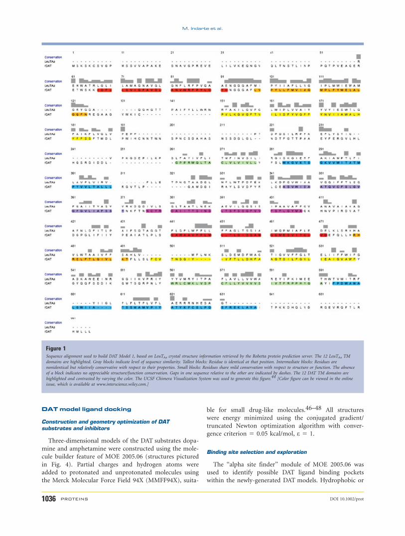
Figure 1Sequence alignment used to build DAT Model 1, based on LeuTAa crystal structure information retrieved by the Robetta protein prediction server. The 12 LeuTAa TM
domains are highlighted. Gray blocks indicate level of sequence similarity. Tallest blocks: Residue is identical at that position. Intermediate blocks: Residues are
nonidentical but relatively conservative with respect to their properties. Small blocks: Residues share mild conservation with respect to structure or function. The absence
of a block indicates no appreciable structure/function conservation. Gaps in one sequence relative to the other are indicated by dashes. The 12 DAT TM domains are
highlighted and contrasted by varying the color. The UCSF Chimera Visualization System was used to generate this figure.44 [Color figure can be viewed in the online
issue, which is available at www.interscience.wiley.com.]
M. Indarte et al.
1036 PROTEINS DOI 10.1002/prot
hydrophilic alpha spheres served as ‘‘probes’’ denoting
zones of tight atomic packing. All probe clusters of alpha
spheres not situated in cytoplasmic or phospholipid-
facing regions were used to identify potential binding
sites that were used in the docking simulations. These
alpha spheres were used as centroids for the creation of
dummy atoms used to define potential binding sites dur-
ing the docking process.
MOE-Dock 2005.06
A binding region is identified by a cluster of hydro-
phobic and hydrophilic alpha spheres; hydrophobic
spheres mark hydrophobic environments, and hydrophilic
spheres mark hydrophilic environments. Ligand atoms
are matched to corresponding alpha spheres during the
docking process. The alpha spheres are used to calculate
shape complementarity of small molecules fitting into
macromolecules, as well as binding affinities of these
conformers. Docking methods that employ alpha spheres
may generate bound conformations that approach crys-
tallographic resolution.49 The ligand explores the confor-
mational space to locate the most favorable binding ori-
entation and conformation (denoted as a ‘‘pose’’)24–27
by aligning and matching all triangles of the template
points with compatible geometry and chemistry; the pro-
tein atoms remain fixed during the process. For each
ligand, 100 poses were generated and scored in an effort
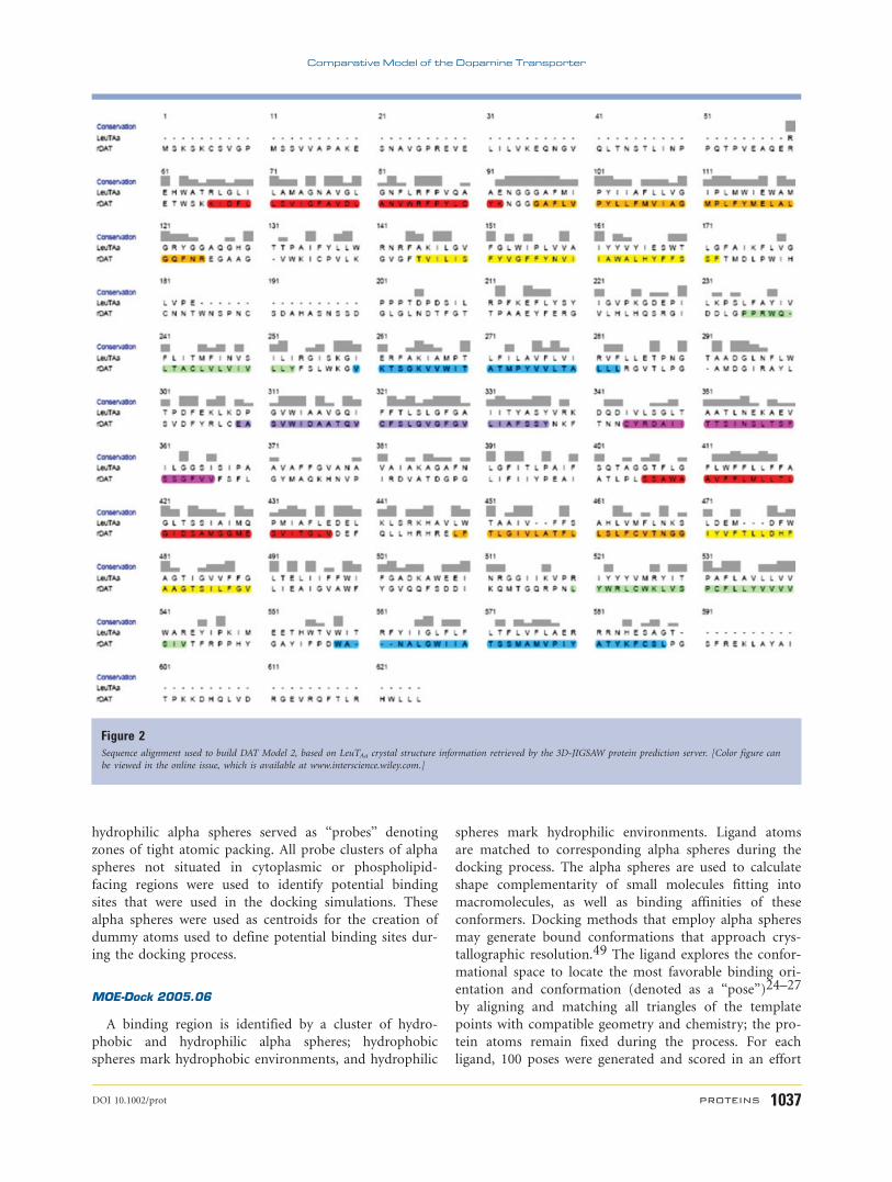
Figure 2Sequence alignment used to build DAT Model 2, based on LeuTAa crystal structure information retrieved by the 3D-JIGSAW protein prediction server. [Color figure can
be viewed in the online issue, which is available at www.interscience.wiley.com.]
Comparative Model of the Dopamine Transporter
DOI 10.1002/prot PROTEINS 1037
to determine favorable binding modes. An affinity scor-
ing function, G, was employed to rank candidate poses.
This pairwise atomic contact scoring methodology esti-
mates the enthalpic contribution to the free energy of
binding using the following linear function:
G 5 Chbfhb 1 Cionfion 1 Cmligfmlig
1 Chhfhh 1 Chpfhp 1 Caafaa
The fx terms represent the fractional atomic contacts
for a specific interaction, x. The Cx terms are coefficients
that weight the interaction contribution of x to the affin-
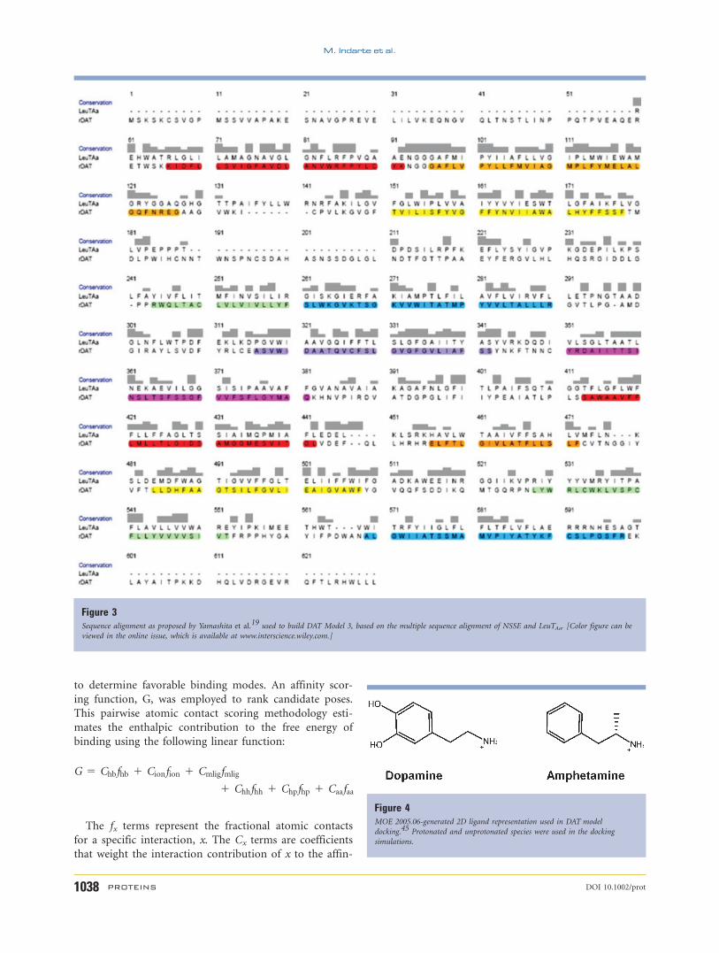
Figure 3Sequence alignment as proposed by Yamashita et al.19 used to build DAT Model 3, based on the multiple sequence alignment of NSSE and LeuTAa. [Color figure can be
viewed in the online issue, which is available at www.interscience.wiley.com.]
Figure 4MOE 2005.06-generated 2D ligand representation used in DAT model
docking.45 Protonated and unprotonated species were used in the docking
simulations.
M. Indarte et al.
1038 PROTEINS DOI 10.1002/prot
ity score. The individual terms are: hb, hydrogen bond
donor-acceptor pair interactions (an optimistic view is
taken; for example, two hydroxyl groups are assumed to
interact in the most favorable way); ion, ionic interac-
tions (a Coulomb-like term is used to evaluate the inter-
actions between charged groups); mlig, metal ligation
interactions (those involving nitrogen and sulfur atoms
and transition metals are so classified); hh, hydrophobic
interactions; hp, interactions between hydrophobic and
polar atoms; aa, an interaction between any two atoms.
Two different placement methodologies for docking DAT
substrates and inhibitors were used. The alpha triangle
placement method generates poses by superposition of
ligand atom triplets and triplet points within the receptor
site. The triangle matcher method generates poses in a
systematic and more accurate way than the alpha triangle
placement method by aligning ligand triplets of atoms
with triplets of alpha spheres in cavities of tight atomic
packing. The docking process accounted for the two pro-
tonation states of the amine group of ligands. Poses from
molecular databases of each ligand were scored based on
complementarity with binding pocket alpha spheres.
ASEDock
Alpha Sphere Based Protein-Ligand Docking (Ryoka
Institutes), or ASEDock, is a novel fast-docking program
written in the SVL language (MOE platform) and based
on the alpha shape method. Ligand atoms have alpha
spheres within 1 A. On the basis of this, concave shape
models can be created, and ligand atoms from a large
number of conformations generated by superposition with
these points can be evaluated and scored by maximum
overlap with the alpha spheres and minimum overlap
(repulsion) with receptor atoms. The initial ligand confor-
mations were subjected to energy minimization using the
MMFF94S force field46 and when converged, reproduced
experimentally bioactive conformations.49 The scoring
function used by ASEDock is based on protein–ligand
interaction energies. The interaction energy of a given
conformation is calculated using the following formula:
Utotal 5 Uele 1 Uvdw 1 Uligand 1 Usolv
Uele and Uvdw represent electrostatic and van der Waals
interactions, respectively, between the protein macromol-
ecule and the ligand. Uligand represents conformation
energy. Usolv represents the energy because of solvation.
The lowest Utotal of the multiple poses generated were
considered optimal poses. All alpha spheres not situated
in cytoplasmic or phospholipid-facing regions of the
DAT model were used as centroids for the creation of
dummy atoms used to dock DAT ligands. The docking
process took into account the two protonation states of
ligand amine groups. Poses from the molecular databases
for each ligand were ranked based on Utotal.
For each ligand, 500 conformations were generated
using the default systematic search parameters in the
ASEDock module. Five thousand poses per conformation
were randomly placed onto the alpha spheres located
within the TM domains. From the resulting 500,000
poses, 200 poses with the lowest Utotal values were
selected, and these poses were further optimized with the
MMFF94S force field. During this refinement step, the
ligand was free to move within the rigid binding pocket
(the transporter atoms were held fixed).
MOE-DOCK 2004.03 GA
A Monte Carlo simulated annealing process is used,
allowing a sampling of the conformational space for the
ligand and an extensive screening of all possible binding
sites in a particular region of the target macromole-
cule.50–52 Docking interaction energy (Utotal) of a given
conformation is estimated from a set of energy grids cen-
tered in the macromolecule binding site using the for-
mula given above for ASEDock. Macromolecule protein
coordinates remain fixed during the process, while the
flexible, mobile ligand moves along the grid to locate the
most favorable binding orientation and conformation
based on the interaction energy. A docking box of 45 345 3 45 grid points was employed with grid spacing of
0.375 A. The alpha spheres generated in the TM domains
by the site finder module were used as the centroids for
the docking box. Once the docking region was defined,
the alpha spheres were deleted (and not used in any sub-
sequent calculations). Minimized ligands were randomly
placed inside the docking box, and the docking process
initiated with an iteration limit of 10,000, cycle number
of 50, and run number of 100. The two protonation
states of the ligand amine group were taken into account
in the docking process. The final molecular database con-
tained 100 docked poses for each ligand as well as all
energy terms discussed earlier.
Validation of the DAT ligand docking process viaLeuTAa-leucine docking
The three docking methods described above were
used to assess the validity of the DAT-ligand docking pre-
dictions by calculating possible bound conformations of
leucine-LeuT complexes. The crystal structure of LeuT
was retrieved from the PDB and prepared for docking:
partial charges and hydrogen atoms were added, cal-
culated and relaxed within the protein structure as
described earlier. No further minimizations of side chains
were carried out. LeuTAa,-leucine docking poses were
obtained using MOE-Dock 2005.06, ASEDock or MOE-
DOCK 2004.03 GA, and compared to the original crystal
structure. The RMSDs of leucine bound in the crystal
versus the predicted bound leucine conformations for the
different methods were calculated using db_crystal_rmsd,
Comparative Model of the Dopamine Transporter
DOI 10.1002/prot PROTEINS 1039
a SVL code custom-written by the Chemical Computing
Group.
RESULTS
Comparative models
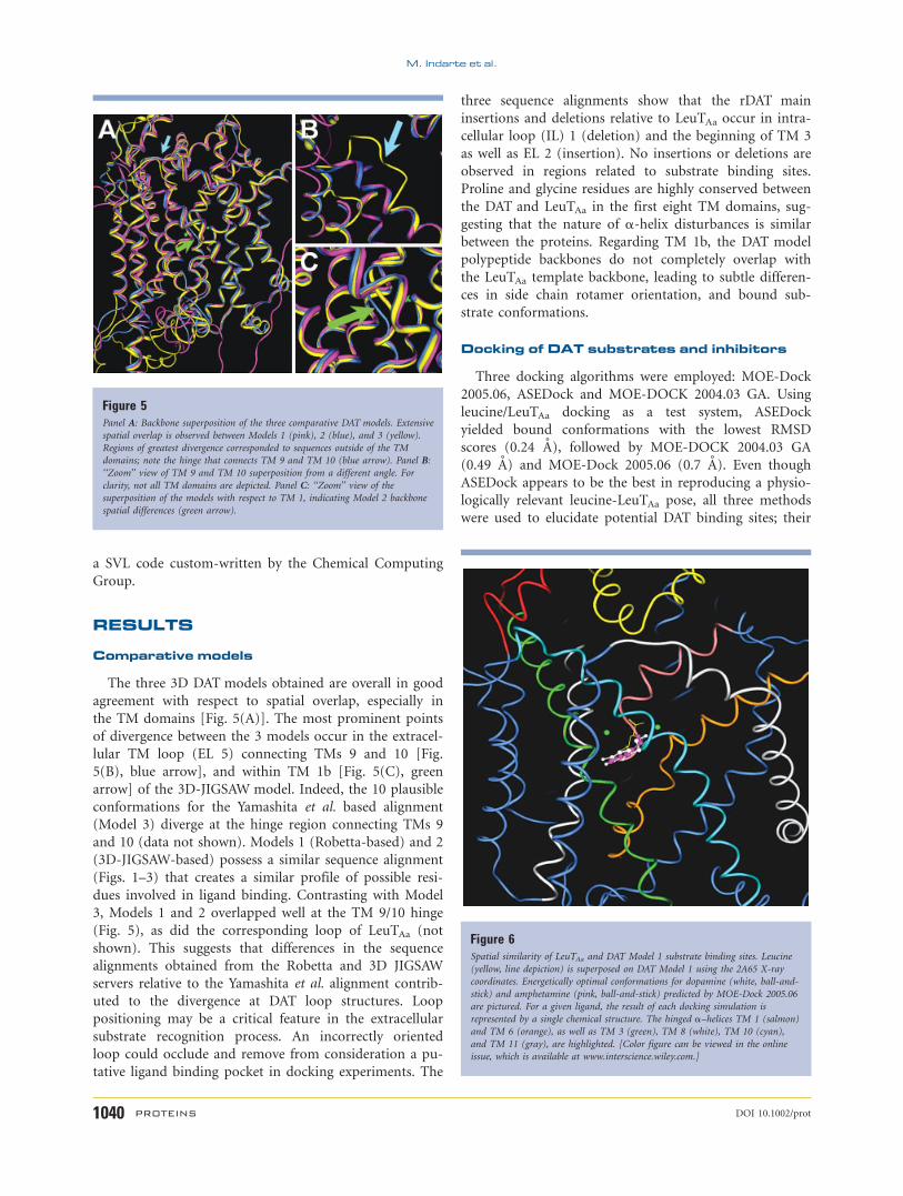
The three 3D DAT models obtained are overall in good
agreement with respect to spatial overlap, especially in
the TM domains [Fig. 5(A)]. The most prominent points
of divergence between the 3 models occur in the extracel-
lular TM loop (EL 5) connecting TMs 9 and 10 [Fig.
5(B), blue arrow], and within TM 1b [Fig. 5(C), green
arrow] of the 3D-JIGSAW model. Indeed, the 10 plausible
conformations for the Yamashita et al. based alignment
(Model 3) diverge at the hinge region connecting TMs 9
and 10 (data not shown). Models 1 (Robetta-based) and 2
(3D-JIGSAW-based) possess a similar sequence alignment
(Figs. 1–3) that creates a similar profile of possible resi-
dues involved in ligand binding. Contrasting with Model
3, Models 1 and 2 overlapped well at the TM 9/10 hinge
(Fig. 5), as did the corresponding loop of LeuTAa (not
shown). This suggests that differences in the sequence
alignments obtained from the Robetta and 3D JIGSAW
servers relative to the Yamashita et al. alignment contrib-
uted to the divergence at DAT loop structures. Loop
positioning may be a critical feature in the extracellular
substrate recognition process. An incorrectly oriented
loop could occlude and remove from consideration a pu-
tative ligand binding pocket in docking experiments. The
three sequence alignments show that the rDAT main
insertions and deletions relative to LeuTAa occur in intra-
cellular loop (IL) 1 (deletion) and the beginning of TM 3
as well as EL 2 (insertion). No insertions or deletions are
observed in regions related to substrate binding sites.
Proline and glycine residues are highly conserved between
the DAT and LeuTAa in the first eight TM domains, sug-
gesting that the nature of a-helix disturbances is similar
between the proteins. Regarding TM 1b, the DAT model
polypeptide backbones do not completely overlap with
the LeuTAa template backbone, leading to subtle differen-
ces in side chain rotamer orientation, and bound sub-
strate conformations.
Docking of DAT substrates and inhibitors
Three docking algorithms were employed: MOE-Dock
2005.06, ASEDock and MOE-DOCK 2004.03 GA. Using
leucine/LeuTAa docking as a test system, ASEDock
yielded bound conformations with the lowest RMSD
scores (0.24 A), followed by MOE-DOCK 2004.03 GA
(0.49 A) and MOE-Dock 2005.06 (0.7 A). Even though
ASEDock appears to be the best in reproducing a physio-
logically relevant leucine-LeuTAa pose, all three methods
were used to elucidate potential DAT binding sites; their
Figure 5Panel A: Backbone superposition of the three comparative DAT models. Extensive
spatial overlap is observed between Models 1 (pink), 2 (blue), and 3 (yellow).
Regions of greatest divergence corresponded to sequences outside of the TM
domains; note the hinge that connects TM 9 and TM 10 (blue arrow). Panel B:
‘‘Zoom’’ view of TM 9 and TM 10 superposition from a different angle. For
clarity, not all TM domains are depicted. Panel C: ‘‘Zoom’’ view of the
superposition of the models with respect to TM 1, indicating Model 2 backbone
spatial differences (green arrow).
Figure 6Spatial similarity of LeuTAa and DAT Model 1 substrate binding sites. Leucine
(yellow, line depiction) is superposed on DAT Model 1 using the 2A65 X-ray
coordinates. Energetically optimal conformations for dopamine (white, ball-and-
stick) and amphetamine (pink, ball-and-stick) predicted by MOE-Dock 2005.06
are pictured. For a given ligand, the result of each docking simulation is
represented by a single chemical structure. The hinged a–helices TM 1 (salmon)
and TM 6 (orange), as well as TM 3 (green), TM 8 (white), TM 10 (cyan),
and TM 11 (gray), are highlighted. [Color figure can be viewed in the online
issue, which is available at www.interscience.wiley.com.]
M. Indarte et al.
1040 PROTEINS DOI 10.1002/prot
RMSD scores are similar yet could potentially yield dif-
ferent poses. An important and attractive feature of the
ASEDock method is indicated by a correlation plot of
RMSD value versus interaction energy (Utotal), revealing
that the lowest RMSD values correlate to the lowest
interaction energies (see online supplementary material).
The three DAT models, refined as described in the Meth-
ods section, were initially employed in MOE-Dock
2005.06 docking simulations with the DAT substrates do-
pamine and d-amphetamine. Given that the two sub-
strates are close structural analogs, it is not surprising
that these ligands were found to dock essentially in the
same primary binding site of the DAT (Fig. 6). Consider-
ing that DAT docking of these substrates employed an
unbiased approach, entirely independent of LeuTAa dock-
ing of its leucine substrate, the substantial overlap
between the dopamine/amphetamine DAT site and the
leucine site of the analogous region of the LeuTAa crystal
structure is remarkable (Fig. 6). The coincidence of sub-
strate binding pockets within the DAT and LeuTAa, pro-
teins largely dissimilar in sequence that recognize struc-
turally dissimilar substrates, in part validates the present
DAT models.
Like the LeuTAa substrate binding pocket, the primary
DAT substrate pocket is at the approximate midpoint of
the lipid bilayer and very close to the two Na1 binding
sites. Regardless of the protonation state, each substrate
optimally fits in to the substrate binding site; however,
protonation introduces a pronounced drop in interaction
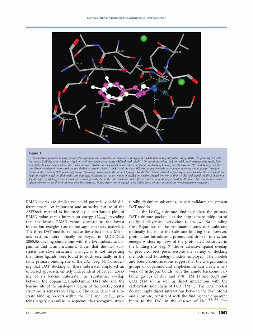
energy. A close-up view of the protonated substrates in
the binding site (Fig. 7) shows extensive spatial overlap
of predicted best poses despite the variety of docking
methods and homology models employed. The models
and bound conformations suggest that the charged amine
groups of dopamine and amphetamine can create a net-
work of hydrogen bonds with the amide backbone car-
bonyl groups of A77 and V78 (TM 1) and S320 and
L321 (TM 6), as well as direct interactions with the
carboxylate side chain of D79 (TM 1). The DAT models
do not imply direct interactions between the Na1 atoms
and substrate, consistent with the finding that dopamine
binds to the DAT in the absence of Na1.53–55 The
Figure 7A representative predicted binding scenario for dopamine and amphetamine obtained with different models and docking algorithms using MOE. The poses represent the
top-ranked DAT-ligand associations based on total interaction energy using ASEDock with Model 1 for dopamine (white, ball-and-stick) and amphetamine (pink, ball-
and-stick). Leucine exported from the crystal structure (yellow, line depiction) demonstrates the spatial similarity of binding pockets between rDAT and LeuTAa and the
considerable overlap of leucine and the two docked structures. Models 1 and 3 and the three different docking methods pose charged substrate amino groups (nitrogen
atoms in blue) close to D79, generating the corresponding interaction in the form of hydrogen bonds. The H-bond network (cyan) depicts and describes the strength of the
bond interaction based on ideal angles and distances, represented as the percentage of possible interaction strength between a given residue and ligand. Model 2 displays a
slightly different docking scenario (data not shown), possibly due to the tilted backbone and different side chain locations predicted by ASEDock. The two sodium atoms
(green spheres) do not directly interact with the substrates. [Color figure can be viewed in the online issue, which is available at www.interscience.wiley.com.]
Comparative Model of the Dopamine Transporter
DOI 10.1002/prot PROTEINS 1041
m-hydroxyl group of dopamine can form hydrogen
bonds with the amide backbone carbonyl group of S421
and A422 (TM 8). The substrate aromatic moiety can
establish favorable hydrophobic interactions with V152
(TM 3) and V327 (TM 6). More importantly, p–p stack-
ing of this substrate group with the phenyl rings of Y156
(TM 3), F319 (TM 6), and F325 (TM 6) are possible
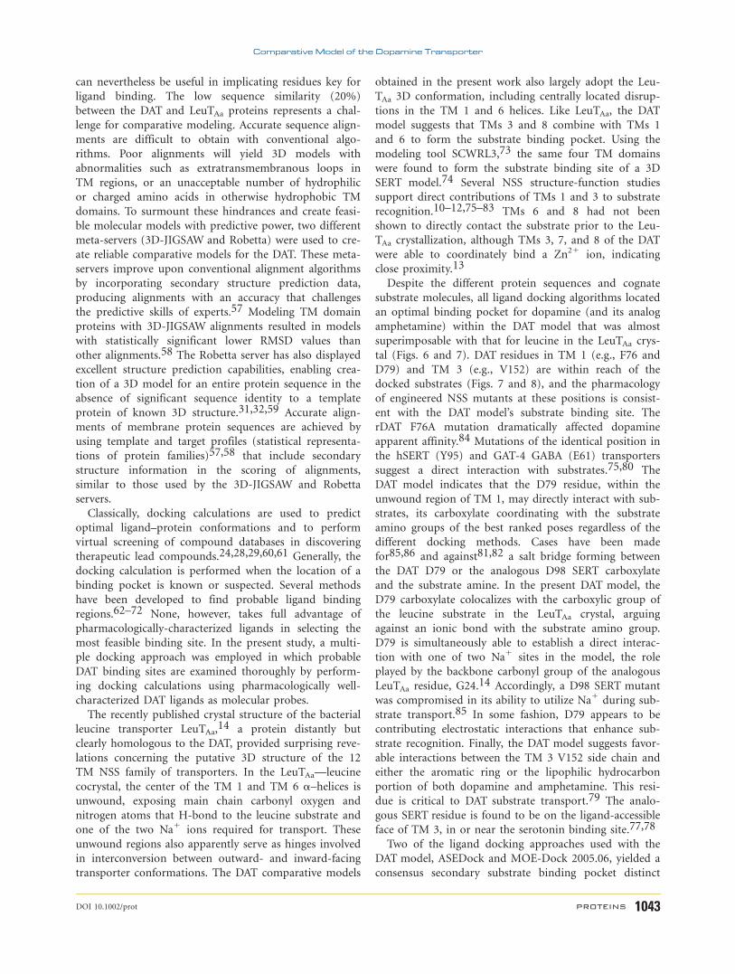
(Fig. 7). Recently developed MOE 2006.07 software was
used to create ligand interaction plots for charged dopa-
mine and amphetamine (Fig. 8), providing a more visu-
ally-digestible arrangement of putatively key intermolecu-
lar interactions that aids in interpreting the 3D juxtaposi-
tion of ligand and transporter protein.
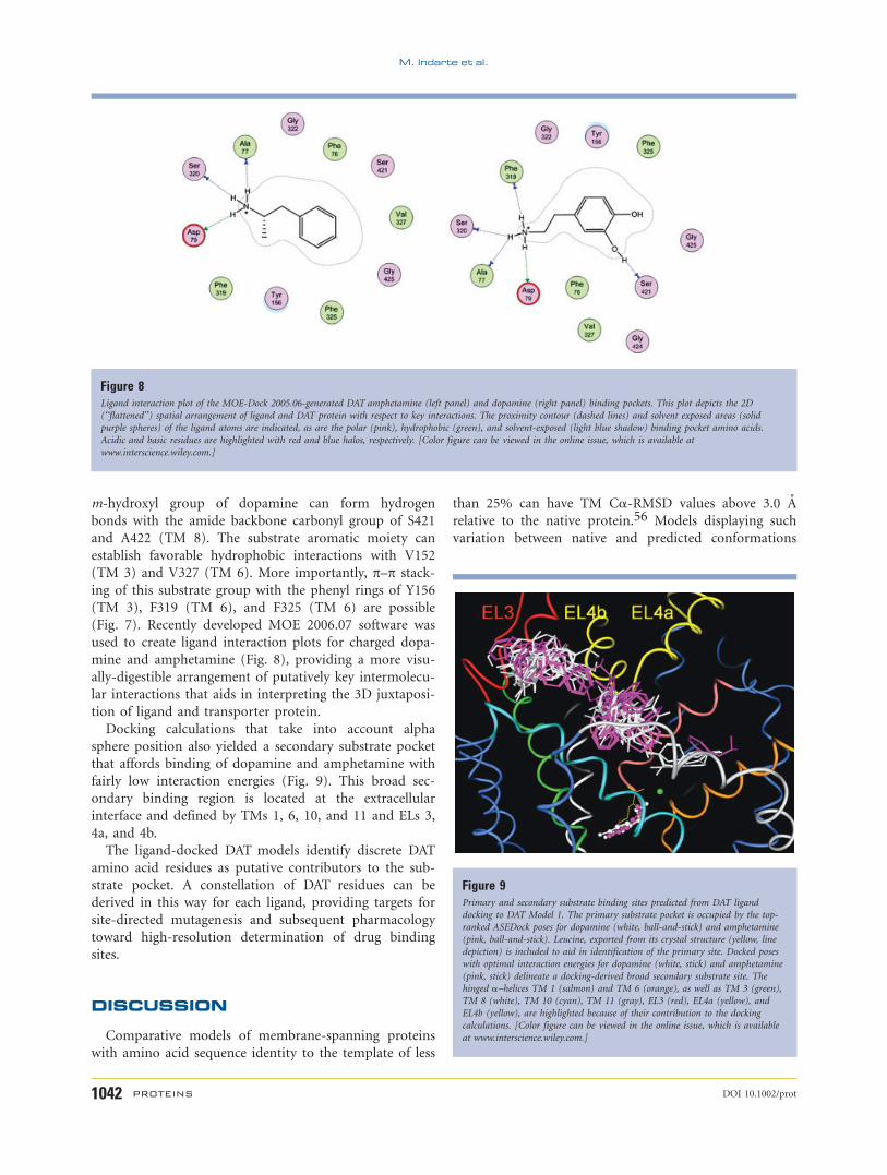
Docking calculations that take into account alpha
sphere position also yielded a secondary substrate pocket
that affords binding of dopamine and amphetamine with
fairly low interaction energies (Fig. 9). This broad sec-
ondary binding region is located at the extracellular
interface and defined by TMs 1, 6, 10, and 11 and ELs 3,
4a, and 4b.
The ligand-docked DAT models identify discrete DAT
amino acid residues as putative contributors to the sub-
strate pocket. A constellation of DAT residues can be
derived in this way for each ligand, providing targets for
site-directed mutagenesis and subsequent pharmacology
toward high-resolution determination of drug binding
sites.
DISCUSSION
Comparative models of membrane-spanning proteins
with amino acid sequence identity to the template of less
than 25% can have TM Ca-RMSD values above 3.0 A
relative to the native protein.56 Models displaying such
variation between native and predicted conformations
Figure 8Ligand interaction plot of the MOE-Dock 2005.06-generated DAT amphetamine (left panel) and dopamine (right panel) binding pockets. This plot depicts the 2D
(‘‘flattened’’) spatial arrangement of ligand and DAT protein with respect to key interactions. The proximity contour (dashed lines) and solvent exposed areas (solid
purple spheres) of the ligand atoms are indicated, as are the polar (pink), hydrophobic (green), and solvent-exposed (light blue shadow) binding pocket amino acids.
Acidic and basic residues are highlighted with red and blue halos, respectively. [Color figure can be viewed in the online issue, which is available at
www.interscience.wiley.com.]
Figure 9Primary and secondary substrate binding sites predicted from DAT ligand
docking to DAT Model 1. The primary substrate pocket is occupied by the top-
ranked ASEDock poses for dopamine (white, ball-and-stick) and amphetamine
(pink, ball-and-stick). Leucine, exported from its crystal structure (yellow, line
depiction) is included to aid in identification of the primary site. Docked poses
with optimal interaction energies for dopamine (white, stick) and amphetamine
(pink, stick) delineate a docking-derived broad secondary substrate site. The
hinged a–helices TM 1 (salmon) and TM 6 (orange), as well as TM 3 (green),
TM 8 (white), TM 10 (cyan), TM 11 (gray), EL3 (red), EL4a (yellow), and
EL4b (yellow), are highlighted because of their contribution to the docking
calculations. [Color figure can be viewed in the online issue, which is available
at www.interscience.wiley.com.]
M. Indarte et al.
1042 PROTEINS DOI 10.1002/prot
can nevertheless be useful in implicating residues key for
ligand binding. The low sequence similarity (20%)
between the DAT and LeuTAa proteins represents a chal-
lenge for comparative modeling. Accurate sequence align-
ments are difficult to obtain with conventional algo-
rithms. Poor alignments will yield 3D models with
abnormalities such as extratransmembranous loops in
TM regions, or an unacceptable number of hydrophilic
or charged amino acids in otherwise hydrophobic TM
domains. To surmount these hindrances and create feasi-
ble molecular models with predictive power, two different
meta-servers (3D-JIGSAW and Robetta) were used to cre-
ate reliable comparative models for the DAT. These meta-
servers improve upon conventional alignment algorithms
by incorporating secondary structure prediction data,
producing alignments with an accuracy that challenges
the predictive skills of experts.57 Modeling TM domain
proteins with 3D-JIGSAW alignments resulted in models
with statistically significant lower RMSD values than
other alignments.58 The Robetta server has also displayed
excellent structure prediction capabilities, enabling crea-
tion of a 3D model for an entire protein sequence in the
absence of significant sequence identity to a template
protein of known 3D structure.31,32,59 Accurate align-
ments of membrane protein sequences are achieved by
using template and target profiles (statistical representa-
tions of protein families)57,58 that include secondary
structure information in the scoring of alignments,
similar to those used by the 3D-JIGSAW and Robetta
servers.
Classically, docking calculations are used to predict
optimal ligand–protein conformations and to perform
virtual screening of compound databases in discovering
therapeutic lead compounds.24,28,29,60,61 Generally, the
docking calculation is performed when the location of a
binding pocket is known or suspected. Several methods
have been developed to find probable ligand binding
regions.62–72 None, however, takes full advantage of
pharmacologically-characterized ligands in selecting the
most feasible binding site. In the present study, a multi-
ple docking approach was employed in which probable
DAT binding sites are examined thoroughly by perform-
ing docking calculations using pharmacologically well-
characterized DAT ligands as molecular probes.
The recently published crystal structure of the bacterial
leucine transporter LeuTAa,14 a protein distantly but
clearly homologous to the DAT, provided surprising reve-
lations concerning the putative 3D structure of the 12
TM NSS family of transporters. In the LeuTAa—leucine
cocrystal, the center of the TM 1 and TM 6 a–helices is
unwound, exposing main chain carbonyl oxygen and
nitrogen atoms that H-bond to the leucine substrate and
one of the two Na1 ions required for transport. These
unwound regions also apparently serve as hinges involved
in interconversion between outward- and inward-facing
transporter conformations. The DAT comparative models
obtained in the present work also largely adopt the Leu-
TAa 3D conformation, including centrally located disrup-
tions in the TM 1 and 6 helices. Like LeuTAa, the DAT
model suggests that TMs 3 and 8 combine with TMs 1
and 6 to form the substrate binding pocket. Using the
modeling tool SCWRL3,73 the same four TM domains
were found to form the substrate binding site of a 3D
SERT model.74 Several NSS structure-function studies
support direct contributions of TMs 1 and 3 to substrate
recognition.10–12,75–83 TMs 6 and 8 had not been
shown to directly contact the substrate prior to the Leu-
TAa crystallization, although TMs 3, 7, and 8 of the DAT
were able to coordinately bind a Zn21 ion, indicating
close proximity.13
Despite the different protein sequences and cognate
substrate molecules, all ligand docking algorithms located
an optimal binding pocket for dopamine (and its analog
amphetamine) within the DAT model that was almost
superimposable with that for leucine in the LeuTAa crys-
tal (Figs. 6 and 7). DAT residues in TM 1 (e.g., F76 and
D79) and TM 3 (e.g., V152) are within reach of the
docked substrates (Figs. 7 and 8), and the pharmacology
of engineered NSS mutants at these positions is consist-
ent with the DAT model’s substrate binding site. The
rDAT F76A mutation dramatically affected dopamine
apparent affinity.84 Mutations of the identical position in
the hSERT (Y95) and GAT-4 GABA (E61) transporters
suggest a direct interaction with substrates.75,80 The
DAT model indicates that the D79 residue, within the
unwound region of TM 1, may directly interact with sub-
strates, its carboxylate coordinating with the substrate
amino groups of the best ranked poses regardless of the
different docking methods. Cases have been made
for85,86 and against81,82 a salt bridge forming between
the DAT D79 or the analogous D98 SERT carboxylate
and the substrate amine. In the present DAT model, the
D79 carboxylate colocalizes with the carboxylic group of
the leucine substrate in the LeuTAa crystal, arguing
against an ionic bond with the substrate amino group.
D79 is simultaneously able to establish a direct interac-
tion with one of two Na1 sites in the model, the role
played by the backbone carbonyl group of the analogous
LeuTAa residue, G24.14 Accordingly, a D98 SERT mutant
was compromised in its ability to utilize Na1 during sub-
strate transport.85 In some fashion, D79 appears to be
contributing electrostatic interactions that enhance sub-
strate recognition. Finally, the DAT model suggests favor-
able interactions between the TM 3 V152 side chain and
either the aromatic ring or the lipophilic hydrocarbon
portion of both dopamine and amphetamine. This resi-
due is critical to DAT substrate transport.79 The analo-
gous SERT residue is found to be on the ligand-accessible
face of TM 3, in or near the serotonin binding site.77,78
Two of the ligand docking approaches used with the
DAT model, ASEDock and MOE-Dock 2005.06, yielded a
consensus secondary substrate binding pocket distinct
Comparative Model of the Dopamine Transporter
DOI 10.1002/prot PROTEINS 1043
from the primary substrate pocket (Fig. 9). It is conceiva-
ble that this secondary site is a temporary ‘‘waiting
room’’ for the substrate, and the substrate is ushered to
its primary binding pocket in the presence of Na1, Cl2,
or the appropriate outward-facing DAT conformation.
The secondary site may be important for recognition of
cocaine and other dopamine uptake inhibitors (unpub-
lished data). Amphetamine and perhaps other uptake
inhibitors may directly compete with dopamine for occu-
pancy of this secondary pocket if the current conforma-
tion resembles a bioactive conformation able to recognize
and bind inhibitors.
The DAT models remained fixed during the docking
process and therefore conformational flexibility of the
macromolecule upon binding was not addressed. This
should be noted as a limitation of the approach given
that the three DAT models are based on the LeuTAa struc-
ture in only one configuration—the transporter with a
tightly bound substrate. Moreover, the modeling effort
may well miss potential ligand binding sites or overstate
minor sites that would be recognized as such if more
flexibility were introduced in the model. Despite the con-
siderable progress achieved in the past few years, accurate
docking methods that predict macromolecular conforma-
tional changes upon ligand binding still remain computa-
tionally challenging.24
This novel DAT model will continue to provide new
DAT mutagenesis targets. The pharmacology from these
mutants will in turn refine the DAT model, affording
high resolution mapping of DAT substrate and inhibitor
binding sites. At that point, the DAT model may be used
for QSAR analysis of putative DAT ligands, involving in
silico screening of structural libraries containing millions
of compounds. The more promising compounds would
be screened at the bench, and then in preclinical and
clinical settings. In this way, rational design of novel DAT
pharmacotherapeutic ligands should be possible. Such
ligands may interfere with actions of abused psychosti-
mulants including cocaine and the amphetamines while
largely sparing normal DAT function. Novel medications
for treating depression, anxiety disorders, attention defi-
cit hyperactivity disorder, narcolepsy, Parkinson’s disease,
and other DAT-related disorders may also result from
rational drug design afforded by this DAT model.
CONCLUSION
Using the LeuTAa crystal structure as a template, three
comparative modeling approaches were used to create
three DAT models. Although quite similar, the nonidenti-
cal sequence alignments led to subtle but significant dif-
ferences between the models. Three docking methods
were applied to the three DAT models to identify poten-
tial binding sites for the substrates dopamine and the
psychostimulant d-amphetamine. The docking calcula-
tions identified two discrete DAT binding regions: a pri-
mary substrate binding site correlating with the binding
site observed in the LeuTAa crystal structure, and a broad
secondary substrate site closer to the extracellular inter-
face. The secondary site may act as a potential staging
area for substrate translocation through the cell mem-
brane. The proposed binding pockets and their function
are consistent with published and unpublished mutagen-
esis data. The DAT models coupled with ligand docking
simulations are refining mutagenesis and other structure-
function investigations, and should aid in the develop-
ment of QSAR as well as pharmacophore models toward
development of novel medications.
ACKNOWLEDGMENTS
Chemical Computing Group is acknowledged for pro-
viding MOE software, especially for access to a beta
release version of MOE. M.I. thanks the technical sup-
port scientists at CCG, especially Dr. Suzanne Schreyer,
Dr. Alain Deschenes and Dr. Andrew Henry for their as-
sistance. Dr. Barry Honig is thanked for helpful com-
ments and discussions. Dr. Junichi Goto is thanked for
granting the Ryoka Institute docking program ASEDock.
REFERENCES
1. Giros B, Jaber M, Jones SR, Wightman RM, Caron MG. Hyperloco-
motion and indifference to cocaine and amphetamine in mice lack-
ing the dopamine transporter. Nature 1996;379:606–612.
2. Ritz MC, Lamb RJ, Goldberg SR, Kuhar MJ. Cocaine receptors on
dopamine transporters are related to self-administration of cocaine.
Science 1987;237:1219–1223.
3. Caine SB, Koob GF. Modulation of cocaine self-administration in
the rat through D-3 dopamine receptors. Science 1993;260:1814–
1816.
4. Caine SB, Koob GF. Pretreatment with the dopamine agonist 7-
OH-DPAT shifts the cocaine self-administration dose-effect function
to the left under different schedules in the rat. Behav Pharmacol
1995;6:333–347.
5. Fischer JF, Cho AK. Chemical release of dopamine from striatal ho-
mogenates: evidence for an exchange diffusion model. J Pharmacol
Exp Ther 1979;208:203–209.
6. Sitte HH, Farhan H, Javitch JA. Sodium-dependent neurotransmit-
ter transporters: oligomerization as a determinant of transporter
function and trafficking. Mol Interv 2004;4:38–47.
7. Saier MH, Jr. A functional-phylogenetic system for the classification
of transport proteins. J Cell Biochem 1999; Suppl 32/33:84–94.
8. Rudnick G. Mechanisms of biogenic amine neurotransmitter trans-
porters. In: Reith MEA, editor. Neurotransmitter transporters: struc-
ture, function, and regulation. Totowa, NJ: Humana Press; 1997.
pp 73–100.
9. Goldberg NR, Beuming T, Soyer OS, Goldstein RA, Weinstein H,
Javitch JA. Probing conformational changes in neurotransmitter
transporters: a structural context. Eur J Pharmacol 2003;479:3–12.
10. Surratt CK, Ukairo OT, Ramanujapuram S. Recognition of psychos-
timulants, antidepressants, and other inhibitors of synaptic neuro-
transmitter uptake by the plasma membrane monoamine transport-
ers. AAPS J 2005;7:E739–E751.
11. Henry LK, Adkins EM, Han Q, Blakely RD. Serotonin and cocaine-
sensitive inactivation of human serotonin transporters by methane-
thiosulfonates targeted to transmembrane domain I. J Biol Chem
2003;278:37052–37063.
M. Indarte et al.
1044 PROTEINS DOI 10.1002/prot

12. Henry LK, Field JR, Adkins EM, Parnas ML, Vaughan RA, Zou MF,
Newman AH, Blakely RD. Tyr-95 and Ile-172 in transmembrane
segments 1 and 3 of human serotonin transporters interact to es-
tablish high affinity recognition of antidepressants. J Biol Chem
2006;281:2012–2023.
13. Loland CJ, Granas C, Javitch JA, Gether U. Identification of intra-
cellular residues in the dopamine transporter critical for regulation
of transporter conformation and cocaine binding. J Biol Chem
2004;279:3228–3238.
14. Yamashita A, Singh SK, Kawate T, Jin Y, Gouaux E. Crystal struc-
ture of a bacterial homologue of Na1/Cl2-dependent neurotrans-
mitter transporters. Nature 2005;437:215–223.
15. Petrey D, Honig B. Protein structure prediction: inroads to biology.
Mol Cell 2005;20:811–819.
16. Bonneau R, Tsai J, Ruczinski I, Chivian D, Rohl C, Strauss CE,
Baker D. Rosetta in CASP4: progress in ab initio protein structure
prediction. Proteins 2001; Suppl 5:119–126.
17. Bradley P, Chivian D, Meiler J, Misura KM, Rohl CA, Schief WR,
Wedemeyer WJ, Schueler-Furman O, Murphy P, Schonbrun J,
Strauss CE, Baker D. Rosetta predictions in CASP5: successes, fail-
ures, and prospects for complete automation. Proteins 2003;53
(Suppl 6):457–468.
18. Bradley P, Malmstrom L, Qian B, Schonbrun J, Chivian D, Kim
DE, Meiler J, Misura KM, Baker D. Free modeling with Rosetta in
CASP6. Proteins 2005;61 (Suppl 7):128–134.
19. Misura KM, Chivian D, Rohl CA, Kim DE, Baker D. Physically
realistic homology models built with ROSETTA can be more accu-
rate than their templates. Proc Natl Acad Sci USA 2006;103:5361–
5366.
20. Rohl CA, Strauss CE, Chivian D, Baker D. Modeling structurally
variable regions in homologous proteins with rosetta. Proteins
2004;55:656–677.
21. Esposito EX, Tobi D, Madura JD. Comparative protein modeling.
In: Lipkowitz KB, editor. Reviews in computational chemistry,
Vol. 22. Hoboken, NJ: Wiley; 2005. pp 57–167.
22. Pieper U, Eswar N, Stuart AC, Ilyin VA, Sali A. MODBASE, a data-
base of annotated comparative protein structure models. Nucleic
Acids Res 2002;30:255–259.
23. Visiers I, Ballesteros JA, Weinstein H. Three-dimensional represen-
tations of G protein-coupled receptor structures and mechanisms.
Methods Enzymol 2002;343:329–371.
24. Brooijmans N, Kuntz ID. Molecular recognition and docking algo-
rithms. Annu Rev Biophys Biomol Struct 2003;32:335–373.
25. Geschwend DA, Good AC, Kuntz ID. Molecular docking towards
drug discovery. J Mol Recognit 1996;9:175–186.
26. Zhou Z, Fisher D, Spidel J, Greenfield J, Patson B, Fazal A, Wigal
C, Moe OA, Madura JD. Kinetic and docking studies of the interac-
tion of quinones with the quinone reductase active site. Biochemis-
try 2003;42:1985–1994.
27. Zhou Z, Madrid M, Madura JD. Docking of non-nucleoside inhibi-
tors: neotripterifordin and its derivatives to HIV-1 reverse tran-
scriptase. Proteins 2002;49:529–542.
28. Perola E, Walters WP, Charifson PS. A detailed comparison of cur-
rent docking and scoring methods on systems of pharmaceutical
relevance. Proteins 2004;56:235–249.
29. Kuntz ID, Blaney JM, Oatley SJ, Langridge R, Ferrin TE. A geomet-
ric approach to macromolecule-ligand interactions. J Mol Biol
1982;161:269–288.
30. Al-Lazikani B, Jung J, Xiang Z, Honig B. Protein structure predic-
tion. Curr Opin Chem Biol 2001;5:51–56.
31. Chivian D, Kim DE, Malmstrom L, Bradley P, Robertson T, Murphy
P, Strauss CE, Bonneau R, Rohl CA, Baker D. Automated prediction
of CASP-5 structures using the Robetta server. Proteins 2003;53
(Suppl 6):524–533.
32. Chivian D, Kim DE, Malmstrom L, Schonbrun J, Rohl CA, Baker
D. Prediction of CASP6 structures using automated Robetta proto-
cols. Proteins 2005;61 (Suppl 7):157–166.
33. Kim DE, Chivian D, Baker D. Protein structure prediction and
analysis using the Robetta server. Nucleic Acids Res 2004;32:
W526–W531.
34. Kilty JE, Lorang D, Amara SG. Cloning and expression of a co-
caine-sensitive rat dopamine transporter. Science 1991;254:578–
579.
35. Chemical Computing Group C. Molecular Operative Enviroment
(MOE), 2006.0706. 1255 University St., Suite 1600, Montreal, Que-
bec, Canada, H3B 3x3; 2006.
36. Ponder JW, Case DA. Force fields for protein simulations. Adv Pro-
tein Chem 2003;66:27–85.
37. Eisenberg D, Luthy R, Bowie JU. VERIFY3D: assessment of protein
models with three-dimensional profiles. Methods Enzymol 1997;
277:396–404.
38. Luthy R, Bowie JU, Eisenberg D. Assessment of protein models
with three-dimensional profiles. Nature 1992;356:83–85.
39. Altschul SF, Madden TL, Schaffer AA, Zhang J, Zhang Z, Miller W,
Lipman DJ. Gapped BLAST and PSI-BLAST: a new generation of
protein database search programs. Nucleic Acids Res 1997;25:3389–
3402.
40. Jones DT. Protein secondary structure prediction based on posi-
tion-specific scoring matrices. J Mol Biol 1999;292:195–202.
41. Bates PA, Kelley LA, MacCallum RM, Sternberg MJ. Enhancement
of protein modeling by human intervention in applying the auto-
matic programs 3D-JIGSAW and 3D-PSSM. Proteins 2001; Suppl
5:39–46.
42. Levitt M. Accurate modeling of protein conformation by automatic
segment matching. J Mol Biol 1992;226:507–533.
43. Fechteler T, Dengler U, Schomburg D. Prediction of protein three-
dimensional structures in insertion and deletion regions: a proce-
dure for searching data bases of representative protein fragments
using geometric scoring criteria. J Mol Biol 1995;253:114–131.
44. Petersen EF, Goddard TD, Huang CC, Couch GS, Greenblatt DM,
Meng EC, Ferrin TE. UCSF Chimera—a visualization system for ex-
ploratory research and analysis. J Comput Chem 2004;25:1605–
1612.
45. Clark AM, Labute P, Santavy M. 2D structure depiction. J Chem
Inf Model 2006;46:1107–1123.
46. Halgren TA. Merck molecular force field. I. Basis, form, scope,
parameterization, and performance of MMFF94. J Comput Chem
1996;17:490–519.
47. Halgren TA. Force fields: MMFF94. In: Schleyer PVR, editor. Ency-
clopedia of computational chemistry, Vol. 2. West Sussex, UK:
Wiley; 1998. p 1033.
48. Maple JR. Force fields: a general discussion. In: Schleyer PVR, edi-
tor. Encyclopedia of computational chemistry, Vol. 2. West Sussex,
UK: Wiley; 1998. p 1015.
49. Goto J, Kataoka R, Hirayama N. Ph4Dock: pharmacophore-based
protein-ligand docking. J Med Chem 2004;47:6804–6811.
50. Morris GM, Goodsell DS, Huey R, Olson AJ. Distributed auto-
mated docking of flexible ligands to proteins: parallel applications
of AutoDock 2.4. J Comput Aided Mol Des 1996;10:293–304.
51. Goodsell DS, Morris GM, Olson AJ. Automated docking of flexible
ligands: applications of AutoDock. J Mol Recognit 1996;9:1–5.
52. Hart TN, Read RJ. A multiple-start Monte Carlo docking method.
Proteins 1992;13:206–222.
53. McElvain JS, Schenk JO. A multisubstrate mechanism of striatal do-
pamine uptake and its inhibition by cocaine. Biochem Pharmacol
1992;43:2189–2199.
54. Chen N, Sun L, Reith ME. Cationic interactions at the human do-
pamine transporter reveal binding conformations for dopamine dis-
tinguishable from those for the cocaine analog 2 a-carbomethoxy-
3a-(4-fluorophenyl)tropane. J Neurochem 2002;81:1383–1393.
55. Li LB, Cui XN, Reith MA. Is Na(1) required for the binding of do-
pamine, amphetamine, tyramine, and octopamine to the human
dopamine transporter? Naunyn Schmiedebergs Arch Pharmacol
2002;365:303–311.
Comparative Model of the Dopamine Transporter
DOI 10.1002/prot PROTEINS 1045
56. Forrest LR, Tang CL, Honig B. On the accuracy of homology mod-
eling and sequence alignment methods applied to membrane pro-
teins. Biophys J 2006;91:508–517.
57. Rychlewski L, Fischer D. LiveBench-8: the large-scale, continuous
assessment of automated protein structure prediction. Protein Sci
2005;14:240–245.
58. Reddy Ch S, Vijayasarathy K, Srinivas E, Sastry GM, Sastry GN.
Homology modeling of membrane proteins: a critical assessment.
Comput Biol Chem 2006;30:120–126.
59. Tai CH, Lee WJ, Vincent JJ, Lee B. Evaluation of domain prediction
in CASP6. Proteins 2005;61 (Suppl 7):183–192.
60. Chen IJ, Neamati N, MacKerell AD, Jr. Structure-based inhibitor
design targeting HIV-1 integrase. Curr Drug Targets Infect Disord
2002;2:217–234.
61. Hancock CN, Macias A, Lee EK, Yu SY, Mackerell AD, Jr, Shapiro
P. Identification of novel extracellular signal-regulated kinase dock-
ing domain inhibitors. J Med Chem 2005;48:4586–4595.
62. Del Carpio CA, Takahashi Y, Sasaki S. A new approach to the auto-
matic identification of candidates for ligand receptor sites in pro-
teins: (I). Search for pocket regions. J Mol Graph 1993;11:23–29.
63. Edelsbrunner H, Facello M, Liang J. On the definition and the con-
struction of pockets in macromolecules. Pac Symp Biocomput
1996:272–287.
64. Edelsbrunner H, Koehl P. The weighted-volume derivative of a
space-filling diagram. Proc Natl Acad Sci USA 2003;100:2203–2208.
65. Goodford PJ. A computational procedure for determining energeti-
cally favorable binding sites on biologically important macromole-
cules. J Med Chem 1985;28:849–857.
66. Hendlich M, Rippmann F, Barnickel G. LIGSITE: automatic and
efficient detection of potential small molecule-binding sites in
proteins. J Mol Graph Model 1997;15:359–363.
67. Liang J, Edelsbrunner H, Fu P, Sudhakar PV, Subramaniam S. Ana-
lytical shape computation of macromolecules. I. Molecular area and
volume through alpha shape. Proteins 1998;33:1–17.
68. Liang J, Edelsbrunner H, Woodward C. Anatomy of protein pockets
and cavities: measurement of binding site geometry and implica-
tions for ligand design. Protein Sci 1998;7:1884–1897.
69. Miranker A, Karplus M. Functionality maps of binding sites: a mul-
tiple copy simultaneous search method. Proteins 1991;11:29–34.
70. Peters KP, Fauck J, Frommel C. The automatic search for ligand
binding sites in proteins of known three-dimensional structure
using only geometric criteria. J Mol Biol 1996;256:201–213.
71. Reynolds CA, Wade RC, Goodford PJ. Identifying targets for biore-
ductive agents: using GRID to predict selective binding regions of
proteins. J Mol Graph 1989;7:103–108.
72. Wade RC, Clark KJ, Goodford PJ. Further development of hydrogen
bond functions for use in determining energetically favorable binding
sites on molecules of known structure. I. Ligand probe groups with the
ability to form two hydrogen bonds. J Med Chem 1993;36:140–147.
73. Canutescu AA, Shelenkov AA, Dunbrack RL, Jr. A graph-theory
algorithm for rapid protein side-chain prediction. Protein Sci 2003;
12:2001–2014.
74. Henry LK, Defelice LJ, Blakely RD. Getting the message across: a
recent transporter structure shows the way. Neuron 2006;49:791–
796.
75. Adkins EM, Barker EL, Blakely RD. Interactions of tryptamine
derivatives with serotonin transporter species variants implicate
transmembrane domain I in substrate recognition. Mol Pharmacol
2001;59:514–523.
76. Barker EL, Perlman MA, Adkins EM, Houlihan WJ, Pristupa ZB,
Niznik HB, Blakely RD. High affinity recognition of serotonin
transporter antagonists defined by species-scanning mutagenesis.
J Biol Chem 1998;273:19459–19468.
77. Chen J-G, Rudnick G. Permeation and gating residues in serotonin
transporter. Proc Natl Acad Sci USA 2000;97:1044–1049.
78. Chen J-G, Sachpatzidis A, Rudnick G. The third transmembrane
domain of the serotonin transporter contains residues associated
with substrate and cocaine binding. J Biol Chem 1997;272:28321–
28327.
79. Lee SH, Chang MY, Lee KH, Park BS, Lee YS, Chin HR. Impor-
tance of valine at position 152 for the substrate transport and 2b-carbomethoxy-3b-(4-fluorophenyl)tropane binding of dopamine
transporter. Mol Pharmacol 2000;57:883–889.
80. Melamed N, Kanner BI. Transmembrane domains I and II of
the g-aminobutyric acid transporter GAT-4 contain molecular
determinants of substrate specificity. Mol Pharmacol 2004;65:1452–
1461.
81. Ukairo OT, Bondi CD, Newman AH, Kulkarni SS, Kozikowski AP,
Pan S, Surratt CK. Recognition of benztropine by the dopamine
transporter (DAT) differs from that of the classical dopamine
uptake inhibitors cocaine, methylphenidate, and mazindol as a
function of a DAT transmembrane 1 aspartic acid residue. J Phar-
macol Exp Ther 2005;314:575–583.
82. Wang W, Sonders MS, Ukairo OT, Scott H, Kloetzel MK, Surratt
CK. Dissociation of high-affinity cocaine analog binding and dopa-
mine uptake inhibition at the dopamine transporter. Mol Pharma-
col 2003;64:430–439.
83. Zomot E, Kanner BI. The interaction of the g-aminobutyric acid
transporter GAT-1 with the neurotransmitter is selectively impaired
by sulfhydryl modification of a conformationally sensitive cysteine
residue engineered into extracellular loop IV. J Biol Chem 2003;
278:42950–42958.
84. Lin Z, Wang W, Kopajtic T, Revay RS, Uhl GR. Dopamine trans-
porter: transmembrane phenylalanine mutations can selectively
influence dopamine uptake and cocaine analog recognition. Mol
Pharmacol 1999;56:434–447.
85. Barker EL, Moore KR, Rakhshan F, Blakely RD. Transmembrane
domain I contributes to the permeation pathway for serotonin
and ions in the serotonin transporter. J Neurosci 1999;19:4705–
4717.
86. Kitayama S, Shimada S, Xu H, Markham L, Donovan DM, Uhl GR.
Dopamine transporter site-directed mutations differentially alter
substrate transport and cocaine binding. Proc Natl Acad Sci USA
1992;89:7782–7785.
M. Indarte et al.
1046 PROTEINS DOI 10.1002/prot


![Mi basics[1]](https://img.pdfslide.us/doc/110x75/55c35d60bb61eb686f8b4618/mi-basics1.jpg)


![Mi Complications[1]](https://img.pdfslide.us/doc/110x75/577d238e1a28ab4e1e9a1f55/mi-complications1.jpg)