Embed Size (px)
Citation preview
MHC antigens
Prof. Ilona Hromadníková, PhD.
Antigens
• Exoantigens – external molecules recognised by immune system (most frequently infectious microorganisms)
• Autoantigens – antigens derived from own cells • Superantigens – exoantigens, non-specific
stimulation of lymphocytes independently on antigen specificity
• Allergens – in predisposed individuals induce pathologic (allergic) immune response
Antigens - structure
Macromolecules or small molecules (haptens) bound to carrier• Soluble and/or expressed on the cell surface• Proteins, glycoproteins, polysaccharides, lipids,
lipoproteins, nucleic acids, etc.• Epitope – a part of antigen recognised by immune
receptors • Immunocomplexes – complex of antigen with
antibody, which may bind complement component
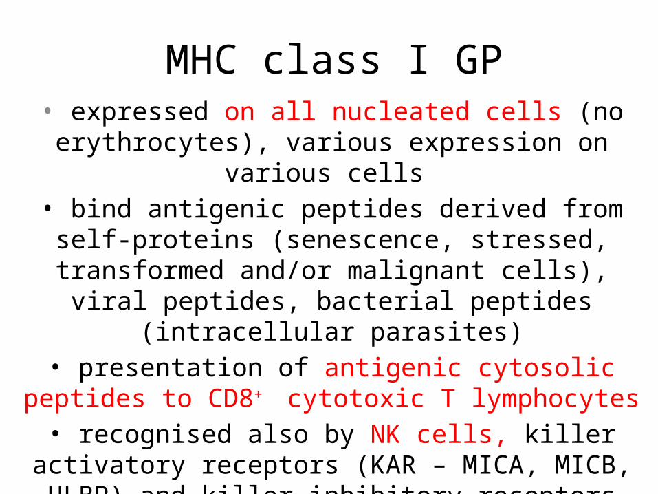
MHC class I GP• expressed on all nucleated cells (no erythrocytes), various
expression on various cells • bind antigenic peptides derived from self-proteins
(senescence, stressed, transformed and/or malignant cells), viral peptides, bacterial peptides (intracellular parasites)• presentation of antigenic cytosolic peptides to CD8+
cytotoxic T lymphocytes• recognised also by NK cells, killer activatory receptors
(KAR – MICA, MICB, ULBP) and killer inhibitory receptors (KIR- HLA-C, HLA-G)
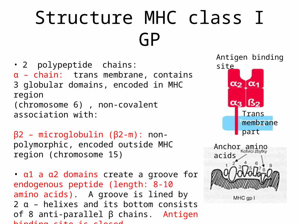
Structure MHC class I GP
• 2 polypeptide chains:α – chain: trans membrane, contains 3 globular domains, encoded in MHC region(chromosome 6) , non-covalent association with:
β2 – microglobulin (β2-m): non-polymorphic, encoded outside MHC region (chromosome 15)
• α1 a α2 domains create a groove for endogenous peptide (length: 8-10 amino acids). A groove is lined by 2 α – helixes and its bottom consists of 8 anti-parallel β chains. Antigen binding site is closed.• peptide origin: product of normal and/or malignant cell, viral peptide
Antigen binding site
Trans membrane part
Anchor amino acids
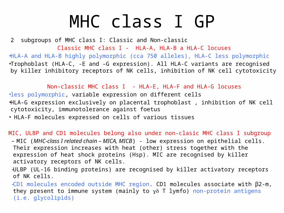
MHC class I GP2 subgroups of MHC class I: Classic and Non-classic
Classic MHC class I - HLA-A, HLA-B a HLA-C locuses• HLA-A and HLA-B highly polymorphic (cca 750 alleles), HLA-C less polymorphic • Trophoblast (HLA-C, -E and –G expression). All HLA-C variants are recognised by killer
inhibitory receptors of NK cells, inhibition of NK cell cytotoxicity
Non-classic MHC class I - HLA-E, HLA-F and HLA-G locuses• less polymorphic, variable expression on different cells • HLA-G expression exclusively on placental trophoblast , inhibition of NK cell cytotoxicity,
immunotolerance against foetus• HLA-F molecules expressed on cells of various tissues
MIC, ULBP and CD1 molecules belong also under non-clasic MHC class I subgroup– MIC (MHC-class I related chain – MICA, MICB) - low expression on epithelial cells. Their
expression increases with heat (other) stress together with the expression of heat shock proteins (Hsp). MIC are recognised by killer activatory receptors of NK cells.
– ULBP (UL-16 binding proteins) are recognised by killer activatory receptors of NK cells. – CD1 molecules encoded outside MHC region. CD1 molecules associate with β2-m, they
present to immune system (mainly to T lymfo) non-protein antigens (i.e. glycolipids)
Binding of peptides into MHC class I groove
• MHC molecules are transported during their synthesis into endoplasmic reticulum (ER). In ER lumen proper folding of both MHC class I chains, creation of antigen binding site for a peptide. Proper folding of MHC class I molecule is enabled by association of α –chain, β2-m and peptide.
• Cytosolic ubiquitinated proteins are cleaved by proteasome into short peptides. Transport of peptides into ER via TAP1 and TAP2 proteins (Transporters Associated with Antigen Processing). TAP proteins prefer transport of peptides containing 8-16 amino acides with hydrophobic or basic amino acides on C-terminus.
• MHC molecule with peptide in antigen binding site is transported from ER through Golgi apparatus on the cell surface
• MHC class I molecules are unstable without peptide in the groove• After dissociation of peptides from MHC class I on the cell surface MHC I
class changes its confirmation, β2-m dissociates and α – chain is internalized by the cell and fastly degraded
MHC class II GP
• Limited tissue distribution comparing with ubiquitous MHC class I GP
• Usually expressed on antigen presenting cells (APC) under normal conditions (monocytes, macrophages, DC, B lymphocytes, Langerhans cells of the skin, Kuppfer cells in the liver, microglia in CNS)
• HLA-DR (highly polymorphic),DQ, DP locuses• Presentation of exogenous peptides (ingested
microorganisms) to CD4+ T lymphocytes
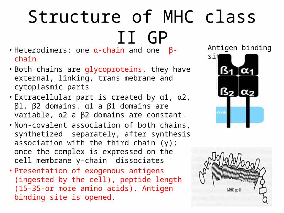
Structure of MHC class II GP• Heterodimers: one α-chain and one β-chain• Both chains are glycoproteins, they have
external, linking, trans mebrane and cytoplasmic parts
• Extracellular part is created by α1, α2, β1, β2 domains. α1 a β1 domains are variable, α2 a β2 domains are constant.
• Non-covalent association of both chains, synthetized separately, after synthesis association with the third chain (γ); once the complex is expressed on the cell membrane γ–chain dissociates
• Presentation of exogenous antigens (ingested by the cell), peptide length (15-35-or more amino acids). Antigen binding site is opened.
Antigen binding site
Binding of peptides into MHC class II groove
• Proteosynthesis of MHC class II GP in ER, association of and chains, binding of invariant Ii chain into antigen binding site (its blockage), transport of the complex into Golgi apparatus, transport secretory vesicle (MHC II + Ii)
• Phagocytosis or endocytosis of exogenous material by APC, proteolytic cleavage into peptide fragments (proteases), fusion of endosome with transport vesicle derived from Golgi apparatus (MHC II + Ii): cleavage of Ii chain into short peptide fragment CLIP (MHC+CLIP), release of CLIP from the groove followed by exogenous peptide binding (MHCII + exogenous peptide), transport into the cell surface, presentation to CD4+ T lymphocytes
Cross-presentation of antigen
• MHC class I molecules are able to present exogenous peptides (cross-presentation)
• Material recognised by cell surface receptors is endocyted, become a part of endosomes, where is partially (but not completely) degraded and transported into cytoplasm, where is cleaved by proteasomes, peptides are transported into ER
• Another way, how extracellular peptides may be presented on MHC class I molecules is fusion of late phagosomes containing peptide fragments with ER
• The ability of cross-presentation differs between various APC cells, the most efficient in DC
Major Histocompatibility Complex• MHC (Major Histocompatibility Complex) in humans is called Human
Leukocyte Antigen (HLA), located on chromosome 6. MHC region is polygenic (the existence of several MHC class I and II isotypes) and polymorphic (the existence of a large number of various variants/alleles of each gene).
• High polymorphism (10 – 100 alleles in certain locus, change of 1 or a few amino acids, able to bind various antigens), most people are heterozygotes for MHC proteins – paternally and maternally inherited alleles for certain MHC isotype. Combination of MHC alleles on one chromosome is called MHC (HLA) haplotype
• MHC gene expression is codominant – products of both alleles from the same locus are in one cell expressed in the same quantity
• Certain MHC molecule (i.e. certain allele from HLA-A2 locus) is able to bind peptides with similar structure – peptides share a binding motif - xLeuxxxxxxLeu(Val)
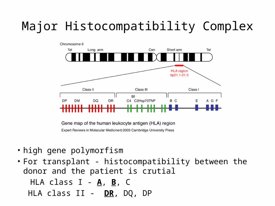
Major Histocompatibility Complex
• high gene polymorfism • For transplant - histocompatibility between the donor and the
patient is crutial HLA class I - A, B, C
HLA class II - DR, DQ, DP
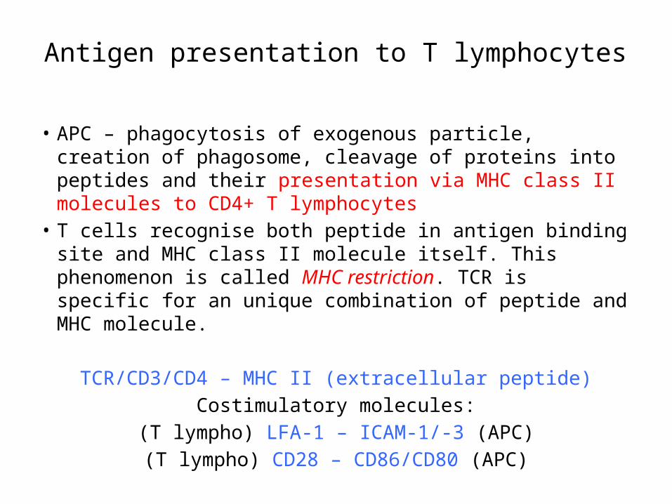
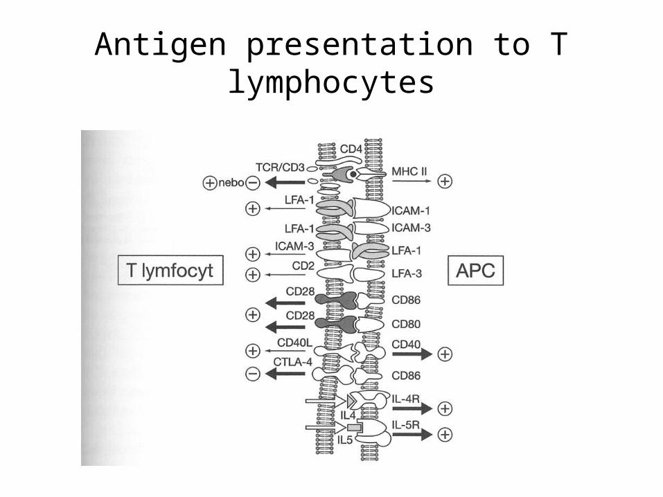
Antigen presentation to T lymphocytes
• APC – phagocytosis of exogenous particle, creation of phagosome, cleavage of proteins into peptides and their presentation via MHC class II molecules to CD4+ T lymphocytes
• T cells recognise both peptide in antigen binding site and MHC class II molecule itself. This phenomenon is called MHC restriction. TCR is specific for an unique combination of peptide and MHC molecule.
TCR/CD3/CD4 – MHC II (extracellular peptide)Costimulatory molecules:
(T lympho) LFA-1 – ICAM-1/-3 (APC)(T lympho) CD28 – CD86/CD80 (APC)
Antigen presentation to T lymphocytes
Antigen presentation to T lymphocytes
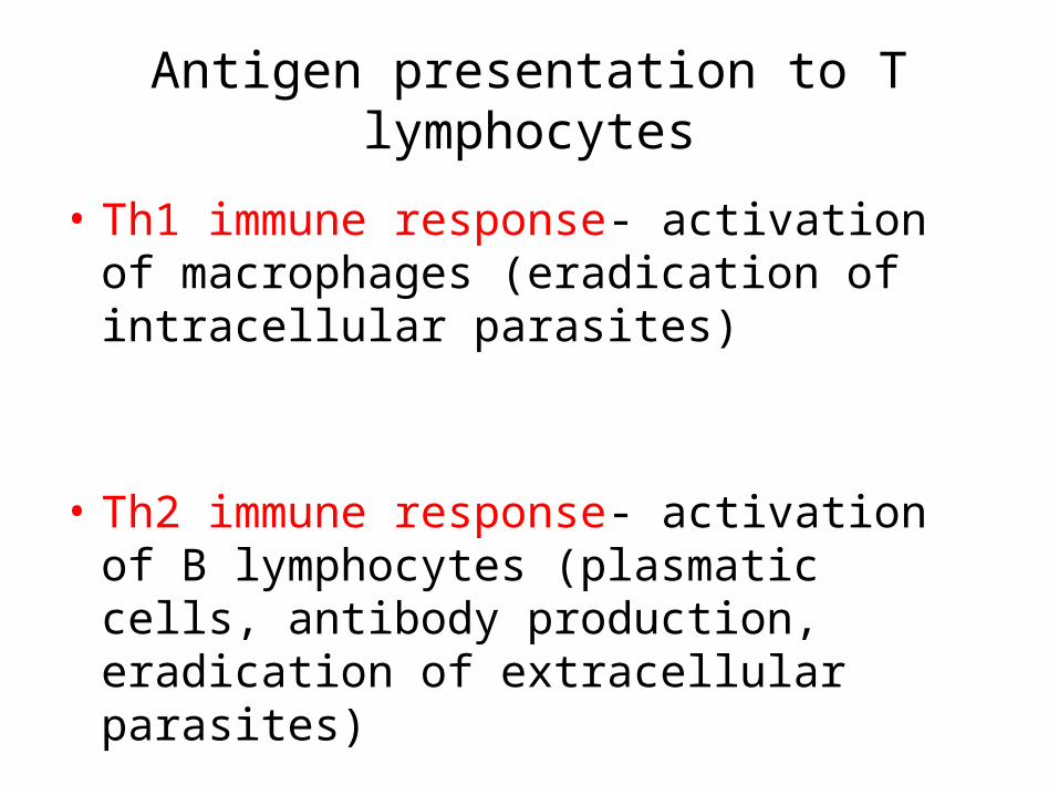
• Th1 immune response- activation of macrophages (eradication of intracellular parasites)
• Th2 immune response- activation of B lymphocytes (plasmatic cells, antibody production, eradication of extracellular parasites)
Antigen presentation to T lymphocytes
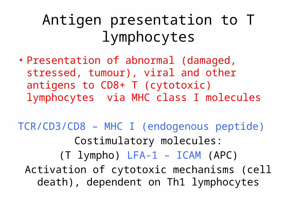
• Presentation of abnormal (damaged, stressed, tumour), viral and other antigens to CD8+ T (cytotoxic) lymphocytes via MHC class I molecules
TCR/CD3/CD8 – MHC I (endogenous peptide)Costimulatory molecules:
(T lympho) LFA-1 – ICAM (APC)Activation of cytotoxic mechanisms (cell death),
dependent on Th1 lymphocytes
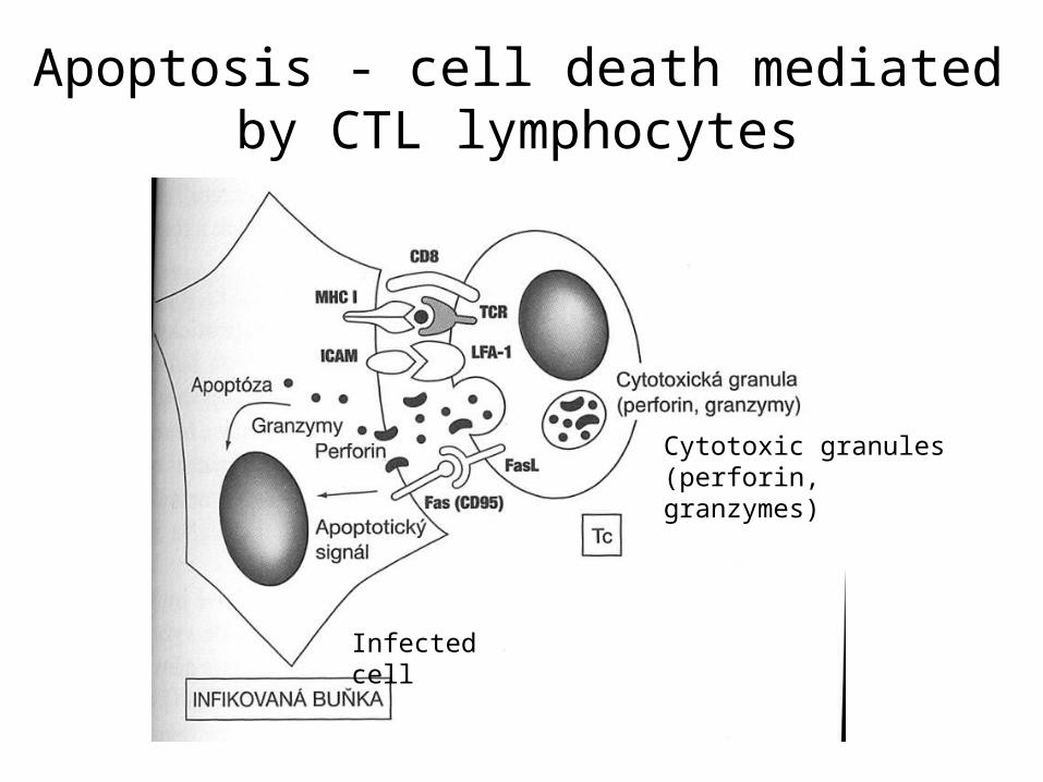
Apoptosis - cell death mediated by CTL lymphocytes
Infected cell
Cytotoxic granules (perforin, granzymes)


![MANUAL DE USUARIO MÁQUINAS DE HIELO...MANUAL DE USUARIO [AUTOCONTENIDAS Y REMOTAS ] MHC-230/506MA - MHC-235/517MA - MHC-280/625MA - MHC-320/706MA MHC-500/1109MAR - MHC-680/1466MAR](https://img.pdfslide.us/doc/110x75/5e93db5530a5a625c35ecff2/manual-de-usuario-mquinas-de-hielo-manual-de-usuario-autocontenidas-y-remotas.jpg)
![Topic (7): Antibodies and Antigens - Doctor 2016...antigens [Ags] (the other two are T-cell receptor [TCR] and major histocompatibility complex [MHC]) {Figure 1}. Antibodies have a](https://img.pdfslide.us/doc/110x75/5f0a54a97e708231d42b2066/topic-7-antibodies-and-antigens-doctor-2016-antigens-ags-the-other-two.jpg)