Embed Size (px)
Citation preview
TOXICOLOGICAL REVIEW
of
METHYLENE DIPHENYL DIISOCYANATE (MDI)
(CAS No. 101-68-8 and 9016-87-9)
In Support of Summary Information on the Integrated Risk Information System (IRIS)
February 1998
U.S. ENVIRONMENTAL PROTECTION AGENCY WASHINGTON, DC
TABLE OF CONTENTS
TOXICOLOGICAL REVIEW FOR
METHYLENE DIPHENYL DIISOCYANATE (MDI)
(CASRN No. 101-68-8 and 9016-87-9)
Disclaimer . . . . . . . . . . . . . . . . . . . . . . . . . . . . . . . . . . . . . . . . . . . . . . . . . . . . . . . . . . . . . . . i
Foreword . . . . . . . . . . . . . . . . . . . . . . . . . . . . . . . . . . . . . . . . . . . . . . . . . . . . . . . . . . . . . . . ii
Contributors and Reviewers . . . . . . . . . . . . . . . . . . . . . . . . . . . . . . . . . . . . . . . . . . . . . . . . iii
1. Introduction . . . . . . . . . . . . . . . . . . . . . . . . . . . . . . . . . . . . . . . . . . . . . . . . . . . . . . . . . . 1
2. Chemical and Physical Information Relevant to Assessments . . . . . . . . . . . . . . . . . . . 2
3. Toxicokinetics Relevant to Assessments . . . . . . . . . . . . . . . . . . . . . . . . . . . . . . . . . . . . 3
4. Hazard Identification . . . . . . . . . . . . . . . . . . . . . . . . . . . . . . . . . . . . . . . . . . . . . . . . . . 4
4.1 Studies in humans - epidemiology, case reports, clinical controls . . . . . . . . . . . . . . . . 44.2 Pre-chronic, chronic studies and cancer bioassays in animals . . . . . . . . . . . . . . . . . . 124.3 Reproductive/developmental studies . . . . . . . . . . . . . . . . . . . . . . . . . . . . . . . . . . . . 194.4 Other studies . . . . . . . . . . . . . . . . . . . . . . . . . . . . . . . . . . . . . . . . . . . . . . . . . . . . . . 204.5 Synthesis and evaluation of major noncancer effects and mode of action . . . . . . . . . 224.6 Weight of evidence evaluation and cancer characterizations - synthesis of human,
animal and other supporting evidence, conclusions about human carcinogenicity, and likely mode of action . . . . . . . . . . . . . . . . . . . . . . . . . . . . . . . . . . . . . . . . . . . . 23
4.7 Susceptible populations . . . . . . . . . . . . . . . . . . . . . . . . . . . . . . . . . . . . . . . . . . . . . . 244.7.1 Possible childhood . . . . . . . . . . . . . . . . . . . . . . . . . . . . . . . . . . . . . . . . . . . . . 244.7.2 Possible gender differences . . . . . . . . . . . . . . . . . . . . . . . . . . . . . . . . . . . . . . . .25
5. Dose Response Assessments . . . . . . . . . . . . . . . . . . . . . . . . . . . . . . . . . . . . . . . . . . . . 25
5.1 Oral Reference Dose (RfD) . . . . . . . . . . . . . . . . . . . . . . . . . . . . . . . . . . . . . . . . . . . 255.2 Inhalation Reference Concentration (RfC) . . . . . . . . . . . . . . . . . . . . . . . . . . . . . . . . 25
5.2.1. Choice of principal study and critical effect . . . . . . . . . . . . . . . . . . . . . . . . . . . 255.2.2. Methods of analysis - including models . . . . . . . . . . . . . . . . . . . . . . . . . . . . . . 265.2.3. RfC derivation - including application of uncertainty factors (UF) and
modifying factors (MF) . . . . . . . . . . . . . . . . . . . . . . . . . . . . . . . . . . . . . . . . . . . 26
CONTENTS (continued)
5.3 Cancer assessment . . . . . . . . . . . . . . . . . . . . . . . . . . . . . . . . . . . . . . . . . . . . . . . . . . 27
6. Major Conclusions in Characterization of Hazard and Dose-Response . . . . . . . . . . 27
6.1 Hazard identification . . . . . . . . . . . . . . . . . . . . . . . . . . . . . . . . . . . . . . . . . . . . . . . . 276.2 Dose-response . . . . . . . . . . . . . . . . . . . . . . . . . . . . . . . . . . . . . . . . . . . . . . . . . . . . . 29
7. References . . . . . . . . . . . . . . . . . . . . . . . . . . . . . . . . . . . . . . . . . . . . . . . . . . . . . . . . . . . 31
8. Appendices . . . . . . . . . . . . . . . . . . . . . . . . . . . . . . . . . . . . . . . . . . . . . . . . . . . . . . . . . . 40
Appendix A: RfC Benchmark Concentration Analyses of Data fromReuzel et al. 1990, 1994b . . . . . . . . . . . . . . . . . . . . . . . . . . . . . . . . . . . . . . . . . . . . . 40
Appendix B: Summary of and Response to External Peer Review Comments . . . . . . . . 43
Appendix C: Regional Deposited Dose Ratios . . . . . . . . . . . . . . . . . . . . . . . . . . . . . . . . 47

DISCLAIMER
Mention of trade names or commercial products does not constitute endorsement or recommendation for use.
i
FOREWORD
The purpose of this review is to provide scientific support and rationale for the hazard identification and dose-response assessments for both cancer and noncancer effects (the inhalation reference concentration [RfC]), from chronic exposure to monomeric methylene diphenyl diisocyanate (MDI) and polymeric MDI (PMDI). It is not intended to be a comprehensive treatise on the chemical or toxicological nature of either MDI or PMDI.
In Section 6, EPA has characterized its overall confidence in the quantitative and qualitative aspects of hazard and dose-response (U.S. EPA, 1995a). Matters considered in this characterization include knowledge gaps, uncertainties, quality of data, and scientific controversies. This characterization is presented in an effort to make apparent the limitations of the individual assessments and to aid and guide the risk assessor in the ensuing steps of the risk assessment process.
For other general information about this assessment or other questions relating to IRIS, the reader is referred to EPA’s Risk Information Hotline at 202-566-1676.
ii
CONTRIBUTORS AND REVIEWERS
Chemical Manager/Author
Mark M. GreenbergNational Center for Environmental AssessmentOffice of Research and DevelopmentU.S. Environmental Protection AgencyResearch Triangle Park, NC 27707
Reviewers
This document and summary information on IRIS have received peer review both by EPA scientists and by independent scientists external to EPA (U.S. EPA, 1994a). Subsequent to external review and incorporation of comments, this assessment has undergone an Agencywide review process whereby the IRIS Program Manager has achieved a consensus approval among the Office of Research and Development, Office of Air and Radiation, Office of Prevention, Pesticides, and Toxic Substances, Office of Solid Waste and Emergency Response, Office of Water, Office of Policy Planning and Evaluation and the Regional Offices.
Internal EPA Reviewers
James W. Holder, Ph.D.ToxicologistNational Center for Environmental AssessmentU.S. Environmental Protection AgencyWashington, DC
Cheryl ScottEpidemiologistNational Center for Environmental AssessmentU.S. Environmental Protection AgencyWashington, DC
Eric D. Clegg, Ph.D.ToxicologistNational Center for Environmental AssessmentU.S. Environmental Protection AgencyWashington, DC
iii
External Peer Reviewers
Charles H. Hobbs, DVMDirector of ToxicologyLovelace Biomedical and Environmental Research InstituteAlbuquerque, NM
Thomas Ledoux, Ph.D.Division of Science and ResearchNew Jersey Department of Environmental ProtectionTrenton, NJ
William E. Brown, Ph.D.Professor and HeadDepartment of Biological SciencesCarnegie Mellon UniversityPittsburgh, PA
Summaries of the external peer reviewers’ comments and the disposition of their recommendations are in Appendix B.
iv
1. Introduction
This document presents the derivation of the noncancer concentration-response assessment for inhalation exposure, the inhalation reference concentration (RfC), and the cancer hazard assessment. There are no data with which to evaluate toxicological effects via the oral route; thus, a derivation of the oral reference dose has not been presented.
The RfC is meant to provide information on long-term toxic effects other than carcinogenicity. The RfC is based on the assumption that thresholds exist for certain toxic effects such as cellular necrosis, but may not exist for other toxic effects such as some carcinogenic responses. The RfC is expressed in mg/m3. In general, the RfC is an estimate (with uncertainty spanning perhaps an order of magnitude) of a daily exposure to the human population (including sensitive subgroups) that is likely to be without appreciable risk of deleterious noncancer effects during a lifetime. The RfC considers toxic effects for both the respiratory system (portal-of-entry) and for effects peripheral to the respiratory system (extrarespiratory or systemic effects).
The carcinogenicity hazard assessment reflects a weight-of-evidence judgment of the likelihood that the agent is a human carcinogen as well as the conditions under which any carcinogenic effects may be manifested. The information available is insufficient to derive a quantitative risk estimate for MDI.
Development of these hazard identifications and the concentration-response assessment for MDI has followed the general guidelines for risk assessments as set forth by the National Research Council (1983). Other EPA guidelines that were used in the development of this assessment include the following: the Risk Assessment Guidelines (U.S. EPA, 1987), the Guidelines for Carcinogen Risk Assessment (U.S EPA, 1986), the (new) Proposed Guidelines for Carcinogen Risk Assessment (U.S. EPA, 1996a), Guidelines for Developmental Toxicity Risk Assessment (U.S. EPA, 1991), (proposed) Interim Policy for Particle Size and Limit Concentration Issues in Inhalation Toxicity (U.S. EPA, 1994b), (proposed) Guidelines for Neurotoxicity Risk Assessment (U.S. EPA, 1995b), Methods for Derivation of Inhalation Reference Concentrations and Application of Inhalation Dosimetry (U.S. EPA, 1994c), Recommendations for and Documentation of Biological Values for Use in Risk Assessments (U.S. EPA, 1988), and the Use of the Benchmark Dose Approach in Health Risk Assessment (U.S. EPA, 1995c).
The literature search strategy employed for this compound was based on the CASRN and at least one common name. At a minimum, the following databases were searched: RTECS, HSDB, TSCATS, CCRIS, GENETOX, EMIC, EMICBACK, DART, ETICBACK, TOXLINE,
1
CANCERLINE, MEDLINE, and MEDLINE backfiles. Recently published information was tracked via Current Contents.
Any pertinent information submitted by the public to the Integrated Risk Information System (IRIS) submission desk also was considered in the development of this document
2. Chemical and Physical Information Relative to Assessments
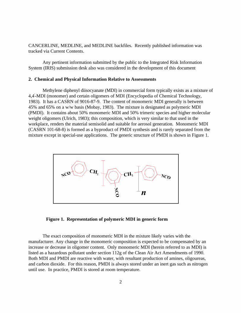
Methylene diphenyl diisocyanate (MDI) in commercial form typically exists as a mixture of 4,4'-MDI (monomer) and certain oligomers of MDI (Encyclopedia of Chemical Technology, 1983). It has a CASRN of 9016-87-9. The content of monomeric MDI generally is between 45% and 65% on a w/w basis (Mobay, 1983). The mixture is designated as polymeric MDI (PMDI). It contains about 50% monomeric MDI and 50% trimeric species and higher molecular weight oligomers (Ulrich, 1983); this composition, which is very similar to that used in the workplace, renders the material semisolid and suitable for aerosol generation. Monomeric MDI (CASRN 101-68-8) is formed as a byproduct of PMDI synthesis and is rarely separated from the mixture except in special-use applications. The generic structure of PMDI is shown in Figure 1.
Figure 1. Representation of polymeric MDI in generic form.
The exact composition of monomeric MDI in the mixture likely varies with the manufacturer. Any change in the monomeric composition is expected to be compensated by an increase or decrease in oligomer content. Only monomeric MDI (herein referred to as MDI) is listed as a hazardous pollutant under section 112g of the Clean Air Act Amendments of 1990. Both MDI and PMDI are reactive with water, with resultant production of amines, oligoureas, and carbon dioxide. For this reason, PMDI is always stored under an inert gas such as nitrogen until use. In practice, PMDI is stored at room temperature.
2
Monomeric MDI is a solid at room temperature; the PMDI mixture is a viscous liquid. The vapor pressure of both the monomer and PMDI is extremely low, about 2 × 10⁻⁶ kPa at 20 C (CMA, 1994). Both MDI and PMDI exist in workplace air primarily as aerosols generated during polyurethane foam formation (Dharmarajan and Weill, 1978; Dharmarajan, 1979). In field studies involving PMDI and conventional airless spraying equipment, these investigators found that, in contrast to impingers without filters, more than 78% of the material collected on Teflon filters with stainless steel backup pads upstream of the impingers was either urethane or MDI urea, since MDI was not detected in the leached material. The size distribution of the particles, determined with a seven-stage cascade impactor, had a mass median aerodynamic diameter (MMAD) of 11.0 µm with a sigma g of 3.6. When pure MDI was heated under laboratory conditions, a 0.5-µm pore-size Teflon filter blocked more than 87% of the MDI entering the impinger. It was recommended that concentrations of MDI should be expressed as mg/m3 and not ppm units.
The strengths and limitations of impingers, bubblers, and filters with respect to collection and detection of both MDI aerosols and vapors have been discussed by Streicher et al. (1994); none of these approaches has been found to be acceptable in all exposure environments. Streicher and colleagues suggested that an impinger or bubbler containing a solution of derivatizing reagent upstream of a reagent-impregnated filter would be suitable. The sampling and detection considerations, as discussed by Streicher and colleagues, are of importance not only with respect to occupational exposure environments, but to chamber exposure of laboratory animals as well. Spanne et al. (1996) have found the use of dibutylamine as a derivatizing agent reduces interferences from other substances in a complex isocyanate exposure mixture, results in a UV spectra determined by the nature of the isocyanates rather than that of the derivatizing regeant, and reduces the instability associated with dilute isocyanate solutions because isocyanate reactions with dibutylamine are fast.
The extent to which MDI, either in monomeric or polymeric form, disperses in air beyond the point of release and exposes general populations is not known. Like other members of the isocyanate family of compounds, MDI is highly reactive with a variety of biological functional groups, such as hydroxyl, amine, and sulfhydryl groups (Brown, 1987).
3. Toxicokinetics Relevant to Assessments
Little is known of the distribution and fate of PMDI/MDI in the human body. Animal inhalation studies have shown that PMDI exposure results in significant deposition both in the nasal region and in the alveolar region of the lungs (Reuzel et al., 1990; 1994a,b) In an unpublished pharmacokinetic study (Saclay, 1977), nose-only exposure of male Sprague-Dawley rats (12) to an aerosol (particle sizes less than 5 µm) of radiolabeled monomeric MDI for 15 min resulted in distribution of radioactivity, predominantly to the lungs and a variety of extrarespiratory sites, principally muscle, liver, kidneys, and the digestive tract. Labeling of the digestive tract was considered to be a result of transference of labeled material from the lungs.
3
After 4 days, 70% of the absorbed dose was eliminated, with fecal elimination predominant (57%). The experiment was terminated at the end of the fourth day.
In both humans (Schutze et al., 1995; Sepai et al., 1995a) and laboratory animals (Sepai et al., 1995b) exposed to PMDI/MDI, 4,4'-methylenedianiline (MDA), a metabolite/reaction product of MDI that is sometimes used in the production of polyurethane foam (Cocker et al., 1988), has been found in urine prior to acid hydrolysis and as an adduct conjugated with hemoglobin. MDA also has been detected in acid-hydrolyzed urine of workers exposed to PMDI/MDI (Skarping and Dalene, 1995a) and thermal degradation products of MDI elastomers (Skarping et al., 1995b). When pregnant Wistar rats were exposed on day 20 post-conception to 20 mg monomeric MDI/m3 for 6 h, MDA was detected in maternal blood, placenta, fetal blood, and amnionic fluid after acid hydrolysis (Bartsch et al., 1996). However, in studies of workers occupationally exposed to either PMDI/MDI or MDA, free MDA was detected in urine prior to acid hydrolysis (Schutze et al., 1995). MDA is a known animal carcinogen and has resulted in alveolar/bronchiolar adenomas and malignant lymphomas in female mice administered the compound in drinking water (Lamb et al., 1986).
The extent to which the results of the Lamb et al. (1986) study relate to inhalation exposure of humans to MDI/PMDI and the formation of MDA remains to be determined. It is not clear if MDA originates as a reaction product after PMDI/MDI uptake, is present in the exposure atmosphere along with PMDI/MDI, or is produced upon thermal degradation of materials made with MDI.
Further evidence in support of translocation of isocyanate material to remote sites is the finding by Maitre et al. (1993) that toluene diamine, a metabolite of toluene diisocyanate (TDI), is present in acid-hydrolyzed urine of TDI-exposed individuals. It has also been postulated that thiolytic acid esters, formed when isocyanates react with thiols in the body, may reverse with regeneration of the isocyanate functional group (Brown, 1987). This potential mechanism may allow for systemic delivery of the isocyanate in reactive form. Because measurable radioactivity was found in extrathoracic tissues and body fluids of guinea pigs exposed to ring-labeled TDI for one hour to levels as low as 4 ppb (Kennedy et al., 1989), some form of MDI may distribute to extrarespiratory sites as well.
4. Hazard Identification
4.1. Studies in Humans - Epidemiology, Case Reports, Clinical Controls
Exposure to isocyanates is a leading cause of occupational asthma worldwide (Mapp et al., 1988). The database associating TDI exposure with occupational asthma is extensive, although quantitative concentration-response relationships are lacking. The database for PMDI/MDI is less extensive but nonetheless compelling. There are a number of case reports and
4
occupational studies that describe sensitivity to PMDI and ascribe hypersensitivity pneumonitis and occupational asthma, but exposure levels are unknown (Zammit-Tabona et al., 1983; Konzen et al., 1966; Tanser et al., 1973; Zeiss et al., 1980; Malo and Zeiss, 1982; O’Brien et al., 1979; Bascom et al., 1985; Mapp et al., 1985; Lob and Boillat, 1981; Baur et al., 1984; Vandenplas et al., 1993). High exposure concentrations, such as might occur during a spill (Brooks, 1982), likely are a risk factor in human sensitization. Support for this view is found in a cross-sectional study (Bernstein et al., 1993). These investigators evaluated MDI-induced sensitization in 243 PMDI/MDI foam workers in a 3-year-old facility in which air levels were monitored continuously by area monitors for 24 h per day, during which time the air levels never exceeded 5 ppm. The average duration of employment was 18.2 mo. Only three cases of occupational asthma were identified, one of which was attributable to a spill.
Genetic predisposition as a function of inter-individual genetic differences in the expression of HLA Class II molecules on antigen-presenting cells has been suggested to play a role in sensitization (Bignon et al., 1994; Balboni et al., 1996). These investigators, using different populations of confirmed TDI asthmatics, found differences from controls in the DQB1 alleles that were interpreted to indicate that some individuals are predisposed to TDI-induced asthma. On the other hand, other groups of investigators (Bernstein et al., 1997; Rihs et al., 1997), with different study populations, found no such differences. All investigators used acceptable molecular biological techniques. Thus, further confirmation is required before any consensus can be reached regarding genetic predisposition as a risk factor in isocyanate-induced asthma.
Immunoglobulin E and G antibodies specific to MDI have been demonstrated in occupationally exposed workers with diagnosis of occupational asthma (Cartier et al., 1989; Liss et al., 1988; Pezzini et al., 1984; Tse et al., 1985; Zeiss et al., 1980). These antibodies are considered biomarkers for exposure only, because the presence of specific antibodies occurs in only a small percentage of individuals with asthmatic symptoms (Baur et al., 1994). MDI-specific immunoglobulin G (IgG) is a more typical finding than immunoglobulin E (IgE), the latter commonly associated with TDI exposure. Occasionally, individuals with sensitivity to one isocyanate exhibit cross-reactivity and subsequent asthmatic symptoms when exposed to another (Malo et al., 1983). MDI exposure of humans has been demonstrated to activate lymphocytes (Bentley et al., 1992; Raulf et al., 1992).
The available human data concerning occupational exposure to PMDI/MDI, coupled with lack of knowledge about mechanism of action and the possible role of genetic predisposition, are insufficient to identify exposure levels and scenarios responsible for isocyanate-induced sensitization. For this reason, PMDI/MDI-induced asthma could not be selected as an endpoint for RfC derivation.
5
Apart from asthma, a few occupational studies have examined exposure-response relationships with respect to pulmonary function decline and exposure to PMDI. However, each of the studies described below has a variety of limitations that preclude identification of exposure-response relationships for nonoccupationally exposed populations.
Musk et al. (1982) followed 107 workers prospectively for 5 years from 2 plants in a polyurethane manufacturing facility in which both TDI and MDI (presumed to be PMDI) were present in the air. [Note: In a later publication, Musk et al. (1985) reported that only 94, not 107, workers were available for reexamination]. Health data were collected during 2 surveys 5 years apart. The cohort was segregated into workers exposed to MDI only (n=25), TDI only (n=17), MDI and TDI (n=6), and no exposure (n=42). Criteria for selection of study participants were not stated. Health status for bronchitis, dyspnea, and smoking status were evaluated using standard questionnaires. It is stated in the discussion that this group did not include any subjects with symptoms suggesting hypersensitivity to isocyanates, but the criteria for this assertion were not provided. Other demographic data were not provided. Spirometric tests were conducted according to applicable criteria of the American Thoracic Society (ATS). Forced expiratory volume in 1-sec (FEV1) values were obtained from the average of 3 best values of 5 spirometric maneuvers. Effects on FEV1 and forced vital capacity (FVC) were stated to be the same, so only FEV1 data were presented. Spirometry was performed on the study subjects as they arrived to work on a Monday morning after a weekend of no exposure and again in the afternoon of that same day. Measurements were also taken on the Monday morning following a 2-week vacation and again in the afternoon of that same day.
For the comparison of changes in FEV1 over the 5-year study period, 94 of the original 107 subjects were tested. There was no evidence of bias resulting from the selective retirement of subjects from the cohort, as evidenced by the similarity of the mean FEV1 value of the original cohort with that of those who remained in the study. Much of the attrition could be attributed to contraction of the operation.
Exposure data were collected over the last 4 years of the 5-year study period, during which a total of 2,043 measurements were taken for TDI. Samplers were hand-held for 20 to 60 min in the breathing zone of workers, and analysis was done by the Marcali method (an impinger flow rate of 0.1 cu. foot/3 min is assumed). The exposure category for each subject was determined from both these measurements and occupational history. MDI, present in the atmosphere only in the last 2 years of the study, was sampled in a total of 470 measurements. Ninety percent of all measurements of TDI taken over the four years prior to the follow-up study contained less than 0.005 ppm in plant 1 and 0.004 ppm in plant 2. MDI levels ranged from <0.0003 - <0.004 ppm. [Note: true values are likely greatly underestimated, as MDI exists as an
6
aerosol and not as a vapor and the low impinger flow rate will not collect 100% of the MDI]. The geometric means of TDI and MDI concentrations, respectively, in the 2 plants were reported as 0.001 ppm and 0.0003 ppm in one and 0.0015 ppm and 0.0006 ppm in the other.
There were no statistically significant differences in the total 5-year decrement in FEV1 among those workers with no exposure (0.11 L), those exposed to TDI alone (0.13 L), those exposed to MDI alone (0.03 L), or those exposed to TDI and MDI (0.06 L). Regression analysis showed that the only decrement detected was due to smoking. The data on FEV1 (with similar ranges of n) also showed no effect in either daily changes (morning to afternoon FEV1) or in pre-and postvacation FEV1 changes. The mean annual rate of decrease in FEV1 was 0.02 L, a decrease expected from aging alone. Reanalysis of these data (Musk et al., 1985) using ATS standards for spirometry promulgated in 1979 confirmed these results and conclusions. It was reported that there was no evidence of bias resulting from attrition due to closure of one facility; the mean FEV1 value of those 152 lost to the study was similar to those 107 who remained. Musk et al. (1985) also stated that the annual FEV1 declines were similar to cross-sectional age regression coefficients of predicted normal populations. However, age-regression coefficients from cross-sectional populations have been described as poor benchmarks for the evaluation of longitudinal spirometric data because cross-sectional populations, unlike longitudinally studied populations, are sensitive to “cohort effects” (Glindmeyer et al., 1982). These effects, such as illness, smoking, and occupational exposures, may have affected lung function in the cross-sectional cohort at an earlier age, but not the longitudinal population in question.
Because of the relatively low number of individuals exposed to isocyanates (48) and to MDI alone (25) in this study, the uncertainty over the actual level of MDI exposure, and the short study duration (5 years), this study offers only limited assurance that MDI is not without effect on pulmonary function.
Sulotto et al. (1990) examined the effect of low-level (reported as less than 1 ppb) MDI (presumed to be PMDI) exposure on lung function parameters (e.g., FEV1) in 27 polyurethane foam workers over a workshift and workweek. All workers were asymptomatic for asthma and were age-matched to nonexposed controls from the same factory. There was no selection of participants from within the exposed group. The duration of employment was similar between exposed (14 years) and controls (16 years). During a portion of their employment individuals had also been exposed to TDI. The exposed group had more smokers (12/27) than controls (5/27). There was no significant difference in spirometric data between groups over the workshift or workweek. Spirometric testing was consistent with ATS criteria.
Pham et al. (1988) prospectively studied a group of workers from two polyurethane manufacturing facilities in 1976 and 1981. In 1976, the first year of the study, isocyanate levels (mainly PMDI/MDI) were described as less than 0.02 ppm. At some point between 1976 and 1981, the last year of the study, a new ventilation system was installed, resulting in levels below
7
0.005 ppm by 1981. There was no further discussion of environmental characterization in the report, and sampling and analytical methodologies were not discussed. Thus, the lack of exposure characterization is a serious limitation of the study that precludes its usefulness in identifying exposure-response relationships. A modified respiratory symptom questionnaire based on the British Medical Research Council’s version was administered, with additional questions on atopy and occupational history. Flow-volume curves and single-breath CO diffusion (DLCO) tests were performed, but no criteria for standardization were described. According to their job category in 1976, workers were classified into three groups: unexposed (62 men and 21 women); indirectly exposed (61 men and 56 women), and directly exposed (91 men and 27 women). Those indirectly exposed are assumed to represent those handling cured foam products but not involved in the foam-making process. No differences in age, height, or weight by gender were present between the groups in 1976. The proportion of smokers in each group was the same for females, but slightly higher (stated as insignificant) in males for groups 2 and 3. The study in 1976 found a significantly increased prevalence (18.5%, p<0.05) of asthma in women in exposure group 3 and of chronic bronchitis in both males and females in groups 2 (males 18%, p<0.01; females 19.6%, p<0.001) and 3 (males 11%, p<0.05; females 29.6%, p<0.001). Criteria for classification of asthma and chronic bronchitis were not described. Pulmonary function testing indicated significant impairments (based on comparison to predicted values from cross-sectional populations) in men only for vital capacity (VC) in group 2 and for VC, FEV1, and DLCO in group 3. Exposure duration was broken down into categories of <20 mo, 20-60 mo, and >60 mo for further analysis of the pulmonary function and diffusion testing. Only VC was reduced in the 20-60 mo exposure category (p<0.05). In the >60 mo category, VC (p<0.01), FEV1 (p<0.05), and DLCO (p<0.01) were less than predicted values.
At follow-up in 1981, only half of the initial cohort remained (114 males, 45 females). Losses were highest (64%) among exposed females. Reasons for attrition were not described and comparisons between the lost group and the remaining cohort were not made. Only the results of males in 1981 were presented because of the low number of remaining females. For the longitudinal analysis, exposure categories were classified as: (A) unexposed at both studies (n=45); (B) indirectly exposed at both studies (n=24); (C) directly exposed at both studies (n=30); and (D) exposed in 1976 but removed from contact with MDI in 1981 (n=15). Pulmonary function test and DLCO values were reported either as percent of predicted or change in predicted values from study 1 (1976) to follow-up (1981). The 5-year longitudinal changes in FEV1 were given for each of the four exposure groups and were stated not to be significantly different between the nonexposed group and each of the three exposed groups. Pulmonary function indices were significantly lower in group C (directly exposed) when compared with predicted values. However, it has been noted previously that cross-sectional declines in predictive values are insensitive in detecting chronic changes in longitudinal studies (Glindmeyer et al., 1982). The lack of exposure characterization, high rate of attrition, and inclusion of asthmatics in the cohorts preclude any quantitative assessment of MDI exposure on lung function.
8
Several studies of polyurethane foam workers examined the role of exposure to isocyanates in relation to cancer incidence and mortality. In these studies, PMDI/MDI was one of a variety of substances in the work environment. Thus, these studies are inadequate for categorizing the carcinogenic potential of MDI. In addition, the cohorts described are relatively young and additional follow-up would be needed before any conclusions could be drawn.
A retrospective mortality and cancer morbidity study was conducted to investigate associations between health risk and exposures from polyurethane foam production, particularly exposures to diisocyanates including MDI (Sorahan and Pope, 1993). The study population was taken from 11 factories in England and Wales that had begun manufacturing foam before 1980. Toluene diisocyanate was the principal isocyanate used; MDI represented about 5% of the amount of TDI. Prior to 1970 only TDI was used.
The cohort consisted of 8,288 workers, of whom 2,465 were women, employed for at least 6 mo and having a portion of this employment between 1958 and 1979. Job histories and descriptions were obtained from personnel records. All jobs were classified into one of four exposure levels: high, low, minimal/zero, and unclassifiable. Higher diisocyanate exposure was defined as above 4 ppb (8-h TWA) or excursions above 10 ppb on most days. Lower exposure was defined as between 1.5 and 4 ppb (8-h TWA) or occasional excursions above 5 ppb. Job history data for each worker were matched to job exposure classification to assign person-years of follow-up to three time-dependent exposure levels. The minimum length of follow-up was 9 years. Only 2% of the cohort was lost to follow-up.
Death certificates were obtained for 803 of the 823 known deaths and were coded according to the 9th revision of the International Classification of Diseases. The National Health Service Central register provided information on 277 incident cancers from 1971 through 1988, the year in which the study ended.
Cause-specific SMRs, concentrating on cancer causes, were calculated by applying gender-specific external rates for England and Wales. Separate SMRs for certain cancers were also presented by gender, by years since first hire, and by relationship to cigarette smoking. The SMR for all cancers was 88 (95% CI=84-100), based on 221 observed and 251.4 expected deaths. For females, a statistically significant excess of pancreatic (SMR=271) and lung (SMR=176) cancer was calculated. However, no trend was seen with time since first hire for either of these cancers. When cancers related to smoking were grouped separately from those unrelated to smoking, the SMRs for women in the entry cohort for cancers related to smoking were 263 and were 67 for cancers unrelated to smoking. Available smoking information as of 1981 on a subset of the study population women revealed that 58% of them were smokers, contrasted with 37% of women in England and Wales in 1980.
9
The relationship between diisocyanate exposure and mortality from all causes of death, respiratory diseases, lung cancer, and pancreatic cancer was evaluated via a multiplicative relative risk model. The internal referent group was person-years at the reference level of all covariates. Exposure was not a statistically significant addition to the model for any of the four causes. There was some suggestion of increased risk at the higher exposure level for lung cancer with a relative risk of 126 (95% CI=39-409) and respiratory diseases(95% CI=54-573). None of the females who died had been classified as receiving either higher or lower diisocyanate exposure during any of their employment history. The authors concluded that excesses in female cancer were probably due to cigarette smoking and other factors unrelated to diisocyanate exposure. Although this well-designed study did not find an association between exposure to diisocyanates and cancer, the cohort was young and follow-up was relatively short.
A retrospective cohort and mortality and cancer incidence study was conducted to determine whether occupational exposure to TDI and PMDI/MDI is associated with excess cancer deaths or incidences or excess deaths from obstructive lung diseases (Hagmar et al., 1993a). The study involved workers employed at any of nine Swedish polyurethane foam manufacturing plants. The plants commenced operations between 1958 and 1973. From 1965 to 1987 personal and area sampling for isocyanates was performed an average total of 15 days on an average of 9 occasions. The TWA for MDI was typically below 0.010 mg/m3. The highest level observed was 0.16 mg/m3.
The cohort consisted of 4,154 workers employed for at least one year before 1987 and having data complete enough for analysis. Average length of follow-up was 10.6 years. Only 6% of workers in follow-up years through 1987 were attributed to the 1958-1970 period because the cohort was young and only 187 deaths occurred. Death certificates were coded according to the 8th revision of the International Classification of Diseases. The National Swedish Tumor Register reported 72 cases of cancer incidence. Each worker’s job history was combined with exposure data and workers were assigned to one of three exposure categories: no exposure, low/intermittent exposure, and apparent exposure. Person-years were also separated into those occurring during the 10 years after first exposure and those occurring later. Cause-specific age/calendar year/gender-specific mortality and incidence rates were obtained from Statistics Sweden and were used to calculate SMRs and the Standardized Incidence Ratios (SIRs).
With 130 observed deaths the overall SMR for all causes of death was 78 (95% CI=66-93). Malignant neoplasms (33 observed, 43 expected) and nonmalignant respiratory diseases (4 observed, 7.3 expected) had SMRs of 77 (95% CI=53-109) and 55 (95% CI=15-141), respectively. The overall SIR for malignant neoplasms (72 observed, 89.4 expected) was 81 (95% CI=63-102), with nonsignificant excess of 166 (95% CI=61-361) for rectal cancer and 153 (95% CI=42-391) for non-Hodgkin’s lymphoma. SIRs for these two cancers appeared higher after, rather than during, the first 10 years from first exposure, with SIR after the first 10 years of 192 (95% CI=55-492) for rectal cancer and 280 (95% CI=76-716) for non-Hodgkin’s lymphoma.
10
Time since exposure did not affect SIRs for lung cancer or all malignant neoplasms. Leukemia (3 observed, 2.4 expected) exhibited an overall SIR of 127 (95% CI=26-370). All other causes of cancer among the 72 incident cases had SIRs less than 1.
To further investigate observations in this cohort, Hagmar et al. (1993b) conducted a nested case-control study of 7,023 members of the previous cohort employed for at least one day during 1958-1987. The study also was initiated to minimize exposure misclassification. Cases were 114 of 119 incident cancers, identified from the National Swedish Tumor Register, having job history data complete enough for exposure evaluation. Information from tumors diagnosed from 1959 to 1987 was coded according to the 7th revision of the International Classification of Diseases. Altogether, there were 313 controls, with each case originally matched to three controls with respect to plant, gender, and year of birth. Five cases and 11 controls were dropped because it was impossible to locate their job history information. Exposure to MDI began between 1964 and 1982, depending on the plant. In addition to personnel records, the basis for exposure assessment was expanded to include information obtained from a selected group of persons with long employment and good personal knowledge of employees and processes. Types of exposure distinguished were MDI only, TDI only, or MDI and TDI combined. Exposure was clarified as high, intermediate, or low. For high or intermediate exposure categories, 18 cases and 65 controls were exposed to MDI only, 9 cases and 16 controls were exposed to MDI/TDI, and 19 cases and 62 controls had TDI exposure only. Exposure levels to MDI were normally below the detection limit of the analytical method (<0.01 mg/cm3) and nearly all were below 0.1 mg/m3.
Results were presented for all cancers combined. No distinction was made in the results between exposure to MDI and TDI. Odds ratios (OR) for diisocyanate exposure with and without a 10-year lag were presented with 90% confidence intervals. Using conditional logistic regression, ORs were calculated separately for (1) any exposure, (2) exposure at intermediate or high levels, (3) high exposure, (4) high exposure for up to 5 years, and (5) high exposure for more than 5 years. For the last two analyses, ORs were produced only for all cancers combined. Potential confounders, with the exception of smoking and alcohol use, were controlled by the matching design of the study.
At the 10% level of significance, no statistically significant association was found between all-cause cancer and diisocyanate exposure using any of the five exposure measures or for non-Hodgkin’s lymphoma and rectal cancer (five cases). The OR for prostatic cancer appeared elevated with high exposure (OR=2.7). However, risk was not enhanced in analyses examining a 10-year latency period. The OR between colon cancer and any exposure and intermediate/high exposure appeared elevated (OR=2.2 and 3.3, respectively). However, there was no odd distribution of colon cancer cases by subsite. All colon cancer cases had less than a 10-year latency. The apparent association of exposure with prostate and colon cancer may be spurious, as there is no plausible biological connection between these cancers and diisocyanates.
11
4.2 Prechronic, Chronic Studies and Cancer Bioassays in Animals
In a range-finding toxicity study, SPF-Wistar rats (15/sex/group) were exposed to PMDI aerosol at 0.35, 1.4, or 7.2 mg/m3, 6 h/day, 5 days/week for 13 weeks (Reuzel et al., 1985a, 1994a). Polymeric MDI was in the form of a dark brown liquid that contained 52% monomeric MDI. The trade name of the mixture used was Desmodur 44 V 20. The PMDI content in test atmospheres was monitored by light scattering, gravimetry, photometry, and high-performance liquid chromatography (HPLC). The content of monomeric MDI in the test material was 52%. Particle sizing, accomplished with cascade impactors, indicated >95% of the particles had an aerodynamic diameter <5 µm. However, sigma g values were not given at each of the concentration ranges. Microscopic evaluation of liver, kidneys, and respiratory tract (nasal cavity, trachea, larynx, and lungs; n = 15) did not reveal any exposure-related lesions. The only exposure-related observation was an increase in yellowish-colored alveolar macrophages in all animals of the high-exposure group, with sporadic signs of a minimal focal inflammatory reaction in the alveolar septa. Urinalysis, hematology, clinical chemistry, and liver enzymes indicated no change in comparison with control animals. It was concluded that no clear adverse effects were observed.
In order to determine appropriate concentrations for a chronic study, a second 13-week, range-finding subchronic toxicity study was performed using specific-pathogen-free (SPF) Wistar rats (30/sex/exposure level) exposed to PMDI aerosol (Reuzel et al., 1985b, 1994a). Rats were exposed to 4, 8, or 12 mg/m3 for 6 h/day, 5 days/week, for 13 weeks. The aerosol was generated by a nebulizer modified to eliminate nonrespirable particles. The MMAD of the throughput, as determined by cascade impaction, was <5 µm, which was representative of 95% of the particles. However, particle size distribution was not reported. Test atmospheres were monitored by gravimetry, photometry, light scattering, and HPLC. Clinical symptoms, ophthalmology, body and organ weights, hematology, biochemical alterations, urine, gross, and histopathologic examination in 10 animals per group at the end of exposure and 4 weeks after the final exposure were evaluated for toxicity.
Lung lavage fluid was analyzed for cell number, cell viability, and survival based on trypan blue exclusion and phagocytic activity in 5 animals per group exposed to 0, 4, and 8 mg/m3. In the animals examined immediately after the exposure, severe respiratory distress was noted in animals exposed to 12 mg/m3, while similar, although less severe, signs were also seen in the 8 mg/m3 group. Mortality occurred only in the 12 mg/m3 groups (15/60 animals) and only during the first 7 weeks of the study. An exposure-related increase in relative lung weight was noted at 8 and 12 mg/m3 in both sexes. Creatinine values in blood plasma increased in an exposure-related manner in both the 8 and 12 mg/m3 groups. Degenerative lesions occurred in the olfactory epithelium of the nasal cavity of both sexes at 12 mg/m3. Hyperplasia of the respiratory epithelium was found in females, but not males, of the 8 and 12 mg/m3 groups. There was a significant increase in accumulation of macrophages, observed histologically, in the lungs and
12
mediastinal lymph nodes of all exposed animals compared with controls. The increase and severity of accumulation at the level of the alveolar septa achieved statistical significance in males at 4 mg/m3 and females exposed to 8 mg/m3. There were no histopathological lesions of other organs examined. No effects were observed on number of lavagable cells, cell viability, or cell survival. In vitro measurements indicated that the phagocytic activity (data not reported) of lavaged alveolar macrophage was reduced in females exposed to 8 mg/m3, suggesting to the investigators that lung clearance might be impaired. The lack of effect on lavageable cell number is not consistent with the histological observation of increased accumulation of macrophages, and suggests incomplete recovery of macrophages in the exposed animals. This study demonstrated adverse effects in the lungs and nasal cavity at levels of 4 mg/m3 and above. However, because of a lack of data on aerosol sizes, a quantitative LOAEL(HEC) cannot be derived. These results are, however, consistent with the findings of the chronic study that subsequently was performed (Reuzel et al., 1990, 1994b).
In a 2-year cancer bioassay, SPF-bred Wistar rats of the Cpb:WU strain (60/sex/exposure level) were exposed whole-body to aerosols of PMDI for 6 hours/day, 5 days/week, for 24 mo (Reuzel et al., 1994b). The nominal chronic exposure levels were 0, 0.2, 1.0, and 6.0 mg/m3. An additional satellite group of 10/sex/exposure level was exposed similarly and used for histopathology at 12 mo. The selection of 6 mg/m3 as the high dose was based on a steep dose-response curve identified in the 13-week study (Reuzel et al., 1994a). In the latter study, severe respiratory effects, including mortality, were observed at 12.3 mg/m3 whereas similar, but much less severe, clinical effects and increased lung weights were seen at the next lowest dose, 8.4 mg/m3. These observations, coupled with significant increases in absolute and relative lung weights in the chronic study, are consistent with 6.0 mg/m3 in the chronic study, approaching a maximum tolerated dose.
The MDI test material (eight different batches) had an average molecular weight of about 400. It was reported that the composition of each batch of the material was analyzed by the supplier (Bayer A.G.) prior to use and, in the case of the first two and last batches, also after three months of storage. The content of monomeric MDI, determined by the supplier, ranged from 44.8% to 50.2%. The remainder, excluding trace impurities, was described as “polymeric” MDI with no further explanation. Test material was kept at room temperature under “common laboratory conditions” prior to use. This is interpreted to mean that the test material was stored under nitrogen or argon, as MDI reacts with water to form carbon dioxide.
Test atmospheres were generated by atomizing the MDI material into droplets using compressed air in a nebulizer fitted with a baffle to remove larger particles from the air stream. The smaller droplets were passed through a cyclone to obtain an aerosol of which 95% of the particles were smaller than 5 µm. Beta attentuation primarily was used, along with intermittent gravimetric determinations, to determine the concentration of the polymeric MDI in the exposure atmosphere. HPLC was used as a secondary standard during the first 3 mo of exposure.
13
However, neither the content of monomeric MDI in the test atmosphere nor other constituents, such as MDA, were determined. Thus, uncertainty as to the amount of monomeric MDI to which the animals were exposed is a limiting feature of this study. The actual mean values were within ± 10% of nominal values. Thus, the nominal values will be used in the following discussion. The duration-adjusted exposure levels are 0, 0.036, 0.180, and 1.100 mg/m3. The MMAD and geometric standard deviation (in parentheses) corresponding to the exposure levels were 0, 0.68 µm (2.93), 0.70 µm (2.46), and 0.74 µm (2.31), respectively.
The animals were observed for clinical signs twice a day during exposure. Hematological analysis, clinical chemistry, and urinalysis were performed on 10 animals/sex/exposure level after 1 year of exposure. Gross observations and organ weights were obtained on all animals. Histopathological observations were made on the lungs, mediastinal lymph nodes, and nasal cavity (6 levels) of all animals. Histological observations in the main group were made on controls and animals exposed to 6 mg/m3 and on low- and mid-concentration decedents for 43 tissues. Examples of the tissues examined included testes, lung, nasal cavity, heart, kidney, pancreas, liver, lymph nodes, mammary glands, stomach, brain, etc. The tissues selected for histopathology are representative of types of tissues that would normally be included in, for example, a protocol of the National Toxicology Program.
There was no exposure-related increase in mortality, clinical signs, clinical chemistry, or body weight. Upon necropsy of the satellite group at 1 year, exposure-related changes were seen in the nasal cavity, lungs, and mediastinal lymph nodes. Males and females at 6 mg/m3 showed a significant increase in relative lung weight. Males only at 6 mg/m3 exhibited statistically significant minimal-to-moderate disarrangement of the olfactory epithelium (0/10, 0/10, 3/10, and 6/10 in the 0, 0.2, 1.0, and 6.0 mg/m3 groups, respectively). The incidence of minimal-to-moderate basal cell hyperplasia was not statistically significant at any exposure level (males, 1/10, 0/10, 2/10, and 2/10; females, 0/10, 0/10, 0/10, and 2/10). Accumulation of macrophages with yellow pigment was seen in the lungs of both sexes at 1 and 6 mg/m3 (males, 0/10, 0/10, 4/10, and 10/10; females, 0/10, 0/10, 3/10, and 9/10). At the site of accumulation, Type II alveolar cells were predominant. Localized fibrosis was significant in males exposed to 6 mg/m3 and in females at 1 and 6 mg/m3. Alveolar duct epithelialization was increased significantly in males and females exposed to 6 mg/m3 and interstitial pneumonitis was significant in males at 1 and 6 mg/m3.
At the 24-mo necropsy of the main group, histologic evidence of damage involved the same tissues as at 12 mo, although severity had increased. Basal cell hyperplasia was evident in the olfactory epithelium of males (14/60, 13/60, 26/60, and 32/60 at 0, 0.02, 1.0, and 6.0 mg/m3, respectively) and females (4/60, 8/60, 8/60, and 49/59 at 0, 0.2, 1.0, and 6.0 mg/m3, respectively). Statistical significance was achieved with males only in the mid-concentration group and with both sexes at in the high-concentration groups. It was often accompanied by Bowman’s gland hyperplasia, which was significant in males at 1 and 6 mg/m3. Olfactory epithelial degeneration was elevated significantly in males and females at 6 mg/m3, incidence at 1 mg/m3 was elevated in
14
males but was not statistically significant. Given the apparent sensitivity of the male to basal cell hyperplasia and the expression of an elevated incidence of both basal cell hyperplasia and olfactory degeneration at both the mid and high concentrations, it is not possible to discern basal cell hyperplasia, on the basis of either the interim or final sacrifice histopathological results, as a compensatory response. Thus, it is prudent to regard basal cell hyperplasia as an adverse response. Because it reached statistical significance at a lower exposure level (male rats only) than olfactory degeneration, the former was chosen as the critical effect.
In the lung there was an increase in the accumulation of pigment-laden macrophage in alveolar duct lumina (incidence in males: 0/60, 3/60, 21/60, and 60/60; females: 0/59, 1/60, 23/60, and 59/59) and in localized fibrotic changes (males: 1/60, 0/60, 9/60, and 44/60; females: 0/59, 0/60, 4/60, and 48/60). Both sexes at 6 mg/m3 exhibited statistically significant increases in mean absolute lung weight, although relative lung weight was increased in males only. Localized alveolar duct epithelialization was increased significantly in both males and females exposed to 1 and 6 mg/m3, and localized alveolar bronchiolization (adenomatous hyperplasia) was significant in both sexes at 6 mg/m3. In animals exposed to 1 mg/m3, there were minimal foci of Type II pneumocyte hyperplasia, which was more extensive in the high-exposure group. An exposure-related accumulation of yellow pigment in the mediastinal lymph nodes was noted in both males and females. The accumulation of macrophages and the localization of tissue damage in the area of macrophage accumulation suggest that the thoracic effect is due primarily to toxicity to the macrophage (with secondary tissue damage), although some of the effects were described as being distributed evenly throughout the lungs. There were no histological effects in any of the other organ systems examined. The information obtained in this chronic study identifies a NOAEL of 0.2 mg/m3 (duration-adjusted concentration = 0.036 mg/m3) and a LOAEL of 1.0 mg/m3 (duration-adjusted concentration = 0.18 mg/m3) for respiratory tract effects in both the pulmonary and extrathoracic regions.
Rather than use the identified NOAEL for calculation of the RfC, a benchmark concentration (BMC) approach was employed, using all the duration-adjusted data for both basal cell hyperplasia and degeneration of the olfactory epithelium. The BMC at the 10% level of risk (BMC10) was calculated (see Appendix A) as the lower bound on the maximum-likelihood estimate. The 10% level of risk was chosen as a result of the findings by Allen et al. (1994) that quantal representation of endpoints from developmental toxicity experiments led to the best match between NOAELs and benchmark concentrations or doses when a 10% level or greater of response was used to define the BMC.
Although there were no compound-related nasal tumors, solitary pulmonary adenomas, described as rare in this strain, were observed in males (6/60) and females (2/59) exposed to 6 mg/m3 compared to controls (0/120). The adenomas, not thought to be the cause of death, were only a few millimeters in size and were located adjacent to areas in which hemorrhage, macrophage accumulations, and fibroblastic reactions were observed. Only one pulmonary
15
adenocarcinoma (approximately 10 mm in size) was observed in one male exposed to 6 mg/m3. Although this study provides clear evidence of a tumorigenic response to treatment, the significance of only one pulmonary adenocarcinoma is insufficient to distinguish PMDI as an animal carcinogen.
Another chronic toxicity/carcinogenicity study (unpublished) was also conducted in female Wistar rats only, strain Crl:[WI]BR, (80/exposure group) exposed to monomeric MDI in aerosol form to nominal levels of 0.23, 0.70, and 2.05 mg/m3 for 17 h/day, 5 days/week for 24 mo (Hoymann et al., 1995). The selection of exposure levels was based on results of a previous 90-day study. The highest concentration can be considered a maximum tolerated dose inasmuch as the body weight gain for this group after 17 mo of exposure was 11% less than that of controls.
The nominal levels of the chamber atmosphere were determined photometrically, after calibration using gravimetric values. However, HPLC analysis of the chamber atmosphere indicated that the amounts (0.097, 0.549, and 1.75 mg/m3, respectively) of monomeric MDI were considerably less than the photometrically determined levels, particularly at the lowest concentration. According to the manufacturer’s specifications, the starting material consisted of 99.5% pure monomeric MDI. It had been heated and mixed with compressed air and passed through a nebulizer to generate the aerosols to which the animals were exposed. The MMAD was determined to be about 1 µm in each of the three exposed groups. A sigma g was not provided; however, the same aerosol generation protocol was used in another study (Buschmann et al., 1996) in which the sigma g was 1.37. Hoymann and colleagues surmised that a portion of the MDI generated with water vapor in the chamber to form reaction products such as urea. This was offered as a likely reason the chemically determined amount of monomeric MDI was less than the gravimetric value, which presumably included reaction products. This observation is consistent with the findings of Dharmarajan (1979). A limitation of the chemical analyses of the exposure atmosphere generated by Hoymann and colleagues is that MDA content was not measured.
Because gravimetric values from the Reuzel et al. (1994b) study had been used (chemical determination of monomeric MDI was not made), the nominal values in the Hoymann et al. study are used throughout the following discussion for comparison purposes. Differences in MDI test material and chemical analyses used by Reuzel et al. (1994b) and Hoymann et al. (1995) preclude assessment of test results on any basis other than gravimetric values. The duration-adjusted values for the Hoymann et al. (1995) study are 0.12, 0.35, and 1.04 mg/m3, respectively. In comparison to the duration-adjusted values from the Reuzel et al. (1994b) study, the lowest two exposure values are about four- and twofold higher, respectively.
In addition to animals exposed for 24 mo, 20 animals per exposure group were examined histopathologically at 12 mo. Smaller numbers of animals were assessed at various time points for
16
lung function and for examination of bronchoalveolar lavage (BAL) fluid for cell counts and protein and enzyme determinations.
Treatment-related and statistically significant pulmonary lesions found in animals exposed for two years included: (1) concentration-related increase in focal/multifocal alveolar and bronchioalveolar hyperplasia of the lungs, (2) concentration-related increase in incidence of focal/multifocal interstitial fibrosis of lungs, (3) concentration-related multifocal accumulation of particle-laden and pigmented macrophages. Alveolar cell hyperplasia, considered preneoplastic, exhibited a concentration-response trend with incidence reaching significance in the high-exposure group (7/80 compared to 0/80 in controls).
The histopathologic effects in the lungs correlated with pulmonary function effects, particularly in the high-exposure group. There were significant differences from controls in forced expiratory flow at 25% (FEF25) and 50% (FEF50) after 6 mo and in CO diffusion after 12 and 17 mo. Bronchoalveolar lavage indicated total protein was significantly elevated after 20 mo in the mid-exposure group; elevation at earlier time points occurred in the high-exposure animals. There was also a concentration-dependent increase in -glutamyl transpeptidase activity that was statistically significant in the mid- and high-concentration groups. All groups evidenced significantly increased relative lung weights at all time periods. This latter observation, coupled with significant increases in hydroxyproline and total protein in BAL fluid, clearly indicates adverse effects on the lungs and that a maximum tolerated dose was achieved.
In contrast to the greater incidence of animals with pulmonary adenomas in the high-exposure group of the Reuzel et al. (1994b) study, only 1 of 80 animals (high-exposure group only) exhibited a single bronchio-alveolar adenoma. Tumor data were presented only for the high-exposure group. Although there was a total of 144 benign tumors in this group, the number in controls was even higher (159). There was no significant difference in the incidence of animals with malignant tumors between the controls and the high exposure-group. The incidence of mammary gland tumors was similar between the high-exposure group and controls. Noncancer histopathologic results for tissues/organs other than the lungs were presented for the high-exposure group only. This lack of data for other exposure groups represents a study deficiency. Goblet cell hyperplasia of the nose and focal/multifocal mucosal inflammatory cell infiltration were significantly elevated above controls for the high-exposure group. No other significant nasal effects were reported. There also were no significant differences in mortality or body weight between exposed and control animals.
In the satellite group examined at 12 mo, there were no statistically significant effects on nasal endpoints, but there was a statistically significant dose-dependent increase in interstitial fibrosis, pigment, and particle-laden macrophage in the lung. This is consistent with the 2-year findings for this endpoint. Increases in alveolar and bronchioalveolar hyperplasia were not statistically significant at 12 mo. With respect to tumor incidence, only the high-exposure group
17
exhibited a greater percentage of tumor-bearing animals (11/15) compared to controls (8/16), but the increase was not statistically significant. The incidence of animals with malignant tumors was similar between controls and the two high-dose groups; no malignant tumors were observed in the low-dose group.
The noncancer and carcinogenic results seen in this study are in general agreement with those of the Reuzel et al. (1994b) study given the difference in duration-adjusted concentration for the two lowest exposure groups, differences in atmosphere generation and exposure duration, the use of only female rats by Hoymann and colleagues, and the difference in the nature of the starting material used by each group of investigators.
The distinctions between the interstudy observations of noncancer effects may also highlight important differences in the toxicological nature and deposition of the inhaled materials and/or their reaction products. In contrast to the increases in basal cell hyperplasia in the nasal tract of female rats exposed in the Reuzel et al. (1994b) study, monomeric MDI in the Hoymann et al. (1995) study was apparently without effect, as no difference between controls and the high-exposure group was observed. In addition, the incidence in accumulation of pigmented macrophages was significantly less than the incidences in females reported by Reuzel et al. (1994b), although the particle sizes in each of the studies were similar. The observation of adverse effects in the lung at 0.23 mg/m3, even though no such effects were noted in the Reuzel et al. study, probably reflects, in part, the longer duration (17 h vs. 5 h) of exposure.
Findings obtained with animals exposed by inhalation to TDI provide a useful comparison to the effects noted in the MDI studies. When Sprague-Dawley rats were exposed whole-body to an 80:20 mixture of 2,4-/2,6-TDI at 0.05 or 0.15 ppm for 6 h/day, 5 days/week for 2 years, the number of tumor-bearing animals in the TDI exposure groups was similar to controls (Loeser, 1983), a situation similar to that seen in the chronic PMDI/MDI studies. The preponderance of the tumors in rats were lung adenomas and incidence was comparable to controls. The amount of TDI ingested as a result of grooming was not investigated. Although there was no treatment-related effect on survival rates or body weights, rhinitis in both sexes suggests that a maximum tolerated dose was achieved. The noncarcinogenic, histopathological results with TDI in the rat were described by Owen (1984a,b). In the nasal tract (three sections representing the anterior, middle, and posterior areas), a concentration-related increase in the incidence and severity of mild to moderate rhinitis characterized by squamous metaplasia and inflammation was observed in male and female rats compared to controls. Olfactory atrophy or degeneration and basal cell hyperplasia were not reported.
In a companion study with CD-1 mice (Loeser, 1983; Owen, 1986), survival rates were also unaffected by treatment. A maximum tolerated dose appears to have been achieved since body weight gain was about 10% lower in the 0.15 ppm group compared to controls and there was a concentration-related increase in the incidence of chronic or necrotic rhinitis in the nasal
18
cavity in both exposure groups. The principal findings in the lungs were an increased incidence of multiple adenomas in male mice: 1/90, 9/90, and 6/90 in controls, low-dose, and high-dose animals, respectively.
In an assessment of this study, Holder (1990) concluded that although the occurrence of mouse lung adenomas was increased in treated groups, there was no progression to malignancy after 2 years of exposure. Thus, TDI was not carcinogenic in CD-1 mice.
In metabolic studies with C14-labeled 2,4-TDI (2 ppm) administered to Fischer 344 rats by inhalation, Timchalk et al. (1994) found that acetylated 2,4-TDA in urine accounted for about 10% of quantitated metabolites, but no free 2,4-TDA was found in the 0-12 h pooled urine specimens. The amount of TDA formed and sequestered in tissues was not determined.
4.3 Reproductive/Developmental Studies
The potential of MDI to cause developmental effects was recently examined in two inhalation studies with laboratory animals.
Prenatal toxicity was evaluated in an inhalation study in which pregnant Wistar rats were exposed to respirable PMDI from day 6 to 15 of gestation (Gamer et al., 1994). Rats (25/exposure group) were exposed to 1, 4, and 12 mg/m3 PMDI with a MMAD below 2.8 µm. Particle sizing was accomplished with cascade impactors. The PMDI was stored under nitrogen in aluminum bottles. Animals were exposed for 6 h/day. Surviving animals were sacrificed on day 20 after a 5-day postexposure observation period. Two dams from the high-exposure group died and death was considered treatment-related. Some survivors in this group exhibited respiratory symptoms that continued into the postexposure period; absolute and relative lung weights were significantly increased. None of the dams in the other two exposure groups exhibited any exposure-related clinical or gross pathological effects. Absolute lung weights were elevated in the 1 and 4 mg/m3 group, but this was not statistically significant. No treatment-related effects on gestational or fetal parameters occurred in the two lowest exposure groups. In the 12 mg/m3 group, statistically significant decreases in placental and fetal weights occurred and fetuses/litters with skeletal variations and retardations occurred at an increased rate. These effects may be the result of maternal toxicity. This study identifies a maternal NOAEL at 4 mg/m3 and a developmental LOAEL at 12 mg/m3.
The Gamer et al. (1994) study suggests that the potential of PMDI to cause prenatal toxicity and teratogenic effects in this strain of rats is low. However, studies in other laboratory animal species and a multigeneration study would be needed to ascertain the potential to cause functional deficits or altered development in the postnatal period.
19
The increase in maternal lung weights in the two lowest exposure groups in the Gamer et al. (1994) study warrants concern inasmuch as the range-finding study preceding the prenatal study also resulted in increased lung weights at the lowest concentration tested, 4.75 mg/m3. In addition, an earlier 14-day study (cited by Reuzel et al., 1994b) reported that rats exposed to 2, 5, or 15 mg/m3 exhibited increased lung weights at all concentrations, with death and severe respiratory distress in the 15 mg/m3 group. These findings are consistent with the results of the Reuzel et al. (1994b) chronic inhalation study, in which lung effects were observed at 1 mg/m3.
Buschmann (1996) reported that gravid Wistar rats were exposed by whole-body inhalation to 1, 3, or 9 mg MDI /m3 for 6 h/day from days 6-15 post conception after which the animals were sacrificed on day 20 and assessed for prenatal effects. A quasi-monodisperse MDI aerosol was generated by an evaporation-condensation technique. The particles had a MMAD of 1.1 µm and a geometric standard deviation of 1.37. Polymerization was precluded by exposing MDI droplets to temperatures above 50 C for a period of 1.5 sec only during droplet evaporation. Absolute and relative lung weights in the high-exposure group were significantly increased compared to controls; these endpoints were not examined in the other exposure groups. There were no clinical symptoms, mortality, or treatment-related effects upon gross pathology in dams. There was, however, a decrease in food consumption in all treatment groups.
The fetal parameters investigated were the number of corpora lutea, implantation sites, pre- and postnatal loss, fetal and placental weights, gross and visceral anomalies, and the degree of ossification. The only anomaly was a significant increase in litters from the high-exposure group with fetuses displaying asymmetric sternebrae. Litters in the lower exposure groups also displayed an increase in asymmetric sternebrae over controls, but apparently failed to reach statistical significance. Although the investigators considered this a minor variation and within the biological variability of the strain, it was regarded as a possible developmental effect at 9 mg/m3. The NOAEL for developmental effects is 3 mg/m3.
4.4. Other Studies
Using a 32P-postlabeling method, Vock et al. (1995) found that monomeric MDI dissolved in acetone forms adducts with DNA in vitro. Analysis of DNA isolated from the epidermis of rats treated dermally with 9 mg MDI (dissolved in acetone) for 90 min showed a similar DNA adduct pattern to that in vitro. Marczynski et al. (1992) reported that MDI exposure is associated with transient damage to white blood cell DNA. Damage suggestive of chromosome breaks and cross-links was found after an MDI-sensitive individual was exposed in a chamber to continuous levels of 5 to 20 ppb. Prior to chamber exposure, the worker had not been exposed occupationally to MDI and no damage to DNA was observed. Because standard molecular biological techniques were not used in this study, the significance of the results is uncertain.
20
MDI yielded mixed results in genotoxicity tests. Technical-grade monomeric MDI was positive in the Salmonella reverse-mutation plate-incorporation assay in strains TA98 and TA100 (only in the presence of exogenous metabolic activation) and was negative in strain TA1537 at concentrations up to 100 µg/plate (Andersen et al., 1980). A prepolymer of MDI tested similarly, although activity was much less than with the technical-grade material. Both test substances had been dissolved in dimethyl sulfoxide (DMSO). Using a plate-incorporation assay, Haskell Laboratories (1976) also found monomeric MDI, in the presence of metabolic activation, to test positive with strains TA98 and TA100, but not TA1535 or TA1537. However, Zeiger et al. (1987) found MDI dissolved in DMSO to test negative both with and without exogenous metabolic activation at concentrations up to 10,000 µg/plate in strains TA98, TA100, TA1535, and TA1537. The conflicting findings for TA98 and TA100 may relate, in part, to instability of MDI in DMSO in which genotoxic degradation products may have formed (Gahlmann et al., 1993). Hengler and Slesinski (1982) also reported negative results for a commercial “prepolymer” of MDI (67% by weight) in these four strains in a plate-incorporation assay. Positive controls tested appropriately for all strains.
A commercial grade of MDI (45% by weight MDI) without exogenous activation was found to induce chromosome aberrations in human whole-blood lymphocyte cultures after 24-h treatment; addition of exogenous metabolic activation significantly increased aberration frequency only at the highest concentration tested (Maki-Paakkanen and Norppa, 1987).
Genotoxic metabolic reaction products of MDI have been identified. When rats were exposed to aerosols of monomeric MDI for 17 h/day, 5 days/week for 12 mo, the reaction products methylenedianiline (MDA) in free form and N-acetylmethylene dianiline (AMD) were found in base-hydrolyzed urine followed by extraction with dichloromethane (Sepai et al., 1995a). The level of AMD was about three times higher than that of MDA. Acid hydrolysis of urine elicited an increased amount of MDA, indicating the presence of other conjugates. MDA, a known animal carcinogen (Lamb et al., 1986), was observed to form adducts with hemoglobin (Sepai et al., 1995a) and with albumin (Sepai et al., 1995b). Free MDA and AMD have also been detected in urine of workers reported to be exposed exclusively to MDI (Schutze et al., 1995; Sepai et al., 1995b). The nature of the adducts formed as well as the dose at the target tissue are likely to affect the bioavailability of MDA, and thus the potential for carcinogenicity.
In the study by Lamb et al. (1986), F344/N rats and B6C3F1 mice were administered MDA in drinking water at levels of 150 ppm and 300 ppm. There was clear evidence of carcinogenicity at both dose levels in rats, as evidenced by carcinomas and adenomas of the thyroid and neoplastic nodules in the liver. Alveolar adenomas, such as those reported by Reuzel and colleagues when rats were exposed to PMDI, were not observed. In mice, principal findings were pheochromocytomas of the adrenals, malignant lymphomas, carcinomas/adenomas of the liver, thyroid gland adenomas, and alveolar/bronchiolar adenomas.
21
4.5 Synthesis and Evaluation of Major Noncancer Effects and Mode of Action
A major health concern of exposure to MDI and isocyanates in general is asthma induction. The mechanism of action and exposure-response characteristics involved in asthma induction are not well elucidated. While immunologically induced pulmonary inflammatory reactions (Maestrelli et al., 1994) are believed to be involved in the asthmatic response, the role of genetic predisposition (Balboni et al., 1996; Bernstein et al., 1997; Rihs et al., 1997; Bignon et al., 1994) as a risk factor of isocyanate-induced asthma is unclear. Difficulties in determining exposure-response relationships are hampered, in part, by the fluctuating exposures that occur in the workplace, where nearly all asthma cases have been identified. Although high peak exposures such as might occur during accidental spills (Brooks, 1982) are a likely factor in initial sensitization, it is not known if “low-level” exposure has the same potential. However, it is widely believed that once an individual has been sensitized, whether or not symptoms are overt, rather low concentrations of isocyanate are required to trigger an asthmatic reaction. The isocyanate database indicates that asthma in a given individual may reverse once exposure ceases, or may remain permanent. Exposure of guinea pigs, a model for human asthma, to TDI levels of 0.36 ppm and above indicates that asthma like reactions in this species are concentration-related (Karol, 1983; Huang et al., 1993). Also consistent are the findings that the prevalence of diisocyanate-induced sensitization is lower in facilities that have low diisocyanate air levels compared with those facilities having higher levels (Bernstein et al., 1993). However, because the period for symptoms to develop in humans can vary from days to years, such long latent periods argue against a simple concentration-related correlation with sensitization. The clinical studies of Vandenplas et al. (1993) with asthmatics already sensitive to TDI suggest that total dose may be more important than concentration in eliciting bronchial responsiveness to a TDI challenge.
The available human data concerning occupational exposure to PMDI/MDI, coupled with lack of knowledge about mechanism of action, are insufficient to identify exposure levels and scenarios responsible for isocyanate-induced sensitization. For this reason, PMDI/MDI-induced asthma could not be selected as an endpoint for RfC derivation. Similarly, the limitations of available occupational studies that have examined pulmonary function in relation to PMDI/MDI exposure also make this endpoint unsuitable for RfC identification. Because exposure of rodents to PMDI has resulted in clear indications of nasal toxicity (olfactory degeneration), this endpoint has been selected as the critical effect for RfC derivation for humans. The nasal region undoubtedly serves as an important site with regard to generation of antigenic determinants involved in asthma induction. The nasal region, by virtue of the first-pass effect, can protect against damage in the thoracic region and perhaps restrict distribution of PMDI/MDI to remote sites. Thus, an RfC based on basal cell hyperplasia serves to protect against other potential and observed effects, such as the pulmonary effects seen upon histopathology in the rat and effects on lung function and PMDI/MDI-induced asthma in humans. However, such an RfC may not be sufficiently protective against isocyanate-induced asthma in individuals already sensitized to isocyanates.
22
Although studies with laboratory animals and workers exposed to TDI have shown that some portion of the molecule translocates, the potential of TDI and MDI to result in developmental effects is considered to be low. Exposure of pregnant rodents to MDI has not been shown to cause prenatal toxicity. Although the effect of MDI on reproductive parameters in rodents is not known, the lack of effect of TDI exposure of rats on reproductive and developmental parameters through the F2 generation (Tyl, 1988; Tyl and Neeper-Bradley, 1989) suggests that the potential of MDI to adversely affect these organs may be low as well.
4.6 Weight of Evidence Evaluation and Cancer Characterization
The role of exposure to PMDI/MDI in cancer induction is not well characterized. The few cohort mortality/cancer incidence case-control studies that have examined cancer incidence and workplace exposure did not identify an exposure-response relationship. None of the studies presents results for PMDI/MDI alone, and it is likely that other chemicals, some of which may be carcinogenic, were also present in the exposure environments. Because of the relatively young age of the cohorts and the short follow-up time, these human studies, therefore, can best be regarded as inconclusive.
On the other hand, the cancer bioassay of Reuzel et al. (1994b) in the rat has demonstrated a tumorigenic potential for PMDI/MDI. This chronic inhalation study is directly relevant to human exposures; the polymeric MDI material to which the rats were exposed is of a composition that is very similar to that to which humans are exposed in polyurethane manufacturing environments, and thus directly relevant to ambient air exposures. However, the aerosol generation techniques and temperatures used in the laboratory and in the workplace are considerably different and result in particle size differences that confound direct extrapolation of results from laboratory exposures to ambient air situations.
The results of the Reuzel et al. (1994b) study are consistent with the hypothesis that recurrent tissue injury, either by direct cell damage or indirect cell signaling, may have been the cause for pulmonary tumor induction. Pulmonary adenomas occurred in high-dose animals only, and only in areas where macrophage accumulation and fibrosis were observed. Although the mechanism by which this occurs is speculative, a possible causal role for MDA cannot be excluded. Although liver, thyroid, and adrenal carcinogenicity predominated in the study by Lamb et al. (1986) in which MDA was administered to rats and mice in drinking water, detection of free MDA in urine (nonhydrolyzed) of workers exposed to MDI (Sepai et al., 1995b; Schutze et al., 1995) underscores concern about the carcinogenic potential of MDI. This concern may best be addressed through additional long-term studies with polymeric MDI in the mouse. The lack of concordance between the results of Reuzel and colleagues and that of Lamb et al. may relate to differences in the route of exposure or in the body burden of MDA.
23
The chronic inhalation study of Hoymann et al. (1995) is of less relevance to human exposures than the study by Reuzel and colleagues because Hoymann’s study used pure MDI monomer as starting material in aerosol generation. The absence of a treatment-related increase in pulmonary adenomas in exposed animals may reflect that the female Wistar rat or the strain is less susceptible, or that the nature of the test material has more bearing on tumor formation.
Both studies generated aerosol particles of a size that is considered a typical of manufacturing environments. During foam production, particle size (about 11 µm) is expected to be significantly greater (Dharmarajan, 1979) than those generated during the Reuzel et al. and Hoymann et al. studies. Larger particles would be expected to settle out of air streams quickly, thus limiting penetrance of the deep lung during ambient exposures. Nasal scrubbing is also important in the removal of particles. However, humans are oronasal breathers and particles as large as or larger than 11 µm can deposit in the deep lung. The extent to which these confounding factors play a role in risk characterization of different ambient human scenarios has not been factored into the assessment.
Because the pulmonary adenomas observed in the Reuzel et al. (1994b) chronic inhalation bioassay occurred only at the high exposure concentration, in which tissue damage also was present, and there was no progression to malignancy (although one pulmonary adenocarcinoma, indicated to be rare in this strain of rat, was detected), the carcinogenic potential of PMDI/MDI cannot be clearly ascertained. An additional confounder is the absence of pulmonary adenomas in the female rat in response to exposure to monomeric MDI, rather than PMDI (Hoymann et al., 1995). Thus, it is concluded that PMDI/MDI should be classified as unclassifiable or a Group D chemical under EPA’s Guidelines for Carcinogen Risk Assessment of 1986 until further information from an additional animal species is forthcoming.
When positive carcinogenicity in the mice administered MDA in drinking water and detection of free MDA in body fluids of workers exposed to PMDI/MDI are also considered, the PMDI/MDI characterization under EPA’s Proposed Guidelines for Carcinogen Risk Assessment of 1996 would be “cannot be determined, but for which there is suggestive evidence that raises concern for carcinogenic effects.” The lack of dose-response data precludes a quantitative estimate of carcinogenic potency.
4.7 Susceptible Populations
4.7.1. Possible Childhood Susceptibility
It has long been recognized that asthma in children is a serious health concern (National Institutes of Health, 1979). The incidence of new cases of asthma is highest in children younger than 5 years of age, with the percentage of self-reported symptoms in those between the ages of 5 and 34 having risen 42% from 1982 to 1992 (Centers for Disease Control, 1995). The prevalence
24
has been reported to be higher in black than in white children in a total population of 15,224 children aged 0 to 17 years in 1981, and 17,110 in 1988 (Weitzman et al., 1992). There are a multitude of extrinsic agents in the home, school, and ambient air that can induce or exacerbate asthma in children. Although there are no data or information associated with exposure of children to PMDI/MDI, recent findings (Bignon et al., 1994; Balboni et al., 1996) of possible genetic determinants of susceptibility to isocyanate-induced asthma suggest that some children may be at risk if exposed.
4.7.2. Possible Gender Differences
Possible gender differences have been reported in young children, with boys more often diagnosed with asthma than are girls (Weitzman et al., 1992). There are no human data or information to suggest gender differences upon exposure to PMDI/MDI. Chronic exposures of the rat to PMDI/MDI suggest that the female rat may be less predisposed to pulmonary adenomas than the male, but the influence of the mixture components on development of pulmonary adenomas has not been ascertained, and other laboratory animal species have not been tested.
5. Dose-Response Assessments
5.1. Oral Reference Dose (RfD)
Not applicable.
5.2. Inhalation Reference Concentration (RfC)
5.2.1. Choice of Principal Study and Critical Effect
In the absence of adequate chronic data in humans for either asthma or pulmonary function, the Reuzel et al. (1990, 1994b) chronic inhalation study in the rat was selected for RfC derivation with basal cell hyperplasia of the olfactory epithelium as the critical effect. The incidence was statistically significant only in male rats in the mid- and high-concentration exposure groups, whereas the incidence of olfactory degeneration was significant only in the high-concentration groups. The subchronic and chronic inhalation studies by Reuzel and colleagues utilized a test material (a mixture of monomeric MDI and polymeric MDI) of a composition to which individuals in the workplace (and perhaps ambient populations) are exposed. The particle size range of the aerosols generated in this study and to which the animals were exposed was less than 1 µm. Although an MMAD of less than 1 µm is considerably smaller than that believed to be associated with particles generated under workplace conditions (Dharmarajan, 1979) and in ambient air, particles considerably larger than 1 µm are inhalable and would be expected to deposit in both the deep lung and in the nasal cavity of humans.
25
A limiting feature of the Reuzel et al. chronic study is the lack of data on the content of monomeric MDI and other constituents in the exposure atmospheres. Thus, it is difficult to determine if polymeric MDI, monomeric MDI, other chemical species, or all the above relate best to the histopathological findings. When one considers that: (1) about 80% or more of the exposure atmosphere of the Hoymann et al. (1995) study was in the form of monomeric MDI at the two highest levels, (2) the noncarcinogenic pulmonary effects observed were of a nature and severity similar to those resulting from exposure to the PMDI mixture used by Reuzel and colleagues, and (3) there was an absence of basal cell hyperplasia/degeneration, the implied toxicity of MDI oligomers for nasal effects may be a subject for future research.
5.2.2. Methods of Analysis
The regional deposited dose ratio (RDDR) was first calculated for the extrathoracic region of the respiratory tract according to a computer program based on the rationale and empirical data described in U.S. EPA (1994b). The default body weight value from Table 4.5 (U.S. EPA, 1994b) for the male Wistar was 462 g. This value was used as input to the RDDR program. The RDDR calculation has been computerized to replace the table of empirical and interpolated values present in tabular form in previous drafts of U.S. EPA (1994b). The RDDR for a particle with a MMAD of 0.68 µm and a sigma g of 2.93 is 0.453 for the extrathoracic region (see Appendix C for printout).
The RfC was derived using BMC analysis of the incidence of basal cell hyperplasia in the olfactory epithelium of the male rat from the Reuzel et al. (1994b) study. This critical effect was chosen over localized pulmonary fibrosis because, after conversion to human equivalent concentrations (HECs), it would yield a more conservative RfC. The THRESH polynomial regression model, developed by Clement International, specific for quantal endpoints, was used in the BMC analysis. It was used to calculate extra risk, defined as the fraction of animals that would respond to a dose among animals who otherwise would not respond. Extra risk was chosen, as opposed to additional risk, because it is believed that it is more conservative for a fixed response level. The models were run at two specified levels of risk, 5% and 10%, using the duration-adjusted concentrations from the Reuzel et al. (1994b) study. Maximum likelihood estimates (MLE) and BMC values (i.e., lower 95% confidence bound on MLE) were derived for each risk level. Results indicated that the model fit to the data was acceptable. The output files for basal cell hyperplasia and olfactory degeneration from the THRESH model are shown in Appendix A.
5.2.3. RfC Derivation
The 95% lower bound on the MLE (BMC10) was calculated as 0.14 mg/m3. The BMC10
(HEC) was determined by multiplying the RDDR of 0.453 (see Section 5.2.2) by the BMC10. The BMC10 (HEC) thus calculated is 0.06 mg/m3. Three uncertainty factors (UFs) were then applied.
26
The criteria for selection of these UFs are found in U.S. EPA (1994b). Researchers used a 10 for intra-individual variation (to include possible genetic differences), 3 for the lack of reproductive data, and a 3 for interspecies variation, as opposed to a full factor of 10, inasmuch as dosimetric adjustments were made. The threefold factor represents the reduction of the usual 10-fold factor by half (i.e. 100.5) since the default dosimetry accounted for variability in disposition. The two UFs of 3 each coalesce to a 10, yielding a total UF of 100. The RfC calculated from BMC10(HEC)/UF = 0.06 mg/m3 /100, is 6E-4 mg/m3. This value is 30-fold higher than the previous RfC value of 2E-5 in the IRIS database.
5.3 Cancer Assessment
The human data are inadequate to judge the carcinogenic potential of PMDI/MDI. The laboratory animal data are limited to two chronic studies in the Wistar rat in which rats were exposed to PMDI in one study and to predominantly monomeric MDI in the other. The principal response was an increased incidence of pulmonary adenomas in both male and female rats exposed to PMDI in the high-concentration group. One pulmonary adenocarcinoma was found in a male rat in the high-concentration group. On the basis of these findings, coupled with detection of free MDA in nonhydrolyzed urine and in blood of PMDI-exposed individuals, the weight of evidence suggests that, under the U.S. EPA 1996 Proposed Guidelines for Carcinogen Risk Assessment (U.S. EPA, 1996), the carcinogenic potential of MDI/MDI would be classified as “cannot be determined, but for which there is suggestive evidence that raises concern for carcinogenic effects.”
6. Major Conclusions in Characterization of Hazard Identification and Dose-Response Assessments
6.1 Hazard Identification
Monomeric MDI is a solid at room temperature, has very low volatility, and is not utilized in workplace scenarios involving generation of polyurethane foam. The commercial form of MDI, which is exclusively used in foaming operations, is polymeric MDI or PMDI and typically is mixture of about 50% monomeric MDI and 50% higher molecular weight oligomers of MDI. Although PMDI is a viscous liquid, it also has very low volatility.
As a result of the widespread workplace use of PMDI as the starting material for polyurethane foaming operations, concern about ambient releases has focused research attention on the possible health effects of inhalation.
There are no studies, human or laboratory animal, that have examined the oral route of exposure. It is unlikely that humans would be exposed to MDI by the oral route. Because PMDI/MDI deposited in the lung is subjected to clearance mechanisms, it is possible that some
27
deposited material can be swallowed and thus may form MDA. Although some MDA may also be formed if PMDI/MDI comes in contact with drinking water, the use patterns of commercial MDI and its high reactivity and low volatility suggest that exposure by this means is unlikely.
The health endpoint of most concern is an association between exposure to PMDI/MDI and occupationally induced asthma. Although the isocyanate literature clearly identifies asthma as the principal health concern for polyurethane foam workers, there is insufficient information to describe the concentration-response relationship and the mechanism of action. Anecdotal information related to TDI suggests that levels consistent with accidental spills may be necessary for asthma sensitization (Brooks, 1982). However, lower levels of exposure cannot be excluded as causal to chronically induced asthma. Thus, an RfC for MDI based on this endpoint cannot be derived. However, an RfC based on nasal toxicity is considered to be protective against asthma and other potential effects that may result at exposure concentrations higher than the RfC. The nasal region has been selected for several reasons. It is likely to be an important region with regard to antigenicity of MDI and, as a first-pass organ, can protect against damage in the thoracic region and restrict distribution to extra-respiratory compartments.
The principal study (Reuzel et al., 1994b) used large group sizes, was well designed, and the test material was similar in composition to that used in the workplace where PMDI is transformed into polyurethane foam. Thus, there is confidence that the results obtained in the critical study are relevant for extrapolation to humans. Although the lack of analytical characterization of the exposure atmosphere prevented identification of chemical species cause-effect relationships, the material used, PMDI, is relevant to human exposure scenarios. However, the potential of PMDI use in the workplace to result in nasal and respiratory effects upon PMDI release to ambient air is not known, given the lack of understanding of transport parameters and atmospheric reactions. In contrast to the Hoymann et al. (1995) study, the composition of the PMDI mixture used by Reuzel and colleagues is very similar to that involved in workplace exposure scenarios, although the pattern of exposure (intermittent) in the workplace differs from that (continuous) in the animal study. Available data from humans or animals are insufficient for identification of sensitive subpopulations. Overall confidence in the RfC is medium. An area of scientific uncertainty, aside from the data gaps associated with asthma induction, is the potential of MDI to cause reproductive effects. Studies have not been conducted in which both male and female animals have been exposed to PMDI prior to conception and in which offspring are followed over two generations. It should be noted that rats have been exposed to TDI prior to conception, with evaluation of offspring in a multigeneration study. No effects of TDI on reproductive parameters were observed.
The evidence from cancer incidence and mortality studies of workers occupationally exposed to isocyanates is insufficient to determine if exposure to PMDI/MDI is causally associated with cancer. No such studies have examined workers exposed to PMDI/MDI alone. The studies cited have a number of limitations, including short duration of exposure, which limits
28
the power of such studies to detect cancer occurrence in target organs of interest, e.g., the respiratory tract. Further studies would be needed as these cohorts age.
The observation of pulmonary adenomas in the male and female rat, 6/60 and 2/59, respectively, and one pulmonary adenocarcinoma in 60 males in the high-exposure group in the Reuzel et al. (1994b) study, coupled with the observation that MDA is a known animal carcinogen via drinking water, causing alveolar/bronchiolar adenomas and malignant lymphomas, suggest that PMDI/MDI may have tumorigenic potential in humans. Inasmuch as the mechanism by which PMDI/MDI induces pulmonary tumors is unknown, but may relate to tissue damage and repair caused by the high PMDI/MDI concentration or possibly in situ generation of MDA, the tumorigenic potential of PMDI/MDI in human exposure scenarios is an area for further research. Thus, the relevance of high-dose findings in one laboratory animal species to human exposures in which the levels of PMDI/MDI are believed to be at or below limits of detection of field analytical equipment can be questioned. There are no published exposure data that document the presence or absence of PMDI/MDI beyond the fenceline of polyurethane manufacturing facilities.
Another data gap is the carcinogenic potential of PMDI/MDI in laboratory animal species other than the rat. Loeser (1983) found that chronic inhalation exposure of mice to TDI resulted in pulmonary adenomas. Thus, there is a question whether the rat is the most sensitive species for lung tumor detection where PMDI/MDI is concerned. The finding from a recent unpublished study (Bartsch et al., 1995) involving exposure of gravid Wistar rats to monomeric MDI raises additional cause for concern, since MDA was detected in a variety of body fluids after acid hydrolysis. The mutagenic potential is only of marginal value with respect to assessment of cancer potential. MDI has been shown to be mutagenic in some Salmonella strains, but not others. Further studies of the mutagenic potential of “polymeric” MDI are needed.
6.2 Dose Response
No dose-response information is available with respect to asthma. The quantitative estimates of human risk as a result of low-level chronic exposure to PMDI/MDI are based on animal experiments involving extrathoracic endpoints because no human data exist.
The RfC was calculated, using a BMC approach at the 10% level of risk, on basal cell hyperplasia in male rats from the Reuzel et al. (1990, 1994b) chronic exposure study. Although thoracic effects also occurred in rats at the same duration-adjusted study concentration, the nasal effects were selected for RfC derivation because they would yield a more conservative value. The daily exposure to the human population that is likely to be without an appreciable risk of deleterious effects during a lifetime (the RfC) is 6E-4 mg/m3. This value is higher than the previous value on IRIS, 2E-5 mg/m3, because of current application of the BMC approach and a reduction in the total UF applied. Three UF were applied to the BMC10(HEC): a 10 for intraindividual variation (including the possibilitity of genetic predisposition), 3 for the lack of
29
reproductive data, and a 3 for interspecies variation, inasmuch as dosimetric adjustments have been made. The two UFs of 3 each coalesce to a 10, yielding a total UF of 100.
Overall confidence in the RfC is medium. Confidence in the principal study is high because the study was well designed, used an adequate number of animals, and indicated a concentration-response relationship for the critical effect. Confidence in the database is also medium owing to data limitations regarding (1) developmental effects in the postnatal period and reproductive parameters in laboratory animals, (2) incomplete characterization of active agent(s) in test atmospheres of PMDI and monomeric MDI, and (3) exposure-response data for PMDI/MDI-associated asthma in humans. Although studies with TDI have not shown any reproductive or developmental effects (Tyl, 1988; Tyl and Neeper-Bradley, 1989), the potential of PMDI/MDI has not been evaluated.
30
7. References
Allen, BC; Kavlock, RJ; Kimmel, CA; Faustman, EM. (1994) Dose response assessments for developmental toxicity: II. Comparison of generic benchmark dose estimates with NOAELs. Fundam Appl Toxicol 23:487-495.
Andersen, M; Binderup, M-L; Kiel, P; Larsen, H; Maxild, J. (1980) Mutagenic action of isocyanates used in the production of polyurethanes. Scand J Work Environ Health 6:221-226.
Balboni, A; Baricordi, OR; Fabbri, LM; Gandini, E; Ciaccia, A; Mapp, CE. (1996) Association between toluene diisocyanate-induced asthma and DBQ1 markers: a possible role for aspartic acid at position 57. Eur Respir J 9:207-210.
Bartsch, W; Buschmann, J; Hoymann, HG; Heinrich, U. (1996) Untersuchungen zur Chronischen Toxizitat/Kanzerogenitat von 4,4'-Methyl endiphenyl-Diisocyanat (MDI). Band 3. Untersuchung des Transplazentaren Ubergangs von MDA nach ganzkorpersition eines monomeren 4,4'-methylendiphenyl-Diisocyanat (MDI) Aerosols. Fraunhofer Institut Toxikologie und Aerosolforschung, Hannover.
Bascom, R; Kennedy, TP; Levitz, D; Zeiss, CR. (1985) Specific bronchoalveolar lavage IgG antibody in hypersensitivity pneumonitis from methylene diphenyl diisocyanate. Am Rev Respir Dis 131:463-465.
Baur, X; Dewair, M; Rommelt, H. (1984) Acute airway obstruction followed by hypersensitivity pneumonitis in an isocyanate (MDI) worker. J Occup Med 26(4):285-287.
Baur, X; Marek, W; Ammon, J; Czuppon, AB; Marczynski, B; Raulf-Heimsoth, M; Roemmelt, H; Fruhmann, G. (1994) Respiratory and other hazards of isocyanates. Int Arch Occup Environ Health 66:141-152.
Bentley, AM; Maestrelli, P; Saetta, M; Fabbri, LM; Robinson, DS; Bradley, BL; Jeffry, PK; Durham, SR; Kay, AB. (1992) Activated T-lymphocytes and eosinophils in the bronchial mucosa in isocyanate-induced asthma. J Allergy Clin Immunol 89:821-829.
Bernstein, DI; Korbee, L; Stauder, T; Bernstein, JA; Scinto, J; Herd, ZL; Bernstein, IL. (1993) The low prevalence of occupational asthma and antibody-dependent sensitization to methylene diphenyl diisocyanate in a plant engineered for minimal exposure to diisocyanates. J Allergy Clin Immunol 92:387-396.
31
Bernstein, JA; Munson, J; Lummus, ZL; Balakrishnan, K; Leikauf, G. (1997) T-cell receptor V gene segment expression in diisocyanate-induced occupational asthma. J Allergy Clin Immunol 99:245-250.
Bignon, JS; Aron, Y; Ju, LY; Kopferschmitt, MC; Garnier, R; Mapp, C; Fabbri, LM; Pauli, G; Lockhart, A; Charron, D; Swierczewski, E. (1994) HLA class II alleles in isocyanate-induced asthma. Am J Respir Crit Care Med 149:71-75.
Brooks, SM. (1982) The evaluation of occupational airways disease in the laboratory and workplace. J Allergy Clin Immunol 70:56-66.
Brown, WE. (1987) The Chemistry and Biochemistry of Isocyanates: An Overview. Chapter 8. In: Current Topics in Pulmonary Pharmacology and Toxicology. Volume 1. Hollinger, MA, ed. Elsevier.
Buschmann, J; Koch, W; Fuhst, R; Heinrich, U. (1996) Embryotoxicity study of monomeric 4,4'-methylenediphenyl diisocyanate (MDI) aerosol after inhalation exposure in Wistar rats. Fundam Appl Toxicol 32:96-101.
Cartier, A; Gammer, L; Malo, J-L; Lagier, F; Ghezzo, H; Harris, K; Patterson, R. (1989) Specific serum antibodies against isocyanates: Association with occupational asthma. J Allergy Clin Immunol 84:507-514.
Centers for Disease Control. (1995) Centers for Disease Control and Prevention: Asthma --United States, 1982-(1992) J Am Med Assoc 273:451-452.
Chemical Manufacturers Association. (1994) OECD screening information data set (SIDS) dossier for MDI. Submitted to the U.S. EPA submissions desk, May 2, 1996.
Clement International. (1990) THC: a computer program to compute a reference dose from quantal animal toxicity data using the benchmark dose method. K.S. Crump division, Ruston, LA.
Cocker, J; Boobis, AR; Davies, DS. (1988) Determination of the N-acetyl metabolites of 4,4'-methylene dianiline and 4,4'-methylene-bis(2-chloroaniline) in urine. Biomed Environ Mass Spectrom 17:161-167.
Dharmarajan, V; Weill, H. (1978) Physical state of airborne p,p’-methylene diphenyl diisocyanate (MDI) and its measurement. Am Ind Hyg Assoc J 39:737-744.
Dharmarajan, V. (1979) Occupational exposures to methylene bisphenylisocyanate (MDI): gaseous or aerosol? J Environ Pathol Toxicol 2:1-8.
32
Encyclopedia of Chemical Technology. (1983) Kirk-Othmer, 3rd edition. New York: Wiley Interscience, John Wiley and Sons.
Gahlmann, R; Herbold, B; Ruckes, A; Seel, K. (1993) Untersuchungen zur Stabilitat aromatischer Diisocyanate in dimethylsulfoxid (DMSO): Toluylendiisocynat (TDI) und Diphenylmethandiisocyanat (MDI) im Ames-Test. Zbl. Arbeitsmed. 43:34-38.
Gamer, AO; Hellwig, J; Kuttler, K; Hildebrand, B. (1994) Prenatal toxicity of polymeric MDI in rats: aerosol inhalation. BASF Aktiengesellschaft, Ludwigshafen/Rhein, FRG. Project No. 31R0354/92046.
Glindmeyer, HW; Diem, JE; Jones, RN; Weill, H. (1982) Noncomparability of longitudinally and cross-sectionally determined annual change in spirometry. Am Rev Respir Dis 125:544-548.
Hagmar, L; Welinder, H; Mikoczy, Z. (1993a) Cancer incidence and mortality in the Swedish polyurethane foam manufacturing industry. Br J Ind Med 50:537-543.
Hagmar, L; Stromberg, U; Welinder, H; Mikoczy, Z. (1993b) Incidence of cancer and exposure to toluene diisocyanate and methylene diphenyldiisocyanate: a cohort based case-referent study in the polyurethane foam manufacturing industry. Br J Ind Med 50:1003-1007.
Haskell Laboratory. (1976) In vitro microbial mutagenicity studies of isocyanic acid, methylenedipara-phenylene ester. E.I. du Pont de Nemours and Company. Haskell Laboratory report No. 463-76.
Hengler, WC; Slesinski, RS. (1982) Methylene diphenyl diisocyanate prepolymer Salmonella/microsome (Ames) bacterial mutagenicity assay. Bushy Run Research Center, Union Carbide. Project Report 45-65.
Holder, JW. (1990) Health Assessment Document for Toluene Diisocyanate (TDI). Draft. Chapter 6. U.S. EPA, July 9.
Hoymann, H-G; Buschmann, J; Heinrich, U. (1995) Examinations about the chronic toxicity/carcinogenicity of 4,4'-methylene diphenyl diisocyanate (MDI). Fraunhofer-Institut fur Toxikologie and Aerosolforschung, Hannover, Germany. Report No. 116-06-084.
Huang, J, Aoyama, K; Ueda, A. (1993) Experimental study on respiratory sensitivity to inhaled toluene diisocyanate. Arch Toxicol 67:373-378.
Integrated Risk Information System. (1994) RfC summary for toluene diisocyanate. Accessible at Internet address www.epa.gov/iris/.
33
Karol, M. (1983) Concentration-dependent immunologic response to toluene diisocyanate (TDI) following inhalation exposure. Toxicol Appl Pharmacol 68:229-241.
Kennedy, AL; Stock, MF; Alarie, Y; Brown, WR. (1989) Uptake and distribution of 14C during and following inhalation exposure to radioactive toluene diisocyanate. Toxicol Appl Pharmacol 100:280-292.
Konzen, RB; Craft, BF; Scheel, LD; Gorski, CH. (1966) Human response to low concentrations of p,p’-methylene diphenyl diisocyanate (MDI). Am Ind Hyg Assoc J 27(2):121-127.
Lamb, JC; Huff, JE; Haseman, JK; Murthy, ASK; Lilja, H. (1986) Carcinogenesis studies of 4,4'-methylenedianiline dihydrochloride given in drinking water to F344/N rats and B6C3F1 mice. J Toxicol Environ Health 18:325-337.
Liss, GM; Bernstein, DI; Moller, DR; Gallagher, JS; Stephenson, RL; Bernstein, IL. (1988) Pulmonary and immunologic evaluation of foundry workers exposed to methylene diphenyldiisocyanate (MDI). J Allergy Clin Immunol 82(1):55-61.
Lob, M; Boillat, M-A. (1981) Asthme retarde au methylene diphenyl diisocyanate (MDI). Schweiz. Med. Wschr. 111:150-154.
Loeser, E. (1983) Long-term toxicity and carcinogenicity studies with 2,4/2,6-toluene-diisocyanate (80/20) in rats and mice. Toxicol Lett 15:71-81.
Maestrelli, P; Calcagni, PG; Saetta, M; Di Stefano, A; Hosselet, J-J; Santonastaso, A; Fabbri, LM; Mapp, CE. (1994) Sputum eosinophilia after asthmatic responses induced by isocyanates in sensitized subjects. Clin Exp Allergy 24:29-34.
Maitre, A; Berode, M; Perdrix, A; Romazini, S; Savolainen, H. (1993) Biological monitoring of occupational exposure to toluene diisocyanate. Int Arch Occup Environ Health (Germany). 65(2):97-100.
Maki-Paakkanen, J; Norppa, H. (1987) Chromosome aberrations and sister-chromatid exchanges induced by technical grade toluene diisocyanate and methylene diphenyl diisocyanate in cultured human lymphocytes. Toxicol Lett 36:37-43.
Malo, J-L; Zeiss, CR. (1982) Occupational hypersensitivity pneumonitis after exposure to methylene diphenyl diisocyanate. Am Rev Respir Dis 125:113-116.
Malo, J-L; Ouimet, G; Cartier, A; Levitz, D; Zeiss, CR. (1983) Combined alveolitis and asthma due to hexamethylene diisocyanate (HDI) with demonstration of crossed respiratory and
34
immunologic reactivities to methylene diphenyl diisocyanate (MDI). J Allergy Clin Immunol 72:413-419.
Mapp, CE; Dal Vecchio, L; Boschetto, P; Fabbri, LM. (1985) Combined asthma and alveolitis due to methylene diphenyl diisocyanate (MDI) with demonstration of no crossed respiratory reactivity to toluene diisocyanate (TDI). Ann Allergy 54:424-429.
Mapp, CE; Boschetto, P; Dal Vecchio, L; Maestrelli, P; Fabbri, LM .(1988) Occupational asthma due to isocyanates. Eur Respir J 1:273-279.
Marczynski, B; Czuppon, AB; Hoffarth, HP; Marek, W; Baur, X. (1992) DNA damage in human white blood cells after inhalation exposure to methylene diphenyl diisocyanate (MD) - case report. Toxicol Lett 60:131-138.
Mauderly, JL. (1996) Lung overload and its importance. Inhal Toxicol 8(suppl):1-28.
Mobay Chemical Corporation. (1983) Health and Safety Information for MDI Methylene Diphenyl Diisocyanate Monomeric Polymeric Modified; company pamphlet.
Musk, AW; Peters, JM; DiBerardinis, L; Murphy, RLH. (1982) Absence of respiratory effects in subjects exposed to low concentrations of TDI and MDI. J Occup Med 24(10):746-750.
Musk, AW; Peters, JM; Bernstein, L. (1985) Absence of respiratory effects in subjects exposed to low concentrations of TDI and MDI: A reevaluation. J Occup Med 27(12):917-920.
National Institutes of Health (NIH). (1979) Asthma and the other allergic diseases: NIAID Task Force report. U.S. Department of Health, Education, and Welfare, Public Health Service. NIH Publication 79-387.
National Research Council. (1983) Risk assessment in the Federal government: managing the process. Washington, DC; National Academy Press.
O’Brien, IM; Newman-Taylor, AJ; Burge, PS; Harries, MG; Fawcett, IW; Pepys, J. (1979) Toluene diisocyanate-induced asthma: II. Inhalation challenge tests and bronchial reactivity studies. Clin Allergy 9: 7-15.
Owen, PE. (1984a) The toxicity and carcinogenicity to rats of toluene diisocyanate vapour administered by inhalation for a period of 113 weeks. Volume 1. Hazleton Laboratories Europe.
35
Owen, PE. (1984b) The toxicity and carcinogenicity to rats of toluene diisocyanate vapour administered by inhalation for a period of 113 weeks. Addendum report. Volume 2. Hazleton Laboratories Europe.
Owen, PE. (1986) The toxicity and carcinogenicity of toluene diisocyanate vapour when administered to mice over a period of approximately two years. Volume 1. Hazleton Laboratories Europe. Report No. 2519-484/2.
Pezzini, A; Riviera, A; Paggiaro, P; Spiazzi, A; Gerosa, F; Filieri, M; Toma, G; Tridente, G. (1984) specific IgE antibodies in twenty-eight workers with diisocyanate-induced bronchial asthma. Clin Allergy 14:453-461.
Pham, QT; Teculescu, D; Meyer-Bisch, C; Mur, JM. (1988) Effects of chronic exposure to diisocyanates. Bull Eur Physiopathol Respir 23:561-564.
Raulf, M; Marczynski, B; Liebers,V; Langhof, T; Baur X. (1992) Expression of lymphocyte activation in diisocyanate-exposed subjects after occupational-type exposure tests. J Allergy Clin Immunol 89(1) part 2:189.
Reuzel, PGJ; Arts, JHE; Kuypers, MHM; Kuper, CF. (1990) Chronic toxicity/carcinogenicity inhalation study of polymeric methylene diphenyl diisocyanate aerosol in rats. Prepared by the Civo Institute for the International Isocyanate Institute. Report No. V 88.122, 1990.
Reuzel, PGJ; Bosland, MS; Appelman, LM; de Jong, AWJ; Bruyntjes, JP. (1985a) Sub-chronic (13 week) inhalation toxicity study of polymeric MDI aerosol in rats (part B1) (Final Report). Prepared by Civo Institute for the International Isocyanate Institute. January 1985. Report No. V83.290/220758.
Reuzel, PGJ; Kuper, CF; Appelman, LM; Hooftman, RN. (1985b) Sub-chronic (13 week) inhalation toxicity study of polymeric MDI aerosol in rats (part B2) (Final report). Prepared by the Civo Institute for the International Isocyanate Institute. January 1985. Report No. V85.023/240158.
Reuzel, PGJ; Kuper, CF; Feron, VJ; Appelman, LM; Loser, E. (1994a) Acute, subacute, and subchronic inhalation toxicity studies of respirable polymeric methylene diphenyl diisocyanate (polymeric MDI) aerosol in rats. Fundam Appl Toxicol 22:186-194.
Reuzel, PGJ; Arts, JHE; Lomax, LG; Kuijpers, MHM; Kuper, CF; Gembardt, C; Feron, VJ; Loser, E. (1994b) Chronic inhalation and carcinogenicity study of respirable polymeric methylene diphenyl diisocyanate (polymeric MDI) aerosol in rats. Fundam Appl Toxicol 22:195-210.
36
Rihs, H-P; Barbalho, T; Huber, H; Baur, X. (1997) No evidence for the influence of HLA Class II in alleles in isocyanate-induced asthma. Am J Ind Med 32:522-527.
Saclay, R. (1977) Pharmacokinetics of MDI after inhalation exposure of rats to labeled MDI. In: Report by Laboratoire D’Etudes du Metabolisme des Medicaments, Dept. Biologie, to the International Isocyanate Institute.
Schutze, D; Sepai, O; Lewalter, J; Miksche, L; Henschler, D; Sabbioni, G. (1995) Biomonitoring of workers exposed to 4,4'-methylenedianiline or 4,4'-methylenediphenyl diisocyanate. Carcinogenesis 16(3):573-582.
Sepai, O; Schutze, D; Heinrich, U; Hoymann, HG; Henschler, D; Sabbioni, G. (1995a) Hemoglobin adducts and urine metabolites of 4,4'-methylenediphenyl diisocyanate exposure of rats. Chemico-Biological Interactions 97:185-198.
Sepai, O; Henschler, D; Sabbioni, G. (1995b) Albumin adducts, hemoglobin adducts and urinary metabolites in workers exposed to 4,4'-methylenediphenyl diisocyanate. Carcinogenesis 16(10):2583-2587.
Skarping, G; Dalene, M. (1995a) Determination of 4,4'-methylenediphenylaniline (MDA) and identification of isomers in technical-grade MDA in hydrolyzed plasma and urine from workers exposed to methylene diphenyldiisocyanate by gas chromatography-mass spectrometry. J Chromatogr B 663:209-216.
Skarping, G; Dalene, M; Littorin, M. (1995b) 4,4'-methylenedianiline in hydrolysed serum and urine from a worker exposed to thermal degradation products of methylene diphenyl diisocyanate elastomers. Int Arch Occup Environ Health 67:73-77.
Sorahan, T; Pope, D. (1993) Mortality and cancer morbidity of production workers in the United Kingdom flexible polyurethane foam industry. Br J Ind Med 50:528-536.
Spanne, M; Tinnerberg, H; Dalene, M; Skarping, G. (1996) Determination of complex mixtures of airborne isocyanates and amines: Part 1. Liquid chromatography with ultraviolet detection of monomeric and polymeric isocyanates as their dibutylamine derivatives. Analyst 121:1095-1099.
Streicher, RP; Kennedy, ER; Lorberau, CD. (1994) Strategies for the simultaneous collection of vapors and aerosols with emphasis on isocyanate sampling. Analyst 119:89-97.
Sulotto, F; Romano, C; Piolatto, G; Coggiola, M; Polizzi, S; Ciacco, C; Berra, A. (1990) Short-term respiratory changes in polyurethane foam workers exposed to low MDI concentration. Int Arch Occup Environ Health 62:521-524.
37
Tanser, AR; Bourke, MP; Blandford, AG. (1973) Isocyanate asthma: respiratory symptoms caused by diphenyl-methane di-isocyanate. Thorax 28:596-600.
Timchalk, C; Smith, FA; Bartels, MJ. (1994) Route-dependent comparative metabolism of [14C] toluene 2,4-diisocyanate and [14C] toluene 2,4-diamine in Fischer 344 rats. Toxicol Appl Pharmacol 124:181-190.
Tse, KS; Johnson, A; Chan, H; Chan-Yeung, M. (1985) A study of serum antibody activity in workers with occupational exposure to diphenyl methane diisocyanate. Allergy 40:314-320.
Tyl, RW. (1988) Developmental toxicity study of inhaled toluene diisocyanate vapor in CD (Sprague-Dawley) rats. Union Carbide, Bushey Run Research Center. Revised Project Report 50-592.
Tyl, RW; Neeper-Bradley, TL. (1989) Two-generation reproduction study of inhaled toluene diisocyanate in CD (Sprague-Dawley) rats. Union Carbide, Bushey Run Research Center. Project Number 86-33-90704.
Ulrich, H. (1983) Urethane Polymers. Kirk-Othmer Encyclopedia of Chemical Technology, 3rd ed. New York: Wiley-Interscience, Vol 23. pp. 576-608.
U.S. Environmental Protection Agency. (1986) Guidelines for Carcinogen Risk Assessment. Part II. Fed. Reg. 51, No. 185:33992-34003.
U.S. Environmental Protection Agency. (1987) The Risk Assessment Guidelines of 1986. Office of Health and Environmental Assessment, EPA/600/8-87/045.
U.S. Environmental Protection Agency. (1988) Recommendations for and Documentation of Biological Values for Use in Risk Assessment. EPA 600/6-87/008. NTIS PB88-179874/AS, February 1988.
U.S. Environmental Protection Agency. (1991) Guidelines for Developmental Toxicity Risk Assessment, dated December 5, 1991. Fed Reg 56 (234):63798-63826.
U. S. Environmental Protection Agency. (1994) Peer review and peer review involvement at the U. S. Environmental Protection Agency. Signed by the U.S. EPA Administrator, Carol M. Browner, dated June 7, 1994.
U.S. Environmental Protection Agency. (1994a) Interim Policy for Particle Size and Limit Concentration Issues in Inhalation Toxicity: Notice of Availability, dated October 26, 1994. Fed Reg 59 (206):53799.
38
U.S. Environmental Protection Agency. (1994b) Methods for Derivation of Inhalation Reference Concentrations and Applications of Inhalation Dosimetry. EPA/600/8-90/066F.
U.S. Environmental Protection Agency. (1995a) Guidance on Risk Characterization. Memorandum of the Administrator, Carol Browner, dated March 21, 1995.
U.S. Environmental Protection Agency. (1995b) (proposed) Guidelines for Neurotoxicity Risk Assessment, dated October 4, 1995. Fed Reg 60 (192):52032-52056.
U.S. Environmental Protection Agency. (1995c) The Use of the Benchmark Dose Approach in Health Risk Assessment. EPA/630/R-94/007, dated February 1995.
U.S. Environmental Protection Agency. (1996, April 23) Proposed guidelines for carcinogen risk assessment. Federal Register 61 (79):17960-18011.
Vandenplas, O; Malo, J-L; Saetta, M; Mapp, CE; Fabbri, LM. (1993) Occupational asthma and extrinsic alveolitis due to isocyanates: current status and perspectives. Br J Ind Med 50:213-228.
Vock, EH; Cantoreggi, S; Gupta, RC; Lutz, WK. (1995) 32P-postlabeling analysis of DNA adducts formed in vitro and in rat skin by methylenediphenyl-4,4'-diisocyanate (MDI). Toxicol Lett 76:17-26.
Weitzman, M; Gortmaker, SL; Sobol, AM; Perrin, JM. (1992) Recent trends in the prevalence and severity of childhood asthma. J Am Med Assoc 268:2673-2677.
Zammit-Tabona, M; Sherkin, M; Kijek, K; Chan, H; Chan-Yeung, M. (1983) Asthma caused by methylene diphenyl diisocyanate in foundry workers. Am Rev Respir Dis 128:226-230.
Zeiger, E; Anderson, B; Haworth, S; Lawlor, T; Mortelmans, K; Speck, W. (1987) Salmonella mutagenicity test: III. Results from testing 255 chemicals. Environ Mutagen 9:1-110.
Zeiss, CR; Kannellakes, TM; Bellone, JD; Levitz,D; Pruzansky, JJ; Patterson, R. (1980) Immunoglobulin E-mediated asthma and hypersensitivity pneumonitis with precipitating antihapten antibodies due to methylene diphenyl diisocyanate (MDI) exposure. J Allergy Clin Immunol 65(5):346-352.
39
8. Appendices
Appendix A: RfC Benchmark Concentration Analyses of Data from Reuzel et al. (1990, 1994b)
(1) Computational Models - Discontinuous (Quantal) Data
The polynomial mean response regression model (THRESH, Clement International, 1990) was used to fit data by the maximum likelihood method. Because of the linear nature of the data distribution, both the Weibull model (THRESHW) and THRESH models were identical in output. THRESH results in the same output as the Weibull model when the degree of the polynomial of THRESH is 1. Thus, only the results of THRESH are presented.
THRESH P(d) = 1-exp{-q0-q1*d-q2*d2}
THRESHW P(d) = 1 - exp{- - *d }
where:d = doseP(d) = probability of a response (health effect) at dose dq1 , q2 , , , = estimated parameters
The response type was extra [P(d) - P(0)] / [1- P(0)]. The model was constrained that no threshold was estimated.
(2) Data
The incidence of basal cell hyperplasia of the olfactory epithelium and olfactory degeneration in the male rat from the Reuzel et al. (1994b) study was modeled. These incidence data are presented in Tables 1 and 2.
(3) Model Fit
Model fit was judged by the p-values resulting from the Chi-Square goodness-of-fit test.
40
(4) Results
The BMC10 (i.e., at the 10% risk level) for basal cell hyperplasia in the olfactory epithelium (both sexes) is 0.14 mg/m3 with an acceptable model fit to the data (P=0.09) (Table 1). The BMC10 for olfactory degeneration in the male rat was 0.18 mg/m3 (P=0.38) (Table 2).
(5) Discussion
Basal cell hyperplasia of the olfactory epithelium was judged to be adverse. Because it reached statistical significance at a lower exposure level (male rats only) than olfactory degeneration, the former was chosen as the critical effect. Localized pulmonary fibrosis was not chosen because the human equivalent concentration (HEC) would have been significantly less conservative than the HECs associated with the nasal effects. A comparison of the graphical representation (not shown) of the data for these endpoints as well as Bowman’s gland hyperplasia indicated consistent BMC trend lines.
The BMC10 of 0.14 mg/m3 derived from duration-adjusted study concentrations for basal cell hyperplasia of the olfactory epithelium was used for RfC derivation.
41
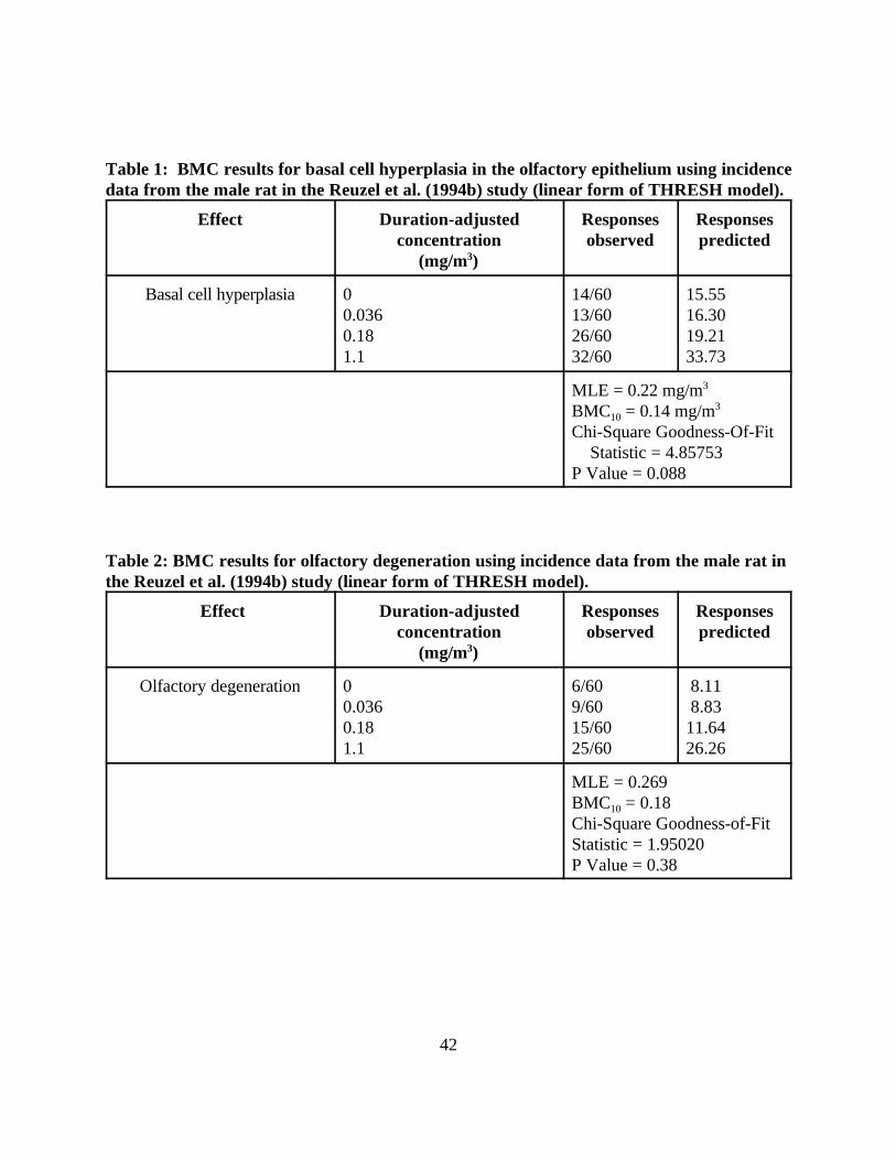
Table 1: BMC results for basal cell hyperplasia in the olfactory epithelium using incidence data from the male rat in the Reuzel et al. (1994b) study (linear form of THRESH model).
Effect Duration-adjusted concentration
(mg/m3)
Responses observed
Responses predicted
Basal cell hyperplasia 0 0.036 0.18 1.1
14/60 13/60 26/60 32/60
15.55 16.30 19.21 33.73
MLE = 0.22 mg/m3
BMC10 = 0.14 mg/m3
Chi-Square Goodness-Of-Fit Statistic = 4.85753
P Value = 0.088
Table 2: BMC results for olfactory degeneration using incidence data from the male rat in the Reuzel et al. (1994b) study (linear form of THRESH model).
Effect Duration-adjusted concentration
(mg/m3)
Responses observed
Responses predicted
Olfactory degeneration 0 0.036 0.18 1.1
6/60 9/60 15/60 25/60
8.11 8.83
11.64 26.26
MLE = 0.269 BMC10 = 0.18 Chi-Square Goodness-of-Fit Statistic = 1.95020 P Value = 0.38
42
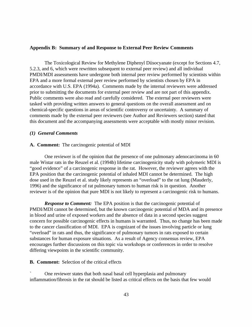
Appendix B: Summary of and Response to External Peer Review Comments
The Toxicological Review for Methylene Diphenyl Diisocyanate (except for Sections 4.7, 5.2.3, and 6, which were rewritten subsequent to external peer review) and all individual PMDI/MDI assessments have undergone both internal peer review performed by scientists within EPA and a more formal external peer review performed by scientists chosen by EPA in accordance with U.S. EPA (1994a). Comments made by the internal reviewers were addressed prior to submitting the documents for external peer review and are not part of this appendix. Public comments were also read and carefully considered. The external peer reviewers were tasked with providing written answers to general questions on the overall assessment and on chemical-specific questions in areas of scientific controversy or uncertainty. A summary of comments made by the external peer reviewers (see Author and Reviewers section) stated that this document and the accompanying assessments were acceptable with mostly minor revision.
(1) General Comments
A. Comment: The carcinogenic potential of MDI
One reviewer is of the opinion that the presence of one pulmonary adenocarcinoma in 60 male Wistar rats in the Reuzel et al. (1994b) lifetime carcinogenicity study with polymeric MDI is “good evidence” of a carcinogenic response in the rat. However, the reviewer agrees with the EPA position that the carcinogenic potential of inhaled MDI cannot be determined. The high dose used in the Reuzel et al. study likely represents an “overload” to the rat lung (Mauderly, 1996) and the significance of rat pulmonary tumors to human risk is in question. Another reviewer is of the opinion that pure MDI is not likely to represent a carcinogenic risk to humans.
Response to Comment: The EPA position is that the carcinogenic potential of PMDI/MDI cannot be determined, but the known carcinogenic potential of MDA and its presence in blood and urine of exposed workers and the absence of data in a second species suggest concern for possible carcinogenic effects in humans is warranted. Thus, no change has been made to the cancer classification of MDI. EPA is cognizant of the issues involving particle or lung “overload” in rats and thus, the significance of pulmonary tumors in rats exposed to certain substances for human exposure situations. As a result of Agency consensus review, EPA encourages further discussions on this topic via workshops or conferences in order to resolve differing viewpoints in the scientific community.
B. Comment: Selection of the critical effects
` One reviewer states that both nasal basal cell hyperplasia and pulmonary inflammation/fibrosis in the rat should be listed as critical effects on the basis that few would
43
argue that the pulmonary lesions are not significant pathology, whereas the significance of the nasal lesions is more questionable. A clear reason why the nasal lesions were selected for RfC derivation should be provided both in the summary and the Toxicological Review document.
Response to Comment: Although the pulmonary lesions indicated by the reviewer are indeed adverse, use of basal cell hyperplasia for RfC derivation results in a more conservative RfC value. Thus, only basal cell hyperplasia of the olfactory epithelium is listed as the critical effect.
C. Comment: The potential for reproductive effects
One reviewer suggests that the amount of data pertaining to potential effects of MDI (and TDI) on reproductive effects is sufficient to eliminate the UF of 3 for reproductive effects.
Response to Comment: EPA feels that the uncertainty factor of 3 should remain inasmuch as the effect of monomeric or polymeric MDI on reproductive endpoints has not been examined in the rat or other laboratory animal species.
D. Comment: Effect of particles with large MMAD versus those with smaller MMAD
One reviewer indicated that the discussion contrasting MDI particles with a MMAD of 11 µm in the workplace and those generated during laboratory exposure of rats should be rewritten to more accurately reflect the relationship between particle size and nasal/pulmonary outcomes.
Response to Comment: As a result of Agency consensus review, EPA agrees that this aspect required a more accurate discussion. Changes to the text reflect that particles as large as or greater than 11 µm are inhalable by humans and can deposit both in the deep lung and nasal tract.
E. Comment: Confidence and weight-of-evidence statements
One reviewer indicated that confidence and weight-of-evidence statements inadequately describe the lack of analytical characterization of the MDI exposure atmospheres in the key publications. A second reviewer is of the opinion that confidence in the RfC should be lowered because of lack of data from a second species.
Response to Comment: EPA has expanded discussion of the confounding factors in analytical characterization of MDI in the key studies and placed in better context those aspects of the exposure generation techniques, particle sizes, and chemical nature (or lack thereof) of the test material in appropriate areas of text in both the summary and Toxicological Review. With regard to the request to lower the confidence in the RfC, there is little expectation that the mouse or another rodent species would be a better surrogate for humans than the rat for nasal endpoints.
44
Furthermore, (1) a UF of 3 for interspecies variation has already been included and (2) the presence of PMDI/MDI in ambient environments has not been documented. Thus, the total UF of 100 is believed to be sufficiently conservative.
F. Comment: Discussion of the derivation and application of the RDDR value
One reviewer urged that the calculation of the RDDR values be made transparent to the reader. At a minimum, the RDDR printout of the computer program be included as an appendix.
Response to Comment: A printout of the RDDR calculations is included as Appendix C. Although the printout lists the calculated RDDR values, the reader is referred to U.S. EPA (1994c) for a more complete discussion of the definition of the RDDR, how it is calculated, and how it is applied. Expanded discussion concerning the RDDR has been included in the Toxicological Review and, to a lesser extent, in the RfC summary sheet. In prior drafts of U.S. EPA (1994c), empirically derived and extrapolated data were in tabular form. Comments received indicated the need to computerize the RDDR calculation. The software for the calculation is included with the U.S. EPA (1994c) document.
(2) Comments on Chemical-Specific Questions
Comment 1: Is the RfC protective for asthma?
One reviewer states that with a UF of 100 and dosimetric adjustments it is very likely that the RfC is sufficiently protective for asthma. However, it is doubtful that nasal lesions and asthma have a common mechanism. The document needs to clearly state these points and indicate there is no information on which to base an RfC that will totally predict prevention of asthma.
Response to Comment: EPA agrees that it is doubtful that the nasal lesions described (e.g., basal cell hyperplasia and olfactory degeneration) and asthma share a common mechanism. However, deposition of PMDI/MDI in the nasal tract offers a means by which antigenic determinants responsible for PMDI/MDI-induced asthma can be generated. The RfC is considered by EPA to be protective against PMDI/MDI-induced asthma, but may not be totally protective for those individuals who are already sensitized to this material.
Comment 2: Lack of analytical characterization of exposure atmospheres
One reviewer states that the causative agent(s) has(have) not been identified; exposure is based on total mass.
Response to Comment: EPA agrees that the lack of analytical characterization is a data limitation and has responded to the reviewer’s concerns by providing additional text on this issue.
45
However, as PMDI is the mixture used in occupational foaming applications, it is also the relevant complex mixture, upon release to ambient air, by which effects in ambient exposure environments can be gauged. Thus, the conclusions reached for noncancer and cancer effects relate to the mixture. For this reason, text references are to PMDI/MDI to avoid confusion that could result if only the term MDI or monomeric MDI were used.
Comment 3: The native state of in vivo MDA.
One reviewer suggests that the text clarify, where appropriate, whether MDA in body fluids is present in free or conjugated form. There is a need to distinguish between hydrolyzed and nonhydrolyzed samples when reporting MDA levels.
Response to Comment: EPA agrees that the discussion of sampling MDA in urine, for example, should distinguish between hydrolyzed and non-hydrolyzed samples. The text has been reviewed and the reviewer’s suggestion adopted.
Comment 4: Lack of data for MDA in exposure atmospheres in animal studies
One reviewer suggests that the lack of data on MDA in the exposure atmosphere in the Hoymann et al. (1995) study should be cited as an important area in analytical characterization.
Response to Comment: EPA agrees that mention should be made in appropriate areas of the text that MDA was not measured. Accordingly, the text has been revised to include this aspect of the analytical results.
46
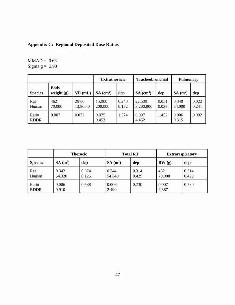
Appendix C: Regional Deposited Dose Ratios
MMAD = 0.68 Sigma g = 2.93
Extrathoracic Tracheobronchial Pulmonary
Species Body weight (g) VE (mL) SA (cm2) dep SA (cm2) dep SA (m2) dep
Rat Human
462 70,000
297.6 13,800.0
15.000 200.000
0.240 0.152
22.500 3,200.000
0.051 0.035
0.340 54.000
0.022 0.241
Ratio RDDR
0.007 0.022 0.075 0.453
1.574 0.007 4.452
1.452 0.006 0.315
0.092
Thoracic Total RT Extrarespiratory
Species SA (m2) dep SA (m2) dep BW (g) dep
Rat Human
0.342 54.320
0.074 0.125
0.344 54.340
0.314 0.429
462 70,000
0.314 0.429
Ratio RDDR
0.006 0.910
0.588 0.006 2.490
0.730 0.007 2.387
0.730
47