Embed Size (px)
Citation preview

THE JDURNAI. OF BIOLOGICAL CHEMISTRY Vul. 25fi. No 6, Issue of March 25, pp. 3067-3076, 1981 Printed in C‘. S. A .
Methylation of Membrane Proteins in Human Erythrocytes IDENTIFICATION AND CHARACTERIZATION OF POLYPEPTIDES METHYLATED IN LYSED CELLS*
(Received for publication, August 21, 1980, and in revised form, October 30, 1980)
Thomas C. Terwilligert and Steven Clarke3 From the Department of Chemistry and the Molecular Biology Institute, University of California, Los Angeles, California 90024
An in vitro system was developed for studying pro- tein methylation reactions in human red blood cells. Packed erythrocytes were lysed by freeze-thawing in the presence of S-adeno~yl[methyZ-~H]methionine. Spe- cific incorporation of base-labile methyl groups into the band 3 anion transport protein and the major sial- oglycoprotein (glycophorin, periodic acid-Schiff re- agent-1) was demonstrated by dodecyl sulfate gel elec- trophoresis at pH 2.4, selective extractions with Triton X-100 and lithium diiodosalicylate, and protease sen- sitivity. Two other unidentified intrinsic membrane proteins with M, = 96,000 and 23,500 were also meth- ylated. Little radioactivity was incorporated into mem- brane proteins when membranes were incubated with S-adeno~yl-L-[methyZ-~H]methionine in the absence of cytosol. No evidence was obtained for incorporation of methyl label into extrinsic proteins including bands 1, 2, 2.1,4, 5, 6, or in zone 4.5.
Proteolytic digestion of intact cells and isolated mem- branes revealed that one site of methylation on the band 3 polypeptide may be at the inner surface of the membrane near the junction of the cytoplasmic domain and the membrane domain.
The rates of hydrolysis of the incorporated methyl groups were characterized over a range of pH values. These rates were compared to those of methyl esterified amino acids and peptides, including aspartic acid p- methyl ester which has been isolated from proteolytic digests of methylated erythrocyte membranes (Janson, C. A., and Clarke, S. (1980) J. Biol. Chem. 255, 11640- 11643). We find that the rates of base-catalyzed hydrol- ysis of &methyl esters of aspartic acid and y-methyl esters of glutamic acid are highly sensitive to the pres- ence of substituents on the a-carboxyl and a-amino groups. The rates of hydrolysis of the membrane-incor- porated methyl groups are consistent with those of internal aspartic acid and glutamic acid methyl ester residues.
The reversible methyl esterification of glutamyl residues on a group of membrane proteins is an essential feat,ure of the mechanism of chemotaxis of enteric bacteria (1,2). It appears that similar protein methylation reactions also occur in many mammalian tissues. In these eucaryotic cells, the existence of
Service Grants GM-26020 and RR-7009 (Biomedical Research Sup- * This research was supported by United States Public Health
port Grant to H. Lewis). The costs of publication of this article were defrayed in part by the payment of page charges. This article must therefore be hereby marked “adoertisement” in accordance with 18 U.S.C. Section 1734 solely to indicate this fact.
$ Recipient of a National Science Foundation Graduate Fellowship. 5 TO whom correspondence should be addressed.
reversible protein methylation systems has generally been surmised from the presence of enzymes which catalyze the transfer of methyl groups from S-adenosylmethionine to ex- ogenous substrate proteins in linkages which may include esters at the COOH-terminal, at aspartyl, or at glutamyl residues (for a recent review, see Ref. 3). Although some functions have been proposed for mammalian carboxyl meth- ylation reactions, their overall physiological role is still poorly understood (3).
Kim (4) has shown that the cytosol of human erythrocytes contains an enzymatic activity which catalyzes the incorpo- ration of methyl groups from S-adenosylmethionine into base- labile linkages on ovalbumin and other proteins. Using meth- yltransferases purified from calf brain or rat red blood cells, and using rat erythrocyte membranes as substrates, Galletti et al. ( 5 ) reported that methyl groups co-migrated with bands 3, 4, and 4.5 when membrane polypeptides were fractionated by dodecyl sulfate gel electrophoresis. However, using the same methyltransferases and human erythrocyte membranes as substrates, they found that methyl groups co-migrated only with band 4.5 and the major sialoglycoprotein (6). Preliminary studies have also indicated that radioactive methyl groups co- migrate with band 4.5 and the major sialoglycoprotein after incubation of intact erythrocytes with [methyZ-:’H]methionine (7).
In order to more precisely identify and characterize poten- tial human red blood cell membrane methyl acceptor species, we have developed an in uitro system where S-adenosylmeth- ionine-dependent methylation reactions can be performed in lysed packed red blood cells under conditions where the concentrations of enzymes, protein substrates, and small molecule effectors can be close to those of in uiuo conditions. In these studies, we have utilized a pH 2.4 electrophoretic system which minimizes hydrolysis and have used several independent criteria to identify methylated species. We report here the methylation of band 3 in human erythrocytes and confirm the previous suggestion (6 ,7) that the major sialogly- coprotein is also methylated in these cells.
Additionally, in this work we have characterized the rates of hydrolysis of the incorporated methyl groups over a range of pH values and have compared them to the rates of hydrol- ysis of methyl esterified peptides and amino acids. We report here the fist evidence that the hydrolysis of methylated membrane proteins proceeds at rates consistent with those of esterified internal aspartyl and glutamyl residues. These re- sults support the recent demonstration of [methyl-”Hlaspartic acid ,&methyl ester in proteolytic digests of methylated red blood cell membranes (8).
EXPERIMENTAL PROCEDURES
Preparation of Methylated Membrane Proteins-Red cells were obtained from recently outdated blood (UCLA Blood Bank) and were
3067

3068 Protein Methylation in Erythrocyte Membranes
washed three times in 150 mM NaCI, 5 mM sodium phosphate, pH 8.0. Unsealed ghost membranes were prepared from washed cells by hypotonic lysis in 5 mM sodium phosphate, pH 8.0, as described by Steck and Kant (9).
Except as otherwise noted, methylation reactions were performed by incubating lysed packed red cells with S-adenosyl-~-[methyl-~H]- methionine. Typically, 1.0 ml of packed, washed erythrocytes were mixed with 0.1 ml of 40 p~ S-adenosyl-L-[methyl-.'H]methionine (33 Ci/mmol in H2S04 at pH 2.5 to 3.5, Amersham). Cells were lysed by freezing a t -70°C in dry ice/isopropanol, and were then incubated on a 37°C shaking water bath for 35 min. In this way, cell constituents were maintained at near-physiological levels and the radioactive substrate was accessible to the cell interior.
["Hlmethylated erythrocyte membranes were prepared from the incubation mixture by an initial dilution with 30 volumes of 5 mM sodium phosphate, pH 8.0, for 1 min a t 2°C to insure hemoglobin release from the membrane (10). This solution was then diluted with 4 volumes (compared to the original incubation) of 46 mM sodium citrate, pH 5.4, to reduce the pH to 6.8 and minimize hydrolysis of methyl ester linkages. Membranes were collected by centrifugation for 10 min a t 22,000 X g a t 2°C in a Sorvall SS-34 rotor. The supernatant and the hard "button" (9) underneath the cells were removed and the entire washing procedure was repeated four addi- tional times. The final pellet, a t a protein concentration of -3.3 mg/ ml in 4.3 mM NaP04, 5.3 mM Na citrate, pH 6.8 was frozen at -70'C. Approximately 0.5 ml of membranes were obtained from 1 ml of packed cells.
Separation of Polypeptides by Dodecyl Sulfate Gel Electrophore- sis-Electrophoresis in polyacrylamide gels was performed in the 50 mM sodium phosphate, pH 2.4, 1% sodium dodecyl sulfate buffer system described by Fairbanks and Avruch (11). The low pH of this gel system minimized ester hydrolysis during electrophoresis. Samples were prepared in 25 mM sodium phosphate, pH 2.4, 2% sodium dode- cy1 sulfate, 30 mM P-mercaptoethanol, 8% glycerol, and 0.03% pyronin Y and heated 3 min a t 100°C. For the gels shown in Figs. 1-3, samples were electrophoresed overnight (60 V, 25 mA) on 3-mm thick slab gels containing 10% acrylamide and 0.34% N,N'-methylene bisacryl- amide in an apparatus similar to that described by Studier (12) until the pyronin Y tracker dye had migrated 12-14 cm. For the gel shown in Fig. 4, samples were electrophoresed for 3 h (75 V, 90 mA) on 1.5- mm thick slab gels containing 7.5% acrylamide and 0.26% N,N'- methylene bisacrylamide until the tracker dye had migrated 10 cm. Marker proteins (Bio-Rad Laboratories) were electrophoresed in parallel lanes and included myosin (M, = 200,000), P-galactosidase (M, = 117,000), phosphorylase (Mr = 94,000), serum albumin (M, = 68,000), ovalbumin (M, = 43,000). carbonic anhydrase (M, = 29,000). soybean trypsin inhibitor (M, = 21,500), and lysozyme (M, = 14,300).
Coomassie blue and periodic acid-Schiff reagent staining reactions were carried out according to Fairbanks et al. (131, except that sodium arsenite was replaced by 10% acetic acid in the PAS' procedure. After staining, gels were scanned with a Joyce-Loebl microdensitometer using unfiltered white light.
Detection of Radiolabeled Polypeptides-Two methods were used to measure the incorporation of radioactivity into polypeptides sep- arated by gel electrophoresis. Fluorography of Coomassie-stained gels using sodium salicylate was carried out as described by Chamberlain (14). Alternatively, wet stained gels were sliced into 1- or 2-mm sections for direct counting. Total radioactivity was measured by incubating gel slices a t 30OC for 18 h in 6 ml of a solution of 10% Protosol (New England Nuclear) in Econofluor (New England Nu- clear). Counts were corrected for quenching using an internal standard method. Base-labile radioactivity in gel slices was determined in parallel experiments by alkaline hydrolysis in 0.15 ml of 2 N NaOH at 25°C for 2 h. After hydrolysis, proteins were precipitated with 1.5 ml of 208 trichloroacetic acid and an aliquot of the supernatant (after centrifugation a t 9OOO X g for 4 min) was counted for radioactivity in 10 ml of Handifluor (Mallinckrodt). This figure, corrected for quench- ing and the volume counted, was taken to be the base-labile radio- activity. Base-stable radioactivity was determined by counting the pellet fraction in 1.5 ml of 16% Protosol in Econofluor after an 18-h incubation at 30°C.
Protein Determinations-Protein concentrations were determined by a modification of the Lowry protocol using bovine serum albumin as a standard (15). Samples containing lithium diiodosalicylate (which produced a deep blue color, interfering with the assay) were precipi- tated twice with ice-cold acetone before analysis. Samples containing
' The abbreviation used is: PAS, periodic acid-Schiff reagent.
Triton X-100 were centrifuged a t 9000 X g for 3 min immediat,ely prior to spectrophotometry.
Fractionation of Membrane Proteins with Triton X-100 and Lith- ium Diiodosalicylate-Membranes were extracted with these com- pounds essentially as described by Yu et al. (16) and Steck and YU (17). T o one volume of packed methylated membranes (at 3-4 mg of protein/&) was added four volumes of a solution containing 5 mM sodium phosphate, pH 6.8, and either 0.28 (v/v) Triton X-100 or 40 mM lithium diiodosalicylate. Both of these compounds were obtained from Sigma. The solutions were incubated a t 0°C for 40 min with occasional vortexing and were then centrifuged a t 32,000 x g for 30 min in a Sorvall SS-34 rotor at 2-4°C. After centrifugation, the supernatants (containing the extracted proteins) were removed, re- centrifuged, and frozen a t -70°C. The pellets were rediluted to the original volume with 5 mM NaP04, pH 6.8, vortexed, and recentri- fuged. The supernatants from this final washing were discarded and the pellets (containing the membrane fraction) were frozen a t - 70°C.
Proteolytic Digestion-Chymotryptic and tryptic digestion of ghost membrane proteins was performed essentially as described by Rao and Reithmeier (18). ["Hlmethylated ghosts were incubated for 1 h a t 0°C in 16 pg/ml of diphenylcarbamyl chloride-treated bovine pancreatic trypsin (Sigma, Type XI) or for 2 h a t 25°C in 200 pg/ml of tosyl-L-lysine chloromethyl ketone-treated bovine pancreatic a- chymotrypsin (Sigma, Type VII). Reactions were terminated by the addition of phenylmethylsulfonyl fluoride to a final concentration of 3 mM, followed by incubation at 0°C for 30 min.
Intact red blood cells were digested with proteases under conditions similar to those described by Drickamer (19). Washed cells in 150 mM NaCI, 5 mM sodium phosphate, pH 8.0, were incubated at 37°C for 3 h with either 5 pg/ml of trypsin or 250 pg/ml of chymotrypsin. Reactions were t.erminated as above by the addition of phenylmeth- ylsulfonyl fluoride to a final concentration of 2 mM. The cells were then washed three times in 150 mM NaCI, 5 mM sodium phosphate, pH 6.8, and used immediately. Control cells were taken through the entire procedure without protease addition.
Determination of Hydrolysis Rate of PHIMethylated Mem- branes-A 10-pl aliquot of [3H]methylated membranes (3-4 mg of protein/ml, 340,000 cpm/ml) in 4.3 mM Na phosphate, 5.3 mM Na citrate, pH 6.8, was diluted into 200 pl of each of the following buffers at 37°C on a shaking water bath: 0.1 M Na citrate, pH 5.49; 0.1 M Na phosphate, pH 6.50; 0.1 M Na phosphate, pH 7.55; 0.1 M Tris. HCI, pH 8.48: 0.1 M Na glycine, pH 9.50; 0.1 M Na glycine, pH 10.51. At various times, 20 pl aliquots were withdrawn from each incubation mixture and the reaction stopped by dilution into 200 p1 of 0.2 M HC1 at 0°C. Samples were then lyophilized to remove the ['Hlmethanol formed during hydrolysis. The nonvolatile material remaining was taken up in 200 pl of H 2 0 and 2 ml of Handifluor and counted by liquid scintillation spectrophotometry. Similar experiments were performed with acid hydrolysis in 6 M HC1 at 37°C; in this case, the reaction was quenched by a 10-fold dilution of the incubation mixture in water at 0°C. Control experiments showed that the rate of hydrolysis in 0.1- 1.0 M HCI a t 0°C was less than 0.2%/h.
Determination of Hydrolysis Rates of Model Compounds-Hy- drolysis rates in strong acid were measured by incubating esters (at concentrations of 0.1-0.5 M ) in 6 M HCl a t 37°C. Aliquots (0.1 ml) were removed a t various times and reacted for 15 min at 25°C with 3.5 ml of 0.6 M NH20H.HCI in 1 M NaOH to form hydroxamic acids from the unhydrolyzed esters (20). The concentration of hydroxamic acids was determined after the addition of 4.0 ml of 10% FeC1.1.6H20 in 0.7 M HCI. The absorbance at 540 nm was read against a water blank. Half-times of hydrolysis were determined from a plot of the logarithm of ester concentration versus time.
Hydrolysis rates in the neutral and alkaline range were determined by the rate of proton release measured in a pH Stat apparatus consisting of a Radiometer Titrigraph, type S B R ~ C , and a Titrator, type TTTlc. Samples were dissolved in 4.0 ml of distilled water in a reaction vessel maintained at 37°C with a circulating water bath, and a stream of purified nitrogen (50 cm'/min) was passed over the surface to prevent uptake of atmospheric carbon dioxide. The amount of H+ produced by ester hydrolysis was calculated from the amount of 0.1 M NaOH added to the solution necessary to maintain the pH. Under conditions where the pH tested was greater than the pK, of the free acid, and where the hydrolysis did not affect the pKn of other functional groups, the rate of proton production was taken as the rate of hydrolysis. Reactions were generally carried out until no additional NaOH was added for several half times, and t m calculated from a semilogarithmic plot of NaOH addition versus time.
At pH values where hydrolysis was slow, the ~ L Z was determined

Protein Methylation in Erythrocyte Membranes 3069
from a plot of the initial rates of hydrolysis versus initial ester concentration. The results were corrected for the nonstoichiometric proton release which occurred in compounds such as leucine methyl ester where the pK, of the amino group was raised upon hydrolysis.
Methyl propionate was obtained from Aldrich, L-glutamic acid y- methyl ester, L-leucine methyl ester, HCl, and L-proline methyl ester. HC1 were obtained from Sigma, and L-aspartic acid P-methyl ester. HCl was obtained from Vega.
N-Benzoyl-L-glutamic acid y-methyl ester and N-benzoyl-L-aspar- tic acid P-methyl ester were synthesized by a procedure similar to that of Vogel (21). The amino acid ester (1.5 mmol) was dissolved in 5 ml of 10% sodium bicarbonate and 3.6 mmol of benzoyl chloride (Aldrich) were added. The mixture was incubated for 1 h at 25°C with stirring. Excess benzoic acid was removed by acidification to pH 2 with HCl. The benzoyl derivatives were crystallized from the aqueous phase at 4°C. Titration of each of these compounds revealed only one equivalent group with a pK, of -3.4; infrared spectra were consistent with that of the benzoylated amino acid methyl ester. N-Acetyl-L- leucine methyl ester was prepared by reacting 1.2 mmol of L-leucine methyl ester. HCl with 5.3 mmol of acetic anhydride (Mallinckrodt) in 5 ml of 10% sodium bicarbonate for 30 min at 25°C. The products were lyophilized, extracted with chloroform, and the insoluble mate- rial (containing NaCl and acetic acid) was used directly in titration studies.
The peptides N-benzoyl-L-glutamyl y-methyl ester glycylamide and N-benzoyl-L-aspartyl P-methyl ester glycylamide were synthe- sized from the benzoyl esters prepared above and glycylamide using a water soluble carbodiimide, l-ethyl-3-(3-dimethylaminopropyl)-car- bodiimide. HC1 (Sigma), as described by Hoare and Koshland (22). In a typical synthesis, 100 pmol of the benzoyl ester, 3000 pmol of glycylamide.HC1 (Sigma), and 200 pmol of the carbodiimide were dissolved in 4 ml of distilled water and the pH maintained at or below 4.75 with a pH Stat apparatus for 2 h at room temperature. The aspartyl peptide was purified from the reaction mixture (dried by flash evaporation at 60°C) by extraction into 5 ml of 2:l (v/v) chloroform/methanol. After removal of solvent by flash evaporation, the peptide was crystallized twice from a minimal volume of water at 4°C. The glutamyl peptide was extracted from the flash evaporated reaction mixture into 5 ml of chloroform. After removal of solvent as above, the peptide was dissolved in water and passed over an ion retardation resin (AG11A8, Bio-Rad Laboratories) to remove charged species. The purity of the products was confirmed by the absence of titratable groups, by stoichiometric recovery of amino acids after hydrolysis in 6 M HC1 for 18 h at 108°C (determined on a Beckman model 120C amino acid analyzer), and by stoichiometric production of protons upon hydrolysis in mild base.
RESULTS
Protein Methylation in Lysed Human Erythrocytes
Fig. 1 shows the distribution of radioactivity in the polypep- tides of red cell membranes prepared from packed lysed cells incubated with S-adenosyl-~-[methyl-~H]methionine as de- scribed under “Experimental Procedures.” Polypeptides with apparent M, = 96,000, 44,000, and 27,000 incorporated sub- stantial quantities of [:3H]methyl groups. The peak of radio- activity at 96,000 daltons co-migrated with the Coomassie blue-staining band 3 region. No Coomassie blue-staining bands co-migrated with the peak of radioactivity at 44,000 daltons. This radioactive peak did coincide, however, with the major PAS staining polypeptide. The peak of radioactivity at -27,000 daltons was not associated with any of the major Coomassie blue- or PAS-staining polypeptides.
AS Table I shows, more than 95% of the total radioactivity incorporated into the red cell membrane is labile under mildly basic conditions (pH 9, 22”C, 4.5 h). The cytosolic fraction, reported to contain a protein methyltransferase (4), was re- quired for alkali-labile protein methylation; when membranes were incubated directly with S-adenosyl-~-[’H-methyl]methi- onine the incorporation of radioactivity into alkali-labile link- ages was greatly diminished while base-stable radioactivity persisted. The latter radioactivity might be accounted for by phospholipid methylation (23). Assuming 7.4 X 10“’ mg of
I 1.2 3
2 4 6 8 10 Gel Migration (cm)
FIG. 1. Membrane protein methylation in lysed packed red cells incubated with 3.6 pM S-aden~syl-L-[rnethyl-~H]methio- nine. [”HIMethylated membranes (60 pg of protein prepared as described under “Experimental Procedures”) were denatured in 2% sodium dodecyl sulfate at pH 2.4 and electrophoresed in 10% poly- acrylamide gels. Identical gel lanes were stained either for protein with Coomassie brilliant blue or for carbohydrate with the periodic acid-Schiff (PAS) reagent. Total radioactivity was determined in 1- mm wet gel slices from the Coomassie-stained lane as described. Coomassie bands of major erythrocyte membrane proteins are iden- tified by number (13). The position of residual hemoglobin chains (Hb) and the tracker dye (pyronin Y) are indicated. Molecular weights of the radiolabeled polypeptides were determined from the migration positions of molecular weight standards run in parallel gel lanes.
membrane protein/red cell (lo), -38,000 methyl groups can be incorporated into base-labile linkages/red cell membrane. This is a minimum estimate of the number of base-labile methyl groups which can be incorporated because there is some isotopic dilution of the S-adenosyl-L-[”H-methyllmethi- onine by endogenous S-adenosylmethionine in red cells (7) and because some proteins may already be partially or fully methylated.
In the experiments performed here, newly outdated human red blood cells were used. However, control experiments with freshly drawn human erythrocytes gave identical results.
Identification of Polypeptides Methylated in Vitro
Selective Extractions with Lithium Diiodosalicylate and Triton X-100”To more precisely identify the radioactive polypeptides which had apparent M , = 96,000 and 44,000 in Fig. 1, [‘Hlmethylated membranes were extracted with either the chaotropic agent lithium diiodosalicylate or the mild nonionic detergent Triton X-100. Lithium diiodosalicylate has been shown to dissociate “extrinsic” proteins from the red cell membrane (17), while Triton X-100 has been shown to bind to and solubilize “intrinsic” red cell proteins (16, 24). In each case, the polypeptides remaining attached to the membranes and those which were dissociated were applied in parallel to dodecyl sulfate polyacrylamide gels and were analyzed as in Fig. 1.
Fig. 2 shows the result of extracting [’Hlmethylated mem- branes with 40 mM lithium diiodosalicylate at p H 6.8. As Steck and Yu have reported (17), bands 4, 5 , and 6 were dissociated from the membranes by this treatment, while band 3 and the major sialoglycoprotein were not. Bands 1 and 2 are poorly resolved in this gel system, but are reported to be
3070 Protein Methylation in Erythrocyte Membranes
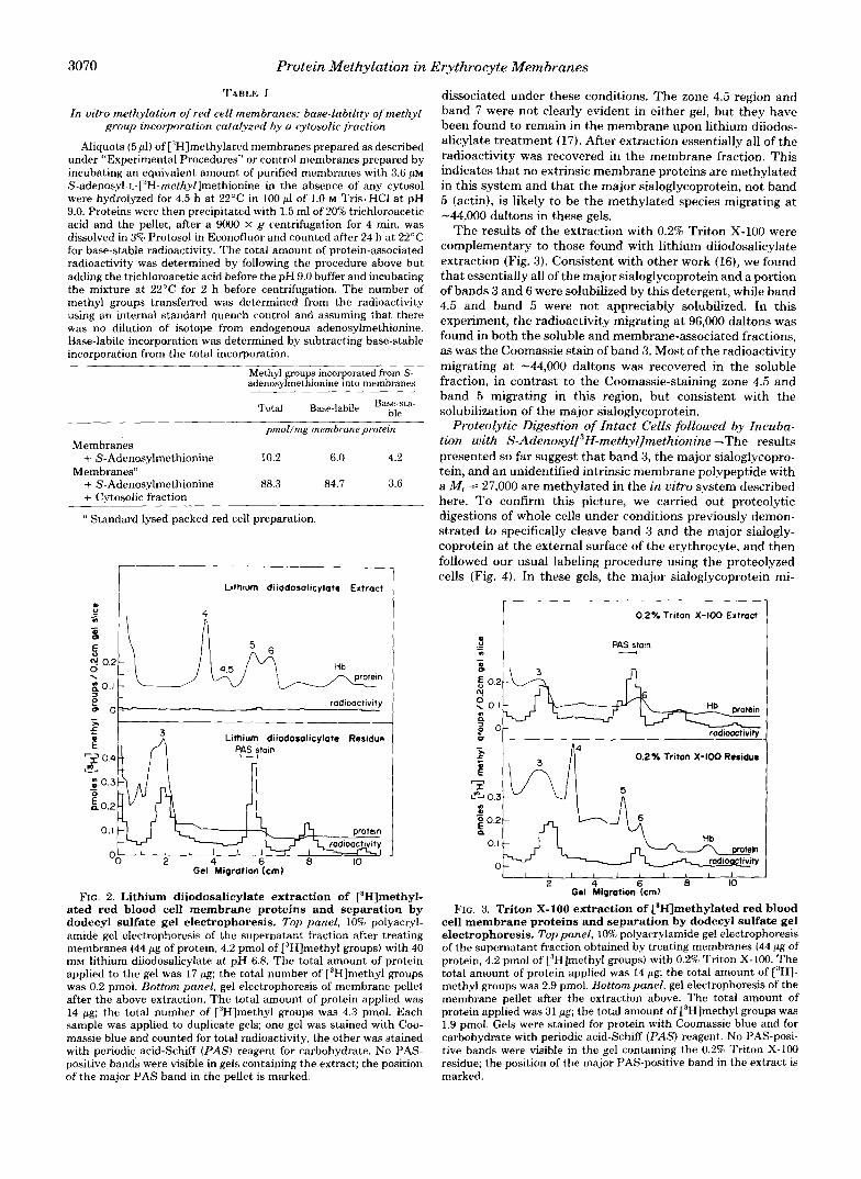
TABLE I In vitro methylation of red cell membranes: base-lability of methyl
group incorporation catalyzed by a cytosolic fraction
Aliquots (5 pl) of ['Hlmethylated membranes prepared as described under "Experimental Procedures" or control membranes prepared by incubating an equivalent amount of purified membranes with 3.6 p~ S-adenosyl-L-["H-methyllmethionine in the absence of any cytosol were hydrolyzed for 4.5 h at 22°C in 100 pl of 1.0 M Tris. HCI at pH 9.0. Proteins were then precipitated with 1.5 ml of 208 trichloroacetic acid and the pellet, after a 9OOO X g centrifugation for 4 min, was dissolved in 3% Protosol in Econofluor and counted after 24 h at 22'C for base-stable radioactivity. The total amount of protein-associated radioactivity was determined by following the procedure above but adding the trichloroacetic acid before the pH 9.0 buffer and incubating the mixture at 22°C for 2 h before centrifugation. The number of methyl groups transferred was determined from the radioactivity using an internal standard quench control and assuming that there was no dilution of isotope from endogenous adenosylmethionine. Base-labile incorporation was determined by subtracting base-stable incorporation from the total incorporation.
adenosylrnethionine into membranes Methyl groups incorporated from S-
Total Base-labile Basbe;esta-
pmol/mg membrane protem Membranes
Membranes" + S-Adenosylmethionine 10.2 6.0 4.2
+ S-Adenosylmethionine 88.3 84.7 3.6 + Cytosolic fraction
' I Standard lysed packed red cell preparation.
*i Lithium diiodosolicylote Extract
g Lithium diiodosalicylate Residue
A 4 0.41 P c s t a l n n Lithium diiodosalicylate Residue
P c s t a l n 0.4 -
A c t / I n
JI 4 b U IU
Gel Migration (cm)
FIG. 2. Lithium diiodosalicylate extraction of VHlmethyl- ated red blood cell membrane proteins and separation by dodecyl sulfate gel electrophoresis. Top panel, 10% polyacryl- amide gel electrophoresis of the supernatant fraction after treating membranes (44 pg of protein, 4.2 pmol of [3H]methyl groups) with 40 mM lithium diiodosalicylate at pH 6.8. The total amount of protein applied to the gel was 17 pg; the total number of ["Hlmethyl groups was 0.2 pmol. Bottom panel, gel electrophoresis of membrane pellet after the above extraction. The total amount of protein applied was 14 pg; the total number of ['Hlmethyl groups was 4.3 pmol. Each sample was applied to duplicate gels; one gel was stained with Coo- massie blue and counted for total radioactivity, the other was stained with periodic acid-Schiff (PAS) reagent for carbohydrate. No PAS- positive bands were visible in gels containing the extract; the position of the major PAS band in the pellet is marked.
dissociated under these conditions. The zone 4.5 region and band 7 were not clearly evident in either gel, but they have been found to remain in the membrane upon lithium diiodos- alicylate treatment (17). After extraction essentially all of the radioactivity was recovered in the membrane fraction. This indicates that no extrinsic membrane proteins are methylated in this system and that the major sialoglycoprotein, not band 5 (actin), is likely to be the methylated species migrating at -44,000 daltons in these gels.
The results of the extraction with 0.2% Triton X-100 were complementary to those found with lithium diiodosalicylate extraction (Fig. 3 ) . Consistent with other work ( X ) , we found that essentially all of the major sialoglycoprotein and a portion of bands 3 and 6 were solubilized by this detergent, while band 4.5 and band 5 were not appreciably solubilized. In this experiment, the radioactivity migrating at 96,000 daltons was found in both the soluble and membrane-associated fractions, as was the Coomassie stain of band 3. Most of the radioactivity migrating at -44,000 daltons was recovered in the soluble fraction, in contrast to the Coomassie-staining zone 4.5 and band 5 migrating in this region, but consistent with the solubilization of the major sialoglycoprotein.
Proteolytic Digestion of Intact Cells followed by Incuba- tion with S-AdenosylfH-methyllmethionine-The results presented so far suggest that band 3, the major sialoglycopro- tein, and an unidentified intrinsic membrane polypeptide with a M, = 27,000 are methylated in the in vitro system described here. To confirm this picture, we carried out proteolytic digestions of whole cells under conditions previously demon- strated to specifically cleave band 3 and the major sialogly- coprotein at the external surface of the erythrocyte, and then followed our usual labeling procedure using the proteolyzed cells (Fig. 4). In these gels, the major sialoglycoprotein mi-
" - s i 0.2Y. Trifon X-100 Extract
PAS stam -
0.2Y. Triton X 4 0 0 Residue
r-7
0.3 I
I I I l l 1 I l l 1 I 2 4 6 a IO
Gel Migrotion (cm)
FIG. 3. Triton X-100 extraction of C3H]methylated red blood cell membrane proteins and separation by dodecyl sulfate gel electrophoresis. Top panel, 10% polyacrylamide gel electrophoresis of the supernatant fraction obtained by treating membranes (44 pg of protein, 4.2 pmol of ["Hlmethyl groups) with 0.2% Triton X-100. The total amount of protein applied was 14 pg; the total amount of ["HI- methyl groups was 2.9 pmol. Bottom panel, gel electrophoresis of the membrane pellet after the extraction above. The total amount of protein applied was 31 pg; the total amount of ["Hlmethyl groups was 1.9 pmol. Gels were stained for protein with Coomassie blue and for carbohydrate with periodic acid-Schiff (PAS) reagent. No PAS-posi- tive bands were visible in the gel containing the 0.2% Triton X-100 residue; the position of the major PAS-positive band in the extract is marked.
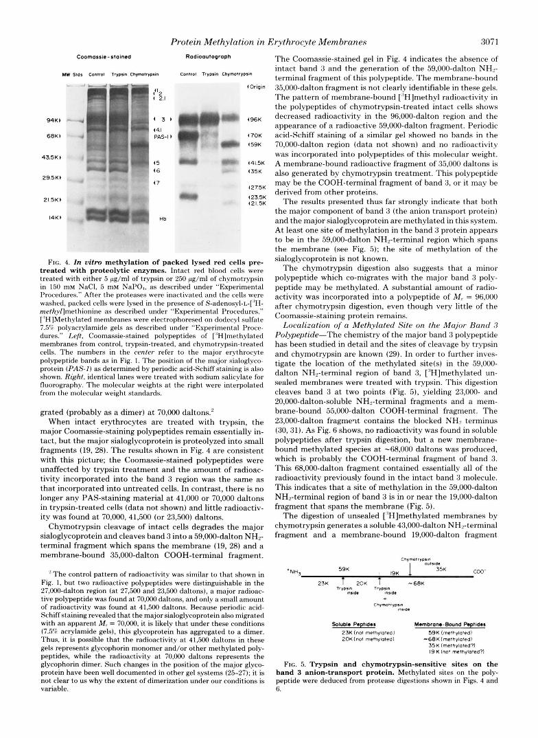
Protein Methylation in Erythrocyte Membranes 3071 Coomassie-stained Radioautograph
MW Stds Control Trypstn Chymotrypsin Control Trypsm Chymotrypsin
9 4 K ) .J
6 8 K 1
, ‘ 3 ’ r-7 (4.1 PAS-I 1
‘1
43.5K 1
29.5K)
21.5K)
(5 ( 6
47
(Origin
4 9 6 K
4 7 0 K
1 (59K
(41.5K
4 35K
( 2 7 5 K
423.5K (21.5K
FIG. 4. In vitro methylation of packed lysed red cells pre- treated with proteolytic enzymes. Intact red blood cells were treated with either 5 pg/ml of trypsin or 250 pg/ml of Chymotrypsin in 150 mM NaCI, 5 mM NaPO,, as described under “Experimental Procedures.” After the proteases were inactivated and the cells were washed, packed cells were lysed in the presence of S-adenosyl-L-[’H- rnethyllmethionine as described under “Experimental Procedures.” [’HIMethylated membranes were electrophoresed on dodecyl sulfate 7.5”; polyacrylamide gels as described under “Experimental Proce- dures.” Left, Coomassie-stained polypeptides of [“Hlmethylated membranes from control, trypsin-treated. and chymotrypsin-treated cells. The numbers in the center refer to the major erythrocyte polypeptide bands as in Fig. 1. The position of the major sialoglyco- protein ( P A S - ] ) as determined by periodic acid-Schiff staining is also shown. Right, identical lanes were treated with sodium salicylate for fluorography. The molecular weights at the right were interpolated from the molecular weight standards.
grated (probably as a dimer) at 70,000 daltons.’ When intact erythrocytes are treated with trypsin, the
major Coomassie-staining polypeptides remain essentially in- tact, but the major sialoglycoprotein is proteolyzed into small fragments (19, 28). The results shown in Fig. 4 are consistent with this picture; the Coomassie-stained polypeptides were unaffected by trypsin treatment and the amount of radioac- tivity incorporated into the band 3 region was the same as that incorporated into untreated cells. In contrast, there is no longer any PAS-staining material a t 41,000 or 70,000 daltons in trypsin-treated cells (data not shown) and little radioactiv- ity was found at 70,000, 41,500 (or 23,500) daltons.
Chymotrypsin cleavage of intact cells degrades the major sialoglycoprotein and cleaves band 3 into a 59,000-dalton NHs- terminal fragment which spans the membrane (19, 28) and a membrane-bound 35,000-dalton COOH-terminal fragment.
” T h e control pattern of radioactivity was similar to that shown in Fig. 1, but two radioactive polypeptides were distinguishable in the 27,000-dalton region (at 27,500 and 23,500 daltons), a major radioac- tive polypeptide was found at 70,000 daltons, and only a small amount of radioactivity was found a t 41,500 daltons. Because periodic acid- Schiff staining revealed that the major sialoglycoprotein also migrated with an apparent M , = 70,000, it is likely that under these conditions (7.5% acrylamide gels), this glycoprotein has aggregated to a dimer. Thus, it is possible that the radioactivity at 41,500 daltons in these gels represents glycophorin monomer and/or other methylated poly- peptides, while the radioactivity a t 70,000 daltons represents the glycophorin dimer. Such changes in the position of the major glyco- protein have been well documented in other gel systems (25-27); it is not clear to us why the extent of dimerization under our conditions is variable.
The Coomassie-stained gel in Fig. 4 indicates the absence of intact band 3 and the generation of the 59,000-dalton NH2- terminal fragment of this polypeptide. The membrane-bound 35,000-dalton fragment is not clearly identifiable in these gels. The pattern of membrane-bound [“Hlmethyl radioactivity in the polypeptides of chymotrypsin-treated intact cells shows decreased radioactivity in the 96,000-dalton region and the appearance of a radioactive 59,000-dalton fragment. Periodic acid-Schiff staining of a similar gel showed no bands in the 70,000-dalton region (data not shown) and no radioactivity was incorporated into polypeptides of this molecular weight. A membrane-bound radioactive fragment of 35,000 daltons is also generated by chymotrypsin treatment. This polypeptide may be the COOH-terminal fragment of band 3, or it may be derived from other proteins.
The results presented thus far strongly indicate that both the major component of band 3 (the anion transport protein) and the major sialoglycoprotein are methylated in this system. At least one site of methylation in the band 3 protein appears to be in the 59,000-dalton NHs-terminal region which spans the membrane (see Fig. 5); the site of methylation of the sialoglycoprotein is not known.
The chymotrypsin digestion also suggests that a minor polypeptide which co-migrates with the major band 3 polv- peptide may be methylated. A substantial amount of radio- activity was incorporated into a polypeptide of M, = 96,000 after chymotrypsin digestion, even though very little of the Coomassie-staining protein remains.
Localization of a Methylated Site on the Major Band 3 Polypeptide-The chemistry of the major band 3 polypeptide has been studied in detail and the sites of cleavage by trypsin and chymotrypsin are known (29). In order to further inves- tigate the location of the methylated site(s) in the 59,000- dalton NH2-terminal region of band 3, [“Hlmethylated un- sealed membranes were treated with trypsin. This digestion cleaves band 3 at two points (Fig. 5), yielding 23,000- and 20,000-dalton-soluble NH2-terminal fragments and a mem- brane-bound 55,000-dalton COOH-terminal fragment. The 23,000-dalton fragmeqt contains the blocked NH2 terminus (30,31). As Fig. 6 shows, no radioactivity was found in soluble polypeptides after trypsin digestion, but a new membrane- bound methylated species a t -68,000 daltons was produced, which is probably the COOH-terminal fragment of band 3. This 68,000-dalton fragment contained essentially all of the radioactivity previously found in the intact band 3 molecule. This indicates that a site of methylation in the 59,000-dalton NH2-terminal region of band 3 is in or near the 19,000-dalton fragment that spans the membrane (Fig. 5).
The digestion of unsealed [‘’Hlrnethylated membranes by chymotrypsin generates a soluble 43,000-dalton NH’-terminal fragment and a membrane-bound 19,000-dalton fragment
Chvmotrvosm w m a e
+NH, 59K I 19K 1 35K COO-
23K T ZOK T - 6 8 K T V V D ~ O ~ TrvDsln
Insla. mrde +
ChvmotryDsm mlnaa
Soluble Peptides Membrane-Bwnd Peptides 23K (not methylatedl 20K (not methylated) -68K(methylaled)
59K (methylatedl
3 5 K (methylated?) 19 K (not memylaled?)
FIG. 5. Trypsin and chymotrypsin-sensitive sites on the band 3 anion-transport protein. Methylated sites on the poly- peptide were deduced from protease digestions shown in Figs. 4 and 6.

3072 Protein Methylation in Erythrocyte Membranes
Control Chymotrypsin-treated Trypsin-treated
Toto1 Soluble Membrone Total Soluble Membrane
i 74
(15 i I 4
kd (25 i
i
blood cell membrane proteins were consistent with the meth- ylation of aspartic (8) or glutamic acid residues, the rates of hydrolysis of ["Hlmethyl groups were compared with those of model compounds. Fig. 7 shows the fraction of radioactivity remaining as a function of time at pH 7.55 a t 37°C for [''HI- methylated membranes prepared as in Fig. 4 from intact and trypsin-treated cells. The data in each case are consistent with the presence of linkages with a t least three rates of hydrolysis ranging from a tlr2 of 3 min to one of 360 min. The half-lives of the three groups of alkali-labile linkages in methylated membranes from trypsin-treated cells are presented as a func- tion of pH in Fig. 8 (left panel).
Comparison of Rates of Hydrolysis of Methylated Mem- branes in Mild Alkali with Those of Small Molecule Methyl Esters-As Fig. 8 shows, the rate of hydrolysis of membrane methyl groups was from one to three orders of magnitude faster in the pH range of 6.5-10.5 than aspartic acid /?-methyl ester, the N-benzoyl derivatives of aspartic acid /?-methyl ester and glutamic acid y-methyl ester, and methyl propionate. The rates of hydrolysis of N-benzoyl-L-aspartyl /?-methyl ester glycylamide, which has its a-amino and a-carboxyl groups in peptide linkages, however, were very similar to those of the bulk of the membrane methyl groups.
FIG. 6. Proteolysis of ['Hlmethylated red cell membranes. Left, fluorograph of a 10% polyacrylamide dodecyl sulfate gel of ["H]methylated membranes prepared by incubating packed, lysed red cells with S-adenosyl-L-[:'H-methyljmethionine (20 pg of protein ap- plied). Molecular weights of the prominent bands given are in kilo- daltons. Center, digestion of control membranes with 0.2 mg/ml of chymotrypsin a t 25'C for 2 h as described under "Experimental Procedures." An aliquot containing 16 pg of protein of the total digest and aliquots of the soluble and membrane fractions obtained by centrifugation a t 100,OOO X g for 10 min corresponding to 100 pg of membrane protein were electrophoresed and fluorographed as above. No radioactivity was detected in the soluble fraction. Right, digestion of control membranes with 16 pg/ml of trypsin for 1 h a t 0°C. Aliquots of the total digest and fractions of soluble and membrane fractions were prepared for fluorography as above. Again, no radioactivity was detected in the soluble fraction.
from the 59,000-dalton NH2-terminal region of band 3 (18,32) (Fig. 5). As Fig. 6 shows, no radioactivity was associated with the soluble cytoplasmic NH2-terminal 43,000-dalton chymo- tryptic fragment or with the 19,000-dalton fragment, although both were clearly visible when the gels were stained with Coomassie (data not shown).
In order to determine whether the absence of radioactivity in the polypeptide fragments of band 3 after chymotrypsin digestion was due to hydrolysis of the radioactive methyl groups or to the production of small peptides, the digestion mixture was applied to a column of Sephadex G-15 in 0.1 M acetic acid. Over 40% of the applied radioactivity was re- covered in peptides smaller than hexaglycine and methanol accounted for less than 35% of the radioactivity (data not shown). Although the recovered peptides could be derived from polypeptides other than band 3, these results suggest that a site of methylation on band 3 is on a small peptide product of chymotrypsin cleavage. A site for this peptide consistent with all of the data presented is at the junction of the 43,000-dalton NH2-terminal fragment and the 19,000-dal- ton membrane-bound fragment (Fig. 5).
Characterization of Residues Methylated in Red Cell Membranes
Rate of Hydrolysis of PHIMethylated Membrane Proteins in Base-In order to determine whether the overall rates of
0.60 o ' m b 4 R k Membranes
0.40 -
-
0.20 - 0
a m .; 010- c e a n a 2 0.05
- 0
a
1 I I I I -
Rbc Membranes from Trypsin-rreared cells
0.20 -
0.10 1 I I I 1 I 0 x) l o o 1 5 0 200 250 xx)
at 37'C pH755 TIME (mid
FIG. 7. Rate of hydrolysis at neutral pH of ['Hlmethylated membranes prepared from red blood cells and trypsin-treated red blood cells. Top panel, rate of hydrolysis of in vitro-labeled. ["Hlmethylated erythrocyte membranes a t pH 7.55 and 37OC. Data were collected as described under "Experimental Procedures." The smooth curve represents the exponential decay of three classes of sites with half-times of hydrolysis of 3, 30, and 360 min and propor- tions of 0.2,0.7, and 0.1, respectively. Bottom panel, rate of hydrolysis of ["Hlmethylated membranes prepared from trypsin-treated cells (cf Fig. 4). The smooth line was calculated for three groups with half- times of hvdrolvsis of 3. 30. and 190 min and proportions of 0.2. 0.3,
hydrolysis of the [:'H]methyl groups incorporated into red and 0.5. respectively. " I . .

Protein Methylation in Erythrocyte Membranes 3073
-9
-0,
-(D
-b
-W
I , I I I I I I n

3074 Protein Methylation in Erythrocyte Membranes
The rates of hydrolysis of both free L-glutamic acid y- methyl ester and its derivative, N-benzoyl-L-glutamyl 7- methyl ester glycylamide, were similar to those of the mem- brane methyl groups with the slowest rates of hydrolysis.
In contrast, N-acetyl-L-leucine methyl ester, chosen to sim- ulate a COOH-terminal methylated amino acid, had a hy- drolysis rate considerably slower than those of the membrane methyl groups.
Comparison of Rates of Hydrolysis of Methylated Mem- branes in 6 M HCl with those of Small Molecule Methyl Esters-The time course of hydrolysis in 6 M HCl of r3H]- methylated membranes prepared from trypsin-treated cells also indicates the presence of linkages with a t least three rates of hydrolysis (ranging from a t 1 / 2 of 3 min to one of 320 min) (Fig. 9). Table I1 gives data for the rates of hydrolysis of small molecule methyl esters in 6 M HC1. These rates (expressed in terms of half-lives) vary from 5 min for methyl propionate to 470 min for L-leucine methyl ester and are consistent with the half-lives of 3-360 min measured for the red cell membrane methyl groups (Figs. 8 and 9).
I.
Rbc Membranes from Trypsin-treated Cells
0.4
.; 0.2
a - t e
o.ozoL
at 37. in 6 M HCI Time (min)
FIG. 9. Rate of hydrolysis in 6 M HCI of [3H]methylated membranes prepared from trypsin-treated red blood cells. For experimental details, see Fig. 7 and “Experimental Procedures.” The smooth curue represents the hydrolysis of three species with half- lives of 3, 60, and 320 min and proportions of 0.25, 0.68, and 0.07, respectively.
TABLE I1 Hydrolysis of methyl esters at 37°C in 6 M HCl
Ester -
t , IL min“
L-Aspartic acid P-methyl ester 108 N-Benzoyl-L-aspartic acid ,@-methyl ester 36 L-Glutamic acid y-methyl ester 16 N-Benzoyl-L-glutamic acid y-methyl ester 10.5 L-Leucine methyl ester 4 70 Methyl propionate 5
‘‘ Determined from a semilogarithmic plot of ester remaining uersus time. Ester concentration was determined by the hydroxylamine/ ferric chloride assay described under “Experimental Procedures.”
DISCUSSION
S-Adenosyl-L ”ethionine-dependent Protein Methylation in Lysed Human Erythrocytes-Kim (4) has described an enzyme from the cytosol of human red cells which catalyzes the transfer of a methyl group from S-adenosyl-L-methionine to exogenous proteins such as ovalbumin and ribonuclease. The methyl group was found to be labile under mildly basic conditions. O’Dea et al. (33) showed that the membrane fraction of erythrocytes contained sites which could be meth- ylated by this enzyme, and that no methyl-accepting proteins were detectable in the cytosol. Galletti et al. (5 , 6 ) have incubated purified methyltransferases with rat and human erythrocyte membranes in order to characterize the methyl- accepting proteins.
We have investigated these methylation reactions under in uitro conditions that paralleled as nearly as possible the conditions in intact red cells. Because biological membranes are generally impermeable to S-adenosylmethionine, it was necessary to disrupt the red cell membrane to allow the S- adenosyl-~-[~H-methyZlmethio~~ine access to the cytosolic en- zyme. This was done in the present study by a freeze-thaw lysis. To keep the concentration of cytoplasmic components, including the methyltransferase(s), methylesterases, activat- ing molecules, and inhibiting molecules as close to physiolog- ical levels as possible, we employed packed red cells. These cells have a very low extracellular volume and were only minimally diluted with the addition of isotopic S-adenosyl-L- methionine.
Identification of Polypeptides Methylated in Human Erythrocyte Membranes in Vitro-The identification of po- tential substrates for the erythrocyte protein methyltransfer- ase is a first step in elucidating the role of this enzymatic system in eucaryotic cells. The human erythrocyte membrane proteins are well characterized and the functions of several have been identified. The bulk of the band 3 polypeptide is homogeneous with respect to its amino acid sequence and other properties (29), spans the membrane, and functions in the transport of anions in intact cells (34). The major sialogly- coprotein (PAS-1, glycophorin) also spans the membrane (35); its function is as yet unclear. The present work shows that both of these polypeptides are methylated in a broken cell system. In the band 3 polypeptide, a t least one site of meth- ylation appears to be at the junction between the cytoplasmic NH2-terminal 43,000-dalton region and the 19,000-dalton in- ternal hydrophobic segment. We have not yet determined whether any other sites of methylation on band 3 or those on the major sialoglycoprotein are internal or external to the erythrocyte and, hence, whether these sites are in fact avail- able to the cytoplasmic protein methyltransferase. As protein methyltransferase activity is very low in the blood plasma (36), external red blood cell sites of methylation are likely to be artifactual.
We have also shown that two minor polypeptide compo- nents of human red cell membranes are methylated in this in vitro system. One of these is a peptide that co-migrates on dodecyl sulfate gel electrophoresis with the band 3 anion transport protein (96,000 daltons), but which can be distin- guished because it is not removed by extracellular chymotryp- sin treatment. Two others migrate a t polypeptide M , = 23,000 and 27,000 and appear to be intrinsic membrane proteins as they can be extracted with Triton X-100, but not with lithium diidosalicylate.
No evidence has been found in this broken cell system for methylation of bands 1 and 2 (spectrin), band 2.1 (ankyrin), band 4, zone 4.5 (which contains the glucose transport pro- tein), band 5 (actin), or band 6 (glyceraldehyde-3-phosphate dehydrogenase). The lack of methylation of these proteins

Protein Methylation in Erythrocyte Membranes 3075
shows that there is specificity in the protein methyltransferase reaction(s).
Galletti et al. (6) have concluded that the major sialogly- coprotein (glycophorin A) and band (zone) 4.5 are the predom- inant methyl acceptors in human erythrocyte membranes when methylation reactions are catalyzed by a purified meth- yltransferase. The results of the present study, in which red blood cell membranes were methylated in a broken cell system with cytosolic components at near-physiological levels, also indicate that the major sialoglycoprotein is a methyl group acceptor. However, zone 4.5 was not found to be methylated in our study. Furthermore, we present evidence here that the primary component of band 3, known to be involved in anion transport, is also a methyl acceptor.
Characterization of the Chemical Site of Methylation-It has recently been shown that at least some of the sites of base-labile methylation in human red blood cell membrane proteins are a t aspartic acid residues (8). The rate of hydrol- ysis of the bulk of the base-labile methyl groups, however, has been found to be much more rapid than that of aspartic acid /?-methyl ester (Figs. 7 and 8) (Refs. 5, 37, 38). In order to determine whether these rapidly hydrolyzable membrane methyl groups might also be carboxylic acid esters, we com- pared the rates of hydrolysis of the membrane methyl groups with those of aspartic acid P-methyl ester and glutamic acid y-methyl ester and their derivatives in which the a-carboxyl and a-amino groups are involved in peptide bonds.
We find that glutamic acid y-methyl ester and its derivative N-benzoyl glutamyl y-methyl ester glycylamide have base hydrolysis rates similar to those of the slowest group of membrane methyl groups. Additionally, while aspartic acid /3-methyl ester has hydrolysis rates considerably slower than those of the membrane methyl groups, N-benzoylaspartyl /?- methyl ester glycylamide has rates of hydrolysis similar to those of the bulk of the membrane methyl groups. Thus, the rates of hydrolysis of the methylated membrane sites are consistent with the methylation of internal aspartic acid and glutamic acid residues, although direct evidence has only been found for aspartyl residues (8).
One explanation for the rapid hydrolysis of internal aspartic acid residues is suggested by the studies of Bernhard et al. (39). They found that the rate of base-catalyzed hydrolysis of /?-benzyl esters of N-carbobenzyloxyaspartyl peptides was up to 107-fold greater than that for benzyl propionate. They demonstrated that the rapid reaction proceeded via an imine intermediate involving the amide nitrogen of the peptide bond on the carboxyl side of the aspartyl residue. Such a large rate enhancement was not found for similar glutamyl peptides (40, 41).
Although it is likely that demethylating enzymes play a role in the hydrolysis of eucaryotic protein-bound methyl esters, it is possible that a rapid spontaneous hydrolysis via an imine intermediate may also be important. If this is the case, such an intermediate can be potentially hydrolyzed to give a free a-carboxyl group and a peptide linkage of the side chain carboxyl group to the rest of the polypeptide chain. Whether this occurs in uiuo has not yet been established. Additionally, the rate of hydrolysis of the methyl esters would be expected to be highly dependent upon the conformation of the protein at the methylated site.
Relation of in Vitro Methylation to Physiological Methyl- ation Reactions in Intact Cells-Because the methylation reactions studied here occurred in a broken cell system, it is possible that qualitative and quantitative differences from the results obtained here occur in in vivo methylation. In the broken cell system, cytoplasmic methyltransferases have ac- cess to polypeptide residues on the exterior membrane surface.
Although we have tentatively shown that at least one site of methylation of band 3 is at the internal surface of the mem- brane, it is possible that some of the methylated sites observed in this study and previously (5,6) are not methylated in intact cells. In a preliminary study, Kim et al. (7) have tentatively shown that the pattern of methylation of red cell membrane proteins in vivo is similar to the in vitro pattern. If the same residues in each polypeptide are methylated in uiuo and in uitro, then the broken cell system will be an appropriate model for studying the physiological role of this covalent modification.
Acknowledgments-We offer special thanks to Prof. David Sigman for many helpful and stimulating discussions and to Prof. Sidney Bernhard for bringing the lability of p-esters of aspartyl peptides to our attention. Densitometry was performed at the facility of the Department of Biology, UCLA, aided by Mrs. June Baumer. We are grateful to Prof. Stephen Feig for his help in supplying red blood cells.
Note Added in Proof-We have recently shown that although band 3 is in fact methylated in intact cells, the major sialoglycoprotein is not (C. Freitag and S. Clarke, manuscript in preparation).
REFERENCES
1. Springer, M. S., Goy, M. F., and Adler, J. (1979) Nature 280,279-
2. Koshland, D. E., Jr. (1980) Bacterial Chemotaxis as a Model
3. Paik, W. K., and Kim, S. (1980) Protein Methylation pp. 202-231,
4. Kim, S. (1974) Arch. Biochem. Biophys. 161,652-657 5. Galletti, P., Paik, W. K., and Kim, S. (1978) Biochemistry 17,
6. Galletti, P., Paik, W. K., and Kim, S. (1979) Eur. J. Biochem. 97,
7. Kim, S., Galletti, P., and Paik, W. K. (1980) J. Biol. Chem. 255,
8. Janson, C. A,, and Clarke, S . (1980) J. Biol. Chem. 255, 11640-
9. Steck, T. L., and Kant, J. A. (1974) Methods Enzymol. 31, 172-
10. Dodge, J. T., Mitchell, C., and Hanahan, D. J. (1963) Arch.
11. Fairbanks, G., and Avruch, J . (1972) J . Supramol. Struct. 1, 66-
12. Studier, F. W. (1973) J. Mol. Biol. 79, 237-248 13. Fairbanks, G., Steck, T. L., and Wallach, D. F. H. (1971) Bio-
14. Chamberlain, J . P . (1979) Anal. Biochem. 98, 132-135 15. Leggett Bailey, J . (1967) Techniques in Protein Chemistry p. 340,
16. Yu, J., Fischman, D. A., and Steck, T. L. (1973) J. Suprumol.
17. Steck, T. L., and Yu, J. (1973) J. Supramol. Struct. 1, 220-232 18. Rao, A., and Reithmeier, R. A. F. (1979) J. Biol. Chem. 254,
19. Drickamer, L. K. (1976) J. Biol. Chem. 251, 5115-5123 20. Hestrin, S. (1949) J. Biol. Chem. 180, 249-261 21. Vogel, A. I. (1956) A Textbook of Practical Organic Chemistry,
3rd Ed, p. 436, John Wiley, New York 22. Hoare, D. G., and Koshland, D. E., Jr . (1967) J. Biol. Chem. 242,
2447-2453 23. Hirata, F., and Axelrod, J . (1978) Nature 275, 219-220 24. Clarke, S. (1975) J. Biol. Chem. 250, 5459-5469 25. Marton, L. S. G., and Garvin, J . E. (1973) Biochem. Biophys. Res.
26. Furthmayr, J., and Marchesi, V. T. (1976) Biochemistry 15,1137-
27. Silverberg, M., Furthmayr, H., and Marchesi, V. T. (1976) Bio-
284
Behavioral System, Raven Press, New York
John Wiley, New York
4272-4276
221-227
338-341
11643
180
Biochem. Biophys. 100, 119-130
75
chemistry 10, 2606-2617
American Elsevier, New York
Struct. 1, 233-248
6144-6150
Commun. 52, 1457-1462
1144
chemistry 15, 1448-1454 28. Triplett, R. B., and Carraway, K. L. (1972) Biochemistry 11,
2897-2903 29. Steck, T. L. (1978) J. Supramol. Struct. 8, 311-324 30. Steck, T. L., Koziarz, J . J., Singh, M. K., Reddy, G., and Kohler,
H. (1978) Biochemistry 17, 1216-1222
3076 Protein Methylation in Erythrocyte Membranes
31. Drickamer, L. K. (1978) J. Biol. Chem. 253,7242-7248 32. Steck, T. L., Ramos, B., and Strapazon, E. (1976) Biochemistry
33. ODea, R. F., Viveros, 0. H., Acheson, A., Gorman, C., and
34. Cabantchik, 2. I., Knauf, P. A., and Rothstein, A. (1978) Biochim.
35. Marchesi, V. T. (1979) Semin. Hematol. 16, 3-20 36. Kim, S., Wasserman, L., Lew, B., and Paik, W. K. (1975) J.
15, 1154-1161
Axelrod, J. (1978) Biochem. Pharmacol. 27,679-684
Biophys. Acta 515, 239-302
Neurochem. 24,625-629
37. Kim, S., and Paik, W. K. (1976) Experientia 32, 982-984 38. Diliberto, E. J., Jr., and Axelrod, J. (1976) J. Neurochem. 26,
1159-1165 39. Bernhard, S. A., Berger, A., Carter, J . H., Katchalski, E., Sela,
M., and Shalitin, Y. (1962) J . Amer. Chem. SOC. 84, 2421- 2434
40. Fosker, A. P., Hanson, R. W., and Law, H. D. (1963) Chem. Ind. 569-570
41. Battersby, A. R., and Robinson, J . C . (1955) J . Chem. SOC. 259- 269
![DNA methylation-linked chromatin accessibility affects ...requires the DNA-methylation reader proteins SUVH2 and SUVH9 [homologs of SU(VAR)3-9], as well as the DDR com-plex (13, 14)](https://img.pdfslide.us/doc/110x75/60e9600d2e83be1a38617ed8/dna-methylation-linked-chromatin-accessibility-affects-requires-the-dna-methylation.jpg)


![Review Histone methylation in DNA repair and clinical ...homology [8, 9]. Besides, the recruitment of NHEJ-related proteins like Ku70, Ku80 and DNA-PKcs and HR-related proteins like](https://img.pdfslide.us/doc/110x75/6082693ddcef7b7cc76793eb/review-histone-methylation-in-dna-repair-and-clinical-homology-8-9-besides.jpg)


![ERYTHROCYTES [RBCs]](https://img.pdfslide.us/doc/110x75/56813dc0550346895da78963/erythrocytes-rbcs-56ea22b2e2743.jpg)