Embed Size (px)
Citation preview

Supplementary information, Data S1 Methods and materials 1
Plasmids 2
The genes encoding full-length human H2A-H2B and CENP-A-H4 were cloned into pET28K 3
and pETDuet-1 vector to yield (His)6-H2A-H2B/pET28K and (His)6-CENP-A-H4/pETDuet-1 4
plasmids, respectively. The (His)8-CENP-N (CENP-N truncations and CENP-N1-214 mutants) 5
plasmids were constructed by cloning CENP-N into pET-28a (+) vector. The full-length CENP-6
N and CENP-L were cloned into Multi-Bac vector1 pACEBac1 (Invitrogen) with a (His)6-tag 7
fused to its C-terminus and pIDC, respectively. The two plasmids were then fused by Cre-8
LoxP reaction for the expression of CENP-LN complex. For in vivo experiments, we cloned 9
the full-length CENP-N into pEGFP-C2-Linker. Point mutants in CENP-N were generated with 10
a site-directed mutagenesis polymerase Q5 (NEB). All plasmids were sequenced for 11
verification. 12
Protein production 13
The histone plasmids were co-transformed into E. coli Rosetta2 (DE3) cells and plated on agar 14
containing 50 μg/mL kanamycin and 100 μg/mL ampicillin followed by grown overnight at 15
37 °C. Picked colonies were inoculated into 5 mL starter culture of Luria-Bertani (LB) medium 16
and grown with shaking for 3 hours at 37 °C. The starter culture was transferred into 50 mL LB 17
medium and incubated at 37 °C for another 3 hours. Fifteen-milliliter of the second culture was 18
transferred into 1L LB medium and incubated at 37 °C to an OD600 of ~0.5. The culture was 19
induced with 0.4 mM isopropyl β-D-1-thiogalactopyranoside (IPTG) and incubated at 37 °C 20
for 12~16 h. Cells were harvested and lysed using high pressure cracker in lysis buffer A (20 21
mM Tris-HCl, 2 M NaCl, 1 mM β-ME, pH 8.0). The human H2A/H2B/CENP-A/H4 octamer 22

was first purified by nickel-nitriloacetic acid (Ni-NTA) affinity chromatography 2, and the 23
(His)8-tag was cleaved by TEV protease. The histone octamer was further purified using a 24
HiLoad Superdex 200 (16/60) (GE Healthcare) column with SEC buffer 1 (20 mM Tris, 2 M 25
NaCl, 1 mM DTT, pH 8.0), and the peaks corresponding to histone octamer were assessed by 26
SDS-PAGE. Purified histone octamers were wrapped with 147 base pair ‘Widom 601’ DNA 27
fragment to reconstitute CENP-A NCP by dialysis 3. The reconstituted NCP was further purified 28
by 60 mL 5% Native-PAGE in Model 491 Prep Cell (Bio-Rad Laboratories) according to the 29
instruction manual in 0.2 x TBE at 10 °C and at a power of 5 W running for 4 hours. Using TCS 30
buffer (20mM Tris-HCl, 1mM EDTA, 1mM DTT, pH7.5) as an elution buffer. The peak 31
fractions measured by UV detector at 254nm were analyzed by 15% SDS-PAGE and 5% 32
Native-PAGE (Figure S1A-S1C). 33
The plasmids of CENP-N truncations or mutants were transformed into E. coli BL21(DE3) RIL 34
(Novagen) cells and grown overnight at 37 °C in a 50 mL starter culture of Luria-Bertani (LB) 35
media containing 50 μg/mL kanamycin. Ten milliliter overnight starter culture was transferred 36
into 1 L LB medium and incubated at 37 °C to an OD600 of ~0.6–0.8. The culture was induced 37
with 0.4 mM IPTG and incubated for ~20 hours at 16 °C. Cells were harvested by centrifugation 38
and lysed using high pressure cracker in a lysis buffer B (50 mM Tris-HCl, 500 mM NaCl, 5 39
mM Imidazole, 5% Glycerol, pH 7.5). The N-terminal (His)8-tagged protein was first purified 40
by nickel-nitriloacetic acid (Ni-NTA) affinity chromatography and further purified using a 41
Superdex 200 (10/300) increase (GE Healthcare) column with SEC buffer 2 (20 mM Tris, 500 42
mM NaCl, 2% glycerol, 1 mM DTT, pH 7.5), and the peaks corresponding to CENP-N were 43
assessed by 15% SDS-PAGE (Figure S9). 44

The CENP-LN complex was expressed in Sf21 insect cells for 48 hours and purified in the 45
same way as CENP-N truncations (Figure S1D-S1E). 46
The CENP-A NCP/CENP-LN complex was obtained by mixing purified CENP-A NCP and 47
CENP-LN complex with a 1:1.5 molar ratio in SEC buffer 3 (20mM Tris-HCl, 50 mM NaCl, 48
1mM DTT, pH7.5) at 4 °C for 30 minutes followed by gel filtration with a Superose6 (10/300) 49
(GE Healthcare) column. The peaks corresponding to CENP-A NCP/CENP-LN complex was 50
assessed by 5% Native-PAGE and 15% SDS-PAGE and concentrated for cryo-EM grid 51
preparation (Figure S2). 52
EM grid preparation and data collection 53
For cryo-EM sample preparation, an aliquot of 2.5μl fresh sample of concentration 0.2~0.3 54
mg/ml (measured by SAM 4000 at 260 nm)was applied to a glow-discharged holey carbon grid 55
(Quantifoil, R1.2/1.3, 300 mesh). The grid was blotted for 6s under 100% humidity at room 56
temperature (20 °C), and plunged into liquid ethane cooled by liquid nitrogen with an FEI 57
Vitrobot (FEI Company). The grids were evaluated in an FEI Tecnai F20 (TF20) microscopy 58
operating at 200 kV with an FEI Eagle’s camera. Good grids were stored in a liquid nitrogen 59
dewar for high resolution imaging. 60
A combination of TF20 and Titan Krios cryo electron microscopes, both equipped with a Gatan 61
K2 Summit camera, were used for data collection. For the TF20 dataset, movies were acquired 62
by SerialEM4 in counting mode with an under-focus range of 1.2~2.5 μm and nominal 63
magnification of 29,000×, corresponding to a pixel size of 1.25 Å at the specimen level. Each 64
movie was dose-fractionated to 40 frames with 0.15s exposure time per frame. With a dose rate 65
of ~12 counts per physical pixel per second, the total dose was ~46 electrons/Å2. For Titan 66

Krios, the cryo-EM data were collected by Leginon5 with the under-focus range of 1.5~2.5μm 67
and at a nominal magnification of 130, 000× in super resolution mode, corresponding to a pixel 68
size of 0.535 Å on the sample level. The dose rate was set to be 7 electrons per physical pixel 69
per second and the total exposure time for each movie was 9 s, fractioned to 45 frames (200 ms 70
per frame), resulting in a total dose of 55 electrons/Å2. 71
72
Image processing 73
The TF20 and Titan datasets were separately processed at beginning and combined for the final 74
refinement. For TF20 data, movie frames were motion-corrected by MotionCorr26 program, 75
generating two datasets summing from all frames or frame 2-21, respectively. The former 76
dataset was used for Contrast transfer function (CTF) estimation, particle picking and initial 77
two-dimensional (2D) classifications, while the latter for further 2D and three dimensional (3D) 78
analysis. Gctf7 was employed to determine defocus parameters. Motion-corrected micrographs 79
were evaluated and selected according to the ice and drift conditions. Particle picking and the 80
following 2D and 3D classifications and refinements were all carried out in Relion2.08, 9. After 81
particle sorting and two rounds of 2D classifications, 583,653 particles from 3,250 micrographs 82
were subjected to 3D analysis. Cryo-EM structure of the nucleosome containing H2B-K34Ub10 83
was low-pass filtered to 50 Å and used as the initial model for 3D classifications and 84
refinements. A round of 3D classification into 6 classes yielded 2 reasonable reconstructions 85
(class 3 and class 4, 124,103 and 128,042 particles respectively). Refinement of class 3 86
generated a partially unpacked CENP-A NCP/CENP-LN complex at 7.5 Å (not shown), while 87
class 4 yielded an intact reconstruction of the complex at an average resolution of 6.4 Å. 88

To improve the resolution of the interfaces, focused refinement on CENP-LN density was 89
carried out. The additional 3D classification yielded 2 good classes with better density for 90
CENP-N. The corresponding 79,797 particles were refined and an improved reconstruction was 91
obtained, at an average resolution of 6.2 Å according to the “gold-standard” Fourier shell 92
correlation (FSC) 0.143 criterion. Local resolution was estimated with ResMap11. 93
For the Titan Krios dataset, frames were motion-corrected and binned over 2× 2 by 94
MotionCorr2, generating summed or dose weighted micrographs with a pixel size of 1.07 Å. 95
Summed micrographs were used for CTF estimation, particle picking and initial 2D 96
classification, and dose weighted data for further 2D and 3D classifications or refinements. CTF 97
parameters was estimated by ctffind412. Micrographs were manually evaluated and those with 98
ice contamination or severe drift were discarded. All subsequent analysis was performed with 99
Relion2.18, 9. Due to the preferred orientation distribution, two rounds of particle picking were 100
carried out using different templates that facilitated top view and side-view particle picking 101
respectively. After particle sorting and 2D classifications 88,227 top view and 136,077 side 102
view particles from 3,062 micrographs were qualified for further 3D analysis. A round of 3D 103
classification into 6 classes with TF20 CENP-A NCP/CENP-LN complex reconstruction as the 104
starting model (low pass filtered to 40 Å) yielded a promising intact complex structure (class 5, 105
refined from 50,801 particles). The subsequent refinement reached an average resolution of 5.6 106
Å. 107
In the end the two datasets were combined to generate the final reconstruction. TF20 particles 108
was rescaled in Fourier space to a pixel size of 1.07 Å, based on the calibrated pixel size by 109
calculating the final reconstructions’ cross-correlation coefficients in UCSF Chimera13. Masked 110

refinement of merged particles generated the final reconstruction with improved resolution for 111
CENP-N. 112
The final resolution was reported 5.8 Å based on the 0.143 FSC criterion. Local resolution was 113
estimated by ResMap11. The 5.8 Å structure was analyzed and interpreted in this paper. 114
Modelling 115
The atomic models of Widom 601 DNA (PDB ID: 4X23)14, H2A/H2B/CENP-A/H4 octamer 116
histones (PDB ID: 3AN2)15 were fitted into the 3D density maps using UCSF Chimera13, 117
yielding a composite atomic model for the CENP-A NCP. 118
Five long (20 Å) and 1~3 short (13 Å) helices and a central β sheet were clearly resolved in our 119
cryo-EM map of CENP-NN (orange in Figure 1A and C). These secondary structures correspond 120
to secondary structures predicted by PSIPRED16 based on the amino-acid sequence of CENP-121
NN. A polyalanine helix model was built with Coot17 based on each helix density resolved in 122
our cryo-EM map of CENP-NN. The NCP-distal density has no interaction with NCP was 123
docked with yeast Chl4C/Iml3 (PDB ID: 4JE3)18 corresponding with the CENP-LNC. All figures 124
were prepared with UCSF Chimera. 125
Gel shift assays 126
The interactions of CENP-LN or CENP-N truncations and mutants with CENP-A NCP were 127
analyzed by 5% Native-PAGE gel electrophoresis. Three microliters of CENP-A NCP (~0.77 128
μM) was mixed with different amounts of CENP-LN (Figure S2A), CENP-N truncations 129
(Figure S6A-S6C) or mutants (Figure 1F) in SEC buffer 3 for 30~60 minutes at 4 °C prior to 130
Native-PAGE gel electrophoresis. 131

The interactions between CENP-N truncations and mutants with 147 bp DNA were analyzed 132
by 1.5% native agarose gel electrophoresis (Figure 1G) or 5% Native-PAGE gel electrophoresis 133
(Figure S6D). One microliter (Figure 1G) or four microliters (Figure S6D) of 0.37 μM 147 bp 134
DNA were incubated with CENP-N1-214 or its mutants in different amounts for 30 minutes at 135
4 °C in 20 mM Tirs-HCl, 350 mM NaCl, 1 mM DTT, pH 7.5. 136
Cell culture, synchronization and transfection 137
HeLa cells, from American Tissue Culture Collection, were maintained as subconfluent 138
monolayers in Dulbecco’s modified Eagle’s medium (DMEM, Gibco) with 10% (vol/vol) fetal 139
bovine serum (FBS; Hyclone) and 100 units/mL penicillin plus 100 μg/mL streptomycin (Gibco) 140
at 37 °C with 5% CO2. For cell synchronization, aliquots of HeLa cells were synchronized at 141
G1/S with 2.5 mM thymidine (Sigma-Aldrich) for 16 h, washed with PBS three times, and then 142
cultured in thymidine-free medium for appropriate time intervals. 143
siRNA 144
CENP-N siRNAs (Dharmacon, M-015872-02-0005) were used according to the manufacturer's 145
instructions. Buffer alone was used in control experiments. All the siRNAs or constructs were 146
transfected into HeLa cells with Lipofectamine 3000 (Invitrogen) following the manufacturer’s 147
protocol. 148
Antibodies 149
For immunofluorescence assay, the following antibodies were used:, ACA (anti-centromere 150
antibodies, gift from Don W. Cleveland, University of California at San Diego), rabbit anti-151
CENP-A (2186, Cell Signaling Technology), rabbit anti-CENP-N (PA5-65747, Invitrogen) and 152
rabbit anti-CENP-L (PA5-60736, Invitrogen). Antibody against CENP-E (HpX) were generated 153

as described previously19, 20. Secondary antibodies were purchased from Jackson 154
ImmunoResearch. 155
Immunofluorescence and live cell imaging 156
HeLa cells transfected with CENP-N siRNA were rinsed for 1 min with PHEM buffer (100 mM 157
PIPES, 20 mM HEPES, pH 6.9, 5 mM EGTA, 2 mM MgCl2, and 4 M glycerol) and 158
permeabilized for 1 min with PHEM plus 0.1% Triton X-100 as described previously21. 159
Extracted cells were then fixed using PHEM buffer supplemented with 3.7% paraformaldehyde. 160
After blocking with 1% bovine serum albumin (Sigma-Aldrich) in PBST (PBS with 0.05% 161
Tween-20) buffer for 45 min at room temperature, the fixed cells were incubated with primary 162
antibodies in a humidified chamber for 1 hour followed by secondary antibodies for 1 hour at 163
room temperature. The DNA was stained with DAPI (Sigma-Aldrich). Images were acquired 164
by DeltaVision softWoRx software (Applied Precision) and processed by deconvolution and z-165
stack projection. 166
For live cell imaging, HeLa cells transfected with CENP-N siRNA and constructs of GFP-167
CENP-N wild type or K10A mutant were cultured in glass-bottom culture dishes (MatTek) and 168
maintained in CO2-independent media (Gibco) supplemented with 10% (vol/vol) FBS and 2 169
mM glutamine22. During imaging, the dishes were placed in a sealed chamber at 37 °C. Images 170
of living cells were taken with a DeltaVision microscopy system (Applied Precision Inc.). 171
Image processing was performed with SoftWoRx (Applied Precision Inc.). To trace 172
chromosomes in mitosis, frames were collected at 3-5 min intervals. Images were prepared for 173
publication using Adobe Photoshop software. 174
Fluorescence intensity quantification 175

Quantification of fluorescence intensity of kinetochore-associated proteins was performed as 176
described previously using ImageJ (NIH)23. In brief, the average pixel intensities from no less 177
than five cells (which were randomly selected) were measured, and background pixel intensities 178
were subtracted. The pixel intensities at each kinetochore pair were then normalized against 179
ACA values to account for any variations in staining or image acquisition. 180
Statistics 181
Two-sided unpaired Student’s t-test was applied for experimental comparisons, using GraphPad 182
Prism. Differences were considered significant when p < 0.05. 183
184
185

Reference: 186
1 Bieniossek C, Imasaki T, Takagi Y, Berger I. MultiBac: expanding the research toolbox for multiprotein 187
complexes. Trends in biochemical sciences 2012; 37:49-57. 188
2 Shim Y, Duan MR, Chen X, Smerdon MJ, Min JH. Polycistronic coexpression and nondenaturing 189
purification of histone octamers. Analytical biochemistry 2012; 427:190-192. 190
3 Dyer PN, Edayathumangalam RS, White CL et al. Reconstitution of nucleosome core particles from 191
recombinant histones and DNA. Methods in enzymology 2004; 375:23-44. 192
4 Mastronarde DN. Automated electron microscope tomography using robust prediction of specimen 193
movements. J Struct Biol 2005; 152:36-51. 194
5 Suloway C, Pulokas J, Fellmann D et al. Automated molecular microscopy: The new Leginon system. J 195
Struct Biol 2005; 151:41-60. 196
6 Zheng SQ, Palovcak E, Armache JP, Verba KA, Cheng YF, Agard DA. MotionCor2: anisotropic 197
correction of beam-induced motion for improved cryo-electron microscopy. Nat Methods 2017; 198
14:331-332. 199
7 Zhang K. Gctf: Real-time CTF determination and correction. J Struct Biol 2016; 193:1-12. 200
8 Scheres SHW. A Bayesian View on Cryo-EM Structure Determination. J Mol Biol 2012; 415:406-418. 201
9 Kimanius D, Forsberg BO, Scheres SHW, Lindahl E. Accelerated cryo-EM structure determination with 202
parallelisation using GPUs in RELION-2. Elife 2016; 5. 203
10 Li JB, He QQ, Liu YT et al. Chemical Synthesis of K34-Ubiquitylated H2B for Nucleosome 204
Reconstitution and Single-Particle Cryo-Electron Microscopy Structural Analysis. Chembiochem 2017; 205
18:176-180. 206
11 Kucukelbir A, Sigworth FJ, Tagare HD. Quantifying the local resolution of cryo-EM density maps. Nat 207
Methods 2014; 11:63-65. 208
12 Rohou A, Grigorieff N. CTFFIND4: Fast and accurate defocus estimation from electron micrographs. 209
J Struct Biol 2015; 192:216-221. 210
13 Pettersen EF, Goddard TD, Huang CC et al. UCSF chimera - A visualization system for exploratory 211
research and analysis. J Comput Chem 2004; 25:1605-1612. 212
14 Kato H, Jiang JS, Zhou BR et al. A Conserved Mechanism for Centromeric Nucleosome Recognition 213
by Centromere Protein CENP-C. Science 2013; 340:1110-1113. 214
15 Tachiwana H, Kagawa W, Shiga T et al. Crystal structure of the human centromeric nucleosome 215
containing CENP-A. Nature 2011; 476:232-235. 216
16 McGuffin LJ, Bryson K, Jones DT. The PSIPRED protein structure prediction server. Bioinformatics 217
2000; 16:404-405. 218
17 Emsley P, Cowtan K. Coot: model-building tools for molecular graphics. Acta Crystallogr D 2004; 219
60:2126-2132. 220
18 Hinshaw SM, Harrison SC. An Iml3-Chl4 heterodimer links the core centromere to factors required 221
for accurate chromosome segregation. Cell reports 2013; 5:29-36. 222
19 Yen TJ, Li G, Schaar BT, Szilak I, Cleveland DW. CENP-E is a putative kinetochore motor that 223
accumulates just before mitosis. Nature 1992; 359:536-539. 224
20 Yao X, Anderson KL, Cleveland DW. The microtubule-dependent motor centromere-associated 225
protein E (CENP-E) is an integral component of kinetochore corona fibers that link centromeres to 226
spindle microtubules. The Journal of cell biology 1997; 139:435-447. 227
21 Dou Z, Liu X, Wang W et al. Dynamic localization of Mps1 kinase to kinetochores is essential for 228

accurate spindle microtubule attachment. Proceedings of the National Academy of Sciences of the 229
United States of America 2015; 112:E4546-4555. 230
22 Ding X, Yan F, Yao P et al. Probing CENP-E function in chromosome dynamics using small molecule 231
inhibitor syntelin. Cell Res 2010; 20:1386-1389. 232
23 Mo F, Zhuang X, Liu X et al. Acetylation of Aurora B by TIP60 ensures accurate chromosomal 233
segregation. Nature chemical biology 2016; 12:226-232. 234
235
236

Supplementary Table and Figures: 237
Table S1: UniProt Entries of the protein sequences used for sequence alignments. 238
239
Figure Species UniProt Entry
S5A
Homo sapiens P49450
Mus musculus O35216
Cricetulus griseus Q8R565
Bos taurus P49449
S5B
Homo sapiens P49450
Xenopus laevis Q569M3
Saccharomyces cerevisiae P36012
Schizosaccharomyces pombe Q9Y812
F
Homo sapiens Q96H22
Mus musculus Q9CZW2
Rattus norvegicus Q5U2W4
Bos taurus Q32LL9
Canis lupus familiaris F1PCB7
Pan troglodytes H2QBK6
Oryctolagus cuniculus G1SCF8
Macaca mulatta G7NR50
Ovis aries W5PED9
Cavia porcellus H0V272
240
241
242
243
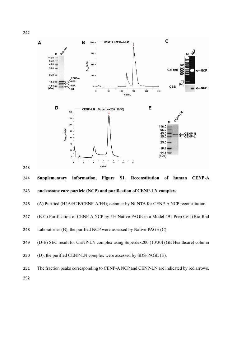
Supplementary information, Figure S1. Reconstitution of human CENP-A 244
nucleosome core particle (NCP) and purification of CENP-LN complex. 245
(A) Purified (H2A/H2B/CENP-A/H4)2 octamer by Ni-NTA for CENP-A NCP reconstitution. 246
(B-C) Purification of CENP-A NCP by 5% Native-PAGE in a Model 491 Prep Cell (Bio-Rad 247
Laboratories (B), the purified NCP were assessed by Native-PAGE (C). 248
(D-E) SEC result for CENP-LN complex using Superdex200 (10/30) (GE Healthcare) column 249
(D), the purified CENP-LN complex were assessed by SDS-PAGE (E). 250
The fraction peaks corresponding to CENP-A NCP and CENP-LN are indicated by red arrows. 251
252

253
254
Supplementary information, Figure S2. Purification of CENP-A NCP/CENP-LN complex. 255
(A) Gel shift assays for the interaction between CENP-A NCP and CENP-LN complex. 256
(B) Purification of CENP-A NCP/CENP-LN complex by Superose6 (10/300) (GE Healthcare) 257
column. The fractions were assessed by Native-PAGE and SDS-PAGE and the samples used 258
for cryo-EM were indicated with red rectangle. 259
260

261
Supplementary information, Figure S3. Cryo-EM structure determination and resolution 262
assessment of CENP-A NCP/CENP-LN complex. 263
(A-F) Cryo-EM structure determination. Representative micrograph of (A) TF20 and (B) Titan 264
Krios cryo-EM data. (C) CTF estimation of image in (A) by Gctf with an estimated image 265
resolution of 3.8 Å. (D) Ctffind showed Thon rings in the Fourier spectrum of the image in (B) 266
extending to 1/3.7 Å-1. Selected 2D class averages of the sample are shown in (E) for TF20 and 267
(F) for Titan datasets. Scale bar in (A) and (B) is 20 nm. (G-I) Validation of CENP-A 268
NCP/CENP-LN complex structure. (G) “Gold-standard” FSC coefficient curve of the final 269
reconstruction showed an overall resolution of 5.8 Å. (H) Local resolution estimation by 270
ResMap11. The core region resolution reached about 4.5 Å. (I) Orientation distribution for 271
particles included in the final reconstruction. 272

273
Supplementary information, Figure S4. Flow chart of cryo-EM data processing. 274
TF20 and Titan Krios data were first separately processed and then combined to generate the 275
final reconstruction. For both datasets particles were picked and sorted, and subjected to 2D 276
classifications in Relion. Good particles were further processed by 3D analysis. For the TF20 277
data an additional 3D classification that focused on CENP-LN density and skipped alignment 278
was carried out. Before combination, particles from TF20 were rescaled to match those from 279
the Titan Krios. 280
281

282
Supplementary information, Figure S5. Superposition of the necleosomal DNA (cyan) from 283
our cryo-EM structure of CENP-A NCP/CENP-LN complex and the previously reported crystal 284
structure of CENP-A nucleosome (wheat, PDB ID: 3AN2). 285

286
Supplementary information, Figure S6. Interactions between CENP-A NCP and CENP-287
N truncations. 288
(A-C) Native-PAGE results of the binding of CENP-A NCP by (A) CENP-N1-260, CENP-N1-289, 289
(B) CENP-N1-221 and (C) CENP-N1-214. 290
(D) Native-PAGE result of the interaction between 147 bp DNA and CENP-N1-214. The bands 291
corresponding to free DNA, free NCP and the complex formed by NCP or DNA with CENP-N 292
are labeled. 293
294
295

296
Supplementary information, Figure S7. Comparison of the sequences and structures of 297
CENP-A RG loops from different species. 298
(A-B) Sequence alignments of human CENP-A against its orthologous from (A) higher 299
mammal species, (B) Xenopus laevis and yeast. the RG loop region is highlighted with red 300
arrows in (A) and red line in (B). The UniProt entries of the CENP-A orthologous used for 301
sequence alignment are listed in Table S1. 302
(C) Structural comparison of the RG loop (red) in human CENP-A (green) and its 303
corresponding regions in human H3 (yellow), S. cerevisiae H3 (pink) and Kluyveromyces lactis 304
Cse4 (cyan). 305
306

307
Supplementary information, Figure S8. Sequence alignments of human CENP-N against 308
its orthologous. 309
The key residues identified to be essential for CENP-A NCP and necleosomal DNA binding are 310
indicated with blue triangle and red dots, respectively. The UniProt entries of the CENP-N 311
orthologous used for sequence alignment are listed in Table S1. 312
313
314
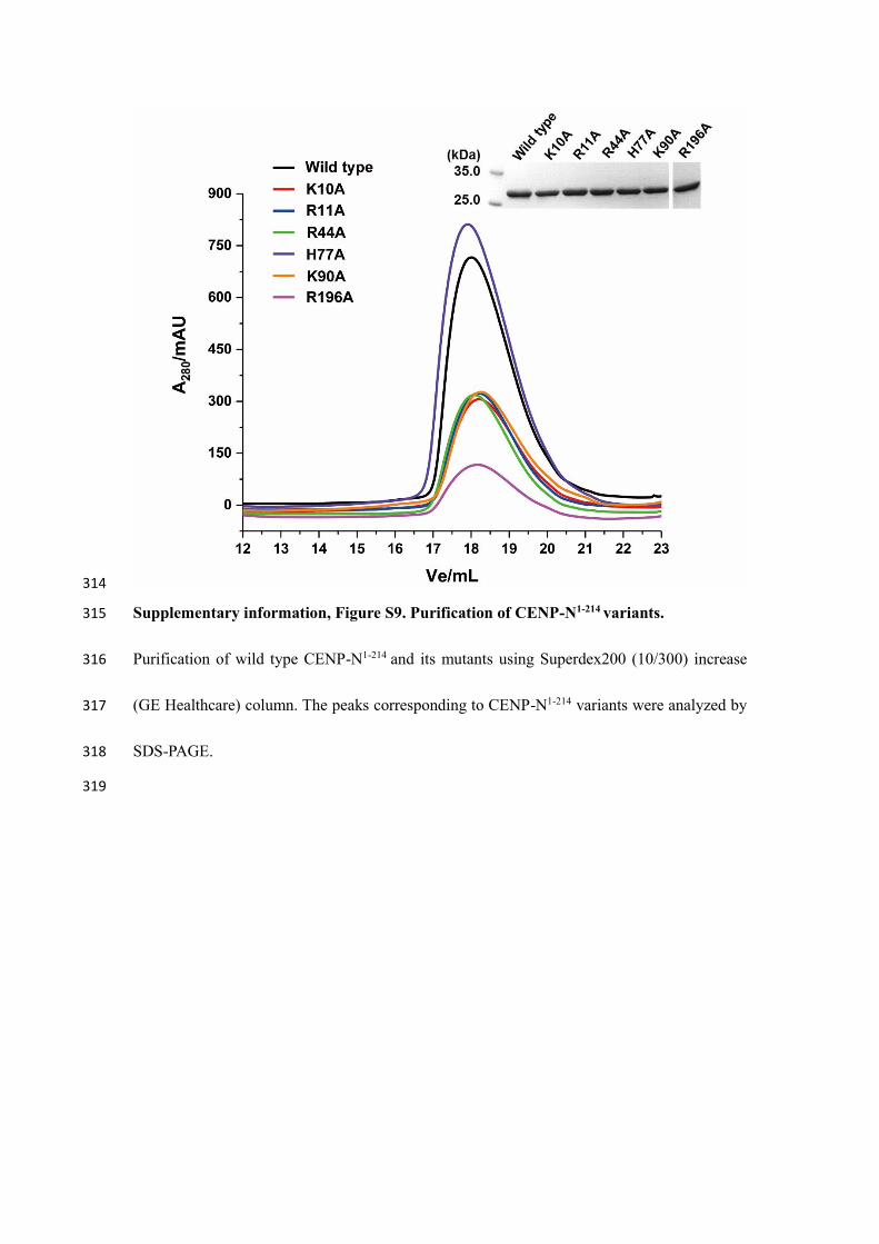
Supplementary information, Figure S9. Purification of CENP-N1-214 variants. 315
Purification of wild type CENP-N1-214 and its mutants using Superdex200 (10/300) increase 316
(GE Healthcare) column. The peaks corresponding to CENP-N1-214 variants were analyzed by 317
SDS-PAGE. 318
319

320
Supplementary information, Figure S10. Quantification of percentage of DNA bound to 321
CENP‑N1-214 WT and mutants relative to Figure 1G. 322
The band intensity was quantified by using ImageJ (https://imagej.nih.gov/ij/index.html). 323

324
Supplementary information, Figure S11. Accurate chromosome segregation in mitosis 325
requires CENP-N DNA-binding activity. 326
Representative mitotic phenotypes in HeLa cells coexpressing CENP-N siRNA and RNAi-327
resistant GFP-CENP-N-WT or GFP-CENP-N-K10A shown by time-lapse microscopy. 328
Chromosomes were visualized by cotransfecting HeLa cells with mCherry-H2B. Scale bar, 10 329
μm. 330
331

332
Supplementary information, Figure S12. Centromere localization of CCAN components 333
is dependent on CENP-N. 334
HeLa cells expressing CENP-N siRNA were fixed and immunostained with CENP-N, CENP-335
L, CENP-A, CENP-E antibodies, respectively. ACA, anti-centromere antibodies. DNA was 336
stained by DAPI. Scale bars, 10 μm. 337
338