Embed Size (px)
Citation preview

Page 1/20
A Comparison of the Analgesic E�cacy of SerratusAnterior Plane Block Vs Paravertebral Nerve Blockfor Video-assisted Thoracic Surgery: a RandomizedControlled TrialYi Zhang
China Medical University First HospitalZe Fu
China Medical University First HospitalTe Fang
China Medical University First HospitalKexin Wang
China Medical University First HospitalZimeng Liu
China Medical University First HospitalHongqing Li
China Medical University First HospitalWenwen Jiang
China Medical University First HospitalXuezhao Cao ( [email protected] )
First Hospital of China Medical University
Research article
Keywords: postoperative pain, serratus anterior plane block, deep serratus plane block, super�cialserratus anterior plane block, paravertebral nerve block, video-assisted thoracic surgery
Posted Date: December 11th, 2020
DOI: https://doi.org/10.21203/rs.3.rs-125277/v1
License: This work is licensed under a Creative Commons Attribution 4.0 International License. Read Full License

Page 2/20
Version of Record: A version of this preprint was published at Videosurgery and Other MiniinvasiveTechniques on January 1st, 2021. See the published version athttps://doi.org/10.5114/wiitm.2021.105725.
Page 3/20
AbstractBackground: Patients who undergo video-assisted thoracic surgery (VATS) that impair the integrity of thechest wall frequently experience moderate to severe postoperative pain. Serratus anterior plane block(SAPB) is a relatively novel technique that can block the lateral cutaneous branches of the intercostalnerves as well as the long thoracic nerve.
Methods: Our study aimed to evaluate the analgesic e�ciency of deep serratus plane block (DSPB) andsuper�cial serratus anterior plane block (SSPB) as well as paravertebral nerve block (PVB) in the patientsundergoing VATS. A total of 74 patients aged from 20-80 undergoing VATS were randomized to receiveeither DSPB or SSPB as well as PVB. Ultrasound (US) guided DSPB or SSPB as well as PVB with 20ml0.5% ropivacaine was performed preoperatively to the patients according to their groups. All patients wereprovided with patient-controlled intravenous analgesia (PCIA) for postoperative analgesia. The primaryoutcomes were the levels of postoperative pain at rest and on coughing evaluated by visual analog scale(VAS), intraoperative and postoperative opioids consumption. The secondary outcomes included PCIApressed times, side effects and satisfaction of analgesia, duration of nerve block, intraoperativehemodynamic changes and vasoactive drug dosage.
Results: No signi�cant differences of VAS score were found in the three groups at each time points.During operation, PVB reduced consumption of opioids (27.23±5.10mg) compared to DSPB(31.20±3.80mg) and SSPB (32.61±5.28mg) (p<0.05). The effective pressed times of PCIA in SSPB group(0.18±0.65) was signi�cantly lower compared to PVB group (1.09±1.50) (p=0.009) at postoperative 12h.Accordingly, SSPB group reduced the dosage of PCIA (26.55±4.72ml) than PVB group (31.45±7.60ml)(p=0.046). Time consuming of PVB procedure was longer (11.14±1.66min) than DSPB (5.68±1.10min)and SSPB (4.77±1.04min) (p<0.001, respectively). PVB group was associated with more intraoperativeatropine consumption (0.14±0.24mg) than DSPB group (0mg) (P=0.043).
Conclusion: DSPB and SSPB are easy to perform and can serve as a promising alternative technique toPVB that may offer comparable analgesic effectiveness and a better side-effect pro�le for patients whoundergoing VATS.
Trial registration: This study was registered to Chinese Clinical Trials Registry on July 20, 2019.(Registration No: ChiCTR1900024678)
IntroductionVideo-assisted thoracic surgery (VATS) is a standard surgical procedure for both minor and majoroncological lung surgery. Over 85% Patients who undergoing VATS often suffer from moderate to severepostoperative pain when coughing and moving(1). Furthermore, 22–63% are converted to chronicpain(2). Uncontrolled postoperative pain, which is attributed to muscle incisions, rib retractions, andintercostal nerve damage, can result in respiratory complications such as hypoxia and atelectasis, whichpreclude early recovery(3, 4).

Page 4/20
Effective analgesia can alleviate the pain of thoracic surgery, which is provided by pleural epidural,paravertebral block, vertical vertebral muscle block, anterior plane block and venous analgesia(5).Thoracic epidural analgesia (TEA) is the current gold-standard analgesia in thoracotomy. However, TEAhas a 30% failure rate, which carries the risk of epidural hematoma or abscess(6). In recent years,paravertebral nerve block (PVB) warrants greater attention with its advantages of small trauma, lowanticoagulation requirements, hemodynamic stability and good analgesic effect(7, 8). However, PVB isnot only technically di�cult to perform especially in obese patients, but also could be less e�cient afterpleurodesis due to pleural in�ammation or surgical dilaceration of the parietal lining of the pleura(9). TEAand PVB exhibit certain di�culties with regard to its administration and side effects, and minimallyinvasive surgery might require less-invasive analgesia. Therefore, the clinical standard minimally invasiveanalgesic technique for VATS is highly recommended.
Serratus anterior plane block (SAPB) which is less di�cult and have fewer severe complications may bemore suitable for VATS(10). SAPB can completely cover the range of the long thoracic nerve as well asthe lateral cutaneous branches of T2 to T5 intercostal nerves, and complement the de�ciency of PVB,TEA, and selective intercostal nerve blocks(11). It was simple to operate, not restricted to the patients whowere obese or use anticoagulants, and also exhibited satis�ed analgesic effect(2, 5, 12, 13). Morerecently, a single-center, double-blinded study with 40 patients showed that compared to PVB, SAPB wasnon-inferior in terms of 48-hour opioid consumption and was associated with improving functionalmeasures in thoracic surgical patients(14). SAPB might be performed at super�cial or deep planes(15–17). Blanco et al showed super�cial serratus anterior plane block (SSPB) had a wider range and longerblock time compared to deep serratus plane block (DSPB)(15). Conversely, Piracha et al found that DSPBcould relieve the uncontrollable pain of SSPB(16). However, whether DSPB and SSPB were as good asPVB remained unclear. This study investigated the analgesic effectiveness of DSPB and SSPB forpatients undergoing VATS and the potential for SAPB as an alternative to PVB for VATS.
MethodsThis randomized, comparative, double-blinded study was performed in Shenyang, from July 2019 toDecember 2019 after receiving the approval of the institutional ethical committee of the First Hospital ofChina Medical University and was registered to Chinese Clinical Trials Registry (Registration No:ChiCTR1900024678). All patients were aged 20–80 years with ASA I or II scheduled for VATS. Thepatients were excluded as follow: (1) allergy to local anesthetics; (2) ASA III or IV; (3) severe obesepatients (BMI > 30 kg/m2); (4) motion sickness; severe cardiorespiratory, hepatic, renal disorders; chronicchest pain; (5) opioid abuse and inability to communicate. According to our preliminary study data,intraoperative opioids consumptions in DSPB, SSPB and PVB groups were 31.22 mg, 34.71 mg and27.5 mg. 22 patients per group were required to achieve a signi�cance level of 0.05 with a power of 80%.The statistical test used for sample size calculation was One Way ANOVA Power Analysis. Powercalculation was completed using PASS (NCSS LLC, Kaysville, Utah). Finally, a total of 74 patients were
Page 5/20
recruited and provided their written informed consent to participate in this study. They were randomlyassigned to receive either DSPB or SSPB as well as PVB.
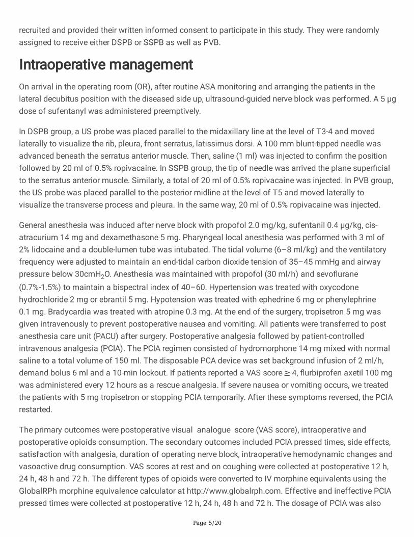
Intraoperative managementOn arrival in the operating room (OR), after routine ASA monitoring and arranging the patients in thelateral decubitus position with the diseased side up, ultrasound-guided nerve block was performed. A 5 µgdose of sufentanyl was administered preemptively.
In DSPB group, a US probe was placed parallel to the midaxillary line at the level of T3-4 and movedlaterally to visualize the rib, pleura, front serratus, latissimus dorsi. A 100 mm blunt-tipped needle wasadvanced beneath the serratus anterior muscle. Then, saline (1 ml) was injected to con�rm the positionfollowed by 20 ml of 0.5% ropivacaine. In SSPB group, the tip of needle was arrived the plane super�cialto the serratus anterior muscle. Similarly, a total of 20 ml of 0.5% ropivacaine was injected. In PVB group,the US probe was placed parallel to the posterior midline at the level of T5 and moved laterally tovisualize the transverse process and pleura. In the same way, 20 ml of 0.5% ropivacaine was injected.
General anesthesia was induced after nerve block with propofol 2.0 mg/kg, sufentanil 0.4 µg/kg, cis-atracurium 14 mg and dexamethasone 5 mg. Pharyngeal local anesthesia was performed with 3 ml of2% lidocaine and a double-lumen tube was intubated. The tidal volume (6–8 ml/kg) and the ventilatoryfrequency were adjusted to maintain an end-tidal carbon dioxide tension of 35–45 mmHg and airwaypressure below 30cmH2O. Anesthesia was maintained with propofol (30 ml/h) and sevo�urane(0.7%-1.5%) to maintain a bispectral index of 40–60. Hypertension was treated with oxycodonehydrochloride 2 mg or ebrantil 5 mg. Hypotension was treated with ephedrine 6 mg or phenylephrine0.1 mg. Bradycardia was treated with atropine 0.3 mg. At the end of the surgery, tropisetron 5 mg wasgiven intravenously to prevent postoperative nausea and vomiting. All patients were transferred to postanesthesia care unit (PACU) after surgery. Postoperative analgesia followed by patient-controlledintravenous analgesia (PCIA). The PCIA regimen consisted of hydromorphone 14 mg mixed with normalsaline to a total volume of 150 ml. The disposable PCA device was set background infusion of 2 ml/h,demand bolus 6 ml and a 10-min lockout. If patients reported a VAS score ≥ 4, �urbiprofen axetil 100 mgwas administered every 12 hours as a rescue analgesia. If severe nausea or vomiting occurs, we treatedthe patients with 5 mg tropisetron or stopping PCIA temporarily. After these symptoms reversed, the PCIArestarted.
The primary outcomes were postoperative visual analogue score (VAS score), intraoperative andpostoperative opioids consumption. The secondary outcomes included PCIA pressed times, side effects,satisfaction with analgesia, duration of operating nerve block, intraoperative hemodynamic changes andvasoactive drug consumption. VAS scores at rest and on coughing were collected at postoperative 12 h,24 h, 48 h and 72 h. The different types of opioids were converted to IV morphine equivalents using theGlobalRPh morphine equivalence calculator at http://www.globalrph.com. Effective and ineffective PCIApressed times were collected at postoperative 12 h, 24 h, 48 h and 72 h. The dosage of PCIA was also
Page 6/20
recorded at the same time. Patients ranked their satisfaction with analgesia in the �rst 72 postoperativehours, from “highly unsatisfactory” to “highly satisfactory”. Intraoperative hemodynamic changescontained the blood pressure and heart rate when the patients entering the OR, before operation, 5minutes after surgical incision, before endotracheal extubation, 5 minutes after extubation and beforeleaving PACU, as well as the duration of intraoperative hypotension. All of the outcomes and perioperativedata were collected by an investigator who was blinded to the group allocation.
Statistical analysisData were analyzed using GraphPad prism 8. Continuous numerical variables were presented as themean and standard deviation or standard error of the mean, categorical variables were presented as theratio or as the number and percentage, and between-group differences were compared using Fisher’sexact test (for nominal data) or the chi-squared test for trend (for ordinal data). Primary and secondaryendpoints for each analgesic technique used were compared using the Kruskal-Wallis test with Dunn’scorrection. The reported P-value is two-sided. P < 0.05 were considered statistically signi�cant. TheCochran–Armitage test was used for satisfaction score analysis.
Results
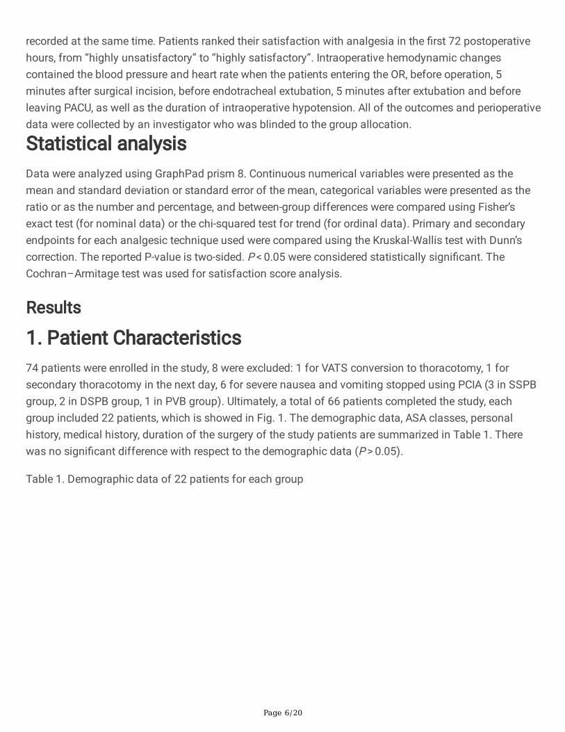
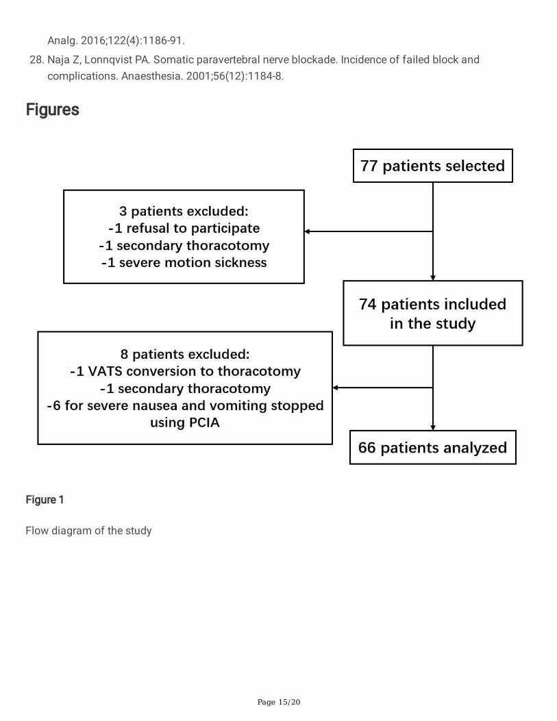
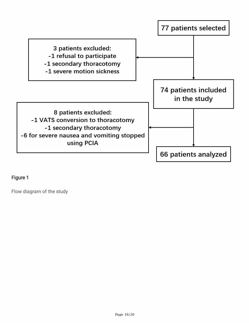
1. Patient Characteristics74 patients were enrolled in the study, 8 were excluded: 1 for VATS conversion to thoracotomy, 1 forsecondary thoracotomy in the next day, 6 for severe nausea and vomiting stopped using PCIA (3 in SSPBgroup, 2 in DSPB group, 1 in PVB group). Ultimately, a total of 66 patients completed the study, eachgroup included 22 patients, which is showed in Fig. 1. The demographic data, ASA classes, personalhistory, medical history, duration of the surgery of the study patients are summarized in Table 1. Therewas no signi�cant difference with respect to the demographic data (P > 0.05).
Table 1. Demographic data of 22 patients for each group

Page 7/20
Notes: Values of age, BMI and duration of surgery are expressed as mean ± standard deviation.
2. Postoperative VAS scoreThere were no signi�cant differences of VAS scores at rest and on coughing among the three groups atpostoperative 12 h, 24 h, 48 h, and 72 h (Table 2). None of the patients suffered severe pain. During the�rst postoperative 24hrs, the patients presented with mild to moderate pain when coughing (13 in DSPBgroup, 13 in SSPB group and 17 in PVB group). At the rest of the time, they presented with mild pain.
Table 2. Postoperative VAS scores at rest and on coughing
Page 8/20
Notes: Values are expressed as the mean [95% con�dence interval]. When the lower limit value of the 95%con�dence interval is less than 0, use 0 instead.
Abbreviations: VAS-R, the values of VAS scores at rest; VAS-C, the values of VAS scores on coughing.
3. Intraoperative dosage of opioidsSufentanil and oxycodone hydrochloride were converted to morphine, and the consumption werecompared in three groups. In intergroup comparisons, PVB group (27.23 ± 5.10 mg) was associated withreducing intraoperative opioid consumption compared to DSPB (31.20 ± 3.80 mg) and SSPB group(32.61 ± 5.28 mg, P < 0.05). Therefore, PVB had better analgesic effect than DSPB and SSPB duringoperation.
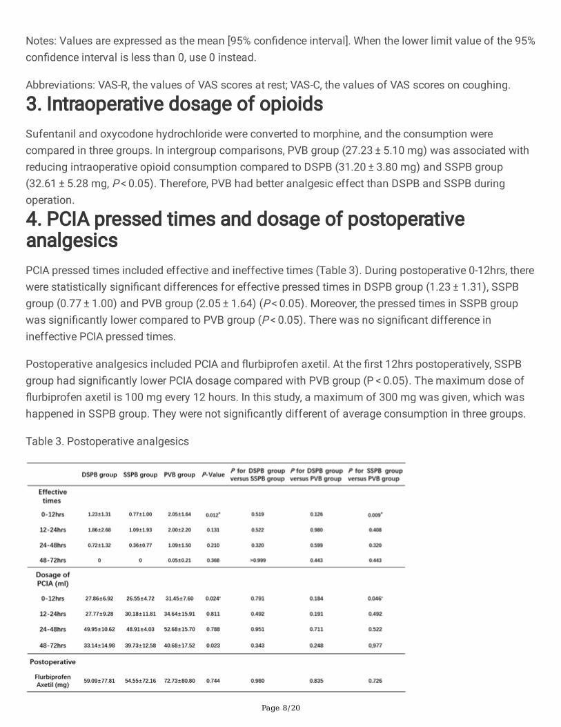
4. PCIA pressed times and dosage of postoperativeanalgesicsPCIA pressed times included effective and ineffective times (Table 3). During postoperative 0-12hrs, therewere statistically signi�cant differences for effective pressed times in DSPB group (1.23 ± 1.31), SSPBgroup (0.77 ± 1.00) and PVB group (2.05 ± 1.64) (P < 0.05). Moreover, the pressed times in SSPB groupwas signi�cantly lower compared to PVB group (P < 0.05). There was no signi�cant difference inineffective PCIA pressed times.
Postoperative analgesics included PCIA and �urbiprofen axetil. At the �rst 12hrs postoperatively, SSPBgroup had signi�cantly lower PCIA dosage compared with PVB group (P < 0.05). The maximum dose of�urbiprofen axetil is 100 mg every 12 hours. In this study, a maximum of 300 mg was given, which washappened in SSPB group. They were not signi�cantly different of average consumption in three groups.
Table 3. Postoperative analgesics
Page 9/20
Notes: * represents signi�cant difference
5. Satisfaction with analgesiaThere were no statistical differences of satisfaction with analgesia in three groups (Table 8). Both DSPBand SSPB, as well as PVB could provide good analgesic effect postoperatively, eighty percent of thepatients showed “neutral” and “satisfactory” approximately. Patients in PVB group showed more “highlyunsatisfactory” and “approximately” than in DSPB group and SSPB group.
6. Side effectsNo patient exhibited block-related complications, such as urinary retention, pneumonia, local anesthetictoxicity, bleeding, or infection. The side effects of opioids included nausea, vomit and dizzy showed nosigni�cant differences among three groups.
7. Duration of operating nerve blockThe duration of operating nerve block recorded from disinfection to nerve block needle extraction, all ofthe groups were done once by the same experienced anesthesiologist. Duration of DSPB (5.77 ± 1.20 min) and SSPB group (4.77 ± 1.04 min) were signi�cantly shorter than that of PVB group (11.14 ± 1.66 min, P < 0.05). PVB was more complex for anesthesiologist to operate.
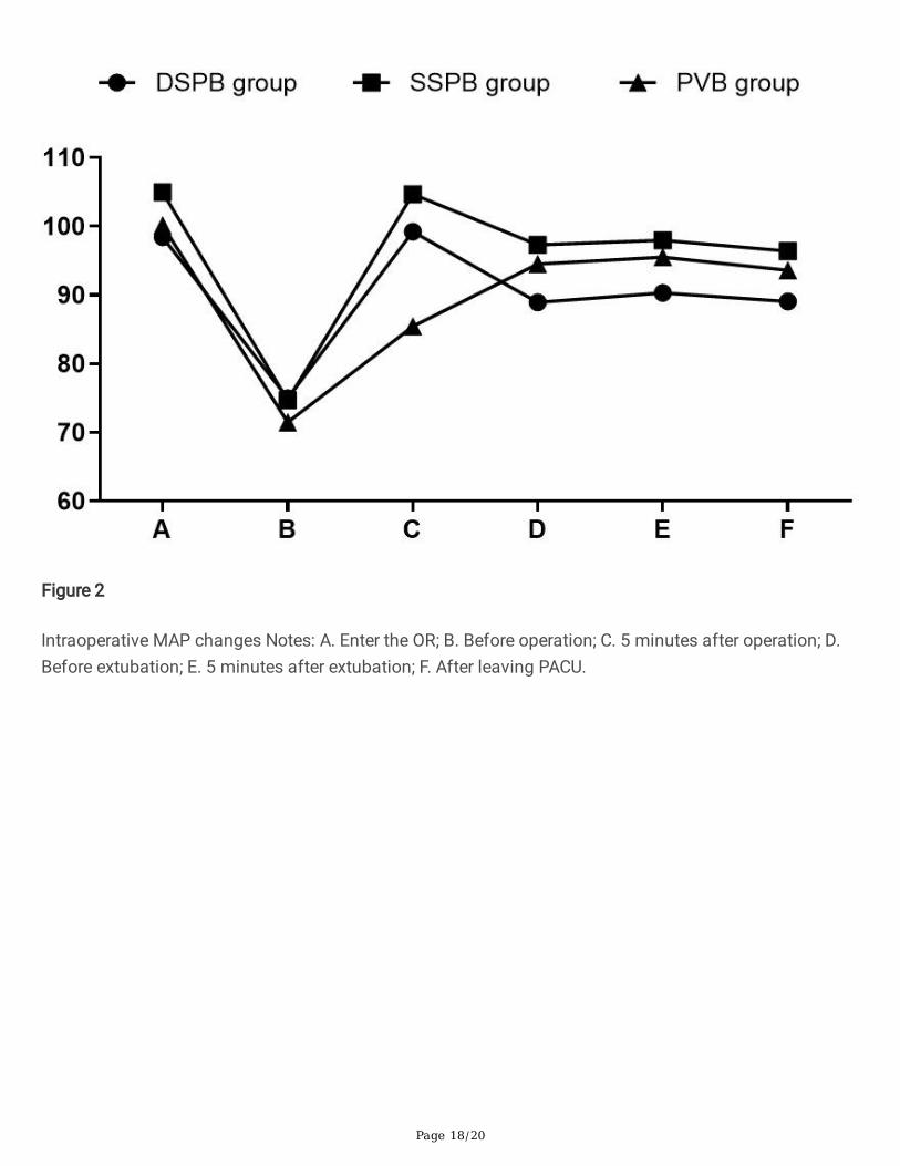
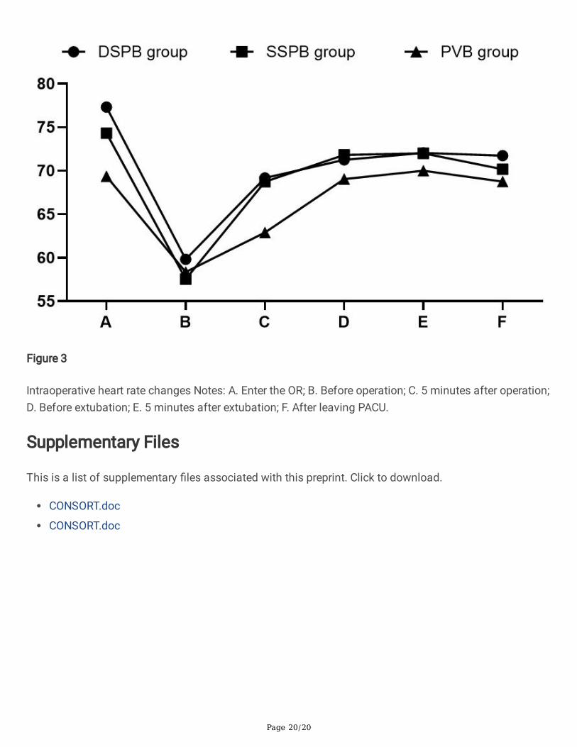
8. Intraoperative hemodynamic changes and the dosage ofvasoactive drugsIntraoperative blood pressure changes were showed with MAP, which were compared at six time points(Fig. 2). At 5 minutes after operation, MAP was lower in PVB group (85.45 ± 16 mmHg) than that in DSPB(99.21 ± 15.27 mmHg, P < 0.05) and SSPB group (104.70 ± 16.33 mmHg, P < 0.05). At 5 minutes afterextubation and before leaving PACU, DSPB group showed lower MAP than SSPB group (P < 0.05). Thethree groups were comparable regarding the changes of heart rate (Fig. 3) and the duration ofintraoperative hypotension, which were no signi�cant differences (P > 0.05).
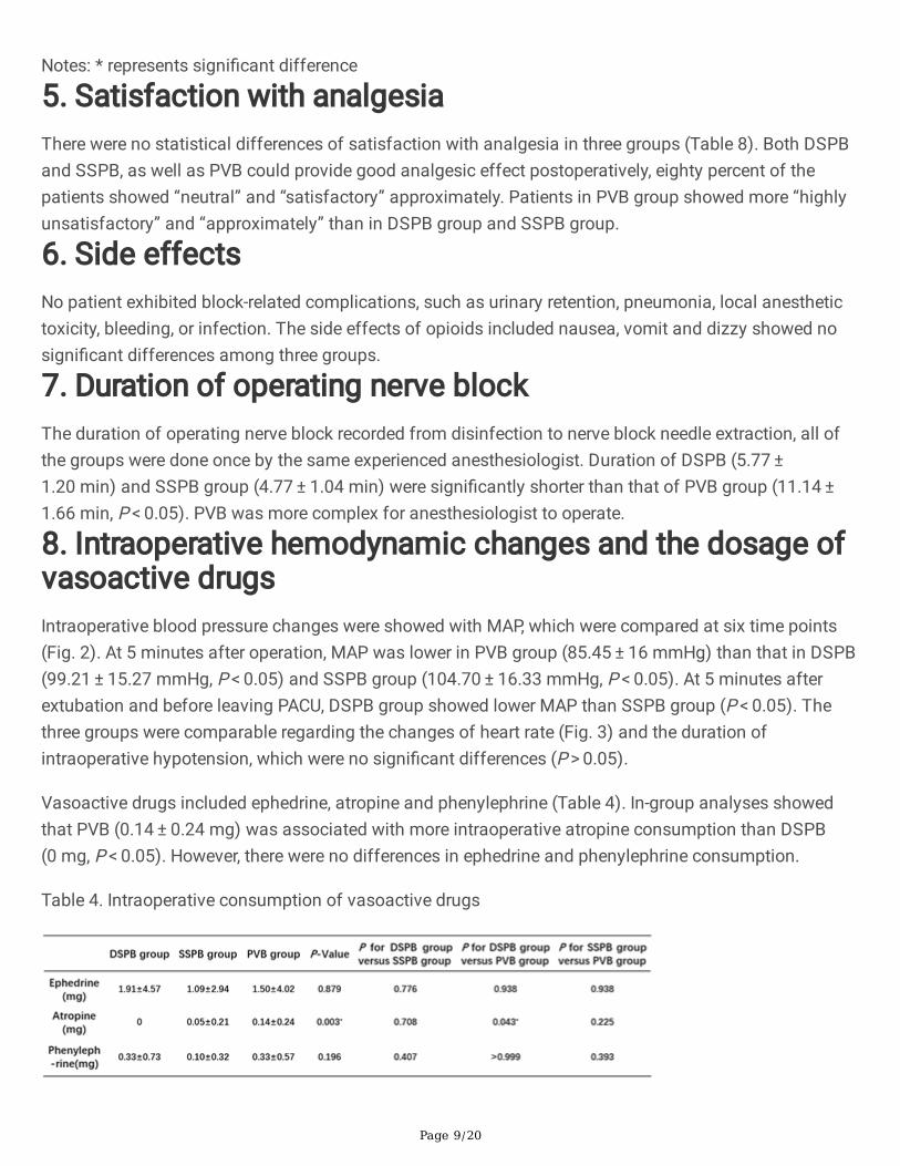
Vasoactive drugs included ephedrine, atropine and phenylephrine (Table 4). In-group analyses showedthat PVB (0.14 ± 0.24 mg) was associated with more intraoperative atropine consumption than DSPB(0 mg, P < 0.05). However, there were no differences in ephedrine and phenylephrine consumption.
Table 4. Intraoperative consumption of vasoactive drugs
Page 10/20
Notes: * represents signi�cant difference
DiscussionThis randomized, double-blind study demonstrated DSPB, SSPB or PVB combined with PCIA reduced thepostoperative pain and showed similar satisfaction with analgesia in patients undergoing VATS.Intraoperative opioid consumption remained signi�cantly lower in PVB. SSPB showed less PCIA pressedtimes and PCIA dosage than PVB. Furthermore, DSPB and SSPB were easy for anesthetist to operate,with signi�cantly lower operating duration than PVB. PVB was associated with maintaininghemodynamic stability. However, PVB consumed more atropine intraoperatively.
There were different opinions about the postoperative analgesic effect of DSPB, SSPB and PVB. PVB haslong been referred as the best possible choice for postoperative analgesia of VATS(18). In recent studies,SSPB proponents have described successful analgesia without the potentially hazardous need foradvancing the needle deeper toward the pleura(15, 17). However, anatomy arguably favored DSPB asinjection in the fascial plane below the serratus muscle which blockade of the lateral cutaneous branchesof the intercostal nerves, might show better analgesic effect(19). In our study, DSPB, SSPB and PVBshowed similar postoperative analgesic effect, and most patients were satis�ed with the analgesic effect.All three can be used for postoperative analgesia of VATS. However, in the early postoperative period(12hrs), SSPB group provided a superior pain relief with signi�cantly lower effective PCIA pressed timesand dosage compared to PVB group. Some studies showed that the duration of the sensory blockadeproduced by SSPB and DSPB was 730–780 min and 380–400 min respectively(20–22). The effectivetime of PVB persisted for 48hrs postoperatively(23). In our study, the duration of postoperative analgesiafor PVB was shorter, probably due to the pharmacological properties of ropivacaine.
During operation, compared with DSPB and SSPB, PVB showed superior analgesic effect. PVBsigni�cantly decreased intraoperative consumption of opioids comparing to DSPB and SSPB, whichindicated the short-term analgesic effect of PVB was better than that of DSPB and SSPB. These �ndingssupport observations from previous reports that showed the effectiveness of PVB(24).
The ideal analgesic techniques should not only have perfect analgesia effect, but also have theadvantages of simple operation, accurate control, high success rate and few complications. The punctureduration of PVB was signi�cantly longer than that of DSPB and SSPB in our study. It might be related tothe difference of anatomical position. The serratus anterior muscle was super�cial which could bescanned by high-frequency linear array ultrasound probe to easily obtain clear images of the serratusanterior muscle and its neighbors. During the puncture, the angle between the needle and skin was smallthat the puncture needle could be imaged clearly(25). J Richardson et al also found that the deep fasciaof the serratus anterior muscle had poor adhesion to the intercostal external muscles and was easier toseparate than the super�cial plane of the serratus anterior muscle, which was also showed in ourstudy(26). The location of thoracic paravertebral nerve was deeper and should be scanned low-frequency
Page 11/20
convex array probe or high-frequency linear array probe. The puncture needle was di�cult to image dueto the large angle.
A few studies have described analgesia effect of SSPB was similar to an epidural but perhaps with lesshemodynamic instability(17). In our study, both DSPB and SSPB, as well as PVB could maintainhemodynamic stability. However, PVB consumed more atropine intraoperatively. Previous studies alsohave shown that PVB can cause the incidence of bradycardia and hypotension with rate of 0.47% ~ 2.2%,which might be related to sympathetic block(27).
In addition, the incidence of side effects did not show signi�cant differences in three groups. There were6 patients who had motion sickness reported severe nausea and vomiting. After stopping PCIA, the sideeffects were disappeared, which indicated that might be associated with the opioid. They withdrew fromour study on the basis of exclusion criteria. We did not report any complication associated with nerveblock, but pneumothorax was potential. The deep surface of the paravertebral area was the pleura, andthere was a risk of puncture of the pleura, pneumothorax and other complications. Naja et al performedPVB in 662 patients, and the probability of developing pneumothorax was about 0.5%(28). J Richardson’sstudy showed that PVB punctures occasionally entered the epidural or puncture the pleura, and had atransient occurrence of Horner syndrome(26). This could explain why many clinicians are reluctant tooperate PVB in daily work. Accordingly, patients with narrow intercostal space, obesity, poor coagulationfunction should use DSPB or SSPB.
Nonetheless, the present study had several limitations. First, as an observational study, our conclusionsmight have been limited by inadequate data collection, the pain of nerve block procedure was notrecorded. Meanwhile, due to the time limitation of preoperative preparation, we could only con�rm thediffusion of local anesthetics by ultrasound, but did not collect the data of spread level of analgesia.Second, the research subjects recruited in this study were not performed by the same surgeon, and therewere uncontrollable differences. Third, it should be noted that during operation, when surgeons cut openthe skin and subcutaneous tissue of patients who received DSPB or SSPB, it showed a slight edema ofsubcutaneous tissue, which indicated the possible loss of local anesthetics. Finally, this study did notexplore the appropriate local anesthetic dose for nerve block, which will be described in further research.
ConclusionIn conclusion, DSPB, SSPB or PVB combined with PCIA could provide good postoperative analgesia forpatients who undergoing VATS. PVB showed better analgesic effect than DSPB and SSPBintraoperatively. However, the operation of PVB was complex and had potential complications. DSPB orSSPB can serve as a promising alternative to PVB in optimal perioperative pain management in VATS. Inthe further study, large-scale prospective randomized controlled trials are required to compare the e�cacyof postoperative analgesia by continuous infusion through a catheter.
List Of Abbreviations
Page 12/20
Video-assisted thoracic surgery (VATS)
Thoracic epidural analgesia (TEA)
Paravertebral nerve block (PVB)
Serratus anterior plane block (SAPB)
Super�cial serratus anterior plane block (SSPB)
Deep serratus plane block (DSPB)
Operating room (OR)
Post anesthesia care unit (PACU)
Patient-controlled intravenous analgesia (PCIA)
Visual analogue score (VAS score)
DeclarationsEthics approval and consent to participate
Study received the approval of the institutional ethical committee of the First Hospital of China MedicalUniversity (No.2018-305-2).
Consent for publication
Availability of data and materials
The datasets used and/or analysed during the current study are available from the corresponding authoron reasonable request.
Competing interests
Not applicable
Funding
This work is supported by the Special Program for Key Research of the Ministry of Science and Technology, China (Grant No. SQ2018YFC200044). Xuezhao Cao was responsible for performing nerveblock.
Authors' contributions

Page 13/20
ZY had full access to all the data in the study and takes responsibility for the integrity of the data and theaccuracy of the data analysis. LHQ and JWW were responsible for collecting research data of previousstudies. FZ and WKX were responsible for collating data of this study. CXZ was responsible forperforming nerve block. FT was responsible for preparation of materials. All authors have read andapproved the manuscript.
Acknowledgements
The authors wish to thank nursing, medical and allied medical colleagues for their co-operation andsupport during the conduct of this study.
References1. Semyonov M, Fedorina E, Grinshpun J, Dubilet M, Refaely Y, Ruderman L, et al. Ultrasound-guided
serratus anterior plane block for analgesia after thoracic surgery. J Pain Res. 2019;12:953-60.
2. Okmen K, Metin Okmen B. Evaluation of the effect of serratus anterior plane block for pain treatmentafter video-assisted thoracoscopic surgery. Anaesth Crit Care Pain Med. 2018;37(4):349-53.
3. Ochroch EA, Gottschalk A. Impact of acute pain and its management for thoracic surgical patients.Thorac Surg Clin. 2005;15(1):105-21.
4. Ochroch EA, Gottschalk A, Augostides J, Carson KA, Kent L, Malayaman N, et al. Long-term pain andactivity during recovery from major thoracotomy using thoracic epidural analgesia. Anesthesiology.2002;97(5):1234-44.
5. Park MH, Kim JA, Ahn HJ, Yang MK, Son HJ, Seong BG. A randomised trial of serratus anterior planeblock for analgesia after thoracoscopic surgery. Anaesthesia. 2018;73(10):1260-4.
�. Rosero EB, Joshi GP. Nationwide incidence of serious complications of epidural analgesia in theUnited States. Acta Anaesthesiol Scand. 2016;60(6):810-20.
7. Hiro K, Sugiyama T, Kurata M, Oi Y, Okuda M. [Postoperative Analgesia for Video-assistedThoracoscopic Surgery--Continuous Intravenous Infusion of Fentanyl Combined with IntercostalNerve Block v.s. Continuous Epidural Analgesia]. Masui. 2016;65(2):114-8.
�. Scarci M, Joshi A, Attia R. In patients undergoing thoracic surgery is paravertebral block as effectiveas epidural analgesia for pain management? Interact Cardiovasc Thorac Surg. 2010;10(1):92-6.
9. Allain PA, Carella M, Agra�otis AC, Burey J, Assouad J, Ha�ani EM, et al. Comparison of severalmethods for pain management after video-assisted thoracic surgery for pneumothorax: anobservational study. BMC Anesthesiol. 2019;19(1):120.
10. Tighe SQ, Karmakar MK. Serratus plane block: do we need to learn another technique for thoracicwall blockade? Anaesthesia. 2013;68(11):1103-6.
11. Chu GM, Jarvis GC. Serratus Anterior Plane Block to Address Postthoracotomy and Chest Tube-Related Pain: A Report on 3 Cases. A A Case Rep. 2017;8(12):322-5.

Page 14/20
12. Corso RM, Piraccini E, Byrne H, Poggi P, Tedesco M. The serratus anterior plane block for pediatricnon-intubated video-assisted thoracoscopic surgery. Minerva Anestesiol. 2017;83(7):775-6.
13. Kim DH, Oh YJ, Lee JG, Ha D, Chang YJ, Kwak HJ. E�cacy of Ultrasound-Guided Serratus PlaneBlock on Postoperative Quality of Recovery and Analgesia After Video-Assisted Thoracic Surgery: ARandomized, Triple-Blind, Placebo-Controlled Study. Anesth Analg. 2018;126(4):1353-61.
14. Hanley C, Wall T, Bukowska I, Redmond K, Eaton D, Ni Mhuircheartaigh R, et al. Ultrasound-guidedcontinuous deep serratus anterior plane block versus continuous thoracic paravertebral block forperioperative analgesia in videoscopic-assisted thoracic surgery. Eur J Pain. 2020;24(4):828-38.
15. Blanco R, Parras T, McDonnell JG, Prats-Galino A. Serratus plane block: a novel ultrasound-guidedthoracic wall nerve block. Anaesthesia. 2013;68(11):1107-13.
1�. Piracha MM, Thorp SL, Puttanniah V, Gulati A. "A Tale of Two Planes": Deep Versus Super�cialSerratus Plane Block for Postmastectomy Pain Syndrome. Reg Anesth Pain Med. 2017;42(2):259-62.
17. Khalil AE, Abdallah NM, Bashandy GM, Kaddah TA. Ultrasound-Guided Serratus Anterior Plane BlockVersus Thoracic Epidural Analgesia for Thoracotomy Pain. J Cardiothorac Vasc Anesth.2017;31(1):152-8.
1�. Steinthorsdottir KJ, Wildgaard L, Hansen HJ, Petersen RH, Wildgaard K. Regional analgesia for video-assisted thoracic surgery: a systematic review. Eur J Cardiothorac Surg. 2014;45(6):959-66.
19. Mayes J, Davison E, Panahi P, Patten D, Eljelani F, Womack J, et al. An anatomical evaluation of theserratus anterior plane block. Anaesthesia. 2016;71(9):1064-9.
20. Bhoi D, Pushparajan HK, Talawar P, Kumar A, Baidya DK. Serratus anterior plane block for breastsurgery in a morbidly obese patient. J Clin Anesth. 2016;33:500-1.
21. Lopez-Matamala B, Fajardo M, Estebanez-Montiel B, Blancas R, Alfaro P, Chana M. A new thoracicinterfascial plane block as anesthesia for di�cult weaning due to ribcage pain in critically illpatients. Med Intensiva. 2014;38(7):463-5.
22. Madabushi R, Tewari S, Gautam SK, Agarwal A, Agarwal A. Serratus anterior plane block: a newanalgesic technique for post-thoracotomy pain. Pain Physician. 2015;18(3):E421-4.
23. Vogt A, Stieger DS, Theurillat C, Curatolo M. Single-injection thoracic paravertebral block forpostoperative pain treatment after thoracoscopic surgery. Br J Anaesth. 2005;95(6):816-21.
24. Chalam KS, Patnaik SS, Sunil C, Bansal T. Comparative study of ultrasound-guided paravertebralblock with ropivacaine versus bupivacaine for post-operative pain relief in children undergoingthoracotomy for patent ductus arteriosus ligation surgery. Indian J Anaesth. 2015;59(8):493-8.
25. Ueshima H, Iwamoto W, Otake H. Serratus Plane Block for a Contraction of the Latissimus DorsiMuscle. Reg Anesth Pain Med. 2016;41(3):411.
2�. Richardson J, Lonnqvist PA, Naja Z. Bilateral thoracic paravertebral block: potential and practice. Br JAnaesth. 2011;106(2):164-71.
27. Pace MM, Sharma B, Anderson-Dam J, Fleischmann K, Warren L, Stefanovich P. Ultrasound-GuidedThoracic Paravertebral Blockade: A Retrospective Study of the Incidence of Complications. Anesth
Page 15/20
Analg. 2016;122(4):1186-91.
2�. Naja Z, Lonnqvist PA. Somatic paravertebral nerve blockade. Incidence of failed block andcomplications. Anaesthesia. 2001;56(12):1184-8.
Figures
Figure 1
Flow diagram of the study
Page 16/20
Figure 1
Flow diagram of the study

Page 17/20
Figure 2
Intraoperative MAP changes Notes: A. Enter the OR; B. Before operation; C. 5 minutes after operation; D.Before extubation; E. 5 minutes after extubation; F. After leaving PACU.
Page 18/20
Figure 2
Intraoperative MAP changes Notes: A. Enter the OR; B. Before operation; C. 5 minutes after operation; D.Before extubation; E. 5 minutes after extubation; F. After leaving PACU.

Page 19/20
Figure 3
Intraoperative heart rate changes Notes: A. Enter the OR; B. Before operation; C. 5 minutes after operation;D. Before extubation; E. 5 minutes after extubation; F. After leaving PACU.
Page 20/20
Figure 3
Intraoperative heart rate changes Notes: A. Enter the OR; B. Before operation; C. 5 minutes after operation;D. Before extubation; E. 5 minutes after extubation; F. After leaving PACU.
Supplementary Files
This is a list of supplementary �les associated with this preprint. Click to download.
CONSORT.doc
CONSORT.doc