Embed Size (px)
Citation preview
Methods For Monitoring Transdermal Drug Delivery
Analytical/Radio/Nuclear (ARN) Seminar
Jivan YewleDepartment of Chemistry
University of Kentucky
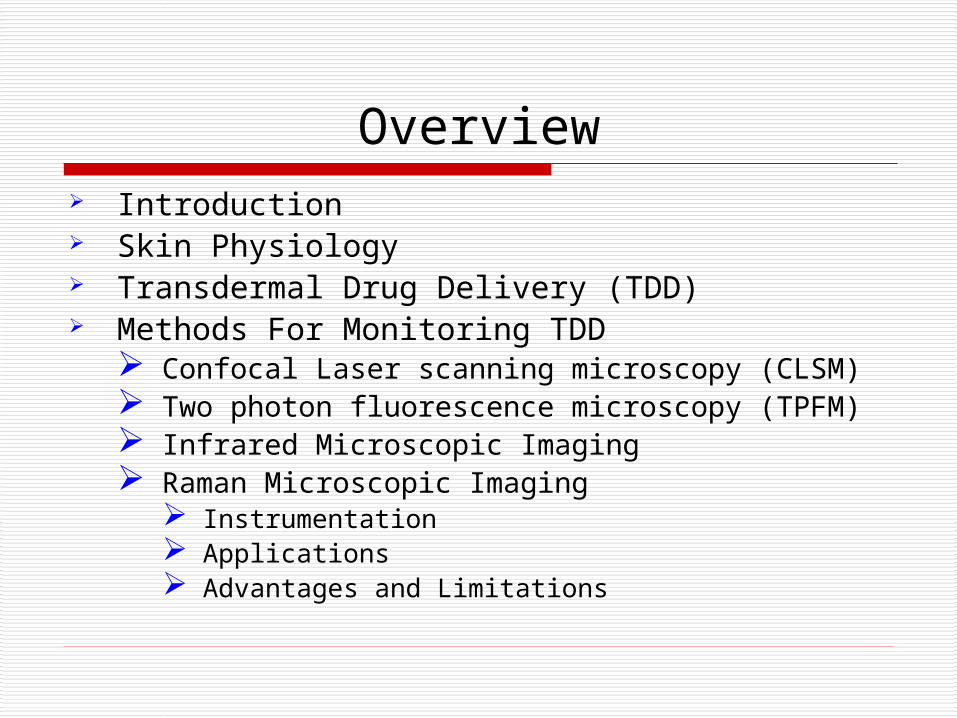
Overview Introduction Skin Physiology Transdermal Drug Delivery (TDD) Methods For Monitoring TDD
Confocal Laser scanning microscopy (CLSM) Two photon fluorescence microscopy (TPFM) Infrared Microscopic Imaging Raman Microscopic Imaging
Instrumentation Applications Advantages and Limitations
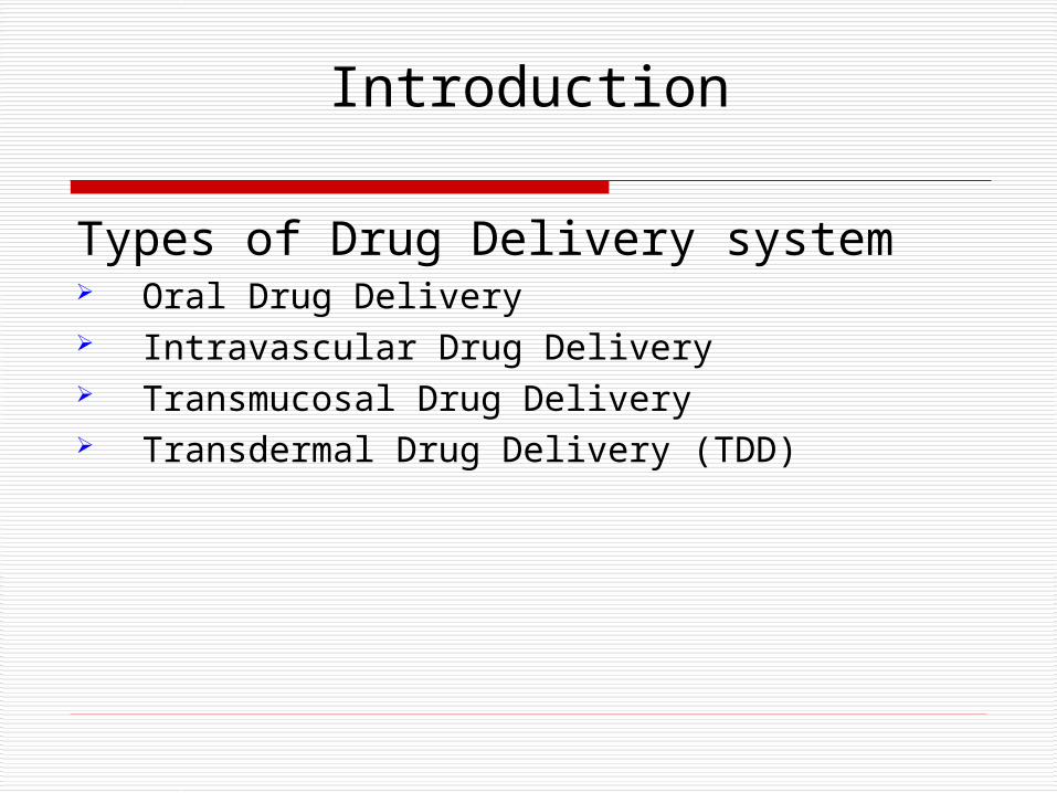
Introduction
Types of Drug Delivery system Oral Drug Delivery Intravascular Drug Delivery Transmucosal Drug Delivery Transdermal Drug Delivery (TDD)
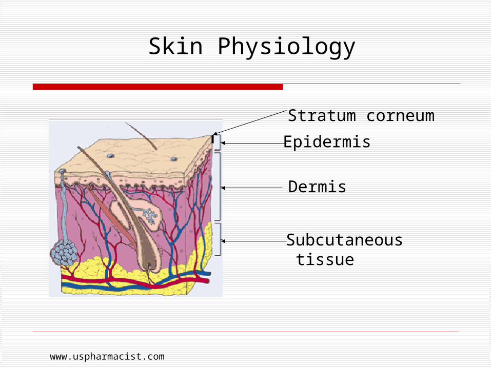
Skin Physiology
Epidermis
Dermis
Subcutaneous tissue
Stratum corneum
www.uspharmacist.com
Skin Physiology
Characteristics: Tough Flexible Poor conductor of electricity
Functions of skin: To protect the body from external insults To contain all body fluids To regulate body temperature To protect from electrical current
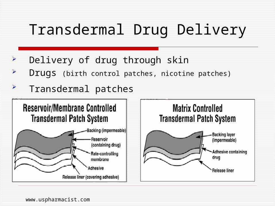
Transdermal Drug Delivery
Delivery of drug through skin Drugs (birth control patches, nicotine patches)
Transdermal patches
www.uspharmacist.com
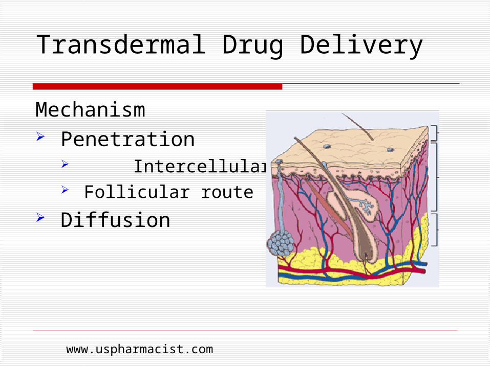
Transdermal Drug Delivery
Mechanism Penetration
Intercellular route Follicular route
Diffusion
www.uspharmacist.com
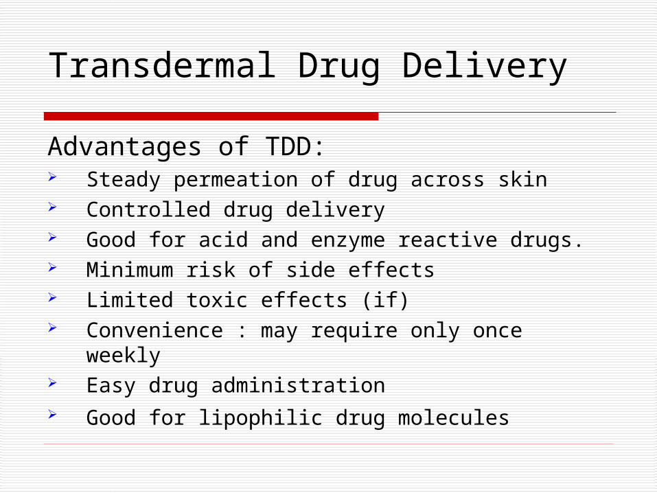
Transdermal Drug Delivery
Advantages of TDD: Steady permeation of drug across skin Controlled drug delivery Good for acid and enzyme reactive drugs. Minimum risk of side effects Limited toxic effects (if) Convenience : may require only once weekly Easy drug administration Good for lipophilic drug molecules

Transdermal Drug Delivery
Disadvantages and limitations of TDD Possibility of a local irritation Allergic reactions are possible Risky for children Skin's low permeability Molecular size and polarity of drug Insufficient bioavailability Damage to a transdermal patch
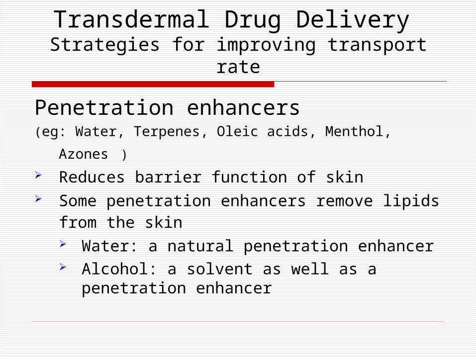
Transdermal Drug Delivery Strategies for improving transport rate
Penetration enhancers(eg: Water, Terpenes, Oleic acids, Menthol, Azones ) Reduces barrier function of skin Some penetration enhancers remove lipids
from the skin Water: a natural penetration enhancer Alcohol: a solvent as well as a penetration
enhancer
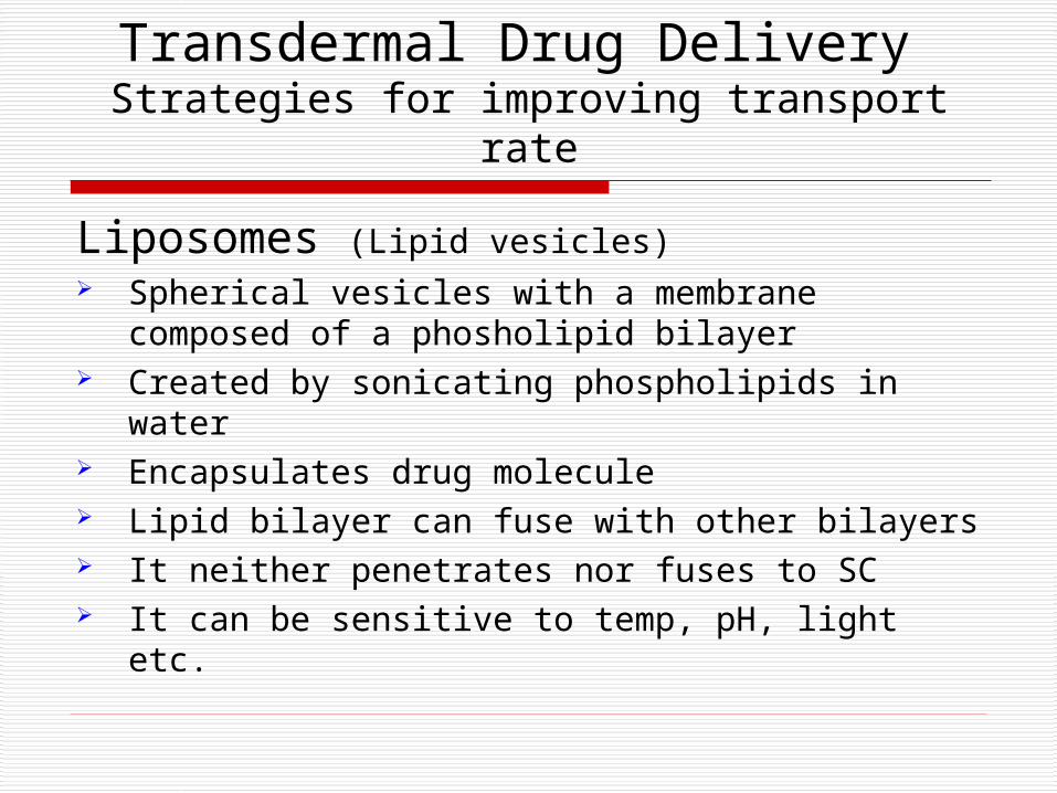
Transdermal Drug Delivery Strategies for improving transport rate
Liposomes (Lipid vesicles) Spherical vesicles with a membrane composed
of a phosholipid bilayer Created by sonicating phospholipids in water Encapsulates drug molecule Lipid bilayer can fuse with other bilayers It neither penetrates nor fuses to SC It can be sensitive to temp, pH, light etc.
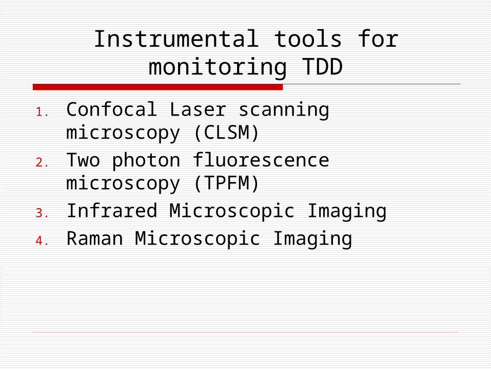
Instrumental tools for monitoring TDD
1. Confocal Laser scanning microscopy (CLSM)
2. Two photon fluorescence microscopy (TPFM)
3. Infrared Microscopic Imaging4. Raman Microscopic Imaging
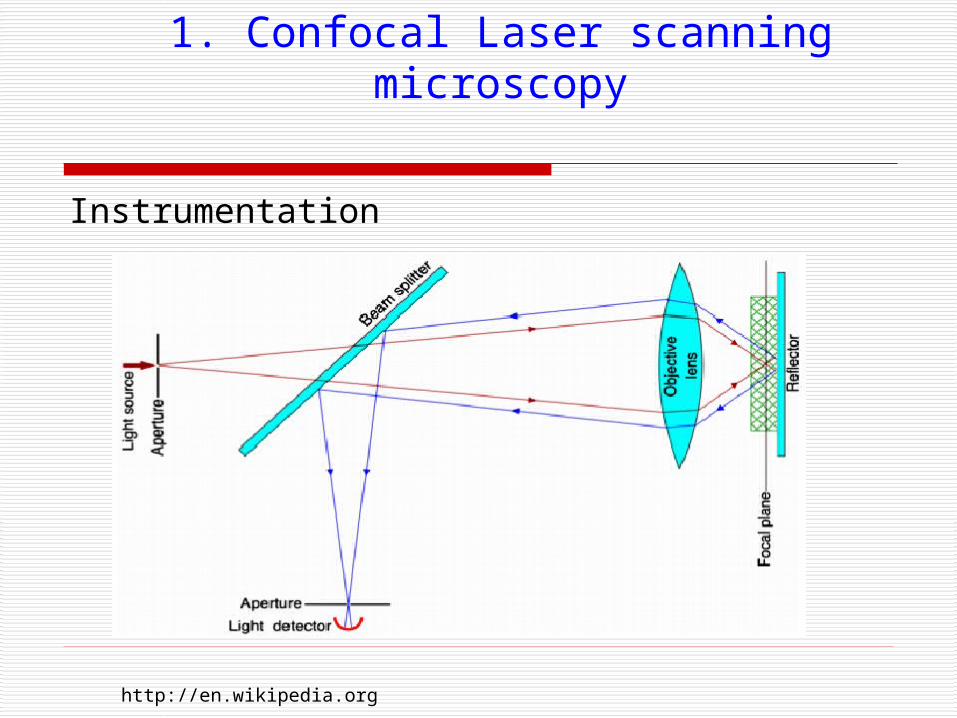
1. Confocal Laser scanning microscopy
Instrumentation
http://en.wikipedia.org
1. Confocal Laser scanning microscopy
Applications in TDD Images parallel to skin surface Position of drug molecule under skin surface Information about penetration of drug
Other applications Evaluation of biological phenomenon Transport studies through biological membrane Surface study of different material
1. Confocal Laser scanning microscopy
Advantages Images of thick specimens at various depth 3D confocal images High degree of precision Blur-free images
Limitations and disadvantages Information about permeation of fluorescent
label only Images up to 25 m depth only Smaller signal to noise ratio
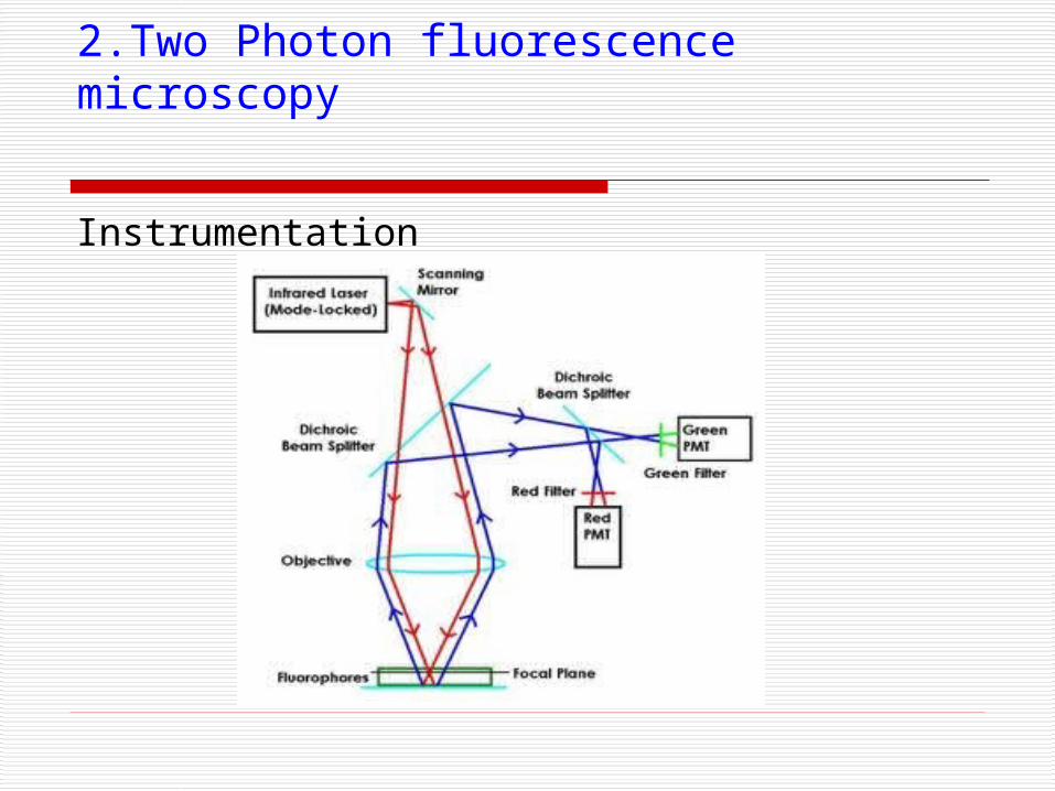
2.Two Photon fluorescence microscopy
Instrumentation
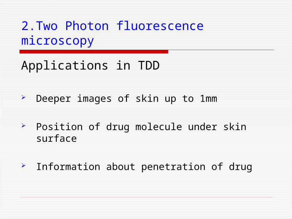
2.Two Photon fluorescence microscopy
Applications in TDD
Deeper images of skin up to 1mm
Position of drug molecule under skin surface
Information about penetration of drug
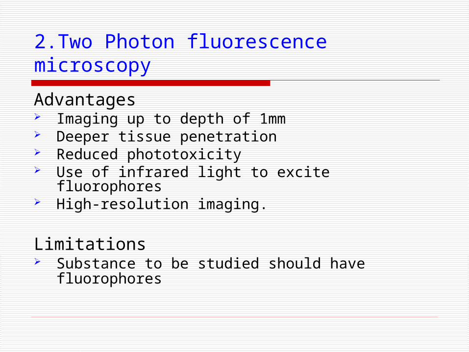
2.Two Photon fluorescence microscopy
Advantages Imaging up to depth of 1mm Deeper tissue penetration Reduced phototoxicity Use of infrared light to excite fluorophores High-resolution imaging.
Limitations Substance to be studied should have
fluorophores
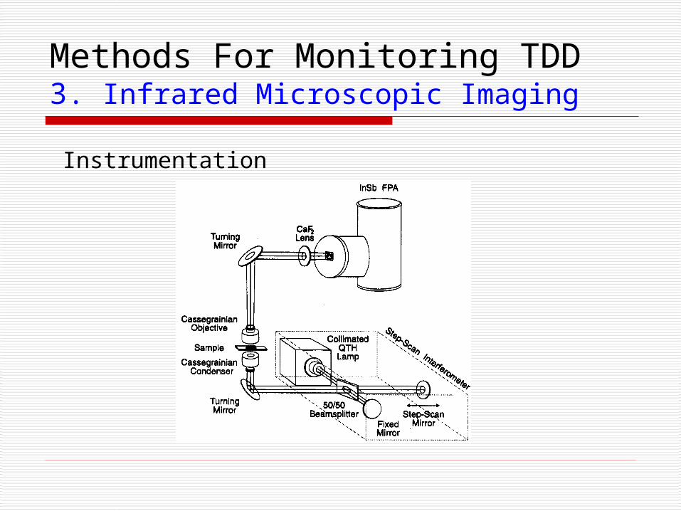
Methods For Monitoring TDD3. Infrared Microscopic Imaging
Instrumentation
3. Infrared Microscopic Imaging
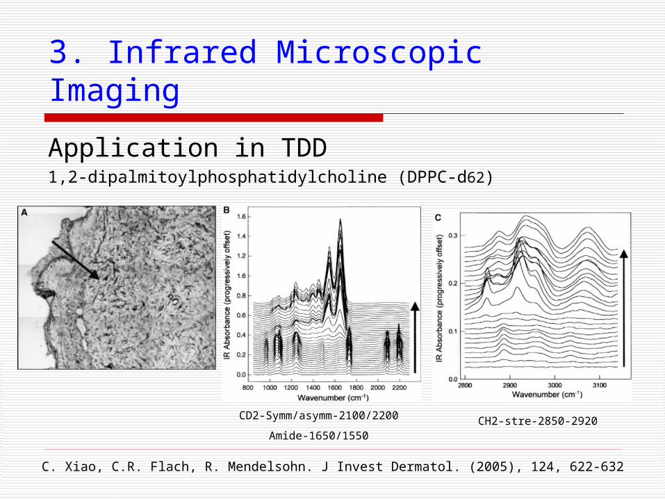
Application in TDD1,2-dipalmitoylphosphatidylcholine (DPPC-d62)
C. Xiao, C.R. Flach, R. Mendelsohn. J Invest Dermatol. (2005), 124, 622-632
CD2-Symm/asymm-2100/2200
Amide-1650/1550CH2-stre-2850-2920
3. Infrared Microscopic Imaging
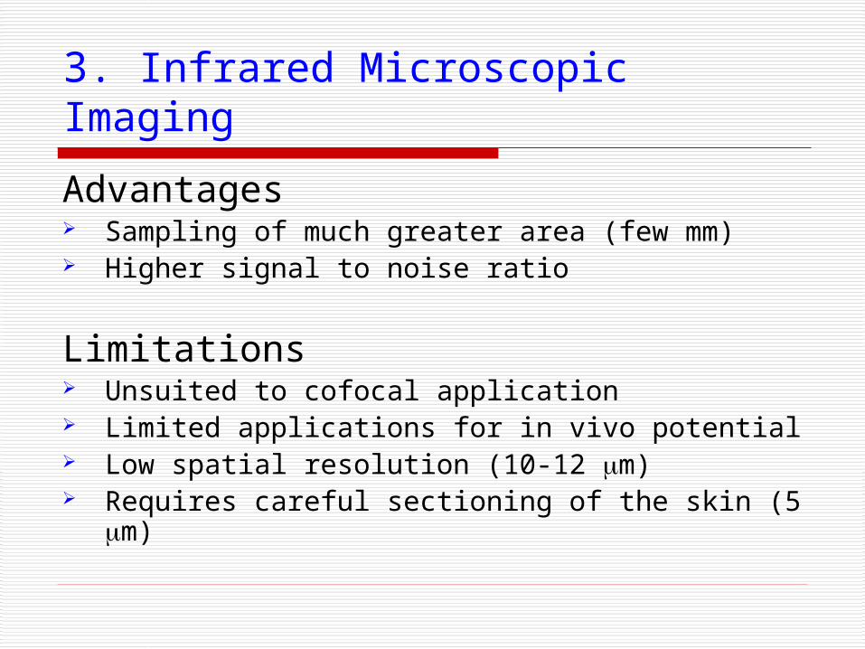
Advantages Sampling of much greater area (few mm) Higher signal to noise ratio
Limitations Unsuited to cofocal application Limited applications for in vivo potential Low spatial resolution (10-12 m) Requires careful sectioning of the skin (5 m)
Methods For Monitoring TDD4. Raman Microscopic Imaging
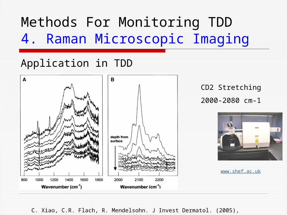
Application in TDD
CD2 Stretching
2000-2080 cm-1
www.shef.ac.uk
C. Xiao, C.R. Flach, R. Mendelsohn. J Invest Dermatol. (2005), 124, 622-632
4. Raman Microscopic Imaging
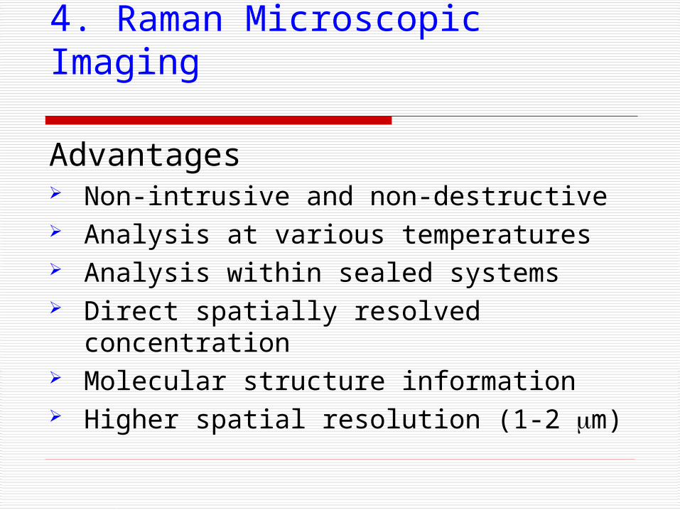
Advantages Non-intrusive and non-destructive Analysis at various temperatures Analysis within sealed systems Direct spatially resolved concentration Molecular structure information Higher spatial resolution (1-2 m)
4. Raman Microscopic Imaging
Applications Surface and materials science Forensic research / investigation Polymer science Geology Pharmaceutical science
Limitations Possibility of errors
Summary
Transdermal drug delivery is an effective technique for steady and consistent drug delivery.
Penetration enhancers and liposomes are good solutions for the slow permeation of the skin.
IR and Raman Microscopic imaging techniques are more useful than others to monitor TDD.

References1. I.V. Zhigaltesv, N. Maurer. Biochemicaet Biophysica Acta. (2002),
1565, 129-135.2. C. Xiao, C.R. Flach, R. Mendelsohn. J Invest Dermatol. (2005), 124,
622-632.3. K.M.Hanson, R.M.Clegg. Biophys J. (2002), 83, 1682–1690.4. E.N.Lewis,P.J.Treado. Anal Chem. (1995), 67, 3377–3381.5. C.Xiao, R.Mendelsohn. Appl Spectrosc. (2004), 58, 382–389.6. P.J.Caspers, G.J.Puppels. Biospectroscopy (1998), 4, S31–S39.
7. www.uspharmacist.com8. http://en.wikipedia.org9. www.shef.ac.uk
Acknowledgements
Dr. Vasilios Gavalas University of Kentucky
Chemistry Department