Embed Size (px)
Citation preview
VOL. 9, NO. 3 - SEPTEMBER, 1998 5 JOURNAL OF BIOMOLECULAR TECHNIQUES
Methods & Reviews
IntroductionBiologic science is in the midst of an information
and technical revolution because of the convergence of de-velopments in many different areas, including genome andproteome sequencing, bioinformatics, and experimental andpredictive structural biology. At the forefront of these de-velopments are new analytic tools for meeting the demandsof increased sequence data production. Prominent amongthese key technologies is biomolecular mass spectrometry(MS) because of the astounding sensitivity and accuracy ofits protein mass determinations and the wealth of informa-tion about molecular composition inherent in a molecularmass. MS provides protein primary structure determinationcapabilities characterized by high accuracy and sensitivityand offers the potential for high throughput. New molecu-lar and structural methods must be developed in order toremain astride the information revolution. The basis for thesemethods is the combined use of proteolytic digestion, massanalysis, and computer-based data analysis.
These methods can identify proteins, such as thosefrom bands in acrylamide gels, using mostly automated pro-tocols and subpicomole quantities. The target protein is com-pletely digested using a sequence-specific protease such astrypsin (eg, within the gel matrix). The resulting fragmentsare extracted and prepared for mass analysis. MS analysiscan yield information on fragment masses with accuracyapproaching ±5 ppm, or ±0.005 Da for a 1000-Da peptide.The protease fragmentation pattern is compared with thepatterns predicted for all proteins within a database, andmatches are statistically evaluated. Because the occurrenceof Arg and Lys residues in proteins is statistically high,trypsin cleavage (specific for Arg and Lys) usually producesa large number of fragments that have a reasonable prob-
Combined Use Of Proteases And Mass SpectrometryIn Structural Biology
Richard W. Kriwacki1 and Gary Siuzdak2
1St. Jude Children�s Research Hospital and 2The Scripps Research Institute
Proteolysis and mass spectrometry methods have been extended to the analysis of higher-order protein structure. Proteases have long been used as probes of native structure,and this approach has been rejuvenated and used in concert with various mass spec-trometry techniques. We discuss the application of protease as probes of native struc-ture, delineate the mass spectrometry methods that are appropriate in these studies, andoffer several innovative case studies to illustrate key concepts in the combined use ofproteolysis and mass spectometry in studies of biomolecular assemblies.
ability of uniquely identifying the target protein. The suc-cess of this strategy relies on the existence of the proteinsequence within the database, and with the sequences ofwhole genomes for several organisms complete (eg, A.fulgidus, Bacillus subtilis, Caenorhabditis elegans, Escheri-chia coli, Saccharomyces cerevisiae) and others well un-derway (eg, Schizosaccharomyces pombe, Homo sapiens),the likelihood for matches is reasonably high. Exact matchesare readily identified, and homologous proteins are identi-fied, albeit with lower statistical significance, placing a tar-get protein within a particular family in the absence of anexact match.
Methods developed for primary sequence identi-fication and elucidation using MS are particularly well suitedfor the analysis of higher-order, native protein structure. MSprotocols used in primary structure analysis are directlytransferable to the analysis of native structure, because theyare used in the readout of information that is indirectly re-lated to structure after the proteolysis reactions are per-formed. Analysis methods, however, must be modified totake into account the added spectral complexity resultingfrom incomplete proteolysis under limiting conditions. Pro-teases have long been used as probes of native structure,and this approach has been rejuvenated because powerfulMS-based methods allow virtually complete identificationof proteolysis reaction products.
We review the application of protease as probes ofnative structure and discuss the MS methods that are ap-propriate in these studies. We provide several innovativecase studies that illustrate key concepts in the combineduse of proteolysis and MS in studies of biomolecular as-semblies.
Corresponding Author: Richard W. Kriwacki, Department of Structural Biology, St. Jude Children�s Research Hospital, 332 N.Lauderdale, Memphis, TN 38105-2794, Tel: (901) 495-2917, Fax: (901) 495-3032, E-mail: [email protected]
JOURNAL OF BIOMOLECULAR TECHNIQUES 6 VOL. 9, NO. 3 - SEPTEMBER, 1998
Methods & Reviews
Limited Proteolysis as a Probe ofHigher-Order Structure
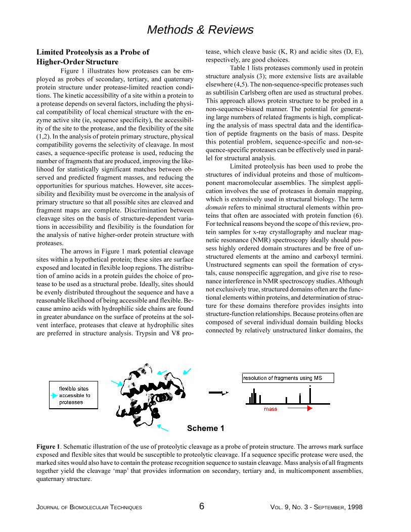
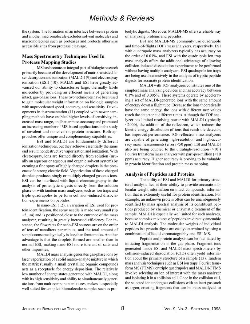
Figure 1 illustrates how proteases can be em-ployed as probes of secondary, tertiary, and quaternaryprotein structure under protease-limited reaction condi-tions. The kinetic accessibility of a site within a protein toa protease depends on several factors, including the physi-cal compatibility of local chemical structure with the en-zyme active site (ie, sequence specificity), the accessibil-ity of the site to the protease, and the flexibility of the site(1,2). In the analysis of protein primary structure, physicalcompatibility governs the selectivity of cleavage. In mostcases, a sequence-specific protease is used, reducing thenumber of fragments that are produced, improving the like-lihood for statistically significant matches between ob-served and predicted fragment masses, and reducing theopportunities for spurious matches. However, site acces-sibility and flexibility must be overcome in the analysis ofprimary structure so that all possible sites are cleaved andfragment maps are complete. Discrimination betweencleavage sites on the basis of structure-dependent varia-tions in accessibility and flexibility is the foundation forthe analysis of native higher-order protein structure withproteases.
The arrows in Figure 1 mark potential cleavagesites within a hypothetical protein; these sites are surfaceexposed and located in flexible loop regions. The distribu-tion of amino acids in a protein guides the choice of pro-tease to be used as a structural probe. Ideally, sites shouldbe evenly distributed throughout the sequence and have areasonable likelihood of being accessible and flexible. Be-cause amino acids with hydrophilic side chains are foundin greater abundance on the surface of proteins at the sol-vent interface, proteases that cleave at hydrophilic sitesare preferred in structure analysis. Trypsin and V8 pro-
tease, which cleave basic (K, R) and acidic sites (D, E),respectively, are good choices.
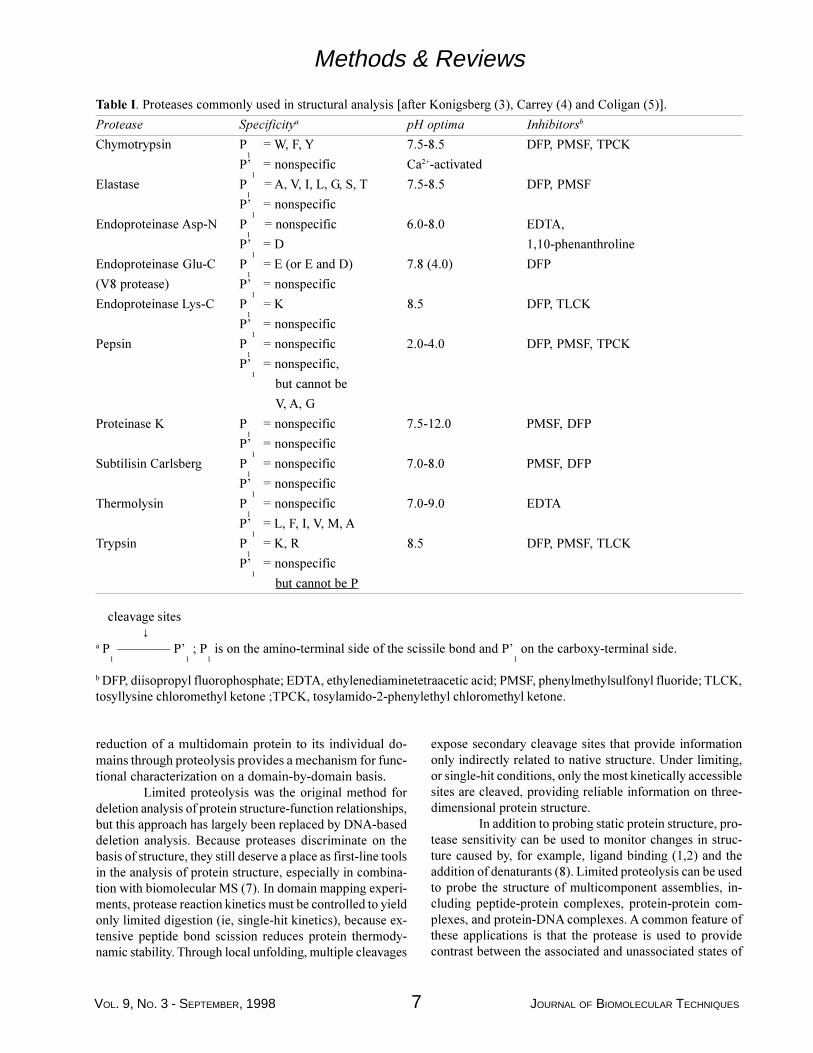
Table 1 lists proteases commonly used in proteinstructure analysis (3); more extensive lists are availableelsewhere (4,5). The non-sequence-specific proteases suchas subtilisin Carlsberg often are used as structural probes.This approach allows protein structure to be probed in anon-sequence-biased manner. The potential for generat-ing large numbers of related fragments is high, complicat-ing the analysis of mass spectral data and the identifica-tion of peptide fragments on the basis of mass. Despitethis potential problem, sequence-specific and non-se-quence-specific proteases can be effectively used in paral-lel for structural analysis.
Limited proteolysis has been used to probe thestructures of individual proteins and those of multicom-ponent macromolecular assemblies. The simplest appli-cation involves the use of proteases in domain mapping,which is extensively used in structural biology. The termdomain refers to minimal structural elements within pro-teins that often are associated with protein function (6).For technical reasons beyond the scope of this review, pro-tein samples for x-ray crystallography and nuclear mag-netic resonance (NMR) spectroscopy ideally should pos-sess highly ordered domain structures and be free of un-structured elements at the amino and carboxyl termini.Unstructured segments can spoil the formation of crys-tals, cause nonspecific aggregation, and give rise to reso-nance interference in NMR spectroscopy studies. Althoughnot exclusively true, structured domains often are the func-tional elements within proteins, and determination of struc-ture for these domains therefore provides insights intostructure-function relationships. Because proteins often arecomposed of several individual domain building blocksconnected by relatively unstructured linker domains, the
Figure 1. Schematic illustration of the use of proteolytic cleavage as a probe of protein structure. The arrows mark surfaceexposed and flexible sites that would be susceptible to proteolytic cleavage. If a sequence specific protease were used, themarked sites would also have to contain the protease recognition sequence to sustain cleavage. Mass analysis of all fragmentstogether yield the cleavage �map� that provides information on secondary, tertiary and, in multicomponent assemblies,quaternary structure.
IOH[LEOH�VLWHV
UHVROXWLRQ�RI�IUDJPHQWV�XVLQJ�06
PDVV
6FKHPH��
DFFHVVLEOH�WR
SURWHDVHV
VOL. 9, NO. 3 - SEPTEMBER, 1998 7 JOURNAL OF BIOMOLECULAR TECHNIQUES
Methods & Reviews
reduction of a multidomain protein to its individual do-mains through proteolysis provides a mechanism for func-tional characterization on a domain-by-domain basis.
Limited proteolysis was the original method fordeletion analysis of protein structure-function relationships,but this approach has largely been replaced by DNA-baseddeletion analysis. Because proteases discriminate on thebasis of structure, they still deserve a place as first-line toolsin the analysis of protein structure, especially in combina-tion with biomolecular MS (7). In domain mapping experi-ments, protease reaction kinetics must be controlled to yieldonly limited digestion (ie, single-hit kinetics), because ex-tensive peptide bond scission reduces protein thermody-namic stability. Through local unfolding, multiple cleavages
expose secondary cleavage sites that provide informationonly indirectly related to native structure. Under limiting,or single-hit conditions, only the most kinetically accessiblesites are cleaved, providing reliable information on three-dimensional protein structure.
In addition to probing static protein structure, pro-tease sensitivity can be used to monitor changes in struc-ture caused by, for example, ligand binding (1,2) and theaddition of denaturants (8). Limited proteolysis can be usedto probe the structure of multicomponent assemblies, in-cluding peptide-protein complexes, protein-protein com-plexes, and protein-DNA complexes. A common feature ofthese applications is that the protease is used to providecontrast between the associated and unassociated states of
Table I. Proteases commonly used in structural analysis [after Konigsberg (3), Carrey (4) and Coligan (5)].
Protease Specificitya pH optima Inhibitorsb
Chymotrypsin P1
= W, F, Y 7.5-8.5 DFP, PMSF, TPCK
P�1
= nonspecific Ca2+-activated
Elastase P1 = A, V, I, L, G, S, T 7.5-8.5 DFP, PMSF
P�1
= nonspecific
Endoproteinase Asp-N P1 = nonspecific 6.0-8.0 EDTA,
P�1
= D 1,10-phenanthroline
Endoproteinase Glu-C P1
= E (or E and D) 7.8 (4.0) DFP
(V8 protease) P�1
= nonspecific
Endoproteinase Lys-C P1
= K 8.5 DFP, TLCK
P�1
= nonspecific
Pepsin P1
= nonspecific 2.0-4.0 DFP, PMSF, TPCK
P�1
= nonspecific,
but cannot be
V, A, G
Proteinase K P1
= nonspecific 7.5-12.0 PMSF, DFP
P�1
= nonspecific
Subtilisin Carlsberg P1
= nonspecific 7.0-8.0 PMSF, DFP
P�1
= nonspecific
Thermolysin P1
= nonspecific 7.0-9.0 EDTA
P�1
= L, F, I, V, M, A
Trypsin P1
= K, R 8.5 DFP, PMSF, TLCK
P�1
= nonspecific
but cannot be P
cleavage sites ↓a P
1 ���� P�
1 ; P
1 is on the amino-terminal side of the scissile bond and P�
1 on the carboxy-terminal side.
b DFP, diisopropyl fluorophosphate; EDTA, ethylenediaminetetraacetic acid; PMSF, phenylmethylsulfonyl fluoride; TLCK,tosyllysine chloromethyl ketone ;TPCK, tosylamido-2-phenylethyl chloromethyl ketone.
JOURNAL OF BIOMOLECULAR TECHNIQUES 8 VOL. 9, NO. 3 - SEPTEMBER, 1998
the system. The formation of an interface between a proteinand another macromolecule excludes solvent molecules andmacromolecules such as proteases and protects otherwiseaccessible sites from protease cleavage.
Mass Spectrometry Techniques Used InProtease Mapping Studies
MS has become an integral part of biologic researchprimarily because of the development of matrix-assisted la-ser desorption and ionization (MALDI) (9) and electrosprayionization (ESI) (10). MALDI and ESI have greatly ad-vanced our ability to characterize large, thermally labilemolecules by providing an efficient means of generatingintact, gas-phase ions. These two techniques have been usedto gain molecular weight information on biologic sampleswith unprecedented speed, accuracy, and sensitivity. Devel-opments in instrumentation (11) coupled with newer sam-pling methods have enabled higher levels of sensitivity, in-creased mass range, and better mass accuracy and promotedan increasing number of MS-based applications in the studyof covalent and noncovalent protein structure. Both ap-proaches offer unique and complementary capabilities.
ESI and MALDI are fundamentally differentionization techniques, but they achieve essentially the sameend result: nondestructive vaporization and ionization. Withelectrospray, ions are formed directly from solution (usu-ally an aqueous or aqueous and organic solvent system) bycreating a fine spray of highly charged droplets in the pres-ence of a strong electric field. Vaporization of these chargeddroplets produces singly or multiply charged gaseous ions.ESI can be interfaced with liquid chromatography in theanalysis of proteolytic digests directly from the solutionphase or with tandem mass analyzers such as ion traps andtriple quadrupoles to perform collision-induced dissocia-tion experiments on peptides.
In nano-ESI (12), a variation of ESI used for pro-tein identification, the spray needle is made very small (tip~5 µm) and is positioned close to the entrance of the massanalyzer, resulting in greatly increased efficiency. For in-stance, the flow rates for nano-ESI sources are on the orderof tens of nanoliters per minute, and the total amount ofsample consumed typically is less than femtomoles. Anotheradvantage is that the droplets formed are smaller than innormal ESI, making nano-ESI more tolerant of salts andother impurities.
MALDI mass analysis generates gas-phase ions bylaser vaporization of a solid matrix-analyte mixture in whichthe matrix (usually a small crystalline organic compound)acts as a receptacle for energy deposition. The relativelylow number of charge states generated with MALDI, alongwith its high sensitivity and ability to simultaneously gener-ate ions from multicomponent mixtures, makes it especiallywell suited for complex biomolecular samples such as pro-
teolytic digests. Moreover, MALDI-MS offers a reliable wayof analyzing proteins and peptides.
ESI and MALDI-MS commonly use quadrupoleand time-of-flight (TOF) mass analyzers, respectively. ESIwith quadrupole mass analyzers typically has accuracy onthe order of 0.01%, and ESI with the quadrupole ion trapmass analysis offers the additional advantage of allowingcollision-induced dissociation experiments to be performedwithout having multiple analyzers. ESI quadrupole ion trapsare being used extensively in the analysis of tryptic peptidedigests for accurate protein identification.
MALDI with TOF analyzers constitutes one of thesimplest mass analyzing devices and has accuracy between0.1% and of 0.005%. These systems operate by accelerat-ing a set of MALDI-generated ions with the same amountof energy down a flight tube. Because the ions theoreticallyhave the same energy, the ions with different m/z valuesreach the detector at different times. Although the TOF ana-lyzer has limited resolving power with MALDI (typically<2000), the addition of the reflectron, which reduces thekinetic energy distribution of ions that reach the detector,has improved performance. TOF reflectron mass analyzersare capable of generating high-resolution and high-accu-racy mass measurements (errors <50 ppm). ESI and MALDIalso are being coupled to the ultrahigh-resolution (>105)Fourier transform mass analyzer with part-per-million (<10ppm) accuracy. Higher accuracy is proving to be valuablein protein identification and protein mass mapping.
Analysis of Peptides and ProteinsThe utility of ESI and MALDI for primary struc-
tural analysis lies in their ability to provide accurate mo-lecular weight information on intact compounds, informa-tion that is extremely useful for protein identification. Forexample, an unknown protein often can be unambiguouslyidentified by mass spectral analysis of its constituent pep-tides produced by chemical or enzymatic treatment of thesample. MALDI is especially well suited for such analyses,because complex mixtures of peptides are directly amenableto MALDI analysis. The molecular weights of individualpeptides in a protein digest are easily determined by using acombination of liquid chromatography and ESI-MS.
Peptide and protein analysis can be facilitated byinitiating fragmentation in the gas phase. Fragment ionsgenerated inside ESI and MALDI mass spectrometers bycollision-induced dissociation (CID) often yield informa-tion about the primary structure of a sample (13). Tandemmass analysis techniques such as ESI ion traps, Fourier trans-form MS (FTMS), or triple quadrupoles and MALDI-FTMSinvolve selecting an ion of interest with the mass analyzerand isolating it in a collision cell. Once in the collision cell,the selected ion undergoes collisions with an inert gas suchas argon, creating fragments that can be mass analyzed to
Methods & Reviews
VOL. 9, NO. 3 - SEPTEMBER, 1998 9 JOURNAL OF BIOMOLECULAR TECHNIQUES
provide information about their sequence. This multiple massanalysis approach is often referred to as tandem MS or MS2.Because the CID behavior of peptides is already well char-acterized, tandem MS with CID can be used to acquire di-rect sequence information on small peptides (<3 kDa).
Two important advantages of MALDI-MS are itssensitivity and ability to analyze complex polypeptide mix-tures. These features also are being used to sequencebiopolymers. The protein ladder sequencing technique origi-nated by Chait et al. (14) allows stepwise removal of eachamino acid in a peptide, a process in which each residue ischemically or proteolytically removed from the amino-ter-minal end to produce sequence-defining peptide fragments.Alternatively, amino acids can be enzymatically removedfrom the carboxyl terminus (15). A MALDI mass spectralreadout enables generation of the resulting protein sequenc-ing ladder. This method, which allows each amino acid tobe identified from the mass difference between successivepeaks, can provide sequence information on peptides ofmore than 30 residues. Sequence data has been obtainedfrom larger proteins by enzymatic cleavage combined withprotein ladder sequencing (15-17).
Software Packages for Spectrographic Analy-sis of Proteolytic Digests
Several software packages have been developedto facilitate the analysis of MS data for proteins and havebeen especially useful in the analysis of multicomponentfragmentation patterns generated through proteolytic diges-tion. Two such software packages are described to illustratesome of the features that are available. These two packagesare available free of charge to not-for-profit organizationsover the World Wide Web.
The software package PAWS (Proteometrics,Rockefeller University, New York, NY, freeware;www.proteometrics.com) offers a user-friendly, intuitiveinterface that allows a wide variety of MS-related, protein-based operations to be performed. Protein sequences canbe loaded from files or entered manually and themonoisotopic or average molecular mass calculated. Theprogram allows specific amino acid sites to be chemicallymodified and the modified mass to be calculated. Theoreti-cal cleavage reactions (enzymatic or chemical) can be per-formed and the resulting fragment masses calculated andpresented in tabular and graphical form. Searching tools havebeen incorporated to aid the analysis of MS data from pro-teolytic or chemical cleavage reactions. A simple search toolidentifies peptide sequences derived from a known proteinsequence that match a particular target mass. Searches con-sider all possible peptide sequences or only sequences con-sistent with fragmentation resulting from a particular se-quence-specific cleavage reaction. Lists of fragment massescan be entered manually or imported from files, and mul-
tiple searches can be performed. Search results are presentedin graphical and tabular forms. The PAWS package offers apowerful and easy-to-use set of tools to predict cleavagepatterns and fragment masses and to search for and identifypeptides derived from known protein sequences that matchexperimentally determined masses.
The Protein Prospector package (University ofCalifornia at San Francisco Mass Spectrometry Facility, SanFrancisco, CA; Drs. Karl Clauser and Peter Baker;prospector.ucsf.edu) offers some of the same capabilitiesas PAWS, along with extensive database searching capa-bilities. The software, which can be used over the Internetor installed locally after appropriate licensing, is dividedinto several modules that perform specific functions. Theseinclude MS-Digest, which can be used to generate theoreti-cal protein digests; MS-Product, which can predict frag-mentation in the mass spectrometer caused by post-sourcedecay and collision-induced dissociation; and MS-Comp,which can suggest amino acid compositions consistent withexperimental MS data, including parent peptide mass andimmonium ion fragmentation data. Database searching toolsinclude MS-Fit, which compares an observed set of pro-teolytic fragment masses with those predicted for all pro-teins in a database; MS-Tag, which compares tandem MSpeptide fragmentation patterns with predicted patterns; andMS-Edman, which compares short segments of a peptidesequence with segments in a protein database. The data-base searching tools in Protein Prospector have been devel-oped to aid the identification of unknown proteins on thebasis of MS data but are also well suited for the analysis ofMS data from protease-based structure mapping experi-ments. The MS-Fit module can be used to match observedfragment masses against those predicted for the protein be-ing studied. Instead of using the entire nonredundant pro-tein database for comparison, the content of the database islimited to the protein under study. Because the proteasecleavage reactions for mapping studies are performed un-der limiting conditions, the fragments that are produced mayspan one or more uncleaved protease sites, increasing thenumber of theoretical fragments that must be consideredduring the comparisons. However, MS-Fit accommodatesthis need with a settable parameter corresponding to themaximum number of missed cleavages. This capability isalso included in MS-Digest, allowing tables of all possiblepeptide fragments to be generated.
Analysis of Protein-DNA InteractionsThe first application of limited proteolysis and
MALDI mass analysis to the study of a multicomponentbiomolecular assembly was published in 1995 by Chait etal. (18). This combined approach was used in structuralanalysis of the transcription factor Max when free in solu-tion and when bound to an oligonucleotide containing its
Methods & Reviews
JOURNAL OF BIOMOLECULAR TECHNIQUES 10 VOL. 9, NO. 3 - SEPTEMBER, 1998
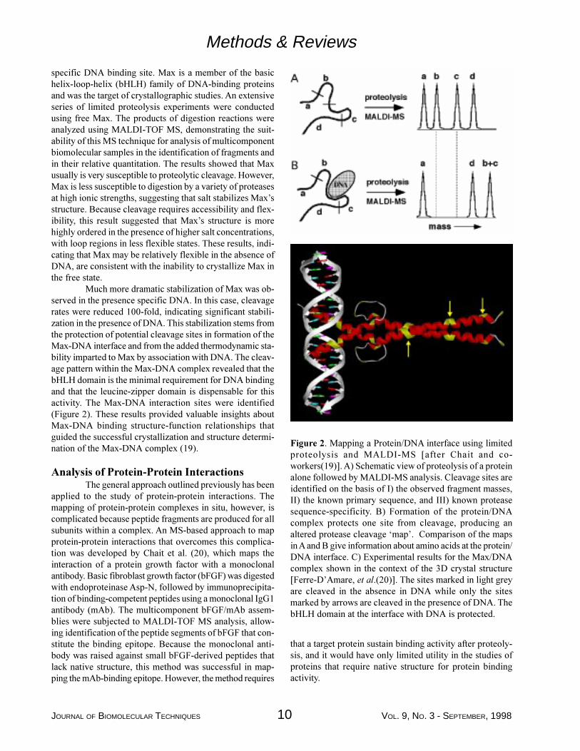
Figure 2. Mapping a Protein/DNA interface using limitedproteolysis and MALDI-MS [after Chait and co-workers(19)]. A) Schematic view of proteolysis of a proteinalone followed by MALDI-MS analysis. Cleavage sites areidentified on the basis of I) the observed fragment masses,II) the known primary sequence, and III) known proteasesequence-specificity. B) Formation of the protein/DNAcomplex protects one site from cleavage, producing analtered protease cleavage �map�. Comparison of the mapsin A and B give information about amino acids at the protein/DNA interface. C) Experimental results for the Max/DNAcomplex shown in the context of the 3D crystal structure[Ferre-D�Amare, et al.(20)]. The sites marked in light greyare cleaved in the absence in DNA while only the sitesmarked by arrows are cleaved in the presence of DNA. ThebHLH domain at the interface with DNA is protected.
specific DNA binding site. Max is a member of the basichelix-loop-helix (bHLH) family of DNA-binding proteinsand was the target of crystallographic studies. An extensiveseries of limited proteolysis experiments were conductedusing free Max. The products of digestion reactions wereanalyzed using MALDI-TOF MS, demonstrating the suit-ability of this MS technique for analysis of multicomponentbiomolecular samples in the identification of fragments andin their relative quantitation. The results showed that Maxusually is very susceptible to proteolytic cleavage. However,Max is less susceptible to digestion by a variety of proteasesat high ionic strengths, suggesting that salt stabilizes Max�sstructure. Because cleavage requires accessibility and flex-ibility, this result suggested that Max�s structure is morehighly ordered in the presence of higher salt concentrations,with loop regions in less flexible states. These results, indi-cating that Max may be relatively flexible in the absence ofDNA, are consistent with the inability to crystallize Max inthe free state.
Much more dramatic stabilization of Max was ob-served in the presence specific DNA. In this case, cleavagerates were reduced 100-fold, indicating significant stabili-zation in the presence of DNA. This stabilization stems fromthe protection of potential cleavage sites in formation of theMax-DNA interface and from the added thermodynamic sta-bility imparted to Max by association with DNA. The cleav-age pattern within the Max-DNA complex revealed that thebHLH domain is the minimal requirement for DNA bindingand that the leucine-zipper domain is dispensable for thisactivity. The Max-DNA interaction sites were identified(Figure 2). These results provided valuable insights aboutMax-DNA binding structure-function relationships thatguided the successful crystallization and structure determi-nation of the Max-DNA complex (19).
Analysis of Protein-Protein InteractionsThe general approach outlined previously has been
applied to the study of protein-protein interactions. Themapping of protein-protein complexes in situ, however, iscomplicated because peptide fragments are produced for allsubunits within a complex. An MS-based approach to mapprotein-protein interactions that overcomes this complica-tion was developed by Chait et al. (20), which maps theinteraction of a protein growth factor with a monoclonalantibody. Basic fibroblast growth factor (bFGF) was digestedwith endoproteinase Asp-N, followed by immunoprecipita-tion of binding-competent peptides using a monoclonal IgG1antibody (mAb). The multicomponent bFGF/mAb assem-blies were subjected to MALDI-TOF MS analysis, allow-ing identification of the peptide segments of bFGF that con-stitute the binding epitope. Because the monoclonal anti-body was raised against small bFGF-derived peptides thatlack native structure, this method was successful in map-ping the mAb-binding epitope. However, the method requires
Methods & Reviews
that a target protein sustain binding activity after proteoly-sis, and it would have only limited utility in the studies ofproteins that require native structure for protein bindingactivity.
VOL. 9, NO. 3 - SEPTEMBER, 1998 11 JOURNAL OF BIOMOLECULAR TECHNIQUES
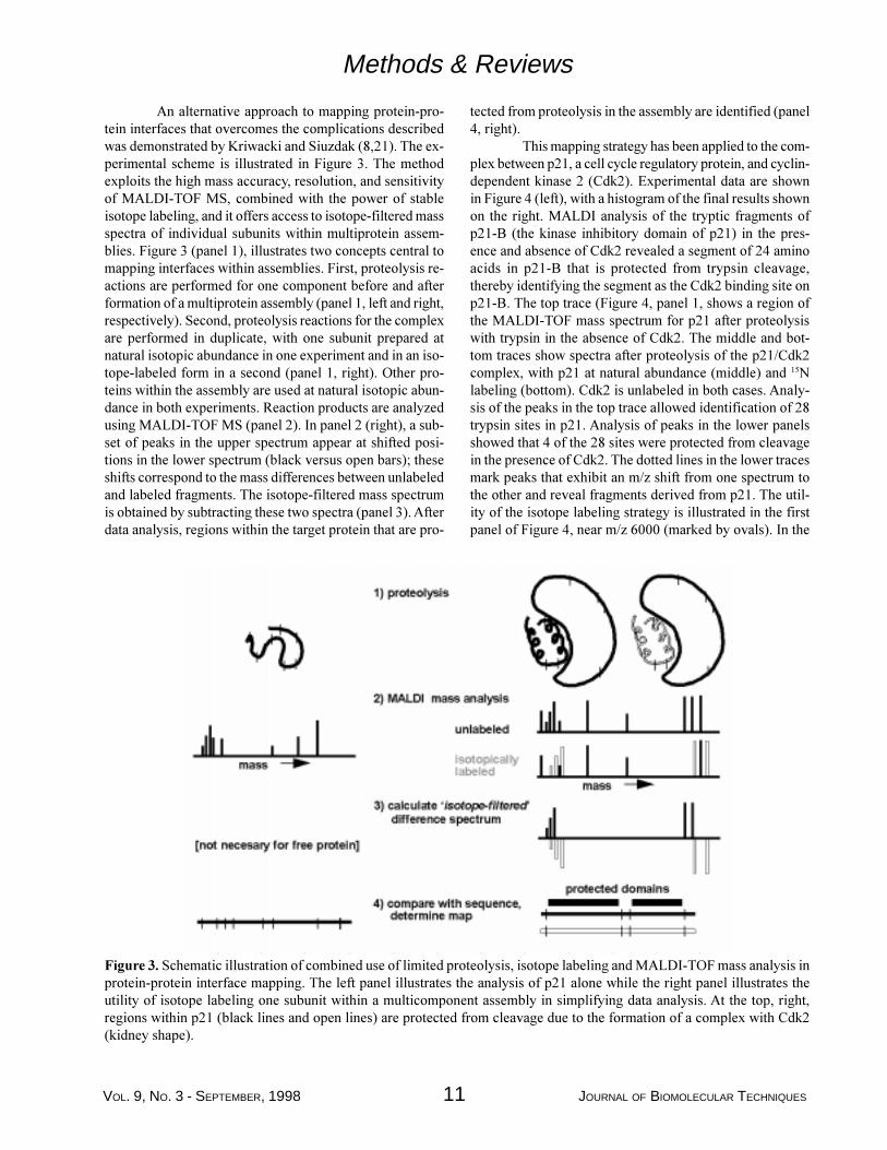
An alternative approach to mapping protein-pro-tein interfaces that overcomes the complications describedwas demonstrated by Kriwacki and Siuzdak (8,21). The ex-perimental scheme is illustrated in Figure 3. The methodexploits the high mass accuracy, resolution, and sensitivityof MALDI-TOF MS, combined with the power of stableisotope labeling, and it offers access to isotope-filtered massspectra of individual subunits within multiprotein assem-blies. Figure 3 (panel 1), illustrates two concepts central tomapping interfaces within assemblies. First, proteolysis re-actions are performed for one component before and afterformation of a multiprotein assembly (panel 1, left and right,respectively). Second, proteolysis reactions for the complexare performed in duplicate, with one subunit prepared atnatural isotopic abundance in one experiment and in an iso-tope-labeled form in a second (panel 1, right). Other pro-teins within the assembly are used at natural isotopic abun-dance in both experiments. Reaction products are analyzedusing MALDI-TOF MS (panel 2). In panel 2 (right), a sub-set of peaks in the upper spectrum appear at shifted posi-tions in the lower spectrum (black versus open bars); theseshifts correspond to the mass differences between unlabeledand labeled fragments. The isotope-filtered mass spectrumis obtained by subtracting these two spectra (panel 3). Afterdata analysis, regions within the target protein that are pro-
Methods & Reviews
tected from proteolysis in the assembly are identified (panel4, right).
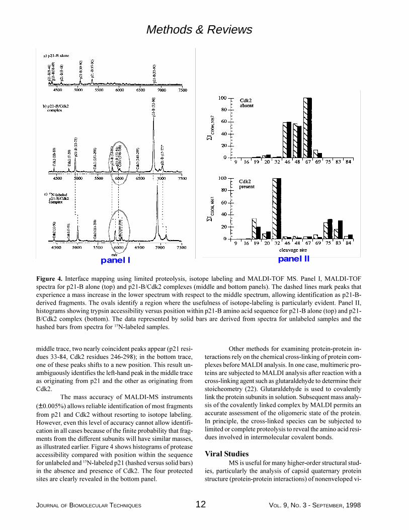
This mapping strategy has been applied to the com-plex between p21, a cell cycle regulatory protein, and cyclin-dependent kinase 2 (Cdk2). Experimental data are shownin Figure 4 (left), with a histogram of the final results shownon the right. MALDI analysis of the tryptic fragments ofp21-B (the kinase inhibitory domain of p21) in the pres-ence and absence of Cdk2 revealed a segment of 24 aminoacids in p21-B that is protected from trypsin cleavage,thereby identifying the segment as the Cdk2 binding site onp21-B. The top trace (Figure 4, panel 1, shows a region ofthe MALDI-TOF mass spectrum for p21 after proteolysiswith trypsin in the absence of Cdk2. The middle and bot-tom traces show spectra after proteolysis of the p21/Cdk2complex, with p21 at natural abundance (middle) and 15Nlabeling (bottom). Cdk2 is unlabeled in both cases. Analy-sis of the peaks in the top trace allowed identification of 28trypsin sites in p21. Analysis of peaks in the lower panelsshowed that 4 of the 28 sites were protected from cleavagein the presence of Cdk2. The dotted lines in the lower tracesmark peaks that exhibit an m/z shift from one spectrum tothe other and reveal fragments derived from p21. The util-ity of the isotope labeling strategy is illustrated in the firstpanel of Figure 4, near m/z 6000 (marked by ovals). In the
Figure 3. Schematic illustration of combined use of limited proteolysis, isotope labeling and MALDI-TOF mass analysis inprotein-protein interface mapping. The left panel illustrates the analysis of p21 alone while the right panel illustrates theutility of isotope labeling one subunit within a multicomponent assembly in simplifying data analysis. At the top, right,regions within p21 (black lines and open lines) are protected from cleavage due to the formation of a complex with Cdk2(kidney shape).
JOURNAL OF BIOMOLECULAR TECHNIQUES 12 VOL. 9, NO. 3 - SEPTEMBER, 1998
Methods & Reviews
middle trace, two nearly coincident peaks appear (p21 resi-dues 33-84, Cdk2 residues 246-298); in the bottom trace,one of these peaks shifts to a new position. This result un-ambiguously identifies the left-hand peak in the middle traceas originating from p21 and the other as originating fromCdk2.
The mass accuracy of MALDI-MS instruments(±0.005%) allows reliable identification of most fragmentsfrom p21 and Cdk2 without resorting to isotope labeling.However, even this level of accuracy cannot allow identifi-cation in all cases because of the finite probability that frag-ments from the different subunits will have similar masses,as illustrated earlier. Figure 4 shows histograms of proteaseaccessibility compared with position within the sequencefor unlabeled and 15N-labeled p21 (hashed versus solid bars)in the absence and presence of Cdk2. The four protectedsites are clearly revealed in the bottom panel.
SDQHO�, SDQHO�,,
Figure 4. Interface mapping using limited proteolysis, isotope labeling and MALDI-TOF MS. Panel I, MALDI-TOFspectra for p21-B alone (top) and p21-B/Cdk2 complexes (middle and bottom panels). The dashed lines mark peaks thatexperience a mass increase in the lower spectrum with respect to the middle spectrum, allowing identification as p21-B-derived fragments. The ovals identify a region where the usefulness of isotope-labeling is particularly evident. Panel II,histograms showing trypsin accessibility versus position within p21-B amino acid sequence for p21-B alone (top) and p21-B/Cdk2 complex (bottom). The data represented by solid bars are derived from spectra for unlabeled samples and thehashed bars from spectra for 15N-labeled samples.
Other methods for examining protein-protein in-teractions rely on the chemical cross-linking of protein com-plexes before MALDI analysis. In one case, multimeric pro-teins are subjected to MALDI analysis after reaction with across-linking agent such as glutaraldehyde to determine theirstoicheometry (22). Glutaraldehyde is used to covalentlylink the protein subunits in solution. Subsequent mass analy-sis of the covalently linked complex by MALDI permits anaccurate assessment of the oligomeric state of the protein.In principle, the cross-linked species can be subjected tolimited or complete proteolysis to reveal the amino acid resi-dues involved in intermolecular covalent bonds.
Viral StudiesMS is useful for many higher-order structural stud-
ies, particularly the analysis of capsid quaternary proteinstructure (protein-protein interactions) of nonenveloped vi-
VOL. 9, NO. 3 - SEPTEMBER, 1998 13 JOURNAL OF BIOMOLECULAR TECHNIQUES
Methods & Reviews
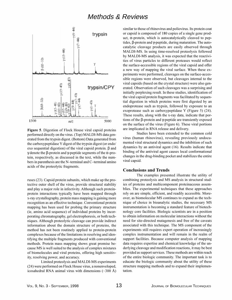
Figure 5. Digestion of Flock House viral capsid proteinsperformed directly on the virus. (Top) MALDI-MS data gen-erated from the trypsin digest. (Bottom) Data generated fromthe carboxypeptidase Y digest of the trypsin digest (or endo/exo sequential digestion) of the viral capsid protein. β andγ denote the β-protein and γ-peptide segments of the α-pro-tein, respectively, as discussed in the text, while the num-bers in parenthesis are the N�terminal and C�terminal aminoacids of the proteolytic fragments.
ruses (23). Capsid protein subunits, which make up the pro-tective outer shell of the virus, provide structural stabilityand play a major role in infectivity. Although such protein-protein interactions typically have been mapped throughx-ray crystallography, protein mass mapping is gaining morerecognition as an effective technique. Conventional proteinmapping has been used for probing the primary structure(ie, amino acid sequence) of individual proteins by incor-porating chromatography, gel electrophoresis, or both tech-niques. Although proteolytic cleavage can provide indirectinformation about the domain structure of proteins, themethod has not been routinely applied to protein-proteincomplexes because of the limitations in resolving and iden-tifying the multiple fragments produced with conventionalmethods. Protein mass mapping shows great promise be-cause MS is well suited to the analysis of complex mixturesof biomolecules and viral proteins, offering high sensitiv-ity, resolving power, and accuracy.
Limited proteolysis and MALDI-MS experiments(24) were performed on Flock House virus, a nonenveloped,icosahedral RNA animal virus with dimensions (~300 Å)
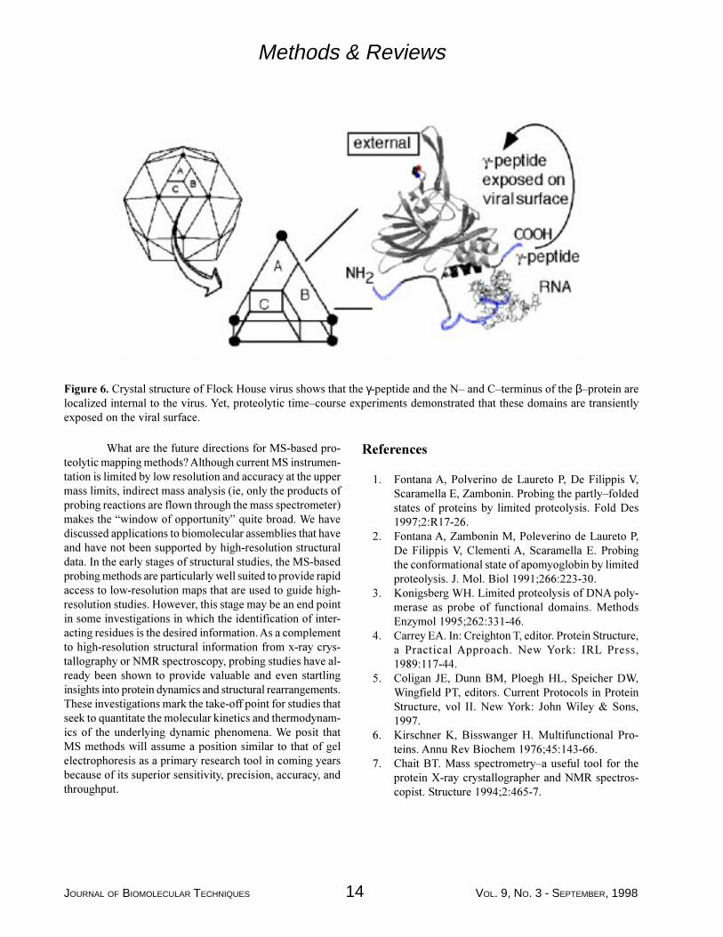
similar to those of rhinovirus and poliovirus. Its protein coator capsid is composed of 180 copies of a single gene prod-uct, α-protein, which is autocatalytically cleaved to pep-tides, β-protein and γ-peptide, during maturation. The auto-catalytic cleavage products are easily observed throughMALDI-MS. In using time-resolved proteolysis followedby MALDI-MS analysis, it was expected that the reactivi-ties of virus particles to different proteases would reflectthe surface-accessible regions of the viral capsid and offera new way of mapping the viral surface. When these ex-periments were performed, cleavages on the surface-acces-sible regions were observed, but cleavages internal to theviral capsids (based on the crystal structure) were also gen-erated. Observation of such cleavages was a surprising andinitially perplexing result. In these studies, identification ofthe viral capsid protein fragments was facilitated by sequen-tial digestion in which proteins were first digested by anendoprotease such as trypsin, followed by exposure to anexoprotease such as carboxypeptidase Y (Figure 5) (24).These results, along with the x-ray data, indicate that por-tions of the β-protein and γ-peptide are transiently exposedon the surface of the virus (Figure 6). These viral portionsare implicated in RNA release and delivery.
Studies have been extended to the common coldvirus (human rhinovirus), revealing previously undocu-mented viral structural dynamics and the inhibition of suchdynamics by an antiviral agent (16). Results indicate thatbinding of the antiviral agent causes local conformationalchanges in the drug-binding pocket and stabilizes the entireviral capsid.
Conclusions and TrendsThe examples presented illustrate the utility of
combining proteolysis and MS analysis in structural stud-ies of proteins and multicomponent proteinaceous assem-blies. The experimental techniques that these approachesrely on are simple, efficient, and readily accessible. More-over, as biomolecular MS continues to expand as the tech-nique of choice in bioanalytic studies, the necessary MSinstrumentation is becoming a standard feature of biotech-nology core facilities. Biologic scientists are in a positionto obtain information on molecular interactions without theneed for site-directed mutagenesis and free of the caveatsassociated with this technique. The MS component of theexperiments still requires expert operation of increasinglycomplex instrumentation and will remain in the realm ofsupport facilities. Because computer analysis of mappingdata requires expertise and chemical knowledge of the un-derlying cleavage and modification reactions, it may be bestprovided as support services. These methods are within reachof the entire biologic community. The important task is toeducate the biologic community about the utility of thesestructure mapping methods and to expand their implemen-tation.
JOURNAL OF BIOMOLECULAR TECHNIQUES 14 VOL. 9, NO. 3 - SEPTEMBER, 1998
What are the future directions for MS-based pro-teolytic mapping methods? Although current MS instrumen-tation is limited by low resolution and accuracy at the uppermass limits, indirect mass analysis (ie, only the products ofprobing reactions are flown through the mass spectrometer)makes the �window of opportunity� quite broad. We havediscussed applications to biomolecular assemblies that haveand have not been supported by high-resolution structuraldata. In the early stages of structural studies, the MS-basedprobing methods are particularly well suited to provide rapidaccess to low-resolution maps that are used to guide high-resolution studies. However, this stage may be an end pointin some investigations in which the identification of inter-acting residues is the desired information. As a complementto high-resolution structural information from x-ray crys-tallography or NMR spectroscopy, probing studies have al-ready been shown to provide valuable and even startlinginsights into protein dynamics and structural rearrangements.These investigations mark the take-off point for studies thatseek to quantitate the molecular kinetics and thermodynam-ics of the underlying dynamic phenomena. We posit thatMS methods will assume a position similar to that of gelelectrophoresis as a primary research tool in coming yearsbecause of its superior sensitivity, precision, accuracy, andthroughput.
Figure 6. Crystal structure of Flock House virus shows that the γ-peptide and the N� and C�terminus of the β�protein arelocalized internal to the virus. Yet, proteolytic time�course experiments demonstrated that these domains are transientlyexposed on the viral surface.
References
1. Fontana A, Polverino de Laureto P, De Filippis V,Scaramella E, Zambonin. Probing the partly�foldedstates of proteins by limited proteolysis. Fold Des1997;2:R17-26.
2. Fontana A, Zambonin M, Poleverino de Laureto P,De Filippis V, Clementi A, Scaramella E. Probingthe conformational state of apomyoglobin by limitedproteolysis. J. Mol. Biol 1991;266:223-30.
3. Konigsberg WH. Limited proteolysis of DNA poly-merase as probe of functional domains. MethodsEnzymol 1995;262:331-46.
4. Carrey EA. In: Creighton T, editor. Protein Structure,a Practical Approach. New York: IRL Press,1989:117-44.
5. Coligan JE, Dunn BM, Ploegh HL, Speicher DW,Wingfield PT, editors. Current Protocols in ProteinStructure, vol II. New York: John Wiley & Sons,1997.
6. Kirschner K, Bisswanger H. Multifunctional Pro-teins. Annu Rev Biochem 1976;45:143-66.
7. Chait BT. Mass spectrometry�a useful tool for theprotein X-ray crystallographer and NMR spectros-copist. Structure 1994;2:465-7.
Methods & Reviews
VOL. 9, NO. 3 - SEPTEMBER, 1998 15 JOURNAL OF BIOMOLECULAR TECHNIQUES
Methods & Reviews
8. Kriwacki RW, Wu J, Tennant L, Wright PE, SiuzdakG. Probing protein structure using biochemical andbiophysical methods. Proteolysis, matrix�assisted la-ser desorption/ionization mass spectrometry, high�performance liquid chromatography and size�exclu-sion chromatography of p21/Waf1/Cip1/Sdi1. JChromatogr A 1997;777:23-30.
9. Hillenkamp F, Karas M, Beavis RC, Chait BT. Ma-trix�assisted laser desorption/ionization mass spec-trometry of biopolymers. Anal Chem 1991;63:A1193-1201.
10. Fenn JB, Mann M, Meng CK, Wong SF, WhitehouseCM. Electrospray ionization�principles and prac-tice. Mass Spectr Rev 1990;9:37-70.
11. Siuzdak G. Mass Spectrometry for Biotechnology.San Diego: Academic Press, 1996.
12. Wilm MS, Mann M. Electrospray and Taylor�conetheory, Dole�s beam of macromolecules at last. Int JMass Spectr Ion Process 1994;136:167-80.
13. Papayannopoulos IA. The interpretation of collision�induced dissociation tandem mass�spectra of pep-tides. Mass Spectr Rev 1995;14:49-73.
14. Chait BT, Wang R, Beavis C, Kent SB. Protein lad-der sequencing. Science 1993;262:89-92.
15. Patterson DH, Tarr GE, Regnier FE, Martin SA. C�terminal ladder sequencing via matrix�assisted laserdesorption mass spectrometry coupled with carbox-ypeptidase Y time-dependent and concentration�de-pendent digestions. Anal Chem 1995;67:3971-8.
16. Thiede B, Wittmann-Liebold B, Bienert M, KrauseE. MALDI-MS for C�terminal sequence determina-tion of peptides and proteins degraded by carbox-ypeptidase Y and P. FEBS Lett 1995;357:65-9.
17. Woods AS, Huang AYC, Cotter RJ, Pasternack GR,Pardoll DM, Jaffee EM. Simplified high�sensitivitysequencing of a major histocompatibility complexclass I�associated immunoreactive peptide using ma-trix-assisted laser desorption/ionization mass spec-trometry. Anal Biochem 1995;226:15-25.
18. Cohen SL, Ferre-D�Amare AR, Burley SK, Chait BT.Probing the solution structure of the DNA�bindingprotein Max by a combination of proteolysis and massspectrometry. Protein Sci 1995;4:1088-99.
19. Ferre-D�Amare AR, Prendergast GC, Ziff EB, BurleySK. Recognition by Max of its cognate DNA througha dimeric b/HLH/Z domain. Nature 1993;363:38-45.
20. Zhao Y, Muir TW, Kent SBH, Tischer E, ScardinaJM, Chait BT. Mapping protein�protein interactionsby affinity�directed mass spectrometry. Proc NatlAcad Sci USA 1996;93:4020-4.
21. Kriwacki RW, Wu J, Tennant L, Siuzdak G, WrightPE. Probing protein/protein interactions with massspectrometry and isotopic labeling: analysis of thep21/Cdk2 complex. J Am Chem Soc 1996;118:5320-1.
22. Farmer TB, Caprioli RM. Mass discrimination inmatrix�assisted laser desorption ionization time�of�flight mass spectrometry�a study using cross�linkedoligomeric complexes. J Mass Spectr 1995;30:1245-54.
23. Siuzdak G. Probing viruses with mass spectrometry.J Mass Spectr 1998;33:203-11.
24. Bothner B, Dong XF, Bibbs L, Johnson JW, SiuzdakG. Evidence of viral capsid dynamics using limitedproteolysis and mass spectrometry. J Biol Chem1998:273:673-6.