Embed Size (px)
Citation preview

Sabnis et al. Journal of Translational Medicine 2014, 12:166http://www.translational-medicine.com/content/12/1/166
METHODOLOGY Open Access
Capillary nano-immunoassay for Akt 1/2/3 and4EBP1 phosphorylation in acute myeloid leukemiaHimalee Sabnis1,2, Heath L Bradley1, Silvia T Bunting1,2, Todd M Cooper1,2 and Kevin D Bunting1*
Abstract
Background: Overall cure rates in acute myeloid leukemia (AML) continue to range between 60-65% with diseaserelapse being a major cause of mortality. The PI3K-Akt-mTOR kinase pathway plays a vital role in pro-survival signalswithin leukemic cells and inhibition of this pathway is being investigated to improve patient outcomes. Trackingactivation of multiple signaling proteins simultaneously in patient samples can be challenging especially withlimiting cell numbers within rare sub-populations.
Methods: The NanoPro 1000 system (ProteinSimple) is built on an automated, capillary-based immunoassayplatform and enables a rapid and quantitative analysis of specific proteins and their phosphorylation states. Wehave utilized this nano-immunoassay to examine activation of Akt 1/2/3 and downstream mTOR target - eukaryoticinitiation factor 4E-Binding Protein 1 (4EBP1).
Results: Assays for Akt 1/2/3 and 4EBP1 were standardized using AML cell lines (MV4-11, MOLM-14, OCI-AML3and HL-60) prior to testing in patient samples. Target inhibition was studied using mTOR 1/2 inhibitor AZD-8055and results were corroborated by Western blotting. The assay was able to quantify nanogram amounts of 4EBP1and Akt 1/2/3 in AML cell lines and primary pediatric AML samples and results were quantifiable, consistent andreproducible.
Conclusion: Our data provides a strong basis for testing this platform on a larger scale and our long term aim isto utilize this nano-immunoassay prospectively in de-novo AML to be able to identify poor responders who mightbenefit from early introduction of targeted therapy.
Keywords: Nano-immunoassay, Biomarker, Leukemia, Capillary electrophoresis, mTOR
IntroductionAcute myeloid leukemia (AML) affects 16,000 -18,000people annually in the United States and approximately75% will succumb to the illness [1]. 6% of all patientsaffected are under the age of 20 years [1]. In spite ofthe advances made in the treatment of acute myeloidleukemia with chemotherapy as well as hematopoieticstem cell transplantation, overall cure rates remain at60-65% with relapse being a major cause of mortality[2]. Of those relapsed patients, only a third are salvage-able with current treatment regimens [3,4]. Discovery ofboth cytogenetic and molecular abnormalities in AMLhas resulted in the development of the current prognos-tic sub-groups in AML [5] and the molecular
* Correspondence: [email protected] of Pediatrics, Aflac Cancer and Blood Disorders Center, EmoryUniversity, 1760 Haygood Drive NE, Atlanta, Georgia, USAFull list of author information is available at the end of the article
© 2014 Sabnis et al.; licensee BioMed CentralCommons Attribution License (http://creativecreproduction in any medium, provided the orDedication waiver (http://creativecommons.orunless otherwise stated.
abnormalities play an important role in leukemogenesis,especially in patients with normal cytogenetics [6].Downstream of these molecular aberrations in leukemic
cells, highly complex and inter-linked networks of signal-ing pathways control cell survival growth, proliferation,self renewal and differentiation. Up-regulation of thePI3K-Akt-mTOR (PI3K-Akt-mammalian target of rapa-mycin) pathway occurs via mutations in surface receptorslike FLT3, c-Kit or by mutations in the genes encodingpathway constituents like PI3K, PTEN or Akt [7,8] . Akt isa serine/threonine protein kinase that exists in three con-served isoforms: Akt 1, 2 and 3. Of the three iso-formspresent, Akt 1 and 2 are expressed to a higher extent inhematopoietic stem cells [9]. Akt is phosphorylated at Thr308 by up-stream phosphoinositide-dependent proteinkinase 1 (PDK-1) and at Ser 473 by mTOR complex 2(mTORC2). Akt plays an important role in key cellular
Ltd. This is an Open Access article distributed under the terms of the Creativeommons.org/licenses/by/2.0), which permits unrestricted use, distribution, andiginal work is properly credited. The Creative Commons Public Domaing/publicdomain/zero/1.0/) applies to the data made available in this article,
Sabnis et al. Journal of Translational Medicine 2014, 12:166 Page 2 of 13http://www.translational-medicine.com/content/12/1/166
processes such as protein translation, cell proliferation,cell cycle, and apoptosis through its multiple downstreamtargets however activating mutations in Akt have not beendescribed in AML [10] . Akt can be constitutively phos-phorylated in AML which results in depletion of normalhematopoietic stem cells [11].Activation of the mTOR pathway is seen in up to 80%
of AML patients and is associated with a shortenedoverall survival. mTOR kinase is also a serine/threoninekinase that complexes with other proteins [12]. mTORC2mainly functions to phosphorylate and activate Aktwhereas mTORC1 plays a central role in the translationalmachinery of normal and leukemic cells via its down-stream targets - p70S6 Kinase and eukaryotic initiationfactor (eIF) 4E binding protein-1 (4EBP1) [12,13]. p70S6Kinase phosphorylates the 40S ribosomal subunit proteinS6 and thereby allows translation of proteins involved incell growth and hypertrophy. 4EBP1 phosphorylationresults in release of the inhibition of eIF4E and enablesthe formation of eIF4F complex. This complex is neces-sary for the cap-dependent translation of highly structuredmRNAs which encode genes such as c-Myc, Mcl-1 andVEGF that are involved in cell survival [13]. In certainsubtypes of AML (FAB M4/M5) eIF4E itself has beenshown to function as an oncogene via transcriptional up-regulation by nuclear factor kappa-light-chain-enhancer ofactivated B cells (NF-κB) [14]. Both p70S6 Kinase and4EBP1 are downstream targets of mTOR however, inhib-ition of 4EBP1 phosphorylation is key for ensuring efficacyof mTOR antagonists [15]. Thus inhibiting downstreammTOR targets has played a prominent role in anti-leukemic therapy for several years and continues to be anactive area of research [12].Molecular differences in Akt-mTOR pathway with
AML patients may provide key information to betterdefine the pathogenesis of disease, especially in patientswith normal cytogenetics. Traditionally, techniques suchas Western Blot and intra-cellular flow cytometry havebeen used for this purpose but these have several limita-tions - they require large number of cells, require tech-nical expertise and quantitative results are difficult toobtain. The NanoPro 1000 system (ProteinSimple) en-ables a rapid and quantitative analysis of specific pro-teins from small quantities of sample (dependent on cellsize and percentage of protein). The NanoPro providesprecise and quantitative data of the multi-site phosphor-ylation states of a specific protein of interest. This degreeof phospho-protein specificity and rapid turn-aroundtime are unique. The system is built on an automated,capillary based immunoassay platform. Proteins are sep-arated by isoelectric focusing separation (PI) to resolvethe various modification states of proteins, immobilized,and probed with specific antibodies. Signal intensity isdetected by a horseradish peroxidase-conjugated chemi
luminescence system and the data is shown as an elec-tropherogram. It can examine numerous proteins withinfew cells using the same antibodies used in traditionalWestern blots. This feature of the NanoPro 1000 makes ita unique tool to look at signaling within rare leukemic cellpopulations as it requires very small amounts (as low as80 ng) of protein per capillary. It is also capable of simul-taneously detecting multi-phosphorylation states of thesame protein which is impossible to do by Western Blotor intracellular flow cytometry. We used the NanoPro1000 platform on AML cell lines to standardize the assaysfor 4EBP1 and Akt 1/2/3.
MethodsCell linesMV4-11, MOLM-14, OCI-AML3 and HL-60 cell lineswere used for this analysis. Cell lines were grown inRPMI (MOLM-14), alpha-MEM (OCI-AML3) or IMDM(MV4-11 and HL-60) and supplemented with 10% or20% fetal bovine serum with 1% penicillin-streptomycin.Cell density was maintained at 0.1 million cells/ml andcultures were split every 48 hours maintaining cell via-bility. Cells were harvested when confluent at 48 hoursfor analysis of baseline signal using the NanoPro 1000assay as outlined below. Cell lines were treated for24 hours in the presence of specific inhibitors prior toanalysis.
Primary patient samplesPrimary AML bone marrow samples were obtainedfrom the Children’s Oncology Group (COG) MyeloidDiseases Reference Laboratory on pilot study protocolAAML12B13. The study was approved by Emory Institu-tional Review Board and the COG Myeloid DiseasesCommittee. These bone marrow samples were obtainedfrom pediatric AML patients after written informedconsent at the time of diagnosis and were de-identifiedprior to storage. Samples were ficolled to isolate mono-nuclear cells and frozen with 10% DMSO. Informationprovided to the investigators included age, gender, sam-ple mono-nuclear cell count and FLT3 mutation status.Samples were thawed at 37°C and placed in RPMI mediasupplemented with 20% fetal bovine serum and 100 ng/ml cytokines (IL-3, G-CSF, GM-CSF, SCF – Gemini BioProducts Cat. #300-151P, #300-123P, #300-124P and#300-185P respectively) for 24 hour drug effects. Forbaseline analysis, samples were analyzed immediatelyafter thawing.
Drug treatmentAZD-8055 was obtained from Chemietek Biochemicals(Cat. No. CT-A8055). It was dissolved in DMSO andstored at −20 C. Both cell lines and primary AML sam-ples were treated with AZD-8055 with concentrations

Sabnis et al. Journal of Translational Medicine 2014, 12:166 Page 3 of 13http://www.translational-medicine.com/content/12/1/166
ranging from 25–1000 nM for varying times to demon-strate target inhibition.
Western blottingMV4-11 cells (5×10⁶ cells) were treated for 1 hour withAZD-8055 at concentrations ranging from 25–1000 nMand with vehicle DMSO. Cell pellets were lysed in150 μl Bicine Chaps lysis buffer (containing protease andphosphatase inhibitor cocktail made as per Protein Sim-ple specifications). Protein concentrations were deter-mined by Bio-Rad protein assay. Proteins were separatedusing SDS-polyacrylamide gels, transferred to polyviny-lidene diflouride membranes (EMD Millipore) andblocked in 5% non-fat dry milk. Primary antibody incu-bations were performed overnight at 4°C, followed byincubation in secondary horseradish peroxidase-linkedanti-rabbit or anti-mouse secondary antibody at roomtemperature for 1 hour. Primary antibodies used weretotal 4EBP1 (Cell Signal Cat #9644 s), phospho-serine65 4EBP1(Cell Signal Cat #9451 s), phospho-threonine37/46 4EBP1 (Cell Signal Cat #2855), total Akt 1/2/3(Santa Cruz Cat #sc-8312), β-2 microglobulin (AbcamCat #ab75853) and β-actin (Sigma Cat #A5441). Thesewere used at a concentration of 1:1000 except for β-actin(conc. 1:10,000) and secondary antibodies (Anti-mouse CellSignal Cat #7076S, Anti-rabbit Cell Signal Cat #7074S)were used at a concentration of 1:2000.
Nano-immunoassayAll isoelectric separations were performed on the Nano-Pro 1000 (ProteinSimple, Santa Clara, CA) by mixing 1part lysate with 3 parts of ProteinSimple’s Generation 2pH 5–8 (nested) separation gradient which contains apH 2–4 plug (Cat #040–972). Standard pI Ladder 3(ProteinSimple Cat #040–646) supplemented with indi-vidual pI Standard 5.5 (ProteinSimple Cat #040–028)diluted 60 fold was added to the ampholyte pre-mix.Lysates were then separated for 40 min at 21,000 μW inindividual capillaries. After separation the proteins in thelysate were immobilized to the capillary wall by subject-ing them to UV exposure for a period of 80 seconds.After two washes of 150 seconds each, primary anti-bodies were introduced into the capillaries for a periodof 2 hours. Antibodies for 4EBP-1 were used at a 1:25dilution, whereas antibodies for AKT 1/2/3 and β-2Microglobulin were used at 1:100 dilutions. After an-other two washes of 150 seconds each, samples were runeither with or without amplification reagents. Secondaryanti-rabbit-HRP-conjugated antibodies (ProteinSimpleCat #040–656) or secondary anti-rabbit-biotin-conju-gated antibodies (ProteinSimple’s amplified rabbit sec-ondary antibody kit - Cat #041–126) were loaded intothe capillary for 1 hour. Amplification was performedonly for 4EBP1 antibodies using primary patient samples
and AML cell lines. After a third set of two washes of150 seconds each, either streptavidin, conjugated withhorse radish peroxidase (ProteinSimple Cat #041–126),or antibody diluent was loaded into the capillary for2 hours or 10 minutes respectively. After a final twowash cycle of 150 seconds each, a luminol-peroxidase1:1 mix (ProteinSimple Cat #040–0652 and 040–684)was flowed through the capillaries and chemilumines-cence was detected at 30, 60, 120, 240, 480, and 960 sec-onds. Primary 4EBP1 antibodies used were similar tothose used for Western blotting and in addition rabbitpolyclonal total Akt1/2/3 (Santa Cruz Cat #sc-8312) wasused for the assay. To determine phospho-peaks, samplelysates were pre-treated with 100 U lambda phosphataseor vehicle according to the manufacturer’s instruc-tions (Millipore, Cat #14-405). Lysates were incubated in1× DTT-containing lambda phosphatase buffer for 1 hourat 37°C before running on the NanoPro.
Statistical analysisAll data was derived as a result of three independentexperiments, unless stated otherwise. Two tailed t-testwas used to calculate p-values and values less than 0.05were considered to be significant.
ResultsThe NanoPro 1000 platform can be used to measure4EBP1 phosphorylation within AML cell lines and todemonstrate target inhibitionWe used AML cell lines to standardize assays for theNanoPro 1000. AML cell lines were analyzed at baselinefor determination of total and phosphorylated forms of4EBP1 (Figures 1 and 2). The pattern of 4EBP1 activa-tion varied across cell lines. The total antibody wascapable of detecting both phosphorylated and non-phosphorylated forms of the protein as depicted by theelectropherogram tracing using the total 4EBP1 antibody(Figure 1-A). All samples were also treated with lambdaphosphatase and analyzed simultaneously to show sup-pression of phosphorylation and an increase in theamount of un-phosphorylated protein. β-2 microglobulinwas used as a loading control. We used the area-under-curve (AUC) for the total 4EBP1 antibody to calculatethe percentage of phosphorylated forms of 4EBP1 in theAML cell lines (Figure 1-B). Each line demonstrateddifferent degrees of phosphorylation – MV4-11 (36.9%),MOLM-14 (34.7%), OCI-AML3 (29%) and HL60 (36.6%).Treatment with lambda phosphatase resulted in a de-creased percentage of phosphorylated forms of the proteinto 10.6% in MV4-11, 11.1% in MOLM-14, 8.1% in OCI-AML3 and 6.7% in HL60 cells and these differences werestatistically significant (p-value <0.05). Interestingly, OCI-AML3 cells showed a unique peak pattern from the other3 cell lines with 2 additional peaks that were resistant to

Figure 1 Measurement of total 4EBP1 in AML cell lines. A) Electropherogram depicting levels of total and phosphorylated 4EBP1 in AML celllines. AML cell lines MV4-11, MOLM-14, OCI-AML3 and HL60 were analyzed at baseline for expression of 4EBP1. 80 ng of protein was used foranalysis. β-2 Microglobulin was used as loading control. Total 4EBP1 antibody detects both phosphorylated and non-phosphorylated protein. Sampleswere treated with lambda-phosphatase or reaction buffer alone and decrease in phosphorylation was noted. X-axis represents iso-electric pH andy-axis represents luminescence units. B) Change in phosphorylation was measured at exposure times varying between 30–960 secs after phosphatasetreatment in all cell lines. The experiments were performed in triplicate (*p < 0.05).
Sabnis et al. Journal of Translational Medicine 2014, 12:166 Page 4 of 13http://www.translational-medicine.com/content/12/1/166
lambda phosphatase treatment. The reasons for this areunknown but may be related to isoform specific expres-sion. This difference highlights the importance of validat-ing phospho-peaks in every cell line or patient sample.Although the treatments in Figure 1 were well con-
trolled for the lambda phosphatase buffer and incuba-tion time, the treatment conditions alone modifiedthe pI of the peaks relative to un-manipulated samplesshown in Figure 2 (compare total 4EBP1 peaks). Samplelysates not treated with lambda phosphatase or vehiclehad the same number of peaks but the isoelectric pointsdiffered. This does not affect the ability of the assay toaccurately determine the percentage of phosphorylatedprotein, however, to further test antibodies that arespecific to phosphorylated 4EBP1 we tested additionalantibodies. Serine 65 4EBP1 antibody is specific for thephosphorylation site and showed a single peak profilewithin each cell line that varied between 4.5 - 4.7 withincell lines. The Threonine 37/46 antibody was able to
detect site specific phosphorylation but also demon-strated capability of detecting non-phosphorylated formsas evident from the electropherogram tracing, althoughthe signal of the phosphorylated peaks was much higher.The intensity of signal using phospho-specific antibodieswas much lower than the signal detected by the total4EBP1 antibody (reasons not completely understood).β-2 microglobulin was used as a loading control.In order to validate the ability of this platform to detect
specific target inhibition, we treated MV4-11 cells withspecific mTOR 1/2 inhibitor AZD-8055 (25–1000 nM) for1 hour. Cell lysates were obtained and analyzed simultan-eously by Western blotting and by nano-immunoassay(Figure 3). The nano-immunoassay was performed using80 ng of protein and was able to demonstrate a dosedependent decrease in phosphorylation with increasingconcentrations of the drug as expected using total andphospho-specific 4EBP1 antibodies (Figure 3-A). Treat-ment with AZD-8055 resulted in a shift of the peak profile

Figure 2 Measurement of phosphorylated 4EBP1 within AML cell lines. AML cell lines MV4-11, MOLM-14, OCI-AML3 and HL60 were analyzedat baseline using total 4EBP1, phospho-specific Serine 65 and Threonine 37/46 4EBP1 antibodies. β-2 Microglobulin was used as loading control.
Sabnis et al. Journal of Translational Medicine 2014, 12:166 Page 5 of 13http://www.translational-medicine.com/content/12/1/166
for the total antibody. The more acidic peaks denotingphosphorylated protein were reduced and there was acompensatory increase in the non-phosphorylated morebasic peaks. The phospho-specific Ser 65 and Thr 37/46antibodies showed a dose dependent decrease in the phos-phorylation with a decrease in the acidic peak profile. β-2microglobulin was used as a loading control for the nano-immunoassay and showed even loading across all samples(Figure 3-B). Western blotting performed using 10 μg ofprotein demonstrated a similar pattern with decreasedphosphorylation with increasing concentration of AZD-8055 (Figure 3-C).
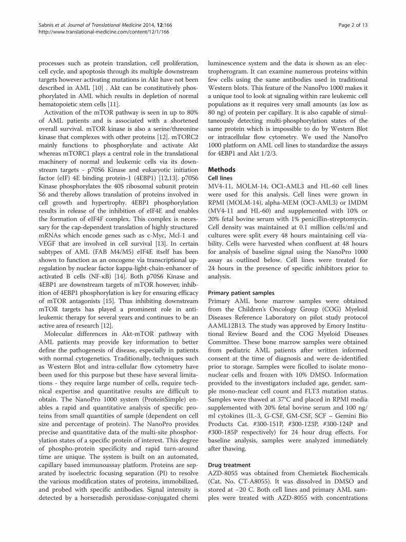
Total Akt 1/2/3 antibody can be used to measure totaland phosphorylated forms using nano-immunoassay inAML cell linesSimilar to 4EBP1 protein, we standardized the nano-immunoassay in AML cell lines for Akt 1/2/3 antibody.The total Akt 1/2/3 antibody was used which was capableof detecting both phosphorylated and non-phosphorylatedforms. Specific phospho-Akt antibody assay is currentlynot standardized in our lab on the nano-immunoassayplatform. AML cell lines were analyzed at baseline forexpression of Akt 1/2/3 (Figure 4). Akt 1/2/3 expressionand activation varied with all cell lines (Figure 4-A). Inorder to determine the specificity of the more acidic peaks
for the phosphorylated protein, each line was treated withlambda-phosphatase concurrently and the electrophe-rogram pattern revealed complete suppression of thephosphorylated peaks as expected and increase in thenon-phosphorylated protein peaks. Our data showed ex-pression of Akt 1 and 2 as expected in hematopoietic cellsand was consistent with recently reported identification ofthe isoforms of the protein [16]. The non-phosphorylatedforms of Akt 1 are detected in the pI range 5.6 – 5.8 andthe non-phosphorylated form of Akt 2 is present in the5.9 – 6.0 pI range. From the electropherogram it appearsas though Akt 2 rather than Akt 1 is the predominantisoform in HL60 cell line and Akt 1 is present in higherquantities in MV4-11, MOLM-14 and OCI-AML3 celllines. β-2 microglobulin was used as a loading control anddemonstrated equal loading in both phosphatase-treatedand non-treated samples. Results were further analyzedby AUC to determine the degree of phosphorylationwithin each cell line (Figure 4-B). Akt 1/2 was found to bephosphorylated similarly in MV4-11 (81.5%), MOLM-14(85.2%) and OCI-AML3 (79.2%) cell lines and phosphory-lated to a lower degree in HL60 (59.6%) cell line. Althoughthe cell lines differed in the degree of Akt 1/2 activation/phosphorylation, treatment with phosphatase showed asignificant decrease in the AUC for the phosphorylatedpeaks on phosphatase treatment to 2.3% in MV4-11, 1.8%

Figure 3 Use of 4EBP1 assay to examine target inhibition within MV4-11 cell line. MV4-11 cells were treated with mTOR1/2 inhibitorAZD-8055 (25 – 1000 nM) for one hour and inhibition of 4EBP1 phosphorylation was noted using the NanoPro 1000 and Western blotting.A) Electropherogram shows the decrease in signal using both total and phospho-specific antibodies. B) β-2 Microglobulin was used as loadingcontrol. X-axis represents iso-electric pH and y-axis represents luminescence units. C) Western blotting confirms the same results with GAPDH asloading control.
Sabnis et al. Journal of Translational Medicine 2014, 12:166 Page 6 of 13http://www.translational-medicine.com/content/12/1/166
in MOLM-14, 4.6% in OCI-AML3 and 1.5% in HL60 celllines with the differences being statistically significant(p < 0.05).In order to demonstrate linearity of the signal intensity
for total and phosphorylated 4EBP1 and total Akt, weplotted signal intensity against exposure time rangingfrom 30–960 secs for each antibody. The assay was runin triplicate for each cell line and results are shown inFigure 5. 4EBP1 antibodies are depicted in Figure 5-Aand total Akt 1/2/3 antibody is depicted in Figure 5-B &C. The linearity of the signal was dependent on theduration of exposure and the abundance of the proteinwithin the sample as demonstrated by the pattern foreach antibody. At higher exposure times (240–960 secs),the signal for all antibodies exhibited a logarithmic in-crease in intensity and burn-out at high intensity. Fromdata obtained in the baseline runs, it was evident thatless than 40% of 4EBP1 is phosphorylated within thesecell lines. In samples where the protein of interest waslow (eg. Phospho-4EBP1), lower exposure times (30–120secs) resulted in a linear increase (R-square value >0.94)in signal intensity, as was evident for both Serine 65 andThreonine 37/46 antibodies using 80 ng of protein percapillary. For proteins that are present in higheramounts such as total Akt 1/2/3, lower exposure times
(between 30–120 secs) exhibited a less linear increasein signal intensity (R-square values between 0.8-0.99)and more abundant proteins such as 4EBP1 exhibitedeven less linear increases with R-square values between0.2-0.9. To test the assumption that amount of proteinwas important for signal linearity, we repeated the total4EBP1 assay by further titrating protein loading to 40and 20 ng of protein and demonstrated sequentialimprovement in the linearity of the signal at lower con-centrations of protein (See Additional file 1: Figure S1).Thus we believe that the adjustment of protein quantityper capillary is an important factor to be consideredwhen setting up these assays.
Nano-immunoassay provides a reliable and sensitivemeasurement of 4EBP1 and Akt activation in primarypatient samplesPrimary pediatric bone marrow samples were obtainedfrom patients enrolled on the Children’s Oncology Grouptrial AAML 0531. Samples were examined for baselineactivation of 4EBP1 (Figure 6). A total of 1.6 × 10⁶ cellswere lysed in 18 μl of lysis buffer and used for this baselineanalysis which accounted for approximately 8888 cells percapillary. The signals obtained using the phosphorylatedantibodies was extremely low so the signal from the
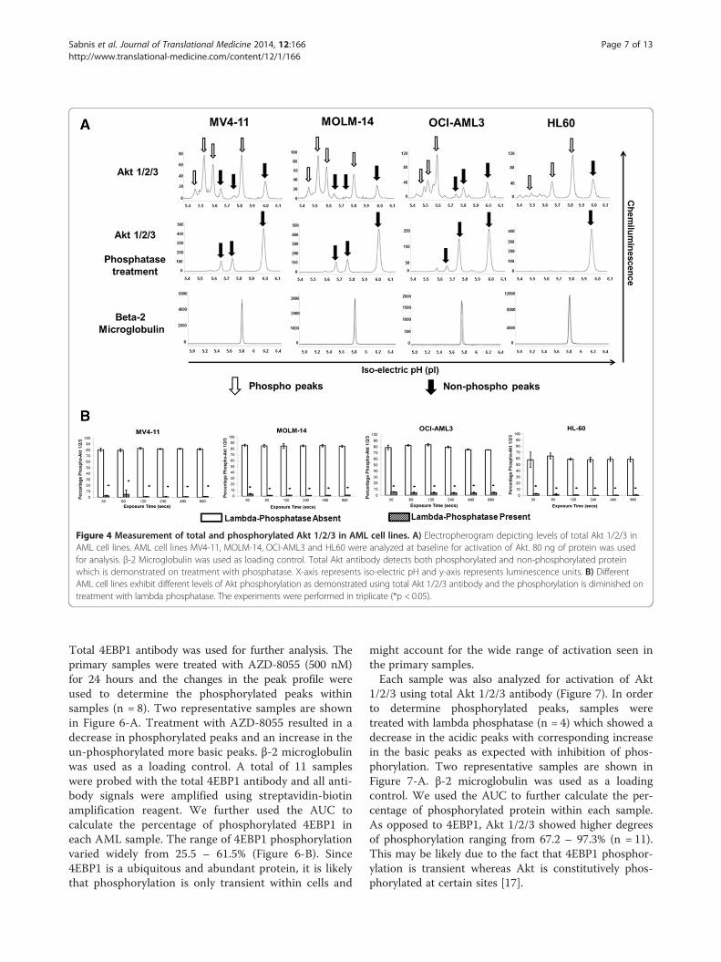
Figure 4 Measurement of total and phosphorylated Akt 1/2/3 in AML cell lines. A) Electropherogram depicting levels of total Akt 1/2/3 inAML cell lines. AML cell lines MV4-11, MOLM-14, OCI-AML3 and HL60 were analyzed at baseline for activation of Akt. 80 ng of protein was usedfor analysis. β-2 Microglobulin was used as loading control. Total Akt antibody detects both phosphorylated and non-phosphorylated proteinwhich is demonstrated on treatment with phosphatase. X-axis represents iso-electric pH and y-axis represents luminescence units. B) DifferentAML cell lines exhibit different levels of Akt phosphorylation as demonstrated using total Akt 1/2/3 antibody and the phosphorylation is diminished ontreatment with lambda phosphatase. The experiments were performed in triplicate (*p < 0.05).
Sabnis et al. Journal of Translational Medicine 2014, 12:166 Page 7 of 13http://www.translational-medicine.com/content/12/1/166
Total 4EBP1 antibody was used for further analysis. Theprimary samples were treated with AZD-8055 (500 nM)for 24 hours and the changes in the peak profile wereused to determine the phosphorylated peaks withinsamples (n = 8). Two representative samples are shownin Figure 6-A. Treatment with AZD-8055 resulted in adecrease in phosphorylated peaks and an increase in theun-phosphorylated more basic peaks. β-2 microglobulinwas used as a loading control. A total of 11 sampleswere probed with the total 4EBP1 antibody and all anti-body signals were amplified using streptavidin-biotinamplification reagent. We further used the AUC tocalculate the percentage of phosphorylated 4EBP1 ineach AML sample. The range of 4EBP1 phosphorylationvaried widely from 25.5 – 61.5% (Figure 6-B). Since4EBP1 is a ubiquitous and abundant protein, it is likelythat phosphorylation is only transient within cells and
might account for the wide range of activation seen inthe primary samples.Each sample was also analyzed for activation of Akt
1/2/3 using total Akt 1/2/3 antibody (Figure 7). In orderto determine phosphorylated peaks, samples weretreated with lambda phosphatase (n = 4) which showed adecrease in the acidic peaks with corresponding increasein the basic peaks as expected with inhibition of phos-phorylation. Two representative samples are shown inFigure 7-A. β-2 microglobulin was used as a loadingcontrol. We used the AUC to further calculate the per-centage of phosphorylated protein within each sample.As opposed to 4EBP1, Akt 1/2/3 showed higher degreesof phosphorylation ranging from 67.2 – 97.3% (n = 11).This may be likely due to the fact that 4EBP1 phosphor-ylation is transient whereas Akt is constitutively phos-phorylated at certain sites [17].

Figure 5 Exposure time and protein amount is important for accurate measurement of signal. The intensity of signal (AUC) for total andphosphorylated 4EBP1 (A) and total Akt 1/2/3 (B & C) was examined at exposure times between 30–960 seconds in MV4-11, MOLM-14, OCI-AML3 andHL60 cells. Higher exposure times (240–960 secs) resulted in signal burn-out which was not observed at lower exposure times (30–120 secs). Moreabundant proteins such as 4EBP1 exhibit improved linearity in signal with titration of protein loading (Additional file 1: Figure S1).
Sabnis et al. Journal of Translational Medicine 2014, 12:166 Page 8 of 13http://www.translational-medicine.com/content/12/1/166
In order to determine the sensitivity of our assays inprimary cells, we performed sequential dilution of theprimary bone marrow samples (n = 3) and analyzed thesignal with total and phosphorylated 4EBP1 antibodiesand total Akt 1/2/3 antibody (Figure 8). A total of 16 ×106 cells were lysed in 140 μl lysis buffer and protein con-centrations were determined. Samples were run in dupli-cate on the assay plate at cell numbers ranging from10,000 to 313 cells per capillary which corresponded toprotein concentrations ranging from 370 ng to 5.9 ng ofprotein per capillary. It is important to note that proteinconcentrations can vary from sample to sample in spite ofhaving similar cell numbers since it is dependent on fac-tors such as percentage of blasts in bone marrow, size ofblasts etc. The AUC for each antibody was plotted againstcell numbers per capillary for each of the samples. A dose-dependent increase in signal was noted with increasingcell numbers (Figure 8A-D) with R-squared values >0.92except for sample A52453 –Thr 37/46 4EBP1 with R-squared value of 0.88. Thus the assay was fairly robustover a wide range of protein concentrations (5.9 - 370 ng)and cell numbers (313–10,000) per capillary.
DiscussionAcute myeloid leukemia continues to be a therapeuticchallenge with low overall survival rates and high inci-dence of relapse. Development of improved biologicalcorrelates that define the disease may allow for riskstratification of patients that can be incorporated intocurrent risk strata based on cytogenetic and molecularabnormalities. Defining the activation status of the4EBP1 and Akt 1/2/3 proteins can serve as an importantindicator of signal transduction in AML and can poten-tially provide information of prognostic significance.Using the nano-immunoassay platform we were able tostandardize assays for both 4EBP1 and Akt 1/2/3 inAML cell lines.Traditionally, Western blotting and intra-cellular flow
cytometry have been used to study individual proteinactivation. However both of these techniques need alarge number of cells, are unable to look at multiple sitesin the same protein in the same assay, and in the case offlow cytometry require significant operator expertise.The NanoPro assays have the advantage of being auto-mated and are amenable to clinical translation due to

Figure 6 Measurement of total and phosphorylated 4EBP1 in primary AML samples. A) Samples (n = 8) were treated with 500 nM AZD-8055and electropherogram demonstrated decrease in phosphorylated forms using total 4EBP1 antibody. Two representative samples are shown. β-2Microglobulin was used as loading control. X-axis represents iso-electric pH and y-axis represents luminescence units. B) Primary AML patient sampleswere examined at baseline for total and phosphorylated forms of 4EBP1 and expressed as percentage phosphorylated against sample number (n = 11).
Sabnis et al. Journal of Translational Medicine 2014, 12:166 Page 9 of 13http://www.translational-medicine.com/content/12/1/166
the rapid turn-around time and the routine methods fordata acquisition. While not capable of providing thedepth of information on single cells that can be achievedfrom flow cytometry analysis, the nano-immunoassayplatform can be combined with flow cytometry sorting
to characterize rare AML sub-populations. Enrichmentof whole AML bone marrow or peripheral blood forimportant cell subsets such as CD34+CD38− populationscan be achieved by flow cytometry and subsequentlythese cells can be lysed and analyzed using the NanoPro
Figure 7 Measurement of total and phosphorylated Akt 1/2/3 in primary AML samples. A) Samples (n = 4) were treated with lambdaphosphatase and electropherogram demonstrated decrease in phosphorylated forms using total Akt 1/2/3 antibody. Two representative samplesare shown. β-2 Microglobulin was used as loading control. X-axis represents iso-electric pH and y-axis represents luminescence units. B) PrimaryAML patient samples were examined at baseline for total and phosphorylated forms of Akt 1/2/3 and expressed as percentage phosphorylatedagainst sample number (n = 11).
Sabnis et al. Journal of Translational Medicine 2014, 12:166 Page 10 of 13http://www.translational-medicine.com/content/12/1/166
platform. Multi-color flow cytometry confers the advan-tage of being able to study different phospho-proteins ingated populations but can also be limited by the number
of phospho-proteins studied simultaneously (typically<12) and can be technically challenging. The NanoProtechnology supplements flow cytometry with the ability

Figure 8 The Nanopro 1000 is capable of detecting signal using low cell numbers in primary AML samples. Three independent primaryAML samples (A47316, A52453 and A52707) were used to test the capability of the assay to detect signal. Antibodies tested were Total 4EBP1(A), Total Akt 1/2/3 (B), Phospho-4EBP1 Thr 37/46 (C) and Phospho-4EBP1 Ser 65 (D) Cell numbers ranged from 10,000-313 cells per capillary.Signal intensity (AUC, log-scale, y-axis) was plotted against cell numbers (log-scale, x-axis) and showed a linear increase with increasing cell numbers.
Sabnis et al. Journal of Translational Medicine 2014, 12:166 Page 11 of 13http://www.translational-medicine.com/content/12/1/166
to quantify phosphorylation patterns and examine otherpost-translational modifications such as acetylation andmethylation. Since the separation of the protein is basedon iso-electric pH, this platform can use total proteinantibodies to determine multi-phosphorylation eventsas demonstrated here for 4EBP1 and Akt1/2/3 sinceheavily phosphorylated proteins tend to have loweriso-electric pH.More recently, Reverse Phase Protein Array (RPPA)
analysis has been used to study the effects of proteinexpression and modification in tumor samples [18].RPPA is a high-throughput antibody based techniquecapable of screening a large number of antibodies in asingle assay however our nano-immunoassay has certainadvantages. Detection of multiple phosphorylation statesof a protein using RPPA requires the use of multiplephospho-specific antibodies while in case of the nano-immunoassay, a single total antibody (eg. Akt 1/2/3) canprovide accurate information about multiple phosphor-ylation sites. RPPA also has a much longer turn-aroundtime of several weeks whereas the NanoPro can provideresults within 24 hours making it a viable option forreal-time analysis of patient samples.
Considering the above mentioned advantages of theNanoPro system, we standardized the assays for 4EBP1and Akt 1/2/3. Depending on the antibody and thedegree of expression, we were able to detect a signal formost antibodies with as low as 80 ng of protein per ca-pillary and in certain cases (total 4EBP1) even as low as20 ng per capillary. The assays were robust with a shortturn-around time of 24 hours providing results that werequantitative and easy to interpret. We further testedthese assays in primary bone marrow samples frompediatric AML patients and found the results to be con-sistent and reproducible. Primary cells are smaller thancell lines but we were able to reliably detect signal in aslow as 40–96 ng of protein. Although the amount ofprotein needed for performing these assays per capillaryis low, there are certain limiting factors that can affectthe sample preparation that need consideration. Firstly,though cell counts for AML blasts might be high insamples, these cells are still fairly small in size as com-pared to AML cell lines. The recommended amount ofprotein per capillary is 80 ng which corresponds roughlyto 2500 primary AML cells and ranged from 900–1400cells from cultured AML cell lines. Secondly, in order to

Sabnis et al. Journal of Translational Medicine 2014, 12:166 Page 12 of 13http://www.translational-medicine.com/content/12/1/166
achieve these numbers, we had to lyse at least 1.6million primary cells in lysis buffer to be able to obtainadequate signal for each antibody. Therefore, futurestudies that focus on leukemia stem cells or minimalresidual disease would require incorporation of methodsfor concentrating protein as part of the sample prepar-ation prior to running on the NanoPro.Both 4EBP1 and Akt are proteins that are phosphory-
lated on multiple sites. Using the nano-immunoassay wewere able to distinguish these multiple phosphorylatedforms in cell lines and primary AML samples. We testedboth AML cell lines and primary AML samples withAZD-8055 mTOR 1/2 inhibitor and found that the drugwas effective at inhibiting 4EBP1 phosphorylation thusmaking this technology useful to determine specifictarget inhibition. We are also currently working on de-veloping assays that cover the entire PI3K-Akt-mTORpathway. Assays for PI3K activation or p70S6K activa-tion have been problematic and require additional devel-opment. Therefore, it is possible that our assay couldmiss some p70S6K activation through the ERK signalingpathway. We could therefore combine the analysis ofAkt 1/2/3 and 4EBP1 phosphorylation with analysisof pERK in future studies. The assay for pERK is welldescribed by several groups in non-hematologic cancers[19] but thus far has not worked on AML samples inour laboratory.Since this was a pilot study utilizing a small number of
samples to highlight an emerging new technology, wewere unable to make any relevant prognostic conclusionscorrelating signal strength to overall survival/relapse rates.Our future studies will involve studying larger numbers ofpatient samples with correlative outcome data as well ascomparison of samples at diagnosis and relapse to deter-mine changes in protein activation. The utility of ouroverall approach to study signal activation is broad andcould apply not only to leukemia but also to other cancerswhere tumor samples might be limiting in number.
Additional file
Additional file 1: Figure S1 Protein amount is important foraccurate measurement of signal. The amount of protein per capillarywas titrated (20–80 ng) in MV4-11, MOLM-14, OCI-AML3 and HL60 celllines and increase in signal linearity was observed with improvement ofR-squared values for total 4EBP1 antibody.
Competing interestsThe authors declare that they have no competing interests.
Authors’ contributionsHSS and HLB performed the experiments. HSS, HLB, STB, TMC, and KDBanalyzed the data. HSS and KDB drafted the manuscript. All authors readand approved the final manuscript.
Funding supportThe content is solely the responsibility of the authors and does notnecessarily represent the official views of the National Institutes of Health.This work was supported by the National Center for Advancing TranslationalSciences of the National Institutes of Health under Award NumberUL1TR000454 and ACTSI KL2 Award number TR000455 (H.S. Sabnis). Thework was also supported by the Aflac Cancer and Blood Disorders Center(K.D. Bunting), Cure Childhood Cancer Foundation (K.D. Bunting) andChildren’s Healthcare of Atlanta Friends Research Fund (S.T. Bunting).All pediatric AML samples were obtained from Children’s OncologyGroup – Myeloid Disease Laboratory. The authors would also like to thankDeborah Pritchett (ProteinSimple) for her invaluable technical support inusing the NanoPro 1000 platform.
Author details1Department of Pediatrics, Aflac Cancer and Blood Disorders Center, EmoryUniversity, 1760 Haygood Drive NE, Atlanta, Georgia, USA. 2Children’sHealthcare of Atlanta, 1405 Clifton Road, Atlanta, Georgia, USA.
Received: 16 January 2014 Accepted: 14 May 2014Published: 12 June 2014
References1. Howlader N, Noone AM, Krapcho M, Garshell J, Miller D, Altekruse SF, Kosary
CL, Yu M, Ruhl J, Tatalovich Z,Mariotto A, Lewis DR, Chen HS, Feuer EJ,Cronin KA (eds). SEER Cancer Statistics Review, 1975-2011. National CancerInstitute: Bethesda, MD, http://seer.cancer.gov/csr/1975_2011/.
2. Fernandez HF: New trends in the standard of care for initial therapy ofacute myeloid leukemia. Hematology Am Soc Hematol Educ Program 2010,2010:56–61.
3. Sander A, Zimmermann M, Dworzak M, Fleischhack G, von Neuhoff C, ReinhardtD, Kaspers GJ, Creutzig U: Consequent and intensified relapse therapyimproved survival in pediatric AML: results of relapse treatment in 379patients of three consecutive AML-BFM trials. Leukemia 2010, 24:1422–1428.
4. Burnett A, Wetzler M, Lowenberg B: Therapeutic advances in acutemyeloid leukemia. J Clin Oncol 2011, 29:487–494.
5. Foran JM: New prognostic markers in acute myeloid leukemia:perspective from the clinic. Hematology Am Soc Hematol Educ Program2010, 2010:47–55.
6. Baldus CD, Mrozek K, Marcucci G, Bloomfield CD: Clinical outcome ofde novo acute myeloid leukaemia patients with normal cytogeneticsis affected by molecular genetic alterations: a concise review.Br J Haematol 2007, 137:387–400.
7. Tamburini J, Green AS, Chapuis N, Bardet V, Lacombe C, Mayeux P, BouscaryD: Targeting translation in acute myeloid leukemia: a new paradigm fortherapy? Cell Cycle 2009, 8:3893–3899.
8. Grossmann V, Schnittger S, Kohlmann A, Eder C, Roller A, Dicker F, SchmidC, Wendtner CM, Staib P, Serve H, Kreuzer KA, Kern W, Haferlach T, HaferlachC: A novel hierarchical prognostic model of AML solely based onmolecular mutations. Blood 2012, 120:2963–2972.
9. Juntilla MM, Patil VD, Calamito M, Joshi RP, Birnbaum MJ, Koretzky GA: AKT1and AKT2 maintain hematopoietic stem cell function by regulatingreactive oxygen species. Blood 2010, 115:4030–4038.
10. Martelli AM, Evangelisti C, Chappell W, Abrams SL, Basecke J, Stivala F, DoniaM, Fagone P, Nicoletti F, Libra M, Ruvolo V, Ruvolo P, Kempf CR, SteelmanLS, McCubrey JA: Targeting the translational apparatus to improveleukemia therapy: roles of the PI3K/PTEN/Akt/mTOR pathway.Leukemia 2011, 25:1064–1079.
11. Kharas MG, Gritsman K: Akt: a double-edged sword for hematopoieticstem cells. Cell Cycle 2010, 9:1223–1224.
12. Altman JK, Sassano A, Platanias LC: Targeting mTOR for the treatment ofAML. New agents and new directions. Oncotarget 2011, 2:510–517.
13. Martelli AM, Evangelisti C, Chiarini F, Grimaldi C, Manzoli L, McCubrey JA:Targeting the PI3K/AKT/mTOR signaling network in acute myelogenousleukemia. Expert Opin Investig Drugs 2009, 18:1333–1349.
14. Hariri F, Arguello M, Volpon L, Culjkovic-Kraljacic B, Nielsen TH, Hiscott J,Mann KK, Borden KL: The eukaryotic translation initiation factor eIF4E is adirect transcriptional target of NF-kappaB and is aberrantly regulated inacute myeloid leukemia. Leukemia 2013, 27:2047–2055.
15. Ducker GS, Atreya CE, Simko JP, Hom YK, Matli MR, Benes CH, Hann B,Nakakura EK, Bergsland EK, Donner DB, Settleman J, Shokat KM, Warren RS:

Sabnis et al. Journal of Translational Medicine 2014, 12:166 Page 13 of 13http://www.translational-medicine.com/content/12/1/166
Incomplete inhibition of phosphorylation of 4E-BP1 as a mechanism ofprimary resistance to ATP-competitive mTOR inhibitors. Oncogene 2014,33:1590–1600.
16. Iacovides DC, Johnson AB, Wang N, Boddapati S, Korkola J, Gray JW:Identification and quantification of AKT isoforms and phosphoforms inbreast cancer using a novel nanofluidic immunoassay. Molecular & cellularproteomics: MCP 2013, 12:3210–3220.
17. Boudeau J, Sapkota G, Alessi DR: LKB1, a protein kinase regulating cellproliferation and polarity. FEBS Lett 2003, 546:159–165.
18. Kornblau SM, Qutub A, Yao H, York H, Qiu YH, Graber D, Ravandi F, Cortes J,Andreeff M, Zhang N, Coombes KR: Proteomic profiling identifies distinctprotein patterns in acute myelogenous leukemia CD34+CD38- stem-likecells. PLoS One 2013, 8:e78453.
19. Chen JQ, Lee JH, Herrmann MA, Park KS, Heldman MR, Goldsmith PK, Wang Y,Giaccone G: Capillary isoelectric-focusing immunoassays to study dynamiconcoprotein phosphorylation and drug response to targeted therapies innon-small cell lung cancer. Mol Cancer Ther 2013, 12:2601–2613.
doi:10.1186/1479-5876-12-166Cite this article as: Sabnis et al.: Capillary nano-immunoassay for Akt 1/2/3and 4EBP1 phosphorylation in acute myeloid leukemia. Journal ofTranslational Medicine 2014 12:166.
Submit your next manuscript to BioMed Centraland take full advantage of:
• Convenient online submission
• Thorough peer review
• No space constraints or color figure charges
• Immediate publication on acceptance
• Inclusion in PubMed, CAS, Scopus and Google Scholar
• Research which is freely available for redistribution
Submit your manuscript at www.biomedcentral.com/submit