Embed Size (px)
Citation preview
394 S. Gundersen et al.
action of MPA also exhibited progestagen/drug synergism. The same group also performed a non-randomised study [9] among patients with advanced breast cancer. They were treated with cyclical sequential administration of an oestrogen, a progestagen and two alternate combinations of cytotoxic drugs. In the 34 patients who completed three double cycles of treatment they obtained an impressive response rate of 91%. The high response rate found in the present trial support these data. The toxicity from high doses of MPA is of concern, however, and lower doses should be considered in future trials.
1. Robustelli Della Cuna G, Pellegrini A. Medroxyprogesterone acetate in combination with chemotherapy for advanced breast cancer: updated results and criticisms. In: Pellegrini A, Robustelli Della Cuna G, Pannuti F, Pouillart P, Jonat W, eds. Role of Medroxyproges- terone in Endocrine-related Tumors, New York, Raven Press, 1984, 3, 91-104.
2. Formelli F, Zaccheo T, Casacca AM, et al. Effect of medr0x$,proges-
terone acetate and doxorubicin on sublines of 13762 mammary adenocarcinoma in rats. Eur J Cancer C lin Onco11981,17 ,1211-1221.
3. EORTC Breast Co.operative group. Revision of the standards for the assessment of hormone receptors in human breast cancer: report of the second EORTC workshop. EurJ Cancer Clin Oncol 1980, 16, 1523-1515.
4. Gundersen S, Kvinnsland S, Klepp O, Lund E, H~st H. Weekly adriamycin versus VAC in advanced breast cancer. A randomised trial. EurJ Cancer Clin Oncol 1986, 22, 1431-1434.
5. Hayward JL, Carbone PP, Heuson J-C, Kumaoka S, Segaloff A, Rubens RD. Assessment of response to therapy in advanced breast cancer. Cancer 1977, 39, 1289.
6. Peto R, Pike MC, Armitage P, et al. Design and analysis of ran- domized clinical trials requiring prolonged observation of each patient. BrJ Cancer 1977, 35, 1-39.
7. Shaikh NA, Owen AM, Gilchik MW, et al. Actions of medroxypro- gesterone acetate on the efficacy of cytotoxic drugs: Studies with human breast cancer cells in culture. In tJ Cancer 1989, 43,458-463.
8. Shaikh NA, Owen AM, Gilchik MW, et al. Adriamycin action on human breast cancer cells: Enhancement by medroxyprogesterone acetate. In tJ Cancer 1989, 43,733-736.
9. Gilchik MW, Shaikh NA, Beranek PA, et al. Cyclical sequential hormonochemotherapy in the treatment of advanced breast cancer. BritMedJ 1987, 295, 1172.
EurJ Cancer, Vol. 28, No. 23. pp. 394-399, 1992. Pnnled m Great Brttaln
0964-1947/9255.00 ~ 0.00 1992 Pergamon Press pie
Metastatic Ovarian or Colonic Cancer: A Clinical Challenge
B.G. Taal, Ph.C. Hageman, J.F.M. Delemarre, J.M.G. Bonfr6r and F.C.A. den Hartog Jager
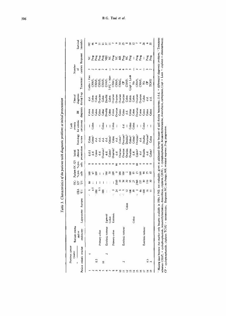
Cl in ica l p r o b l e m s a r i se w h e n h i s t o l o g y is u n a b l e to differentiate between an o v a r i a n c a r c i n o m a in f i l t r a t ing into the rectosigmoid region and a colonic cancer with o v a r i a n m e t a s t a s e s . T o e v a l u a t e the discriminative v a l u e o f
immunohistochemistry we studied four groups: (A) o v a r i a n c a r c i n o m a (n = 21), (B) o v a r i a n c a r c i n o m a wi th
sigmoid s t e n o s i s (n = 18), (C) colonic carcinoma (n = 20) and (D) a g r o u p in which the differential d i a g n o s i s was
a problem (n -- 19). Para f f in sections stained with a p a n e l o f m o n o c l o n a l antibodies revealed specific patterns: in
g r o u p A and B a n e g a t i v e P a r l a m - 4 a n d p o s i t i v e OC-125 ; in g r o u p C the opposite; in group D the 'colonic' pattern in 15 c a s e s , a n d the ' o v a r i a n ' p a t t e r n in on ly 2. T h e c l in ica l d i a g n o s i s in g r o u p D d u r i n g f o l l o w - u p was o v a r i a n
carcinoma in 7, colonic carcinoma in 8, d o u b l e t u m o u r in 1 and sti l l u n k n o w n in 3. T h i s was b a s e d o n h i g h l e v e l s
o f s e r u m t u m o u r m a r k e r s s u c h as c a r c i n o e m b r y o n i c a n t i g e n (n = 5) a n d CA-125 (n = 4), l a p a r o t o m y (n = 4),
autopsy (n = 1), barium enema and/or endoscopy (n = 5). The response to chemotherapy in g r o u p D w a s extremely poor. E u r J Cancer, Vol. 28, N o . 2/3, pp. 394-399 , 1992.
I N T R O D U C T I O N TIIE AETIOLOGY o f a tumour mass in the lower abdomen is sometimes difficult to establish both on clinical findings and histology. The diagnosis of ovarian cancer infiltrating the recto- siganoid region implies long-term aggressive chemotherapy and
Correspondence to B.G. Taal. B.G. Taal and F.C.A. den Hartog Jager are at the Department of Gastroenterology.; Ph.C. Hageman is at the Department of Tumour Biology; J.F.M. Delemarre is at the Department of Pathology; and J.M.G. Bonfrer is at the Department of Clinical Chemistry, Netherlands Cancer Institute, Antoni van Leeuwenhoek Huis, Plesmanlaan 121, 1066 CX Amsterdam, The Netherlands. Revised 10 July 1991 ; accepted 4 Oct. 1991.
a fair prognosis, whereas advanced colonic cancer implies the availability of less effective chemotherapy and a poor prognosis. There are many reports dealing with ovarian metastases derived from colorectal, breast or gastric carcinoma [1, 2], sometimes appearing after a relatively long interval following surgery [3]. Only a few papers describe the diagnostic problem ofa colorectal cancer presenting as an ovarian tumour mass [4]. A clear diagnostic approach and therapeutic policy is lacking. There- fore, we evaluated the discriminative value of immunohistoch- emistry on the impact of response to chemotherapy.
P A T I E N T S A N D M E T H O D S In 19 patients (median age 56 years, range 38-72) seen at the
Netherlands Cancer Institute from 1979 to 1986 it was difficult
Metastatic Ovarian or Colonic Cancer: A Clinical Challenge 395
to differentiate at presentation between ovarian cancer and colorectal carcinoma either on clinical findings (n = 6),histology (n = 6) or both (n = 7). Patients with clear-cut ovarian cancer (n = 21; median age 54 years, range 24-74; Figo stage I = 5, stage II = 2, stage III = 11 and stage IV = 3), patients with ovarian cancer with a documented sigmoid stenosis (n = 20; median age 56 years, range 41-68) and patients with clear-cut colorectal carcinoma (n = 20, median age 53 years, range 27-70; Dukes B = 6 and Dukes C = 14) seen in the same period were randomly selected as control groups.
All medical records were reviewed. A definitive clinical diag- nosis was made using initial and follow-up data acquired at endoscopy, barium enema, laparotomy, autopsy and serum tumour markers (carcinoembryonic antigen, CEA; CA-125) as well as that related to response to chemotherapy.
Histology of conventional slides was reviewed and the degree of differentiation was scored. The presence of psammoma- bodies and squamous metaplasia were suggestive of ovarian cancer, while garland and cribriform growth pattern with necrosis and segmental destruction of glands was characteristic for colonic metastases in the ovaries according to Lash and Hare [3].
A panel of five monoclonal antibodies was used for immunohi- stochemistry. Parlam-4 against purified CEA detects an epitope on CEA (5). 115D8 which is directed against human mill<fat globule membranes [8] detects an epitope on a large sialomucin, present on the majority of epithelial tumour [9]. The antibody 67D11 was prepared against human milkfat globule membranes [8] and recognises an antigenic determinant on glycoproteins involving the bloodgroup Lewis ~ antigen in combination with the stage specific embryonic antigen SSEA-1 [10]. This antigen is found on a number of epithelial tissues and tumours. NS19- 9, which detects a sialytated derivative of Lewis b bloodgroup, was raised against human colon carcinoma cell-line SW1116 [11, 12]. OC-125, made against the epithelial ovarian cancer cell line OVCA433 [13], is directed against a CA-125 antigen occurring primarily on non-mucinous ovarian tumours but absent on colorectal cancers [14]. The immunoperoxidase test was perfor- med on paraffin sections using peroxidase labelled rabbit anti- mouse IgG as an antibody according to the method described previously [6]. For optimal reactions with OC-125 the deparaf- finised sections were pretreated with 0.1% pronase for 20 min at room temperature, as pronase rather than trypsin treatment restores the immunoreactivity for OC-125 in formalin-fixed and embedded tissues [7].
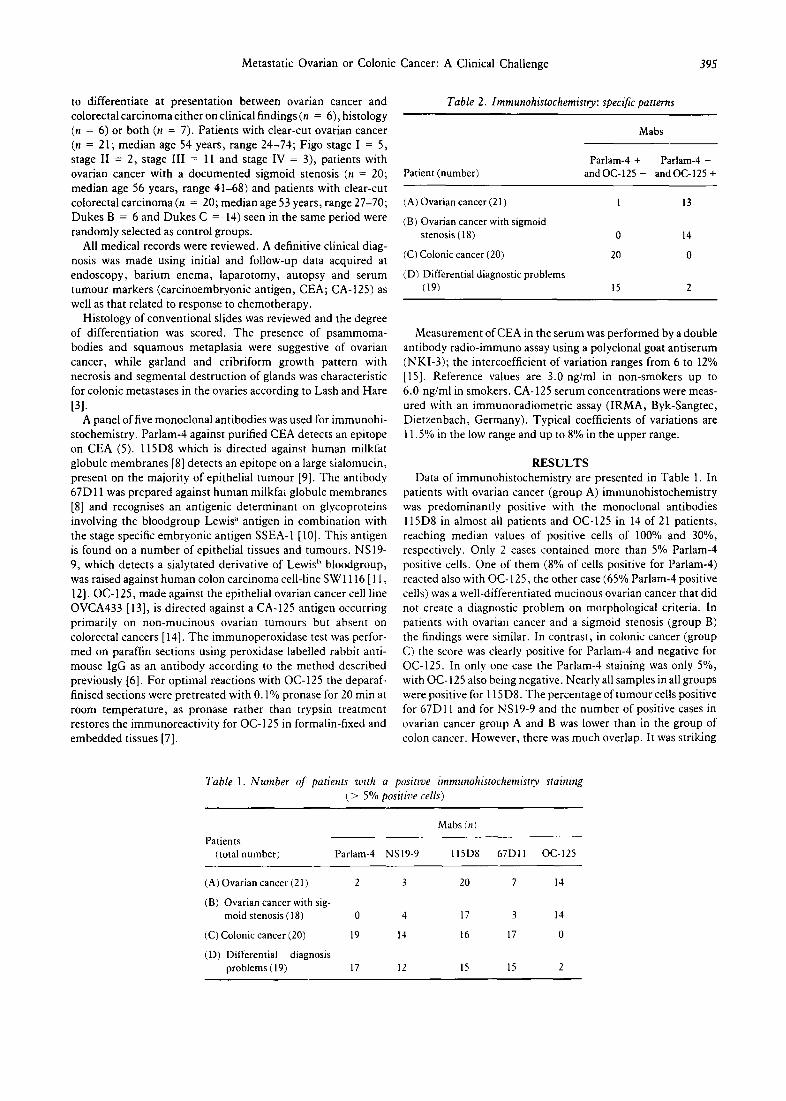
Table 2. Immunohistochemistry: specific patterns
Patient (number)
Mabs
P a r l a m - 4 + P a r l a m - 4 -
a n d O C - 1 2 5 - andOC-125 +
(A) Ovarian cancer (21) 1 13
(B) Ovarian cancer with sigmoid stenosis (18) 0 14
(C~ Colonic cancer (20) 20 0
(D'~ Differential diagnostic problems (19) 15 2
Measurement of CEA in the serum was performed by a double antibody radio-immuno assay using a polyclonal goat antiserum (NKI-3); the intercoefficient of variation ranges from 6 to 12% [15]. Reference values are 3.0 ng/ml in non-smokers up to 6.0 ng/ml in smokers. CA-125 serum concentrations were meas- ured with an immunoradiometric assay (IRMA, Byk-Sangtec, Dietzenbach, Germany). Typical coefficients of variations are 11.5% in the low range and up to 8% in the upper range.
R E S U L T S Data of immunohistochemistry are presented in Table 1. In
patients with ovarian cancer (group A) immunohistochemistry was predominantly positive with the monoclonal antibodies 115D8 in almost all patients and OC-125 in 14 of 21 patients, reaching median values of positive ceils of 100% and 30%, respectively. Only 2 cases contained more than 5% Parlam-4 positive cells. One of them (8% of cells positive for Parlam-4) reacted also with OC-125, the other case (65% Parlam-4 positive cells) was a well-differentiated mucinous ovarian cancer that did not create a diagnostic problem on morphological criteria. In patients with ovarian cancer and a sigmoid stenosis (group B) the findings were similar. In contrast, in colonic cancer (group C) the score was clearly positive for Parlam-4 and negative for OC-125. In only one case the Parlam-4 staining was only 5%, with OC-125 also being negative. Nearly all samples in all groups were positive for 115D8. The percentage of tumour ceils positive for 6 7 D l l and for NS19-9 and the number of positive cases in ovarian cancer group A and B was lower than in the group of colon cancer. However, there was much overlap. It was striking
Table 1. Number of patients zoith a positive immunohistochemist~., staining (> 5% positive cells)
Mabs (n)
Patients (total number) Parlam-4 NS19-9 115D8 67D11 OC-125
(A) Ovarian cancer (21 ) 2 3 20 7 14
(B) Ovarian cancer with sig- moid stenosis (18) 0 4 17 3 14
(C) Colonic cancer (20) 19 14 16 17 0
(D) Differential diagnosis problems (19) 17 12 15 15 2
396 B.G. Taal et al.
e~
Z
• "r- ~
~ N
& ,-.i
~a2
g.
~ ° o ~ ~ ~ z ~ ~ ~ ~ ~
. . . . . . . I . . . . . . I ~ - I ~ , , ,
~ . ~ . ~ . ~ . ~ ~ ~ ~ . . . . ~~
~ . ~ ~
~ I I I I I I I ~ ~ ~
N
~+
~ u
= E
~rt
~ .~ z
_~-=
o " ~
Metastatic Ovarian or Colonic Cancer: A Clinical Challenge 397
"~, ...... ~ # ~ ] ~ 2 ! 4 ; . / . . : , . . . , _ ; . " . . L " --.~ • " ~ I ~ . ~ " ; . - / ; -~ , . . . . , . ' , , ~ ~ . " . ~ . ~ ) ~ a - _ ~ . - - . " .. ,
• "- " " "" ( T , ,,:" " ' • " _~ , , '~ '2~ " . . . . - , , n ~ t - i • . . . . ' , . . . . . . " : ( ~ , ~ . - ,. - ~ i & ~ . . , . ~ . T - . . . - , *" : - " _~
( b ) P 4
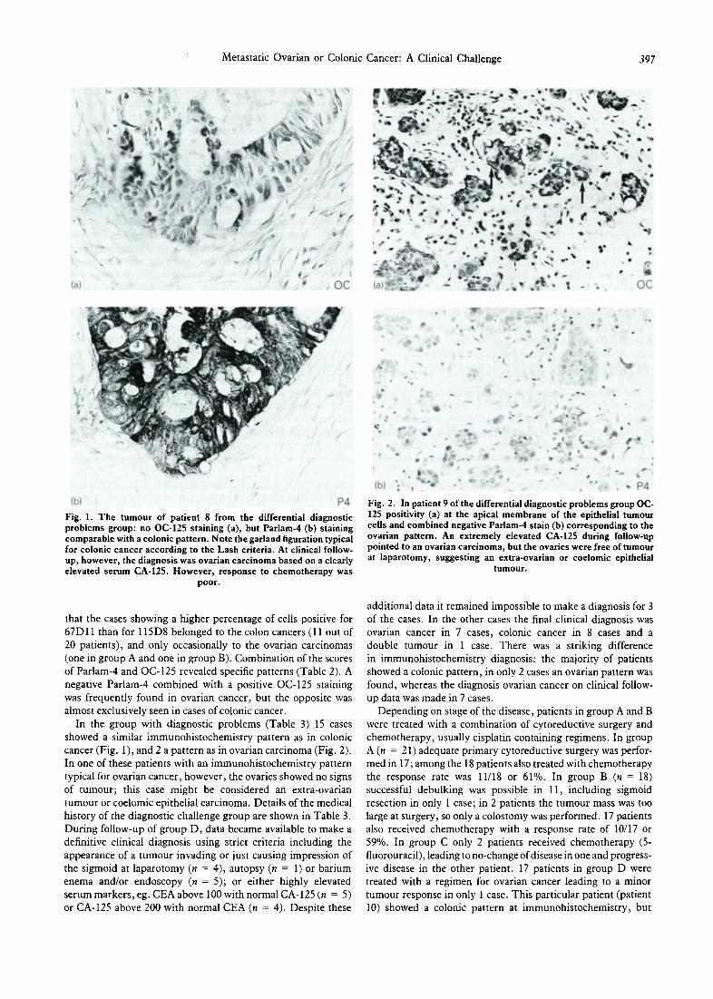
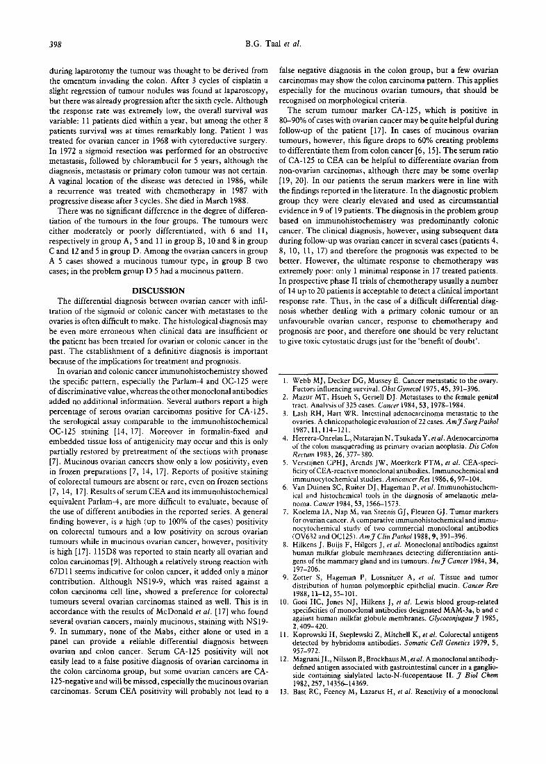
Fig. 1. The tumour of patient 8 from the differential diagnostic problems group: no OC-125 staining (a), but Parlam-4 (b) staining comparable with a colonic pattern. Note the garland figuration typical for colonic cancer according to the Lash criteria. At clinical follow- up, however, the diagnosis was ovarian carcinoma based on a clearly elevated serum CA-125. However, response to chemotherapy was
poor.
, • • , i * ° o
A ~ . . "~ - ' .' . , . ' , •
t
, . _ ~, • . ~ - 4 , ~ , , ' , , r
( b ) t _ " _ • * - • • P 4
Fig. 2. In patient 9 of the differential diagnostic problems group OC- 125 positivity (a) at the apical membrane of the epithelial tumour cells and combined negative Parlam-4 stain (b) corresponding to the ovarian pattern. An extremely elevated CA-125 during follow-up pointed to an ovarian carcinoma, but the ovaries were free of tumour at laparotomy, suggesting an extra-ovarian or coelomic epithelial
t u m o u r ,
that the cases showing a higher percentage of cells positive for 67D11 than for 115D8 belonged to the colon cancers (11 out of 20 patients), and only occasionally to the ovarian carcinomas (one in group A and one in group B). Combination of the scores of Parlam-4 and OC-125 revealed specific patterns (Table 2). A negative Parlam-4 combined with a positive OC-125 staining was frequently found in ovarian cancer, but the opposite was almost exclusively seen in cases of colonic cancer.
In the group with diagnostic problems (Table 3) 15 cases showed a similar immunohistochemistry pattern as in colonic cancer (Fig. 1), and 2 a pattern as in ovarian carcinoma (Fig. 2). In one of these patients with an immunohistochemistry pattern typical for ovarian cancer, however, the ovaries showed no signs of tumour; this case might be considered an extra-ovarian tumour or coelomic epithelial carcinoma. Details of the medical history of the diagnostic challenge group are shown in Table 3. During follow-up of group D, data became available to make a definitive clinical diagnosis using strict criteria including the appearance of a tumour invading or just causing impression of the sigmoid at laparotomy (n = 4), autopsy (n = 1) or barium enema and/or endoscopy (n = 5); or either highly elevated serum markers, eg. CEA above 100 with normal CA-125 (n = 5) or CA-125 above 200 with normal CEA (n = 4). Despite these
additional data it remained impossible to make a diagnosis for 3 of the cases. In the other cases the final clinical diagnosis was ovarian cancer in 7 cases, colonic cancer in 8 cases and a double tumour in 1 case. There was a striking difference in immunohistochemistry diagnosis: the majority of patients showed a colonic pattern, in only 2 cases an ovarian pattern was found, whereas the diagnosis ovarian cancer on clinical follow- up data was made in 7 cases.
Depending on stage of the disease, patients in group A and B were treated with a combination of cytoreductive surgery and chemotherapy, usually cisplatin containing regimens. In group A (n = 21) adequate primary cytoreductive surgery was perfor- med in 17; among the 18 patients also treated with chemotherapy the response rate was 11/18 or 61%. In group B (n = 18) successful debulking was possible in 11, including sigmoid resection in only 1 case; in 2 patients the tumour mass was too large at surgery, so only a colostomy was performed. 17 patients also received chemotherapy with a response rate of 10/17 or 59%. In group C only 2 patients received chemotherapy (5- fluorouracil), leading to no-change of disease in one and progress- ive disease in the other patient. 17 patients in group D were treated with a regimen for ovarian cancer leading to a minor tumour response in only 1 case. This particular patient (patient 10) showed a colonic pattern at immunohistochemistry, but
398 B.G. Taal et al.
during laparotomy the tumour was thought to be derived from the omentum invading the colon. After 3 cycles of cisplatin a slight regression of tumour nodules was found at laparoscopy, but there was already progression after the sixth cycle. Although the response rate was extremely low, the overall survival was variable: 11 patients died within a year, but among the other 8 patients survival was at times remarkably long. Patient 1 was treated for ovarian cancer in 1968 with cytoreductive surgery. In 1972 a sigmoid resection was performed for an obstructive metastasis, followed by chlorambucil for 5 years, although the diagnosis, metastasis or primary colon tumour was not certain. A vaginal location of the disease was detected in 1986, while a recurrence was treated with chemotherapy in 1987 with progressive disease after 3 cycles. She died in March 1988.
There was no significant difference in the degree of differen- tiation of the tumours in the four groups. The tumours were either moderately or poorly differentiated, with 6 and 11, respectively in group A, 5 and 11 in group B, 10 and 8 in group C and 12 and 5 in group D. Among the ovarian cancers in group A 5 cases showed a mucinous tumour type, in group B two cases; in the problem group D 5 had a mucinous pattern.
D I S C U S S I O N The differential diagnosis between ovarian cancer with infil-
tration of the sigmoid or colonic cancer with metastases to the ovaries is often difficult to make. The histological diagnosis may be even more erroneous when clinical data are insufficient or the patient has been treated for ovarian or colonic cancer in the past. The establishment of a definitive diagnosis is important because of the implications for treatment and prognosis.
In ovarian and colonic cancer immunohistochemistry showed the specific pattern, especially the Parlam-4 and OC-125 were of discriminative value, whereas the other monoclonal antibodies added no additional information. Several authors report a high percentage of serous ovarian carcinomas positive for CA-125, the serological assay comparable to the immunohistochemical OC-125 staining [14, 17]. Moreover in formalin-fixed and embedded tissue loss of antigenicity may occur and this is only partially restored by pretreatment of the sections with pronase [7]. Mucinous ovarian cancers show only a low positivity, even in frozen preparations [7, 14, 17]. Reports of positive staining of colorectal tumours are absent or rare, even on frozen sections [7, 14, 17]. Results of serum CEA and its immunohistochemical equivalent Parlam-4, are more difficult to evaluate, because of the use of different antibodies in the reported series. A general finding however, is a high (up to 100% of the cases) positivity on colorectal tumours and a low positivity on serous ovarian tumours while in mucinous ovarian cancer, however, positivity is high [17]. 115D8 was reported to stain nearly all ovarian and colon carcinomas [9]. Although a relatively strong reaction with 67D11 seems indicative for colon cancer, it added only a minor contribution. Although NS19-9, which was raised against a colon carcinoma cell line, showed a preference for colorectal tumours several ovarian carcinomas stained as well. This is in accordance with the results of McDonald et al. [17] who found several ovarian cancers, mainly mucinous, staining with NS 19- 9. In summary, none of the Mabs, either alone or used in a panel can provide a reliable differential diagnosis between ovarian and colon cancer. Serum CA-125 positivity will not easily lead to a false positive diagnosis of ovarian carcinoma in the colon carcinoma group, but some ovarian cancers are CA- 125-negative and will be missed, especially the mucinous ovarian carcinomas. Serum CEA positivity will probably not lead to a
false negative diagnosis in the colon group, but a few ovarian carcinomas may show the colon carcinoma pattern. This applies especially for the mucinous ovarian tumours, that should be recognised on morphological criteria.
The serum tumour marker CA-125, which is positive in 80-90% of cases with ovarian cancer may be quite helpful during follow-up of the patient [17]. In cases of mucinous ovarian tumours, however, this figure drops to 60% creating problems to differentiate them from colon cancer [6, 15]. The serum ratio of CA-125 to CEA can be helpful to differentiate ovarian from non-ovarian carcinomas, although there may be some overlap [19, 20]. In our patients the serum markers were in line with the findings reported in the literature. In the diagnostic problem group they were clearly elevated and used as circumstantial evidence in 9 of 19 patients. The diagnosis in the problem group based on immunohistochemistry was predominantly colonic cancer. The clinical diagnosis, however, using subsequent data during follow-up was ovarian cancer in several cases (patients 4, 8, 10, 11, 17) and therefore the prognosis was expected to be better. However, the ultimate response to chemotherapy was extremely poor: only 1 minimal response in 17 treated patients. In prospective phase II trials of chemotherapy usually a number of 14 up to 20 patients is acceptable to detect a clinical important response rate. Thus, in the case of a difficult differential diag- nosis whether dealing with a primary colonic tumour or an unfavourable ovarian cancer, response to chemotherapy and prognosis are poor, and therefore one should be very reluctant to give toxic cytostatic drugs just for the 'benefit of doubt ' .
1. Webb M J, Decker DG, Mussey E. Cancer metastatic to the ovary. Factors influencing survival. Obst Gynecol 1975, 45, 391-396.
2. Mazur MT, Hsueh S, Gersell DJ. Metastases to the female genital tract. Analysis of 325 cases. Cancer 1984, 53, 1978-1984.
3. Lash RH, Hart WR. Intestinal adenocarcinoma metastatic to the ovaries. A clinicopathologic evaluation of 22 cases. A m J Surg Pathol 1987, 11, 114-121.
4. Herrera-Onrelas L, Natarajan N , Tsukada Y , et aL Adenocarcinoma of the colon masquerading as primary ovarian neoplasia. Dis Colon Rectum 1983, 26,377-380.
5. Verstiinen CJ?HJ, Arends JW, Moerkerk PTM, et al. CEA-speci- ficity ofCEA-reactive monoclonal antibodies. Immunochemical and immunocytochemical studies. Anticancer Res 1986, 6, 97-104.
6. Van Duinen SC, Ruiter D J, Hageman P, et al. Immunohistochem- ical and histochemical tools in the diagnosis of amelanotic mela- noma. Cancer 1984, 53, 1566-1573.
7. Koelema IA, Nap M, van Steenis GJ, Fleuren GJ. Tumor markers for ovarian cancer. A comparative immunohistochemical and immu° nocytochemical study of two commercial monoclonal antibodies (OV632 and OC125). A m J Clin Patho11988, 9,391-396.
8. Hilkens J, Buijs F, Hilgers J, et al. Monoclonal antibodies against human milkfat globule membranes detecting differentiation anti- gens of the mammary gland and its tumours, l n t J Cancer 1984, 34, 197-206.
9. Zotter S, Hageman P, Lossnitzer A, et al. Tissue and tumor distribution of human polymorphic epithelial mucin. Cancer Rev 1988, 11-12, 55-101.
10. Gooi HC, Jones NJ, Hilkens J, et al. Lewis blood group-related specificities of monoclonal antibodies designated MAM-3a, b and c against human milkfat globule membranes. GlycoconjugateJ 1985, 2,409-420.
11. Koprowski H, Steplewski Z, Mitchell K, et al. Colorectal antigens detected by hybridoma antibodies. Somatic Cell Genetics 1979, 5, 957-972.
12. Magnani J L , Nilsson B, Brockhaus M, et al. A monoclonal antibody- defined antigen associated with gastrointestinal cancer in a ganglio- side containing sialylated lacto-N-fucopentaose II. J Biol Chem 1982, 257, 14356-14369.
13. Bast RC, Feeney M, Lazarus H, et al. Reactivity of a monoclonal
Metastatic Ovarian or Colonic Cancer: A Clinical Challenge 399
antibody with human ovarian carcinoma. J Clin Invest 1981, 68, 1331-1337.
14. Kabawat SE, Bast RC, Welch WR, et al. Immunopathologic characterization of a monoclonal antibody that recognizes common surface antigens of human ovarian tumors of serous, endometrioid and clear cell types. A m J Clin Patho11983, 79, 98-104.
15. Persijn JP, Koster CB. The development of a radio-immuno assay for carcinoembryonic antigen with some applications. J Clin Chem Clin Biochem 1976, 14,377-387.
16. UlbrightTM, Roth LM, Stehman FB. Secondary ovarian neoplasia: a clinicopathologic study of 35 cases. Cancer 1984, 53, 1164-1174.
17. McDonald F, Bird R, Stokes H, et al. Expression of CEA, CA125,
CA19-9 and human milk-fat globule membrane antigen in ovarian tumours. J Clin Pathol 1988, 41,260-264.
18. O'Connell G J, Ryan E, Murphy K J, et al. Predictive value of CA125 for ovarian carcinoma in patients presenting with pelvic masses. Obst Gyneco11987 , 70,930-932.
19. Wu JT, Miya T, Knight A, Knight DP. Improved specificity of the CA125 enzyme immunoassay for ovarian carcinomas by use of the ratio of CA 125 of carcino-embryonic antigen. Clin Chem 1988, 34, 1853-1857.
20. Buamab PK, Rake MO, Drake SR, Skillen AW. Serum CA125 concentrations and CA12-5/CEA ratios in patients with epithelial ovarian cancer. J Surg Onco11990, 44, 97-99.
EurJ Cancer, Vol. 28, No. 2/3, pp. 399-403, 1992. Primed m Great Bream
0964-1947/92 $5.00 ¢ 0.00 t~) 1992 Pergamon Press pie
Pilot Study of a Rapid Etoposide-cisplatin Regimen in Paediatric Soft Tissue Sarcomas
Marianne B. Phillips and C. Ross Pinkerton
10 patients with refractory or relapsed soft tissue sarcoma were treated with weekly etoposide (150 mg/m 2 on days 1, 2 and 3) and cisplatin (60 mg/m 2 on day 2). Toxicity was mainly myelosuppression which resulted in deviation from planned weekly chemotherapy scheduling. With this rapid dose-delivery schedule the tolerated median dose intensities were 161 mg/m 2 per week for etoposide and 49 mg/m 2 per week for cisplatin. In 9 evaluable patients there were 7 responses, 2 complete and 5 partial, giving a response rate of 78% (confidence interval 51-100%). The combination of etoposide and cisplatin in this schedule produced a higher response rate than reported with previous schedules and is worthy of further evaluation. E u r J Cancer, Vol. 28, No. 2/3, pp. 399-403, 1992.
INTRODUCTION THE SURVIVAL of children with soft tissue sarcoma has improved over the past decade with 3 year event free survival (excluding stage IV disease) of 53%, overall survival 69%, using combined chemotherapy, radiotherapy and surgery in the SIOP MMT 1984 trials [1], and 70% 5 year survival for this group in the IRS II study [2].
Refractory disease (approximately 10% of cases) and local or metastatic relapse remain problems and further efforts at improving chemotherapy are warranted. Potential new agents are limited and it is therefore important to optimise the schedul- ing of existing drugs.
Single agent etoposide has achieved response rates of 20% in intensively pretreated children with rhabdomyosarcoma [3].
Cisplatin has been shown to have 15% response rate as a single agent in refractory rhabdomyosarcoma and undifferentiated soft tissue sarcoma in phase II trials [4].
Etoposide inhibits DNA synthesis with little effect on RNA
Correspondence to C.R. Pinkerton. The authors are at the Paediatric Department, Royal Marsden Hospital, Downs Road, Sutton, Surrey SM2 5PT, U.K. Revised 18 Oct. 1991; accepted 25 Oct. 1991.
and protein synthesis, whereas cisplatin also causes inhibition of RNA and protein synthesis [3, 4]. This may explain the synergy which exists experimentally and clinically between these two agents. Additionally repair of the DNA damage induced by cisplatin cross linking may be inhibited by the anti-topoisomer- ase II effect of etoposide [5]. A previous phase II trial of etoposide and cisplatin achieved a response rate of 33% in relapsed paediatric rhabdomyosarcoma [6], though another study achieved only a 12.5% response and the efficacy of such a drug combination was questioned [7]. A heterogeneous pretre- ated group of patients receiving higher dose intensities achieved a response rate of 62% [8], and subsequently the incorporation of both agents, with differing schedules, into first-line therapy for children with soft tissue sarcoma showed encouraging response rates of 80, 87 and 100% [8-10], though not without appreciable toxicity.
Dose escalation of cisplatin to 200 mg/m 2 is possible using a 5 day schedule [11] but the toxicity is probably less if a similar total dose is administered weekly over 2 weeks [12, 13]. The least effective way to give etoposide in small cell lung cancer is as a single dose [14, 15]. The higher response rates achieved with weekly administration of the same dose divided over 3 days may be obtainable for other tumours. This reasoning provided