Embed Size (px)
Citation preview
Metastatic disease of the Spine
Jwalant S. Mehta
MS (Orth); MCh (Orth); D Orth; FRCS (Tr & Orth)
Consultant Spine Surgeon
The Royal Orthopaedic Hospital
Birmingham Children’s Hospital
MSCC
Metastatic Spinal Cord Compression
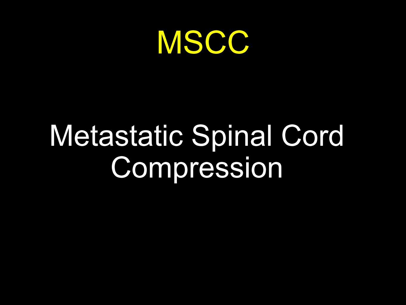
Prevalence
• Bone metastasis seen in 150,000 patients with
solid tumours in England and Wales
• Common sites of metastasis: Lung; Liver; Bone
Aaron AD JAMA 1994; 272: 1208 - 9

Prevalence
• Spine is the commonest site of bone metastasis
• 30 - 70% Cancer patients have spine mets on
autopsy
• 5 - 10% patients with cancer develop spinal
cord compression
Jacobs, Perin Neurosurg Focus 2001
‘ As survival rates for primary cancers improve, the prevalence of spinal metastasis will rise.’

Common sites of primaries
• Adults: Breast, Lung, Prostate, Renal, Melanoma, Thyroid,
Colorectal, Haematologic (MM; Lymphoma)
Constans J Neurosurg 1983; 59: 111 - 118
• Children: Neuroblastomas, Sarcomas
Choi ESJ 2010 19: 215 - 222

Pathology • Reaching the spine:
Haematogenous spread
Direct extension / invasion
Seeding of CSF
• Thoracic Spine 70%
• Lumbar spine 20%
• Cervical and Sacrum 10%

Pathology
• Vertebral body 80%
• Posterior elements 20%
• Most are osteolytic 95%
• Breast and Prostate are osteoblatic
• Usually do not cross dural barrier
exc sarcomas, recurrence, post radiotherapy

Grade Bone Epidural Theca Cord
deformation
Cord
compression
0 + - - - -
1a + + - - -
1b + + + - -
1c + + + + -
2 + + + + +
CSF seen
3 + + + + +
No CSF
Grades of MSCC
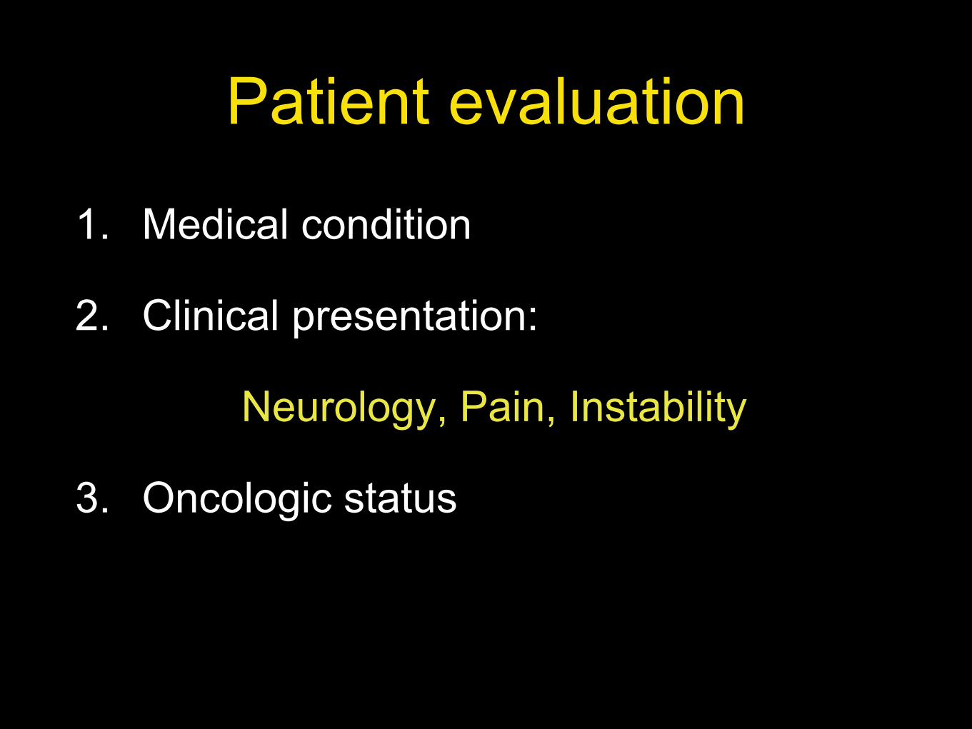
Patient evaluation
1. Medical condition
2. Clinical presentation:
Neurology, Pain, Instability
3. Oncologic status
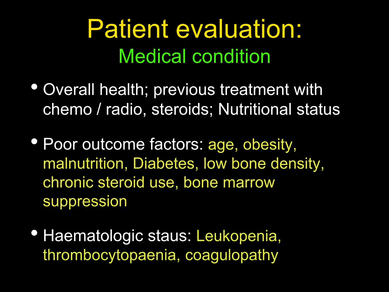
Patient evaluation: Medical condition
• Overall health; previous treatment with
chemo / radio, steroids; Nutritional status
• Poor outcome factors: age, obesity,
malnutrition, Diabetes, low bone density,
chronic steroid use, bone marrow
suppression
• Haematologic staus: Leukopenia,
thrombocytopaenia, coagulopathy

Patient evaluation
1. Medical condition
2. Clinical presentation:
Neurology, Pain, Instability
3. Oncologic status

Patient evaluation: Neurology
• Sensory (including fine touch, pin prick,
vibration, temperature)
• Motor
• Reflexes (including pathologic reflexes)
• Autonomic

Patient evaluation: Neurology
• Cord v nerve root
• Myelopathy v radiculopathy
• Ambulation status (important predictor)
• Degree of cord compression
• 5-10% of all MSCC
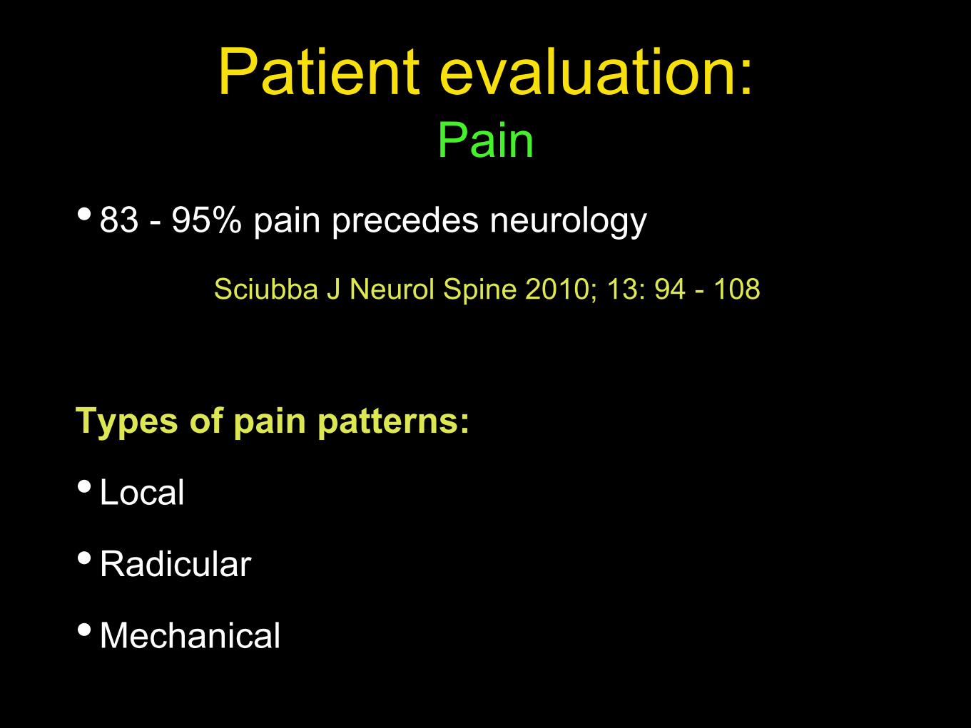
Patient evaluation: Pain
•83 - 95% pain precedes neurology
Sciubba J Neurol Spine 2010; 13: 94 - 108
Types of pain patterns:
•Local
•Radicular
•Mechanical

Patient evaluation: Local Pain
• Causes: Periosteal strech, endosteal pressure,
inflammation by tumour growth
Gokaslan Curr Opin Oncol 1996
• Localised, constant, not related to
activities, ‘deep ache’
• Responds to: NSIAD’s, Steroids, radiotherapy

Patient evaluation: Radicular Pain
• Root compression alongs its course (Dermatomal pattern)
• Sharp, shooting, stabbing
• Constant, not related to activity
• Response to NSAID’s, steroids, chemo and radiotherapy (tumour shrinkage)
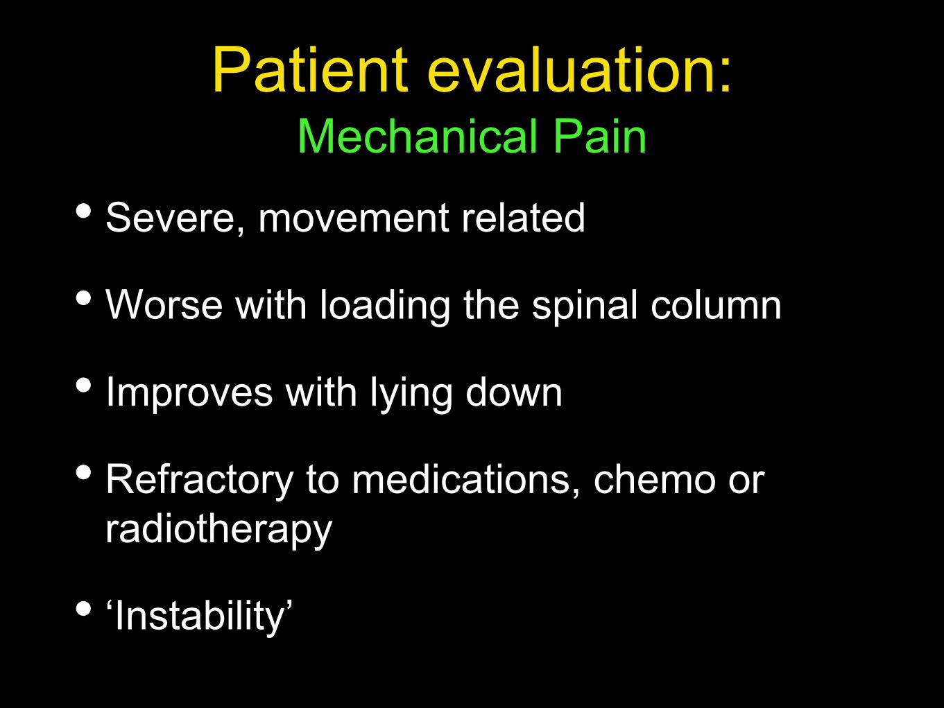
Patient evaluation: Mechanical Pain
• Severe, movement related
• Worse with loading the spinal column
• Improves with lying down
• Refractory to medications, chemo or
radiotherapy
• ‘Instability’
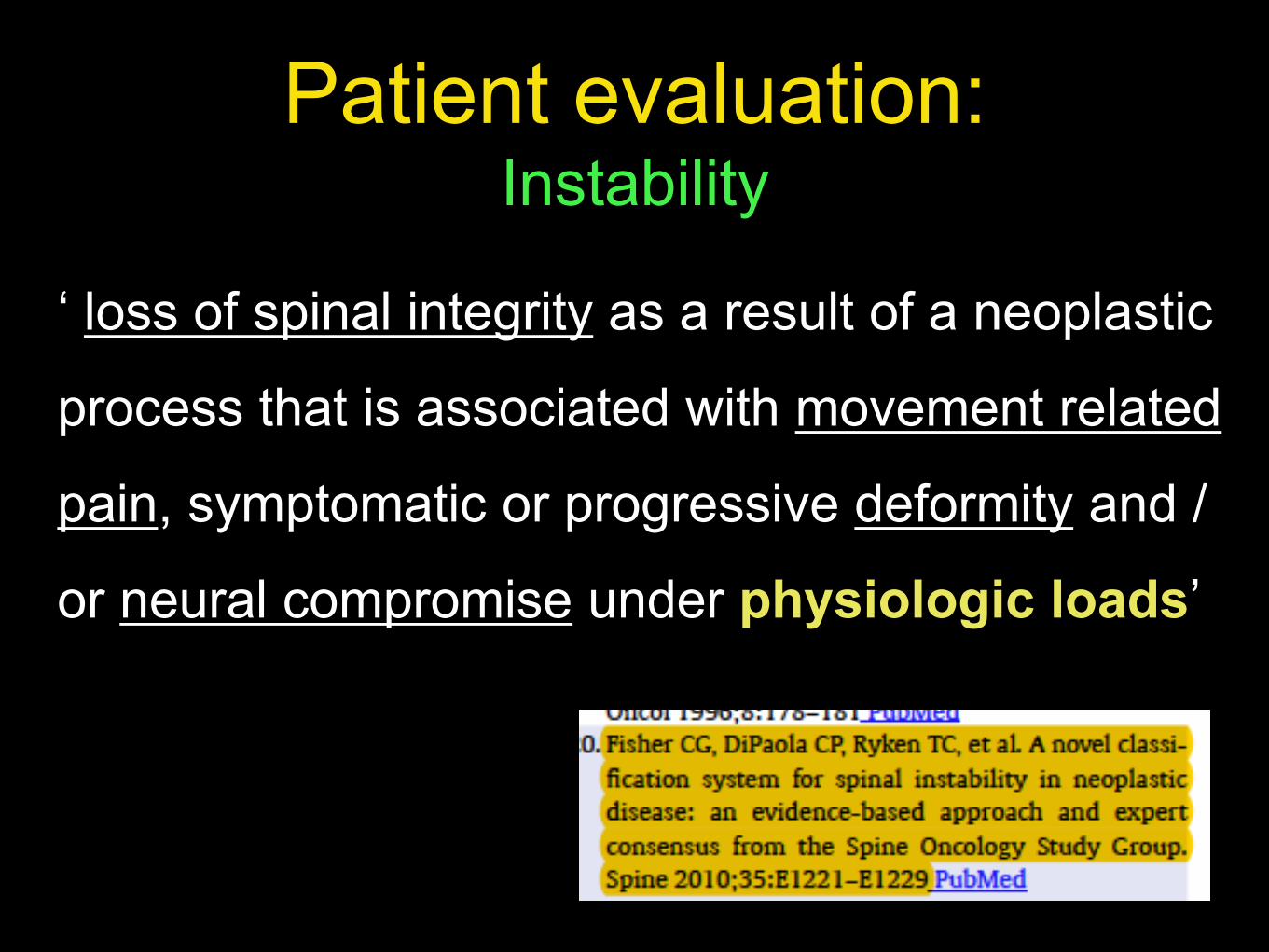
Patient evaluation: Instability
‘ loss of spinal integrity as a result of a neoplastic
process that is associated with movement related
pain, symptomatic or progressive deformity and /
or neural compromise under physiologic loads’
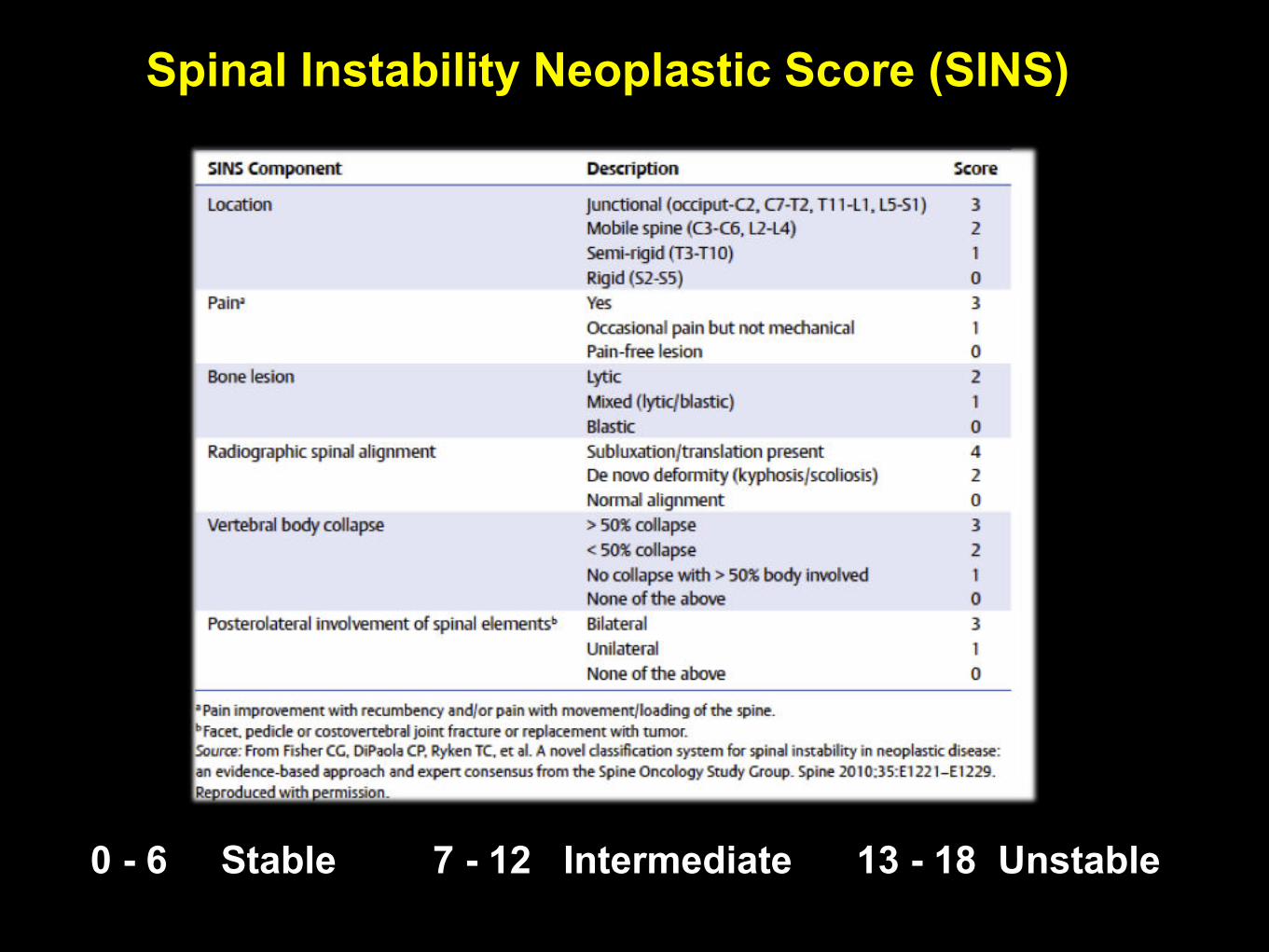
0 - 6 Stable 7 - 12 Intermediate 13 - 18 Unstable
Spinal Instability Neoplastic Score (SINS)
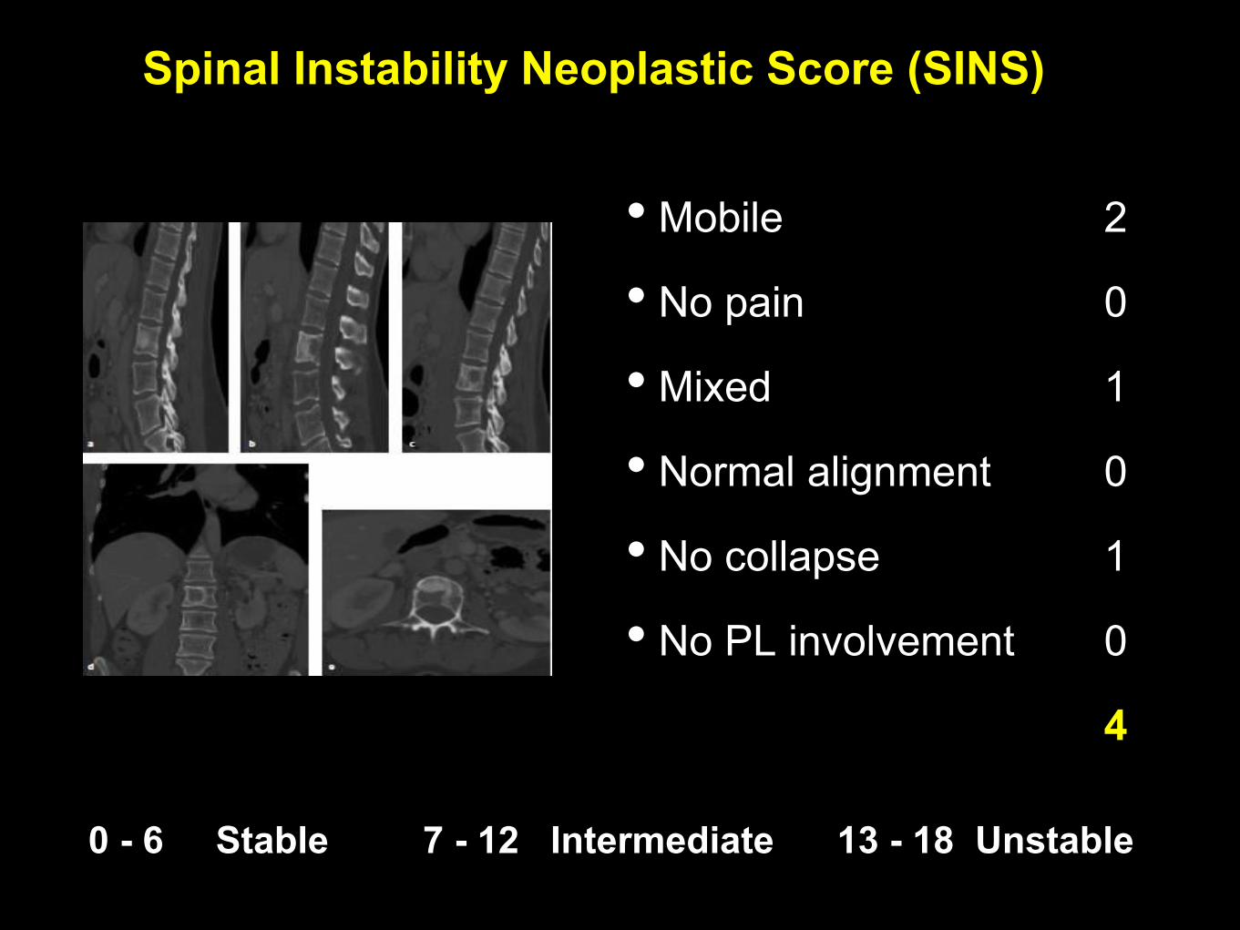
0 - 6 Stable 7 - 12 Intermediate 13 - 18 Unstable
Spinal Instability Neoplastic Score (SINS)
• Mobile 2
• No pain 0
• Mixed 1
• Normal alignment 0
• No collapse 1
• No PL involvement 0
4

0 - 6 Stable 7 - 12 Intermediate 13 - 18 Unstable
Spinal Instability Neoplastic Score (SINS)
• Junctional 3
• Pain (not mech) 1
• Lytic 2
• Normal alignment 0
• < 50% collapse 2
• Right pedicle 1
9

0 - 6 Stable 7 - 12 Intermediate 13 - 18 Unstable
Spinal Instability Neoplastic Score (SINS)
• Mobile 2
• Mech pain 3
• Lytic 2
• Kyphotic 2
• > 50% collapse 3
• Bil PL involced 3
15

• Cancellous involvement with intact cortical shell may not lead
to instability
• Taneichi Risk factors:
• Multivariate logistic regression model
• Thoracic: Costo-vertebral joint destruction v size of lesion
• TL / L: Size of lesion and pedicle involvement
• Bone mineral density v size of lesion
Biomechanics of collapse

Patient evaluation
1. Medical condition
2. Clinical presentation:
Neurology, Pain, Instability
3. Oncologic status

Patient evaluation Oncologic status
• Tumour histology
• Single strongest predictor of survival
• Vascularity: Renal, Thyroid, Hepato-cellular, Melanoma, GCT (hypervascular; prep embolisation)
• Tomita stratification of tumour histology

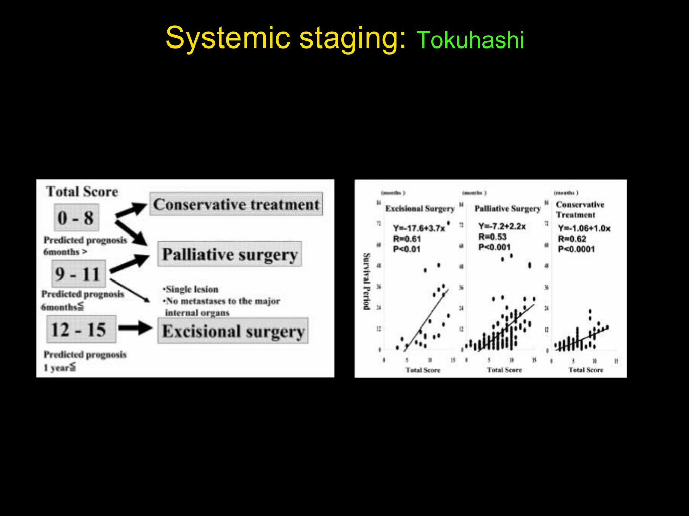
Systemic staging
• Tomita score
• Tokuhashi score
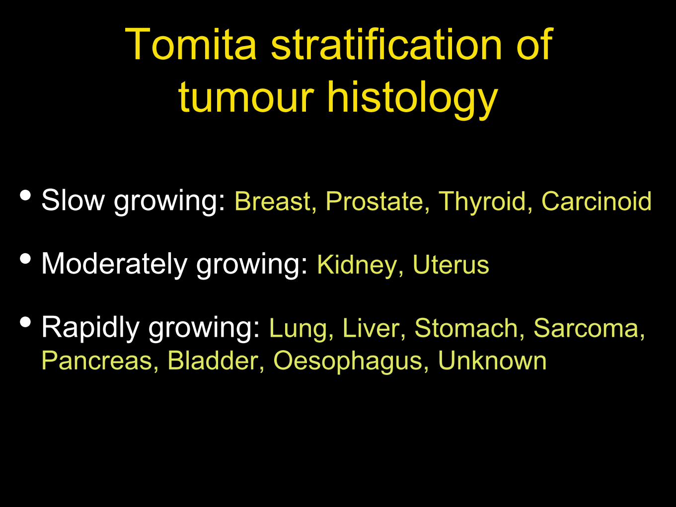
Tomita stratification of
tumour histology
• Slow growing: Breast, Prostate, Thyroid, Carcinoid
• Moderately growing: Kidney, Uterus
• Rapidly growing: Lung, Liver, Stomach, Sarcoma,
Pancreas, Bladder, Oesophagus, Unknown
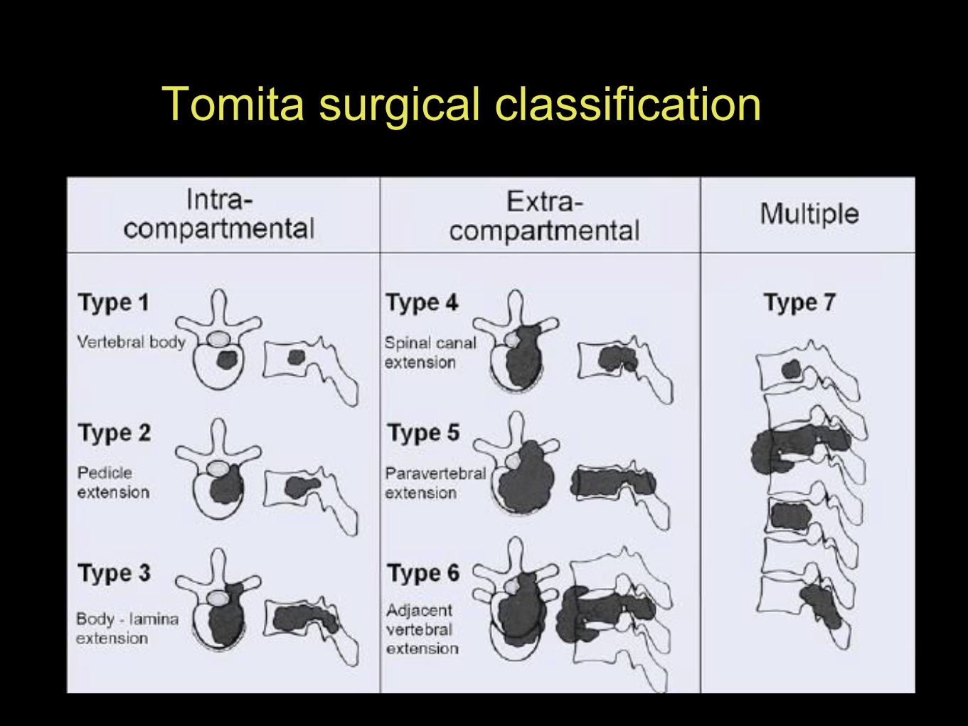
Tomita surgical classification

Weinstein - Boriani - Biagini surgical staging

Systemic staging: Tomita
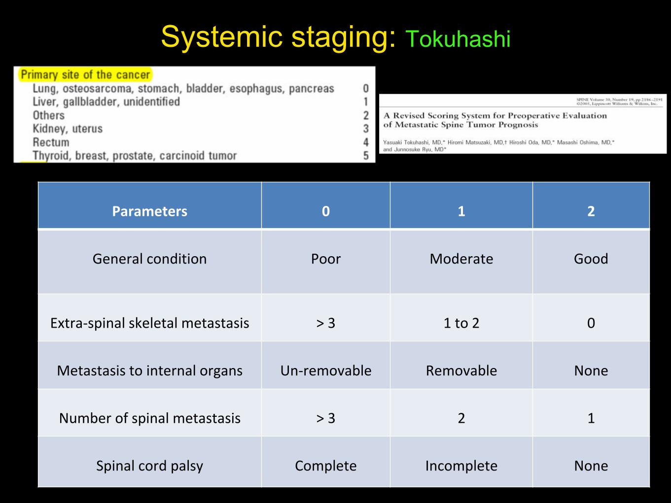
Systemic staging: Tokuhashi
Parameters 0 1 2
General condition Poor Moderate Good
Extra-spinal skeletal metastasis > 3 1 to 2 0
Metastasis to internal organs Un-removable Removable None
Number of spinal metastasis > 3 2 1
Spinal cord palsy Complete Incomplete None
Systemic staging: Tokuhashi
• Chemotherapy
• Radiotherapy (CXT, IMRT)
• Surgery (en bloc, palliative)
• End of life pathway
Treatment options
• Asymptomatic / minimal symptoms
• Haematologic malignancies
• Hormone sensitive tumours (if no surgical
indication)
• Newer drugs (named clinical oncologist)
Treatment options: Chemotherapy
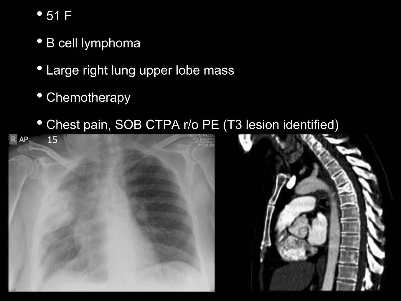
• 51 F
• B cell lymphoma
• Large right lung upper lobe mass
• Chemotherapy
• Chest pain, SOB CTPA r/o PE (T3 lesion identified)
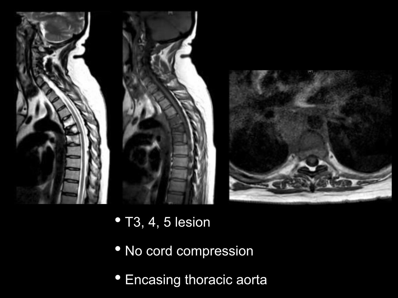
• T3, 4, 5 lesion
• No cord compression
• Encasing thoracic aorta
• Conventional (CRT)
• CRT limited by cord tolerance
• Radio-sensitive: Response to doses within the
cord tolerance
• Radio-resistant: Requires higher doses than
cord tolerance
Treatment options: Radiotherapy

• Intensity-modulated radiation therapy
• Higher dose of conformal radiation
• Easing of distinction between sensitive and
resistant tumours
Sensitive: Breast, prostate, ovarian, neuro-endocrine
cancers
Resistant: Renal, Thyroid, Hepato-cellular, Non Small cell,
Colon, Melanoma, Sarcomas
Treatment options: Radiotherapy

Problems:
• Compression fractures
• Pain flare: Transient increase after CRT
• Visceral (esophagus), plexus / root
susceptible to ‘collateral damage’
Treatment options: Radiotherapy

• 55 M
• Recent diagnosis of Lung Ca
• Pre-morbid normal mobility
• Neurology:
Right L2 - S1 4/5
Left L2 - S1 3/5
Normal PR, Sensory level ill defined
Palliation
IV Steroids
Radiotherapy
Pain management

• En-bloc
• Stabilisation / decompression
• Goals and timing of surgery
• Role of fusion
• Complications
Treatment options: Surgery
• Manage expectations
• Discuss with patient and family
• Discuss with oncologist
• Reduce pain
• Protect, restore neurology
• Maintain stability for ‘rest of the life’ (QoL)
Treatment options: Goals of Surgery

• Single level lesion (look for skip lesions)
• Vertebrectomy, sagittal resection, posterior arch
resection, spondylectomy
• Pre-operative embolisation
• Assess epidural spread
• Ligate Hoffmann’s ligaments
Treatment options: En Bloc Resection
• Stabilisation, Decompression
• Anaesthetic assessments
• Surgical risk stratifications
• Anterior column reconstruction
• Minimally invasive options
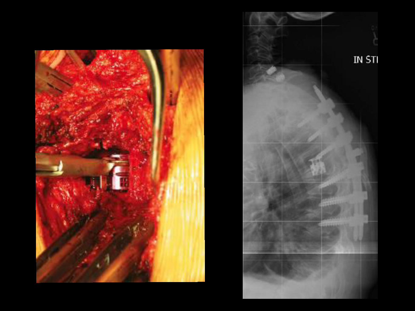
Treatment options: Palliative Surgery
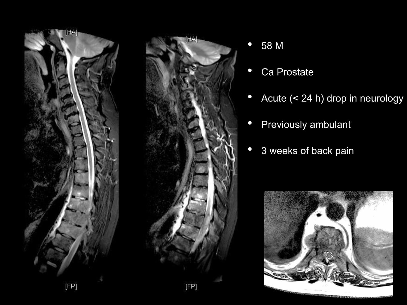
• 58 M
• Ca Prostate
• Acute (< 24 h) drop in neurology
• Previously ambulant
• 3 weeks of back pain
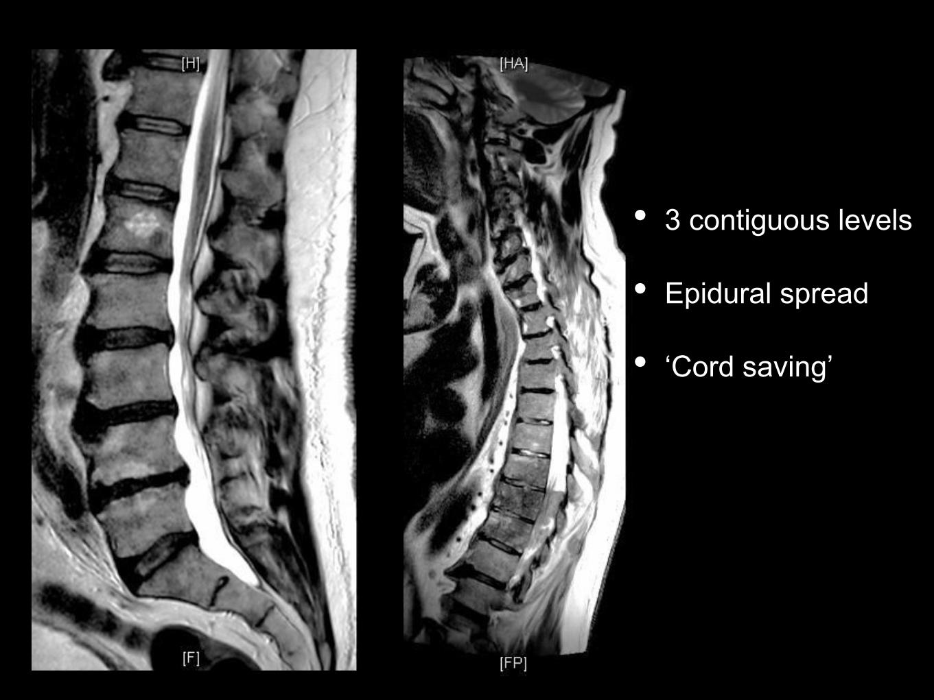
• 3 contiguous levels
• Epidural spread
• ‘Cord saving’

New lesions within 3 months
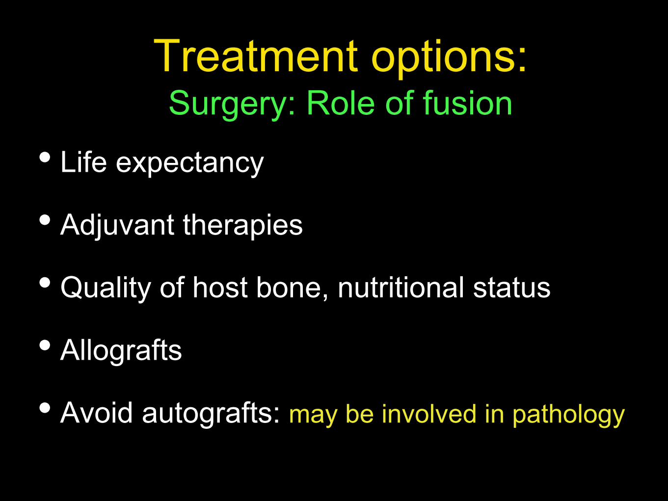
• Life expectancy
• Adjuvant therapies
• Quality of host bone, nutritional status
• Allografts
• Avoid autografts: may be involved in pathology
Treatment options: Surgery: Role of fusion
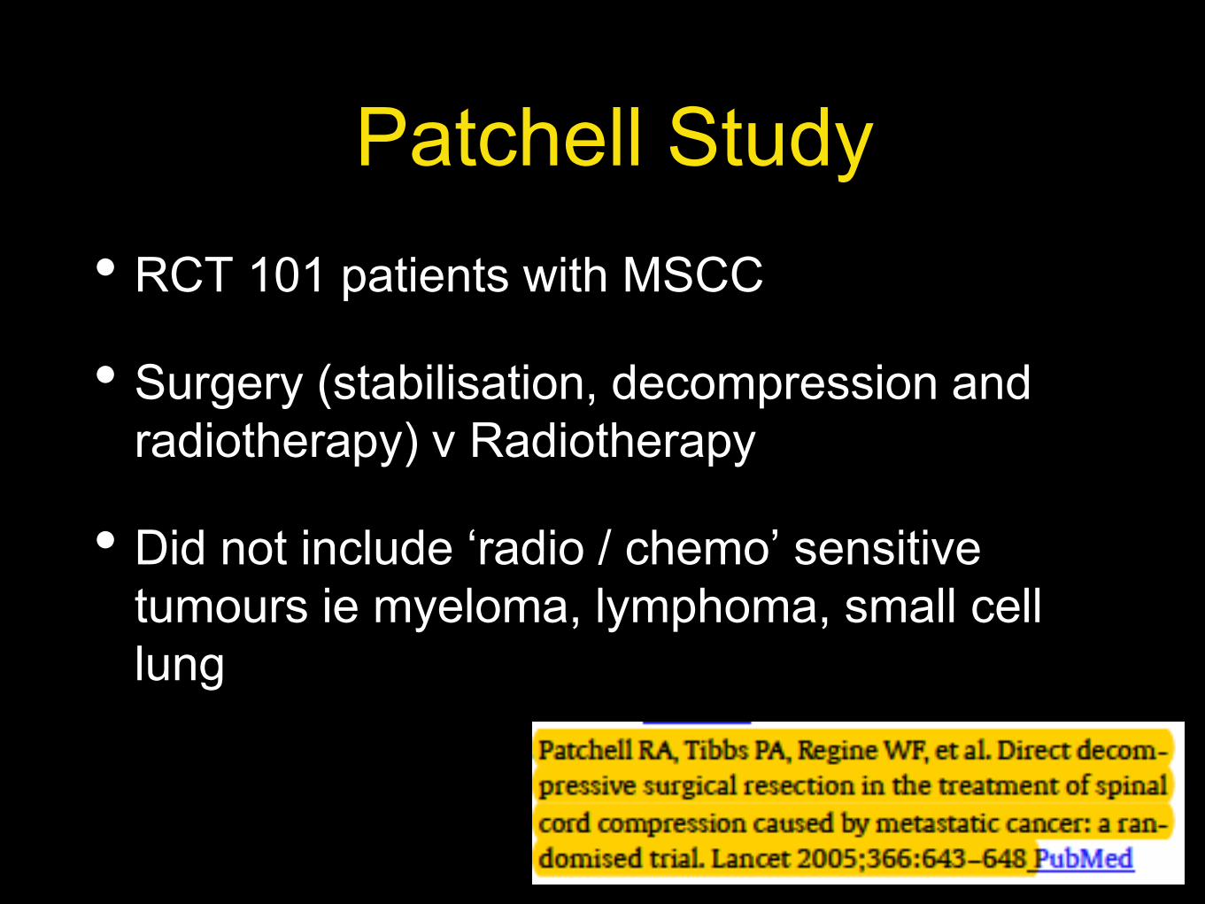
• RCT 101 patients with MSCC
• Surgery (stabilisation, decompression and
radiotherapy) v Radiotherapy
• Did not include ‘radio / chemo’ sensitive tumours ie myeloma, lymphoma, small cell
lung
Patchell Study

• Ambulation better in surgery group (84%)
than in radiotherapy group (57%) OR 6.2 p =
0.001
• Maintained ambulation for longer in surgery
group (122 d) v radiotherapy (13d) p = 0.003
Patchell Study

• Highly sensitive radio / chemo sensitive
tumours may respond to cord compression
without surgery
• Solid tumours with cord compression (grade
2, 3) require surgery and radiotherapy
• Grade 1 MSCC may not require surgery
(unless unstable)
Patchell Study

• Reduce instability pain
• Image guidance; Minimally invasive
• Local control of pain
Treatment options: Vertebral augmentation
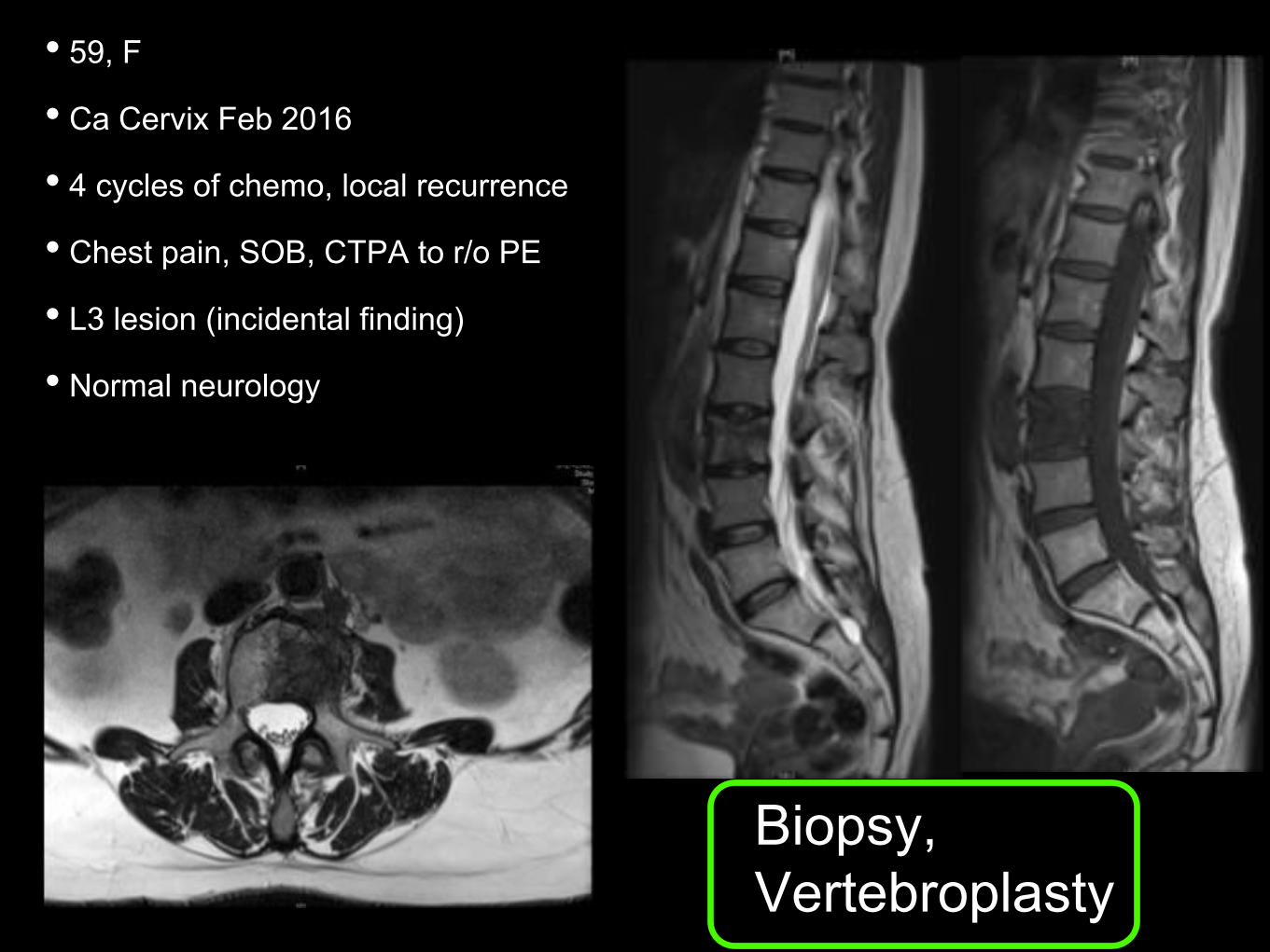
• 59, F
• Ca Cervix Feb 2016
• 4 cycles of chemo, local recurrence
• Chest pain, SOB, CTPA to r/o PE
• L3 lesion (incidental finding)
• Normal neurology
Biopsy,
Vertebroplasty
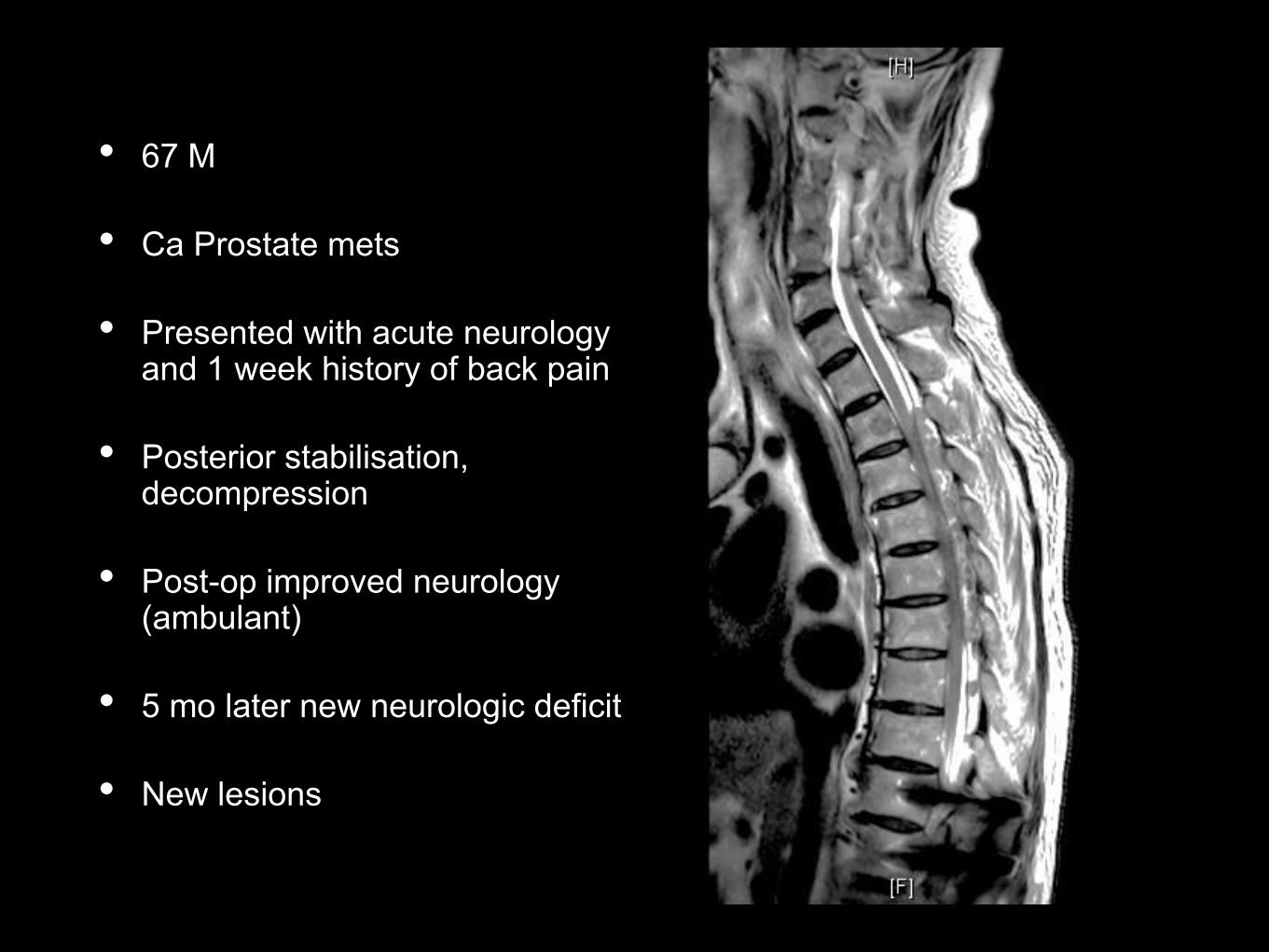
• 67 M
• Ca Prostate mets
• Presented with acute neurology and 1 week history of back pain
• Posterior stabilisation, decompression
• Post-op improved neurology (ambulant)
• 5 mo later new neurologic deficit
• New lesions
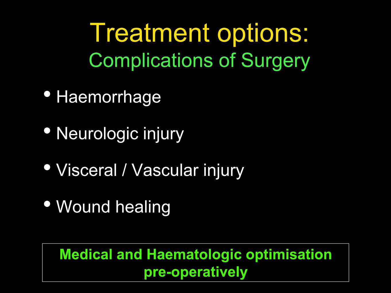
• Haemorrhage
• Neurologic injury
• Visceral / Vascular injury
• Wound healing
Treatment options: Complications of Surgery
Medical and Haematologic optimisation
pre-operatively

• Pain, Neurology, Suspected MSCC: Nurse flat
• TEDS, Flowtrons, Steroids, Bloods
• Maintain and update neurologic assessments
• Discuss with Family, Oncologists, Spinal Surgeon
and Anaesthetists
• Identify imaging requirements and related logistics
Initial management
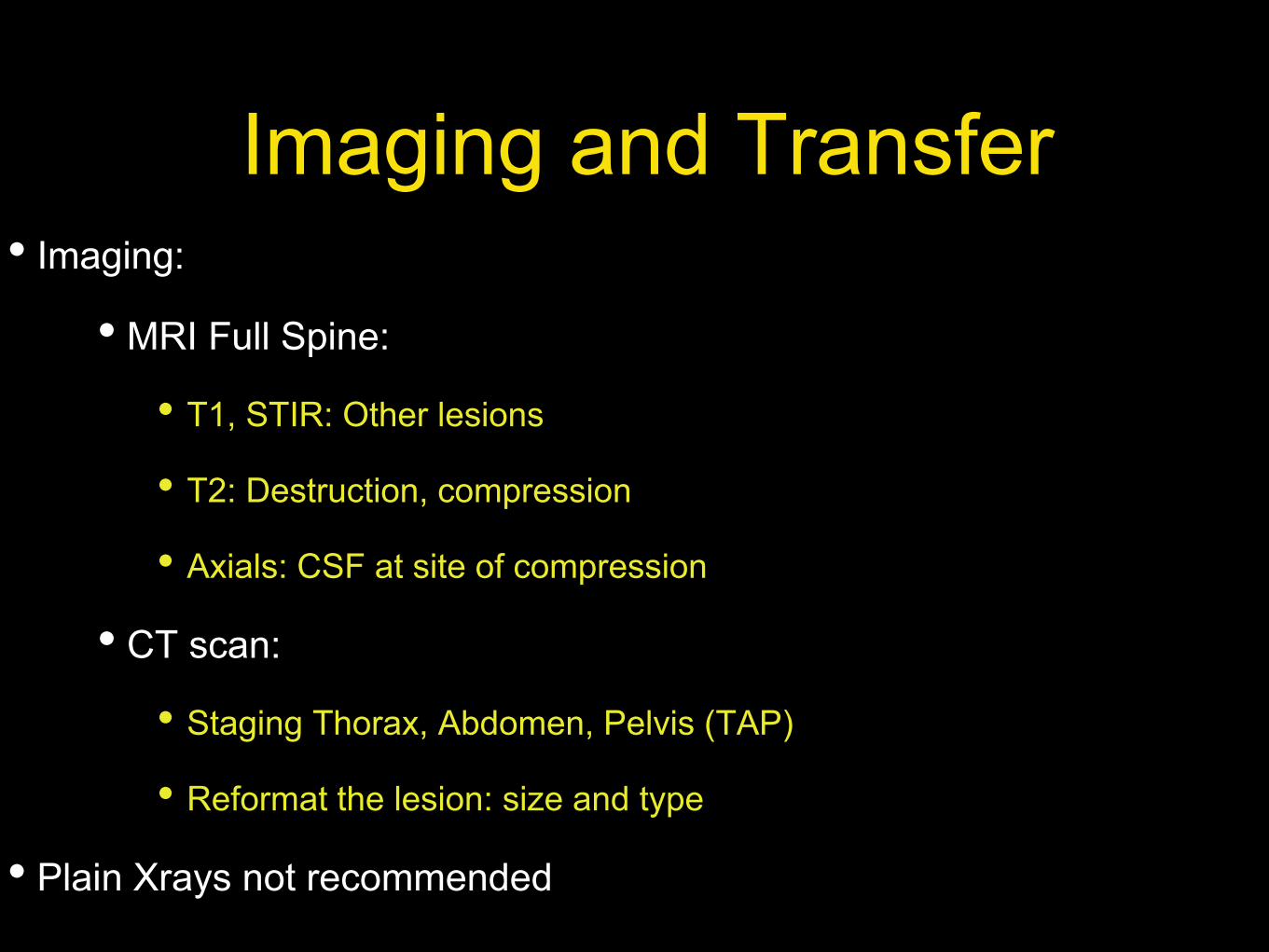
• Imaging:
• MRI Full Spine:
• T1, STIR: Other lesions
• T2: Destruction, compression
• Axials: CSF at site of compression
• CT scan:
• Staging Thorax, Abdomen, Pelvis (TAP)
• Reformat the lesion: size and type
• Plain Xrays not recommended
Imaging and Transfer
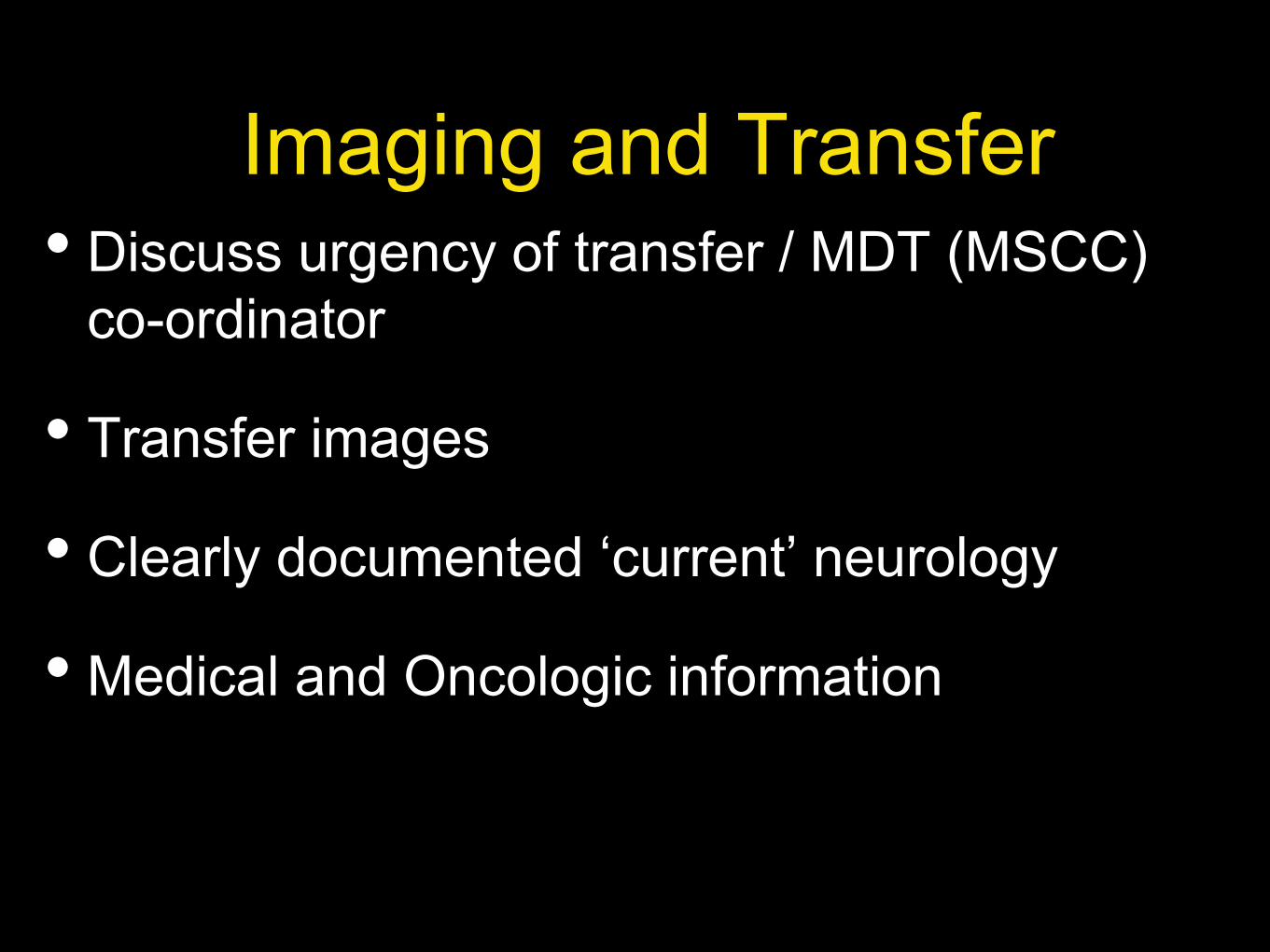
• Discuss urgency of transfer / MDT (MSCC)
co-ordinator
• Transfer images
• Clearly documented ‘current’ neurology
• Medical and Oncologic information
Imaging and Transfer
Selected references
Fischer CG et al Spine 2010; 35: E1221 – 1229
Tomita K et al Spine 2001; 26: 298 – 306
Tokuhashi Y et al Spine 2005; 30: 2186 – 2191
Fourney DR, et al J Clin Ocol 2011; 29: 3072 – 3077
Enneking Clin Orth Rel Res 1986; 204: 9 – 24
Boriani S et al Spine 1997; 22: 1036 – 1044
Patchell RA et al Lancet 2005; 366: 643 – 648
Gokaslan ZL, et al J Neurosurg 1998; 89: 599 - 609


![Home > [bec.uac.bj]...Home > Archives > Vol 12, No 29 (2016) VOL 12, NO 29 (2016) ESJ OCTOBER EDITION TABLE OF CONTENTS ARTICLES ESJ October Cover Page Editorial Office PDF Criminological](https://img.pdfslide.us/doc/110x75/5f33d6be5f9262283047833f/home-becuacbj-home-archives-vol-12-no-29-2016-vol-12.jpg)