Embed Size (px)
Citation preview

2

3
Metaphyseal Fractures
Of the
Proximal Tibia
Kaye E. Wilkins MD
Professor of Orthopedics and Pediatrics
University of Texas Health Science
Center
at San Antonio
e-mail:
4
METAPHYSEAL FRACTURES OF THE TIBIA
K.E. Wilkins
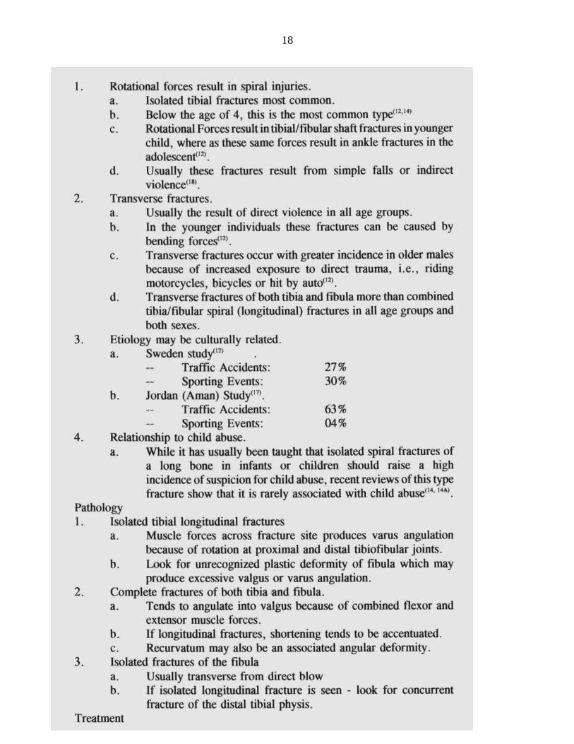
Fractures of the proximal tibial metaphysis can be deceiving in that what appears to be
initially a simple undisplaced fracture can lead to a significant cosmetic deformity even with the
best of treatment. Thus the message of this chapter is that when explaining the outcome of this
type of fracture to the parents, the treating surgeon must emphasize before the treatment is
initiated that the child may develop a significant valgus angulation regardless of the adequacy of
the treatment. The cause can be a biological phenomenon and not necessarily the result of
inadequate treatment by the surgeon. Explaining the potential for the deformity before the
treatment is initiated helps greatly when it occurs. The surgeon would be considered
knowledgeable in predicting the deformity before it developed. Trying to explain why it happened
after the deformity has occurred often only adds to the parents’ skepticism about the skill of the
treating surgeon.
This chapter will explore the pathogenesis, treatment options - including attempts at
prevention or minimization of the deformity, and the long-term prognosis of the valgus angulation
deformity once it develops.
HISTORICAL PERSPECTIVE
The first article in the English literature warning of this complication was published over
40 years ago, when Cozen (8) described four cases of valgus deformity following greenstick
fractures of the proximal tibial metaphysis. Almost 20 years later, he and Jackson (15) added
seven more cases, with an 18- to 20-year follow-up in three of the original cases. Following
Cozen’s initial warning of the development of this deformity after fractures of the proximal tibial
metaphysis, many other case reports and series have been reported in the literature (3-6, 8, 22,
25-27, 29). Thus the danger of a valgus deformity developing following a greenstick proximal
tibial metaphyseal fracture has become firmly established in the recent orthopedic literature.
INCIDENCE
Maximum valgus alignment appears to occur when the child undergoes the valgus stage
of lower extremity development, which usually is manifest between the ages of 2 and 8 years
(24, 26).
When considering all nonphyseal fractures of the tibia, that is, metaphyseal and
diaphyseal, the incidence of fractures involving only the proximal tibial metaphysis is about 3%
(19, 29).
PATHOGENESIS
5
There has been no one explanation as to why the valgus deformity occurs following
proximal metaphyseal fractures. It must be remembered that a valgus angulation can develop
spontaneously without fractures of the tibia. This deformity previously was observed when bone
grafts were originally obtained from the proximal tibial metaphysis for the Grice-Green technique
of subtalar arthrodesis. It has also developed following acute hematogenous osteomyelitis of the
tibia (28).
There are two broad categories of etiological conditions which can be factors in the
subsequent development of valgus angulation following proximal metaphyseal fractures. The first
group is composed of iatrogenic conditions, because they can he controlled by the surgeon. The
second group involves various biological parameters which produce asymmetrical overgrowth
contributing to valgus angulation .These various etiological factors are summarized in Table I.
Iatrogenic Conditions
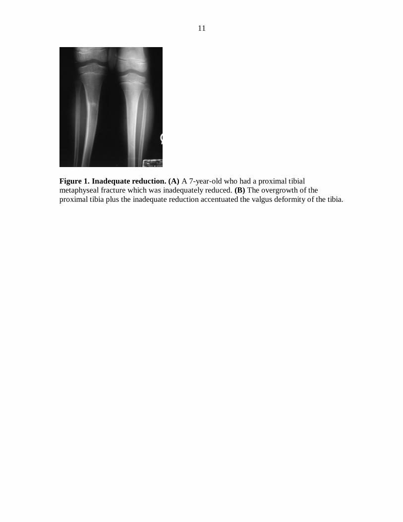
Inadequate Reduction
Inadequate reduction is most commonly due to failure to manipulate the leg into the
reduced position. This usually results in an immediate valgus deformity which is accentuated
when the secondary biological overgrowth occurs (Fig.1). Placing the leg in a bent-knee,
long-leg cast often makes it difficult to evaluate the adequacy of the reduction both clinically and
radiographically (21).
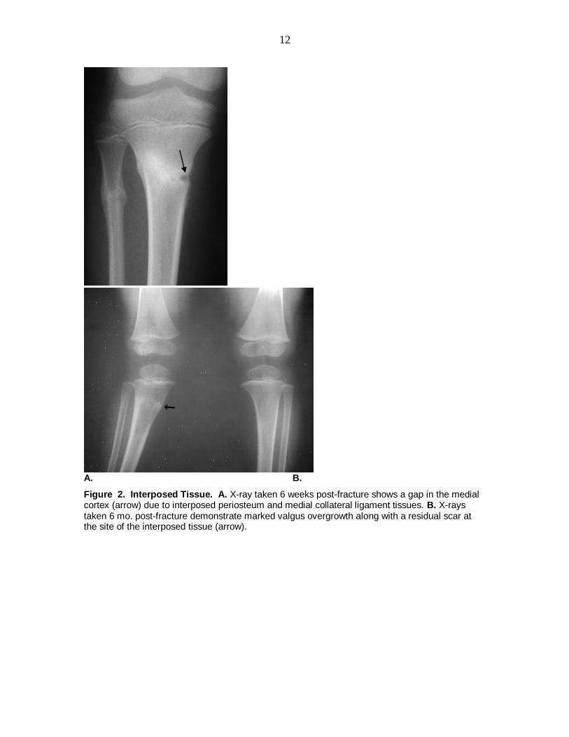
Interposed Tissue
Tissue interposed medially at the fracture site has been incriminated as a factor in failing
to achieve an adequate initial reduction ( Fig.2). Weber (31) found the tendinous insertion of the
pes anserinus plus the periosteum which also had been stripped off the metaphysis distally to be
interposed in the fracture site in his four cases. In addition, he theorized that this disruption of the
medial periosteum released its inhibition to growth medially, allowing the medial physis to grow
more rapidly, thus producing a delayed valgus angulation. This interposition of the pes anserinus
was confirmed by the work of Bassey (4), who routinely repaired the tendon.
Coates (7) found in two of his cases that the interposed material was the superficial part
of the medial collateral ligament instead of the pes anserinus tendon.
Premature Weight Bearing
Pollen (20) felt that too early weight bearing allowed the reduction to be lost, creating
recurrence of the initial valgus angulation. Bahnson and Lovell (2) theorized that weight bearing
produced compression or inhibition on the proximal tibial physis laterally along with distraction
forces medially which allowed more rapid growth in this area. This cause of localized lateral
growth inhibition produced by weight bearing is no longer mentioned in the recent literature.
Biological Conditions
6
Biological factors usually are responsible for the gradual increase in valgus deformity that
occurs over a 6- to 18-month period of time after the initial fracture has healed.
There are many explanations as to why this unequal growth develops.
Tibial Overgrowth
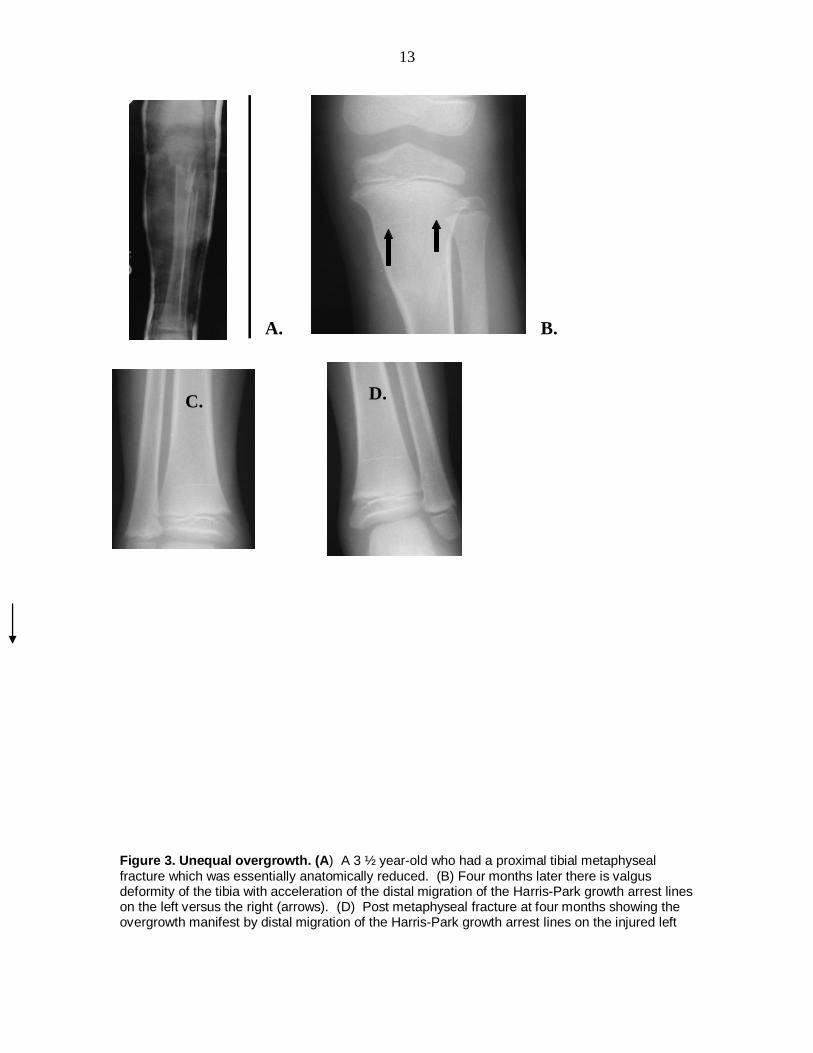
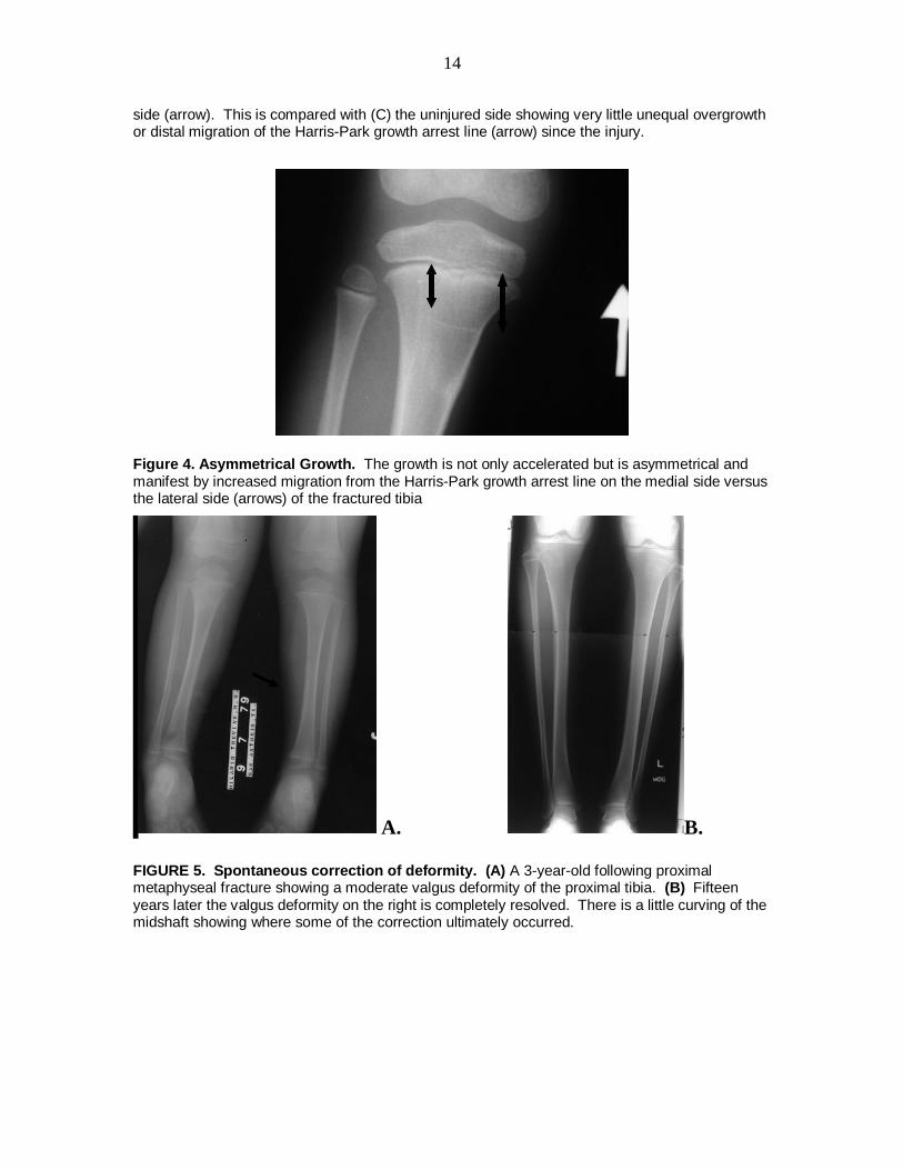
The fact that the tibia overgrows in length and does so asymmetrically has now been well
established by various studies (3, 10, 16, 22, 28, 33). First, there is an increased migration of the
Harris-Park growth arrest lines. Not only is migration greater on both the proximal and distal
fracture sites (Fig. 3), but it is also asymmetrical, being greater on the medial versus the lateral
side of the proximal tibia (10) (Fig.4). This asymmetrical growth has been confirmed by unequal
uptake of technetium using quantitative bone scans (16, 17, 33).
The exact cause of why the medial side grows faster than the lateral side is still not
completely known. Various theories have been proposed such as increased blood supply
medially, temporary loss of lateral growth and loss of the inhibition factor of the intact periosteum.
The data supporting each of these various theories will be examined in detail
Ogden (18) has demonstrated in cadaver studies that the blood flow to the medial side of
the proximal tibial metaphysis is significantly greater than on the lateral side. Thus, he feels that
the hyperemia caused by the fracture is asymmetrical, producing an increased stimulation to the
medial side of the proximal tibial metaphysis.
Ogden (18) has also speculated about temporary cessation of lateral physis growth as a
mechanism; however, no permanent osseous bridges have been demonstrated.
Most of the data supporting the theory that intact periosteum inhibits longitudinal growth
come from animal studies. Early studies show that in chickens (9) and rats (30) circumferential
release of the periosteum resulted in overgrowth of the long bone affected. Originally this was
suspected to be due to increased vascularity from the surgical release per se of the periosteum.
Subsequent studies in rabbits (1, 12) have shown that the release of only the medial metaphyseal
periosteum can produce asymmetrical valgus growth stimulation. In Aronson’s ( I ) study,
isolated lateral release of the periosteum produced a varus deformity. In none of the above
animal studies were structural changes able to be demonstrated in histological examination of the
physes.
Thus, there is some experimental evidence that release of the periosteim per se may
release its restricting effect and contribute to the asymmetrical growth. However, Jordan, (16)
feels he negates this theory, because in cases where he carefully repaired the medial
periosteum, valgus overgrowth still occurred.
Tethering by the Fibula
The theory of tethering by the fibula was popularized by the original Studie’s of Taylor
(28) in which he studied valgus overgrowth after tibial osteoteotomies. He noted that when the
fibula was not simultaneously osteotomized, the tibia often drifted into a late-onset valgus after
7
the Osteotomy had healed. When the fibula was simultaneously osteotomized there was very
little valgus angulation. However, prophylactic fibular osteotomy following a proximal tibial
metaphyseal fracture has failed to prevent the subsequent valgus overgrowth (16). In addition.
this author and others (16) have seen valgus angulation develop in those cases in which the
fibuIa is concurrently fractured.
Iliotibial Band Forces
The theory of iliotibial band forces arises from the polio era, when Irwin (14) attributed the
valgus deformity of the tibia in these paralytic patients to the lateral or valgus pull of the iliotibial
band. There has been no support for this theory in the recent literature.
AUTHOR’S PREFERRED ETIOLOGIES
It is this author’s opinion that the causes are multifactorial. The cause of an immediate
valgus alignment is due to failure to achieve an adequate reduction, which may or may not be
due to interposed tissue at the fracture site. The cause of the late-onset deformity is due to two
major biological factors, namely, asymmetrical hyperemia and, probably, asymmetrical loss of the
tethering effect of the periosteum. There is not much good evidence to support the other
proposed etiologies.
DEVELOPMENT OF THE DEFORMITY
Two long-term studies by Skak (27) and Zionts and MacEwen (32) have documented the
rate and patterns of deformity that develop following these types of fractures. First, the deformity
begins to develop during the healing process of the original fracture. It seems to develop most
rapidly during the first year postinjury, but some increase in angulation can be expected for as
long as 18 months. According to Herring and Moseley ( I I ), the valgus deformity becomes
clinically apparent when it reaches 10-15°.
In the Zionts and MacEwen study (32), the average overgrowth was I cm, but ranged as
high as 1.7 cm. The diaphyseal-metaphyseal angle increased an average of 9.6°. The average
time to obtain the maximum angulation was 12.6 months.
CORRECTION OF THE DEFORMITY
Many of the long-term studies show that most of the deformities resolve with time (Fig.
5). A great deal of the deformity corrects proximally, but some of it corrects distally, producing an
S-shaped type of tibia. Only on rare occasions does an operative correction need to be
performed, and then it should be done only close to the termination of growth.
TREATMENT
Treatment of these fractures is divided into two stages: First, there is the treatment of the
acute fracture. The second stage is how to manage the late valgus angularion.
Acute Fractures
8
Initially, the fracture must be reduced anatomically to minimize the amount of valgus
angulation. The parents of the patient must be told that this can best be done under either very
heavy sedation or a general anesthesia so a good varus molding can be applied to the extremity
with the knee in extension. Parsch (19) found that when this was done, the incidence of
cosmetically apparent valgus was essentially nonexistent.
The parents need to understand before the manipulation that a minor surgical procedure
may be necessary to remove any interposed tissue should an anatomical reduction not be
obtained. Some authors (22, 27) question whether removing the interposed tissue makes any
difference in the ultimate outcome.
Treatment of the Valgus Deformity
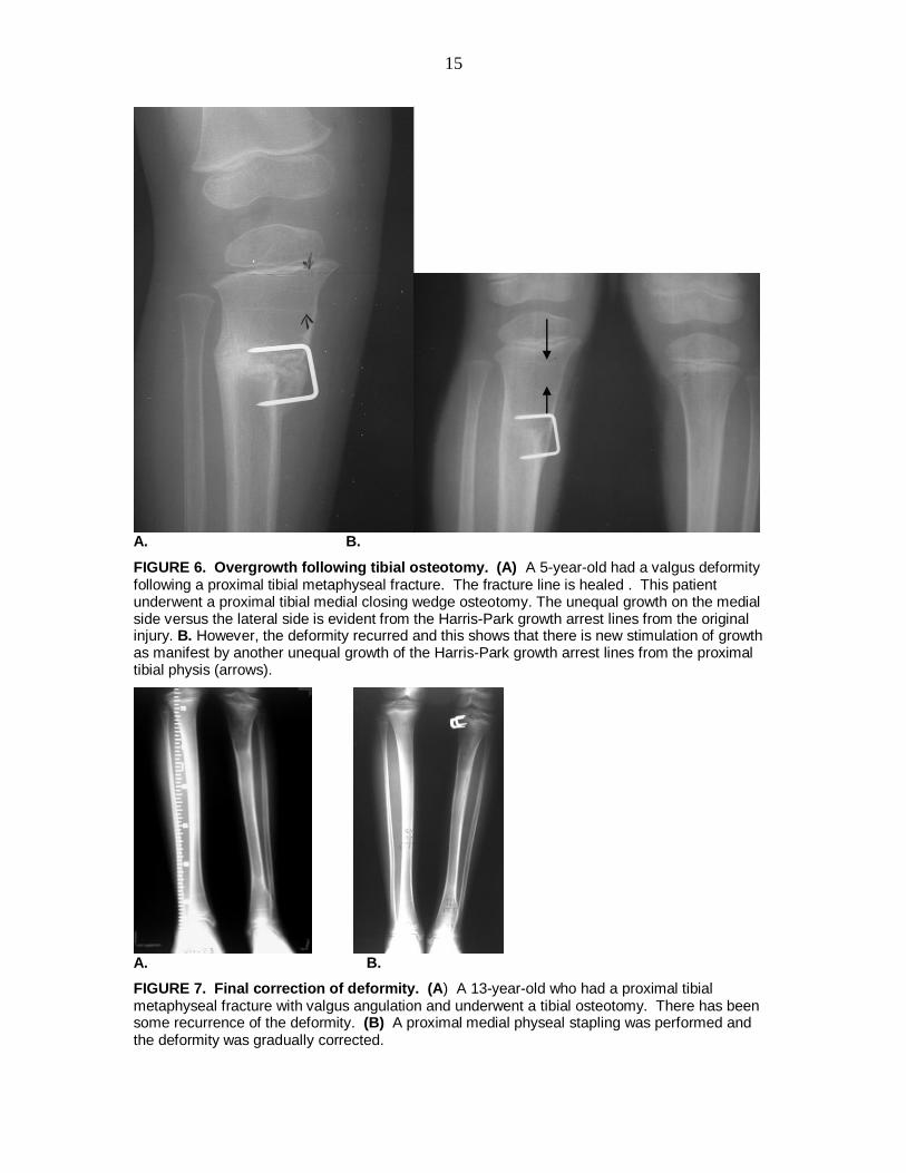
It has been shown by many (3, 11, 13, 31) that performing a corrective osteotomy may produce a
temporary correction, but in the following months, the deformity will recur due to the stimulating
effect of the osteotomy (Fig. 6). Since most of these ultimately have a satisfactorily functional
and cosmetic outcome at the termination of growth, it is felt that watching and waiting is the best
approach ( See Fig.5). Should the deformity persist into the early adolescent years, selective
medial proximal tibial hemiepiphysiodesis is felt by many (3, 23, 32) to be the best way to obtain
a correction of the angular deformity(Fig.7).
CONCLUSION
Tibia valga following greenstick proximal metaphyseal fractures involves both iatrogenic
and biological factors. The iatrogenic problems can be eliminated by obtaining an anatomic
reduction and ininiobilizing the leg in a well-molded, long-leg, extended-knee cast for 4-6 weeks.
The resultant deformity is best managed only at the termination of growth. A corrective osteotomy
soon after the fracture may result in a recurrent deformity and should be avoided. The parents
should be warned before treatment is initiated that a valgus deformity may develop despite the
best of care.
REFERENCES
1. Aronson DD, Stewart MC, Crissman JD. ,Experimental tibial fractures in rabbits
simulating proximal tibial metaphyseal fractures in children. Clin Orthop Relat Res 1990; 255:61-67. Bahnson
2. Bahnson DH, Lovell WW. ,Genu valgum following fractures of the proximal tibial metaphysis in children. Orthop Trans 1980; 4:306.
3. Balthazar DA, Pappas AM. Acquired valgus deformity of the tibia in children. J Pediatr Orthop 1984; 4:538.
4. Bassey LO. Valgus deformity following proximal metaphyseal fractures in children: Experiences in the African tropics. J Traurna 1990; 30:102.
5. Ben Itzhak I, Eiken EH, Malkin C. Progressive valgus deformity after juxta epiphyseal fracture of the upper tibia in children. Injury 1997; 18:169-173.
6. Brougham DI, Nicol RO. Valgus deformity after proximal tibial fractures in children. J Bone Joint Surg (Br) 1987; 69:482.
9
7. Coates R. Knock-knee deformity following upper tibial “greenstick” fractures. J Bone Joint Surg (Br) 1977; 59:516.
8. Cozen L. Fracture of the proximal portion of the tibia in children followed by valgus deformity. Surg Gynecol Obstet 1953; 97:183.
9. Crilly RG. Longitudinal overgrowth of chicken radius. J Anat 1972; 112:11-18. 10. Green NE. Tibia valga caused by asymmetrical overgrowth following a nondisplaced
fracture of the proximal tibia metaphysis. J Pediatr Orthop 1983; 3:235. 11. Herring JA, Moseley C. Posttraumatic valgus deformity of the tibia: Instructional case. J
Pediatr Orthop 1981;1: 435-439. 12. Houghton GR, Rooker GD. ,The role of the periosteum in the growth of long bones. An
experimental study in the rabbit. J Bone Joint Surg (Br) 1979; 61:218-220. 13. Ippolito E, Pentimalli C. Post-traumatic valgus deformity of the knee in proximal tibial
metaphyseal fractures in children. Ital J Orthop Traumatol 1984; 10:103-108. 14. Irwin CE. Iliotibial band. Its role in producing deformity in poliomyelitis. J Bone Joint
Surg (Am) 1949; 34:141-146. 15. Jackson DW, Cozen L. Genu valgum as a complication of proximal tibial metaphyseal
fractures in children. J Bone Joint Surg (Am) 1971; 53:1571. 16. Jordan SE, Alonso JE, Cook, FF. The etiology of valgus angulation after metaphyseal
fractures of the tibia in children. J Pediatr Orthop 1987; 7:450. 17. Keret D, Harcke HT, Bowen JR. Tibia valga after fracture: Documentation of mechanism.
Arch Orthop Trauma Surg 1991; 110:216-219. 18. Ogden JA. Skeletal Injury in the Child. Philadelphia: Sanders & Saunders, 1990; 822. 19. Parsch K. Proximal metaphyseal fractures of the tibia. How can progressive valgus
angulation be prevented? 2nd
Swedish International Seminar on Fractures in Children Stockholm, Sweden; September, 23-25, 1992.
20. Pollen AG. Fractures and Dislocations in Children. Baltimore: Williams & Wilkins, 1973; 179.
21. Rang M. Tibia. Children’s Fractures 2nd
ed. Philadelphia: J.B. Lippincott, 1983; 189. 22. Robert M, Khouri N, Carlioz H, Alain JL. Fractures of the proximal tibial metaphysis in
Children: Review of a series of 25 cases. J Pediatr Orthop 1987; 7:444. 23. Rooker G, Salter R. Presentation of valgus deformity following fracture of the proximal
metaphysis of the tibia in children. Bone Joint Surg (Br) 1980; 62:527. 24. Salenius P, Vankaa E. The development of the tibiofemoral angle in children. J Bone
Joint Surg (Am) 1975; 57:259-261. 25. Salter RB, Best T. The pathogenesis and prevention of valgus deformity following
fractures of the proximal metaphyseal region in the tibia in children. J Bone Joint Surg (Am) 1973; 55:1324.
26. Salter RB, Best T N. Pathogenisis of progressive valgus deformity following fractures of the proximal metaphyseal region of the tibia in young chiIdren. Instr Course Lect 1992; 41:409-411.
27. Skak SV. Valgus deformity following proximal tibial metaphyseal fracture in children. Acta Orthop Scand 1982; 53:141.
28. Taylor SL. Tibial overgrowth: A cause of genu valgum. J Bone Joint Surg (Am) 1963; 45:659.
29. Visser J, Veldhvizen A. Valgus deformity after fractures of the proximal tibial metaphysis in childhood. Acta Orthop Scand 1982; 53:663.
30. Warrell E, Taylor JF. The effect of trauma on tibial growth. J Bone Joint Surg (Br) 1970; 58:375.
31. Weber BG. Fibrous interposition causing valgus deformity after fracture of the upper tibial metaphysis in children. J Bone Joint Surg (Br) 1977; 59:290.
32. Zionts LE, MacEwen GD. Spontaneous improvement of post traumatic tibia valga. J Bone Joint Surg (Am) 1986;68:680.
33. Zionts LE, Harcke HT, Brooks KM, MacEwen GD. Posttraumatic tibia valga: A case demonstrating asymmetric activity at the proximal growth plate on technetium bone scan. J Pediatr Orthop 1987; 7:458.
10
Table I. Etiological factors producing tibia valga
I. Iatrogenic conditions Inadequate reduction Interposed tissue Premature weight bearing
II. Biological conditions Asymmetrical tibial growth due to:
Increased medial blood supply Temporary cessation of lateral physeal growth Loss of periosteal inhibition of growth Tethering of the fibula lliotibial band forces
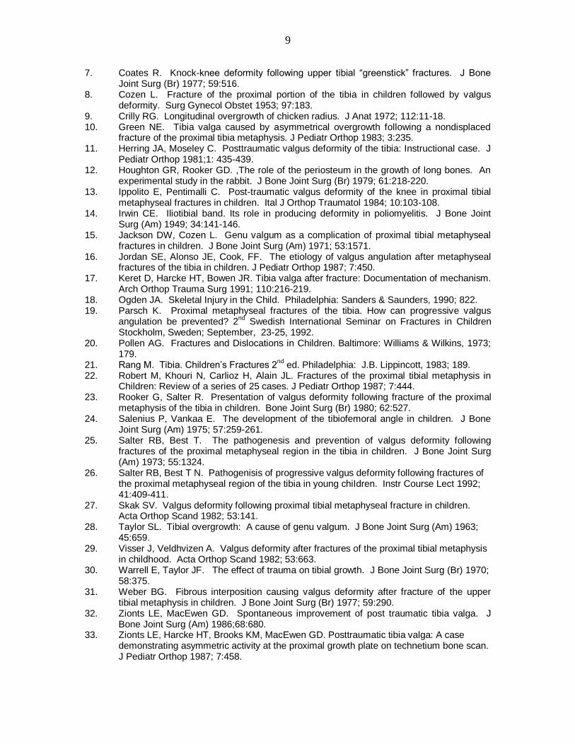
Illustrations A.
B.
11
Figure 1. Inadequate reduction. (A) A 7-year-old who had a proximal tibial
metaphyseal fracture which was inadequately reduced. (B) The overgrowth of the
proximal tibia plus the inadequate reduction accentuated the valgus deformity of the tibia.
12
A. B.
Figure 2. Interposed Tissue. A. X-ray taken 6 weeks post-fracture shows a gap in the medial cortex (arrow) due to interposed periosteum and medial collateral ligament tissues. B. X-rays taken 6 mo. post-fracture demonstrate marked valgus overgrowth along with a residual scar at the site of the interposed tissue (arrow).
13
A A. B.
Figure 3. Unequal overgrowth. (A) A 3 ½ year-old who had a proximal tibial metaphyseal fracture which was essentially anatomically reduced. (B) Four months later there is valgus deformity of the tibia with acceleration of the distal migration of the Harris-Park growth arrest lines on the left versus the right (arrows). (D) Post metaphyseal fracture at four months showing the overgrowth manifest by distal migration of the Harris-Park growth arrest lines on the injured left
C.
D.
14
side (arrow). This is compared with (C) the uninjured side showing very little unequal overgrowth or distal migration of the Harris-Park growth arrest line (arrow) since the injury.
Figure 4. Asymmetrical Growth. The growth is not only accelerated but is asymmetrical and manifest by increased migration from the Harris-Park growth arrest line on the medial side versus the lateral side (arrows) of the fractured tibia
A. B.
FIGURE 5. Spontaneous correction of deformity. (A) A 3-year-old following proximal metaphyseal fracture showing a moderate valgus deformity of the proximal tibia. (B) Fifteen years later the valgus deformity on the right is completely resolved. There is a little curving of the midshaft showing where some of the correction ultimately occurred.
15
A. B.
FIGURE 6. Overgrowth following tibial osteotomy. (A) A 5-year-old had a valgus deformity following a proximal tibial metaphyseal fracture. The fracture line is healed . This patient underwent a proximal tibial medial closing wedge osteotomy. The unequal growth on the medial side versus the lateral side is evident from the Harris-Park growth arrest lines from the original injury. B. However, the deformity recurred and this shows that there is new stimulation of growth as manifest by another unequal growth of the Harris-Park growth arrest lines from the proximal tibial physis (arrows).
A. B.
FIGURE 7. Final correction of deformity. (A) A 13-year-old who had a proximal tibial metaphyseal fracture with valgus angulation and underwent a tibial osteotomy. There has been some recurrence of the deformity. (B) A proximal medial physeal stapling was performed and the deformity was gradually corrected.
16
17
18
19
20

21
22
23
24
25